613
© 2018 by the Serbian Biological Society How to cite this article: Gavrilović M, Tešević V, Đorđević I, Rajčević N, Bakhia A, Garcia Jacas N, Susanna A, Marin PD, Janaćković P. Leaf micromorphology, antioxidative activity and a new record of 3-deoxyamphoricarpolide of relict and limestone endemic Amphoricarpos elegans Albov (compositae) from Georgia. Arch Biol Sci. 2018;70(4):613-20.
Leaf micromorphology, antioxidative activity and a new record of 3-deoxyamphoricarpolide
of relict and limestone endemic
Amphoricarpos elegans
Albov (Compositae) from Georgia
Milan Gavrilović1,*, Vele Tešević2, Iris Đorđević3, Nemanja Rajčević1, Arsena Bakhia4, Núria Garcia Jacas5, Alfonso Susanna5, Petar D. Marin1 and Peđa Janaćković1
1University of Belgrade, Faculty of Biology, Institute of Botany and Botanical Garden “Jevremovac”, Studentski trg 16,
11 000 Belgrade, Serbia
2University of Belgrade, Faculty of Chemistry, Studentski trg 12-16, 11000 Belgrade, Serbia
3University of Belgrade, Faculty of Veterinary Medicine, Bulevar oslobođenja 18, 11000 Belgrade, Serbia
4Ilia State University, School of Natural Sciences and Engineering, Cholokashvili Avenue 3/5, 0160 Tbilisi, Georgia 5Botanic Institute of Barcelona (IBB,CSIC-ICUB), Pg. del Migdia s. n., 08038 Barcelona, Spain
*Corresponding author: [email protected]
Received: March 9, 2018; Revised: May 5, 2018; Accepted: May 14, 2018; Published online: June 4, 2018
Abstract: We examined for the first time the leaf micromorphology, phytochemistry and biological activity of the rare and stenoendemic Amphoricarpos elegans Albov (Compositae) from Georgia. Scanning electron microscopy (SEM) revealed the presence of glandular trichomes on the leaves, which appeared as glandular dots that are considered the main sites of bio-synthesis and accumulation of sesquiterpene lactones. Using high-performance liquid chromatography (HPLC) and nuclear magnetic resonance (NMR) spectroscopy analyses, we identify and characterized 3-deoxyamphoricarpolide, a known ses-quiterpene lactone for the genus Amphoricarpos Vis. Regarding chemotaxonomic significance, 3-deoxyamphoricarpolide represents a link between Balkan and Caucasian species of the genus. The antioxidative capacity of different leaf extracts, obtained using a Soxhlet extractor, was evaluated by two radical scavenging assays: DPPH (1,1-diphenyl-2-picrylhydrazyl) radical and the 2,2΄-azino-bis-3-ethylbenzthiazoline-6-sulphonic acid (ABTS), and ferric ion reducing antioxidant power (FRAP). The total phenolic and flavonoid contents were also determined. The highest antioxidative activity and the high-est phenolic and flavonoid contents were detected in the methanol fraction, as a result of the contribution of not only phenols, but probably also lactones. The considerable antioxidative potential indicates possible applications in pharmacy and medicine.
Key words: Amphoricarpos elegans; glandular trichomes; 3-deoxyamphoricarpolide; antioxidative activity
INTRODUCTION
Compositae is one of the largest angiosperm families, containing more than 1600 genera and 23000 species with an almost global distribution [1]. In the Com-positae, glandular trichomes of short-stalked capitate type, usually seen as glandular dots, are microchar-acters that synthesize and accumulate sesquiterpene lactones [2]. Sesquiterpene lactones are secreted into the extracellular and subcuticular secretion storage space at the apical ends of multicellular trichomes and excreted to the plant surface. This type of gland is widely distributed in Compositae [2]. Compositae taxa produce a wide range of specialized metabolites
with significant biological effects [3-4], and many are well-known medicinal plants showing antimicrobial and antioxidant activities due to the presence of phe-nolic compounds (e.g. flavonoids) and sesquiterpene lactones in different organs [5-12].
eudesmanolides, which are classified on the basis of their carbocyclic skeletons. A characteristic of these compounds is the γ-lactone function. Because of their biological activity and ecological functions, as well as their chemotaxonomic significance, sesquiterpene lactones are the subject of constant research world-wide [14-17].
The genus Amphoricarpos Vis. (Compositae-Car-dueae-Carduinae) belongs to the Xeranthemum group [18], which includes annual plants, such as species of the genera Xeranthemum L., Chardinia Desf. and
Siebera J. Gay, and perennial plants,including species
of the genera Amphoricarpos Vis.and Shangwua Yu J. Wang, Raab-Straube, Susanna & J. Quan Liu [18]. The group is well characterized based on its molecular and morphological features [19]. Phylogenies based on plastid and nuclear analysis confirm that it is a natural group [20]. Amphoricarpos species are heterocarpic perennial chasmophytic plants, mountain endemics in the eastern Mediterranean (the Balkans, Anatolia and the Caucasus) [18].
The Caucasus is distinguished by different phy-tolandscapes and species genetic diversity due to its edaphic-climatic conditions, high hypsometric levels, well-expressed geographical isolation, etc. [21]. High endemism is characteristic of the Caucasus and it rep-resents one of the world’s biodiversity hotspots [22]. The flora of Georgia is represented by 4130 species of vascular plants, of which 4034 are angiosperms [23]; 1304 (32.3%) species are endemics for the Caucasus and 261 (6.6%) are endemics for Georgia [24]. In the Cau-casus flora, there are 17 endemic genera, most of them represented by one species, Amphoricarpos elegans Al-bov [22]. Among the calciphytes, in the geographical province of Colchis, the most distinguished is A. elegans
[21], which belongs to the group of “limestone endem-ics” of the western Caucasus Mountains [25].
The Balkan species of Amphoricarpos has been the subject of some phytochemical [26-28], biological activ-ity [14,29,11], morphological [31] and taxonomic stud-ies [32]. To the best of our knowledge, there is only one study describing the morphology and anatomy of A.
elegans [31], but there are no data dealing with
micro-morphological, phytochemical and examinations of the biological activities of this species. Thus, A. elegans is almost unexplored. Therefore, the present study, for the
first time, aimed to (i) examine leaf micromorphology, (ii) determine the total phenol and flavonoid contents and to evaluate the antioxidative potential of various leaf extracts, and (iii) to identify the most dominant sesquiterpene lactones of A. elegans.
MATERIALS AND METHODS Plant material
The plant material (leaves) of A. elegans was collected in 2015 during the flowering period from plants grow-ing in their natural habitat, Mt. Migaria, Samegrelo, Georgia (N 42.6479800; E 42.63992537). A voucher specimen was deposited in the Herbarium of the University of Belgrade, Faculty of Biology, Institute of Botany and Botanical Garden “Jevremovac” (ac-cession number: BEOU 17420).
Micromorphological methods
Micromorphological analysis was carried out using scanning electron microscopy (SEM). Small parts of dry leaves were sputter-coated with gold for 180 s at 30 mA (BAL-TEC SCD 005) and observed using a JEOL JSM-6460LV electron microscope at an acceleration voltage of 20 kV.
Soxhlet extraction
Powdered dried leaves (1.00 g) were extracted in a Soxhlet extractor using pure n-hexane, ethyl acetate and methanol as solvents for 6 h. The solvent (3x120 mL) was successively changed after 2, 4 and 6 h. The extracts were evaporated to dryness.
Determination of total phenolics and flavonoids in the plant extracts
the content of phenolics in the extracts was expressed in terms of the gallic acid (GA) equivalent (GAE) or mg of GA/g of extract. The flavonoid contents of the methanol, ethyl acetate and n-hexane extracts were determined by UV spectroscopy [33]. The sample contained 0.1 mL of the methanol solution of the ex-tract at a concentration of 1 mg/mL, 0.41 mL 80% ethanol, and 0.01 mL and 0.1 mL of Al(NO3)3x9H2O and CH3COOK each. The samples were incubated for 40 min at room temperature. Absorbance was determined at λmax=415 nm. Based on the measured absorbance, the flavonoid contents of the extracts were expressed in terms of the quercetin (QU) equivalent (QUE), or mg of QU/g of extract.
Evaluation of antioxidant activity DPPH assay
The ability of the plant extracts (dry methanol, ethyl acetate and n-hexane extracts) to scavenge the DPPH free radical was assessed by UV spectroscopy [34-35]. Dilutions of stock methanolic solution were made to obtain concentrations of 500, 350, 275, 250, 200, 100, 50 μg/mL DPPH. After 40 min in the dark at room temperature, the absorbance was measured at 517 nm. The control samples contained all the reagents except the extract. The percentage inhibition was calculated using the equation:
%inhibition=100x(A of control – A of the sample)/A of control).
IC50 values were estimated from the % inhibition vs. the concentration sigmoidal curve, using nonlin-ear regression analysis. The antioxidant effects of the extract increased with the decrease in IC50 values.
ABTS assay
The ABTS radical scavenging method [36] with some modifications was used. The ABTS radical cation is generated by the oxidation of ABTS with potassium persulfate and its reduction in the presence of hydro-gen donating antioxidants is measured by UV spec-troscopy at 734 nm. The reaction mixture of 5 mg of ABTS was dissolved in potassium persulfate before the experiment. This solution was dissolved in distilled
water in order to calibrate working solution absorb-ance to 0.700 at 734 nm. The sample concentration was 1 mg/mL. The reaction mixture was prepared by mixing 15 μL of a test sample and 4-1.5 mL ABTS. After a 4-min incubation at room temperature, the absorbance of the mixture was measured at 734 nm. The radical scavenging activity for each extract was determined based on the linear calibration curve of ascorbic acid and was expressed as mg ascorbic acid/g of dry extract (mg Vitamin C/g d.e.).
FRAP assay
The reducing power of methanol, ethyl acetate and n -hexane extracts was determined using the FRAP assay [37]. This assay is based on the reducing power of a compound (antioxidant). The potential antioxidant is expected to reduce the ferric ion (Fe3+) to the ferrous ion (Fe2+); the latter forms a blue complex (Fe2+ /2,4,6-Tris(2-pyridyl)-s-triazine (TPTZ), which increases the absorption at 593 nm. Briefly, the FRAP reagent was prepared by mixing acetate buffer (200 μL, pH 3.6), a solution of 20 μL TPTZ and 20 μL FeCl3. The sample solution (30 μL) and the reagent (0.9 mL FRAP) were mixed thoroughly and incubated at 37°C. Absorbance was taken at 593 nm after 5 min. A standard cali-bration curve was prepared using different concen-trations of FeSO4×7H2O. All solutions were freshly prepared. The results were expressed in mmol Fe/mg dried extract (mmol Fe/mg d.e.).
All the assays described above were performed us-ing a Perkin Elmer Lambda Bio UV-Vis spectrometer.
Extraction and Isolation for HPLC and NMR analysis
equipped with a diode array detector (DAD; λ=210 nm) and autosampler. The separation column was a Zorbax Eclipse XDB C18 (250×9.4 mm; 5 μm). The column heater was set at 20°C and the mobile phase flow rate was maintained at 0.5 mL min-1. The gradient (solvent A water, solvent B, 100% acetonitrile) was 0-5 min, 10% B; 5-20 min, 10-35% B; 20-30 min, 35% B, 30-40 min, 35-50% B; 40-60 min, 50% B; 60-61 min, 50-10% B; 61-66 min, 10% B. The injection volume was 1 µL.
Statistical analysis
All the experiments were carried out in triplicate. The results are expressed as mean values and standard er-rors of the mean. The existence of significant differ-ences among the results for total phenol and flavonoid contents and the antioxidant properties of the extracts were analyzed by analysis of variance (ANOVA) [38]. The IC50 values obtained in the antioxidant assays es-tablished the regression equation between the sample concentrations and the scavenging effect.
RESULTS
Leaf micromorphology
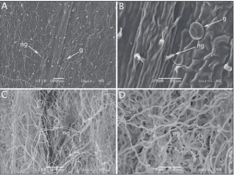
Scanning electron micrographs of the adaxial and abaxial leaf blade of A. elegans are presented in Fig. 1. Epidermal cells show sinuous anticlinal walls (Fig. 1B). Densely distributed, uniseriate, nonglandular
curly trichomes were noticed on both sides on the leaf blade, but there were much more on the abaxial side (Fig. 1C and D); thus, the leaves are velvety un-derneath. Rare glandular trichomes were observed on the adaxial side (Fig. 1A and B). The glandular trichomes are of the capitate type, seen as glandular dots (Fig. 1A and B). On the abaxial side, glandular trichomes were not recorded as the abaxial side was densely covered with nonglandular curly trichomes.
Total phenol and flavonoid content
The total phenol contents varied significantly between the extracts, with the methanol extract containing the largest amount of phenolics (Table 1). No phenols or flavonoids were detected in the n-hexane fraction.
Antioxidative activity
Since there were no flavonoids or phenols present in the n-hexane fraction (Table 1), ethyl acetate and methanol fractions were used in the antioxidative as-says. In all three assays, the methanol extract showed the highest antioxidative potential. As there is little to no difference between the total flavonoid contents in both extracts, most of the antioxidative potential can be attributed to phenols. However, the antioxidative potential of the methanol fraction, as determined by the DPPH assay, was more than ten times higher than that of the ethyl acetate fraction.
Identification and characterization of 3-deoxyamphoricarpolide
In the HPLC chromatogram of the A. elegans ex-tract, one peak is dominant (Fig. 2). After comparison with chromatograms and retention characteristics of previously isolated and characterized amphoricar-polides from Balkan species of Amphoricarpos, it was concluded that this dominant peak is 3-deoxyam-phoricarpolide [26].
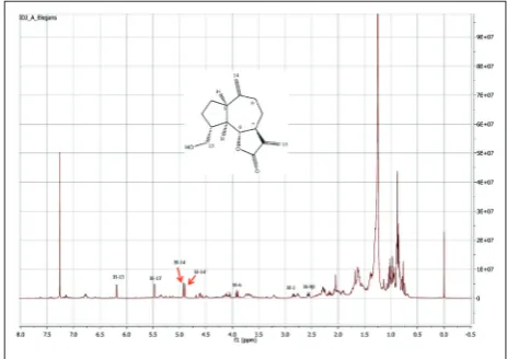
The 1H NMR spectrum of the A. elegans extract (Fig. 3) confirmed the results of HPLC; namely, the signals from H-14, H-14’, H-13, H-13’, H-6, H-1 and H-9β are in agreement with the previously isolated and characterized compound from Balkan
Amphori-carpos taxa [26].
DISCUSSION
Cavities, ducts, idioblasts and glandular trichomes are found inside and on the leaves of many Com-positae members [39-41]. In the whole ComCom-positae family, essential oils, resins, alkaloids, lipids, tannins, pectin-like substances, flavonoids and sesquiterpene lactones are the main products located mostly in ducts and glands [42]. Based on the extraction of the main classes of metabolites of Compositae, glandular tri-chomes are considered the main sites of biosynthesis and accumulation of sesquiterpene lactones, assuming important roles in the biological activity, ecology and chemotaxonomy of the family [12,16,43-48].
In Compositae, especially in the tribe Cardueae, specialized metabolites predominantly include li-pophilic constituents (particularly sesquiterpene lactones), while hydrophilic compounds are scarcely represented [18]. Guaianolide-type sesquiterpene lac-tones representing one of the largest groups among the sesquiterpene lactones from Compositae, generally
exhibit a low complexity [26]. The common feature of all guaianolides isolated from Amphoricarpos,and named amphoricarpolides, is that they have a unique oxygenation pattern that is not found in any Cardueae nor in other tribes of Compositae. All previous phyto-chemical studies [26-28] of Amphoricarpos taxa from the western Balkans revealed thirty amphoricarpolides in total (with the same guaianolide skeleton). Our investigation of the rare and stenoendemic species
A. elegans from western Caucasus shows that
3-de-oxyamphoricarpolide is dominant. This finding, as regards the chemotaxonomic significance of amphori-carpolides, represents a link between Balkan and Cau-casian species of the same genus. Since A. elegans is a stenoendemic species, we used a very small amount of plant material (only 1 g) for lactone isolation, in comparison to previous investigations, where much more of the plant material was used [26,27]. This ob-stacle resulted in the identification and characteriza-tion of only the dominant sesquiterpene lactone with a guaianolide skeleton – 3-deoxyamphoricarpolide.
Table 1. Total phenolic and flavonoid contents and antioxidant activity of n-hexane, ethyl acetate and methanol leaf extracts of Am-phoricarpos elegans.
Extracts Yield Total phenols Total flavonoids DPPH ABTS FRAP
[%] GAE μg/mg QUE μg/mg IC50 mg/mL AAEC mg/mL Fe2+ mmol/mg
n-hexane 6.65 - - - -
-Ethyl acetate* 2.32 19.9a±12.8 86.6a±8.9 3.50 0.03a±0.01 0.13a±0.01
Methanol* 9.67 96.3b±5.5 96.7a±7.7 0.24 0.07b±0.01 0.32b±0.01
*The samples were analyzed in triplicate (n=3) and expressed as the mean±standard deviation, except for the DPPH assay (IC50). Means followed by different superscripts (a-b) within the same column indicate statistically significant differences based on ANOVA (p<0.05)
Fig. 2. HPLC chromatogram of the methanol extract of
Amphori-carpos elegans. Fig. 3.
Literature on the biological activities of Balkan
Amphoricarpos species is scarce; the cytotoxic activity
of A. neumayerianus [14] and the antifungal activity
of the leaf-surface constituents of A. autariatus ssp.
autariatus [29] have been described. Also, our
pre-vious research on the antimicrobial and antioxida-tive activities of various leaf extracts of three Balkan
Amphoricarpos taxa showed a very high antimicrobial
and a considerable antioxidative potential [11]. The highest total flavonoid and phenolic contents, as well as the best antioxidative activity (DPPH assay) were observed in the methanol extract of A. autariatus ssp.
autariatus. This finding is in accordance with current
results, as we also show that the methanol extract of
A. elegans possessed the highest total flavonoid and
phenolic contents and exhibited the best antioxida-tive activity. The antioxidaantioxida-tive potential of the metha-nol fraction, as observed in the DPPH test, was over ten times higher than of the ethyl acetate fraction. This cannot be contributed only to phenols since the amount of total phenolic compounds was only four times higher in the methanol fraction, and this could be linked to the proven presence of lactones.
Guaianolide-type sesquiterpene lactones pro-duce effects related to the prevention and regulation of oxidative cellular damage and inflammation [16]. While the molecular mechanism of the antioxidative protection of sesquiterpene lactones is unknown, their antioxidant effects appear to be associated with the biosynthesis of endogenous antioxidants (e.g. pros-taglandins) [17]. Previous studies have shown that the cyclic esters of hydroxycarboxylic acids containing the sesquiterpene lactones are responsible for their bio-logical activities [49]. It has also been shown that the biological activity displayed by the majority of sesquit-erpene lactones is due to the presence of α-methylene-γ-lactones and an α,β-unsaturated cyclopentenone ring [50]. For these reasons, our results regarding the antioxidative potential of A. elegans extracts and the finding of 3-deoxyamphoricarpolide are of importance for future research into biological activity, therapeutic action and new plant sources of medicinal substances.
CONCLUSION
This study represents a contribution to the leaf micromorphology, antioxidative activity and
phyto-chemistry of an unexplored relict and limestone en-demic species, Amphoricarpos elegans (Compositae) from Georgia. We showed the presence of glandular trichomes, the main sites of biosynthesis and accu-mulation of sesquiterpene lactones. Also, our phy-tochemical investigation provides a new record of 3-deoxyamphoricarpolide, which is a component of the related Amphoricarpos taxa from the Balkan Pe-ninsula. The considerable antioxidative potential of this species indicates the need to continue the study of other species of the genus Amphoricarpos and also other species from related Cardueae genera (Com-positae) and their potential application in pharmacy and medicine.
Funding: Financial support was provided by the Ministry of Edu-cation, Science and Technological Development of the Republic of Serbia (Project Nos. 173029 and 172053).
Author contributions: MG, NR, PJ drafted the manuscript; AB collected the plant material and drafted the manuscript; VT, IĐ, NGJ, AS, PM and PJ supervised the research and critically re-viewed the manuscript. All authors read and approved the final manuscript.
Conflict of interest disclosure: The authors declare no conflict of interest.
REFERENCES
1. Anderberg AA, Baldwin BG, Bayer RG, Breitwieser J, Jeffrey C, Dillon MO, Eldenäs P, Funk V, Garcia-Jacas N, Hind DJN, Karis PO, Lack HW, Nesom G, Nordenstam B, Oberprieler Ch, Panero JL, Puttock C, Robinson H, Stuessy TF, Susanna A, Urtubey E, Vogt R, Ward J., Watson LE. Compositae. In: Kubitzki K, editor. The families and genera of vascular plants. Vol. 8. Springer-Verlag Berlin Heidelberg; 2007. p. 61-588.
2. Robinson H. An introduction to micro-characters of Com-positae. In: Funk VA, Susanna A, Stuessy TF, Bayer RJ, edi-tors. Systematics, Evolution and Biogeography of Composi-tae. Vienna (Austria): IAPT; 2009. p. 89-100.
3. Taleb-Contini HS, Schorr K, Da Costa BF, Camilo D, de Oliveira R. Detection of flavonoids in glandular trichomes of Chromolaena species (Eupatorieae, Asteraceae) by reversed-phase high-performance liquid chromatography. Braz J Pharm Sci. 2007;43:2.
4. Fritz E. and Saukel J. Anatomy of subterranean organs of medicinally used Cardueae and related species and its value for discrimination. Sci Pharm. 2011;79:157-74.
5. Farrag NM, Abd El Aziz EM, El-Domiaty MM, El Shafea
AM. Phytochemical investigation of Centaurea araneosa
6. Font Quer P. Plantas Medicinales: El Dioscórides Renovado. 15th ed. Madrid: Labor; 1995. 1033 p.
7. Barrero AF, Herrador MM, Arteaga P, Cabrera E, Rodrí-guez-García I, García-Moreno M, Grávalos DG. Cytotoxic activity of flavonoids from Carthamus arborescens, Ononis natrix ssp. ramosissima and Centaurea malacitana. Fitote-rapia. 1997;68:281-3.
8. Baba Aissa F. Flore d’Algérie et du Maghreb. Substances végétales d’Afrique, d’orient et d’occident. Encyclopédie des plantes utiles. Rouiba (Algeria): EDAS, Librairie moderne; 1999.
9. Vallejo JR, Pardo de Santayana M, Peral D, Carrasco MC, López D. Uso medicinal de Atractylis gummifera L. en Gua-diana del Caudillo (Badajoz, Espana). Toxicidad y especies afines. Rev Fitoterapia. 2008;8:161-9.
10. Vallejo JR, Peral D, Gemio P, Carrasco MC, Heinrich M, Pardo de Santayana M. Atractylis gummifera and Centaurea
ornata in the Province of Badajoz (Extremadura, Spain).
Ethnopharmacological importance and toxicological risk. J Ethnopharmacol. 2009;126:366-70.
11. Gavrilović M, Soković MD, Stanković M, Marin PD, Stevanović ZD, Janaćković P. Antimicrobial and antioxida-tive activity of various leaf extracts of Amphoricarpos Vis. (Asteraceae) taxa. Arch Biol Sci. 2016;68:803-10.
12. Sokovic M, Ciric A, Glamoclija J, Skaltsa H. Biological activ-ities of sesquiterpene lactones isolated from the genus
Cen-taurea L. (Asteraceae). Curr Pharm Design.
2017;23:2767-86.
13. Milosavljević S, Bulatović V, Stefanović M. Sesquiterpene lactones from the Yugoslavian wild growing plant families Asteraceae and Apiaceae. J Serb Chem Soc. 1999;64:397-442. 14. Atrrog BAA, Natic M, Tosti T, Opsenica Milojković D,
Djordjević I, Tešević V, Jadranin M, Milosavljević S, Lazić M, Radulović S, Tešić Z. Lipophilicity of some guaianolides isolated from two endemic subspecies of Amphoricarpos
neumayeri (Asteraceae) from Montenegro. Biomed
Chro-matogr. 2008;23:250-6.
15. Repetto MG, Llesuy S. Antioxidant properties of natural compounds used in popular medicine for gastric ulcers. Braz J Med Biol Res. 2002;35:523-34.
16. Da Costa F, Terfloth L, Gasteiger J. Sesquiterpene lactone-based classification of three Asteraceae tribes: a study lactone-based on self-organizing neural networks applied to chemosys-tematics. Phytochemistry. 2005;66:345-53.
17. Repetto MG, Boveris A. Bioactivity of sesquiterpenes: com-pounds that protect from alcohol-induced gastric muco-sal lesions and oxidative damage. Mini Rev Med Chem. 2010;10:615-23.
18. Susanna A, Garcia-Jacas N. Cardueae (Carduoideae). In: Funk VA, Susanna A, Stuessy TF, Bayer RJ, editors. System-atics, Evolution and Biogeography of Compositae. Vienna (Austria): IAPT; 2009. p. 293-313.
19. Susanna A, Garcia-Jacas N. Tribe Cardueae Cass. (1819). In: Kubitzki K, editor. The Families and Genera of Vascu-lar Plants. VIII Flowering Plants-Eudicots. Springer-Verlag Berlin Heidelberg; 2007. p. 123-46.
20. Barres L, Sanmartín I, Anderson CL, Susanna A, Buerki S, Galbany‐Casals M, Vilatersana R. Reconstructing the
evolu-tion and biogeographic history of tribe Cardueae (Composi-tae). Am J Bot. 2013;100:867-82.
21. Shetekauri S, Kutateladze L. Diversity, ecotopology and hyp-sometric distribution of the endemic flora in high-mountain phytolandscapes of the Caucasus. Earth. 2017;6:38-48. 22. Akhalkatsi M, Ekhvaia J, Asanidze Z. Diversity and genetic
erosion of ancient crops and wild relatives of agricultural cultivars for food: implications for nature conservation in Georgia (Caucasus). In: Tiefenbacher J, editor. Perspectives on Nature Conservation-Patterns, Pressures and Prospects. Rijeka, Croatia: InTech; 2012. p. 51-92.
23. Nakhutsrishvili G. The vegetation of Georgia (Caucasus). Camerino: Dipartimento di Botanica ed Ecologia dell’ Univ; 1999. p. 74. (Braun-Blanquetia; Vol. 15).
24. Schatz G, Shulkina T, Nakhutsrishvili G, Batsatsashvili K, Tamanyan K, Ali-zade V, Kikodze D, Geltman D, Ekim T. Development of Plant Red List Assessments for the Caucasus Biodiversity Hotspot. In: Zazanashvili N, Mallon D, editors. Status and Protection of Globally Threatened Species in the Caucasus. Tbilisi: CEPF, WWF. Contour Ltd; 2009. p. 188-92.
25. Linczevsky IA. Compositae Tribes Echinopsideae and Cynareae. In: Schischkin BK, Bobrov FG, editors. Flora of the USSR. Vol. 27. Moscow, Leningrad: Akademiya Nauk SSSR; 1962. p. 74-9.
26. Djordjević I, Vajs V, Bulatović V, Menković N, Tešević V, Macura S, Janaćković P, Milosavljević S. Guaianolides from two subspecies of Amphoricarpos neumayeri from Montene-gro. Phytochemistry. 2004;65:2337-45.
27. Djordjević I, Jadranin M, Vajs V, Menković N, Tešević V, Macura S, Milosavljević S. Further Guaianolides from Amphoricarpos neumayeris ssp. murbeckii from Montenegro. Z Naturforsch. 2006;61b:1437-42.
28. Cvetković M, Ðorđević I, Jadranin M, Vajs V, Vučković I, Menković N, Milosavljevic S, Tešević V. Further Amphori-carpolides from the surface extracts of Amphoricarpos Com-plex from Montenegro. Chem Biodivers. 2014;11:1428-37. 29. Jadranin M, Djordjević I, Tešević V, Vajs V, Menković N,
Soković M, Glamočlija J, Milosavljević S. Sesquiterpene Lactones of Amphoricarpos autariatus ssp. autariatus from Montenegro - Antifungal Leaf - Surface Constituents. Rec Nat Prod. 2013;7:234-8.
30. Petit DP. Generic interrelationships of the Cardueae (Com-positae): A cladistic analysis of morphological data. Plant Syst Evol. 1997;207:173-203.
31. Caković D, Stešević D, Schönswetter P, Frajman B. How many taxa? Spatiotemporal evolution and taxonomy of
Amphoricarpos (Asteraceae, Carduoideae) on the Balkan
Peninsula. Org Divers Evol. 2015;15:429-45.
32. Singleton VL, Orthofer R, Lamuela-Raventós RM. Analysis of total phenols and other oxidation substrates and anti-oxidants by means of Folin-Ciocalteu reagent anti-oxidants and antioxidants. Method Enzymol. 1999;299:152-78.
34. Tekao T, Watanabe N, Yagi I, Sakata K. A simple screening method for antioxidant and isolation of several antioxidants produced by marine Bacteria from fish and shellfish. Biosci Biotechnol Biochem. 1994;58:1780-3.
35. Kumarasamy Y, Byres M, Cox PJ, Jasapars M, Nahar L, Sarker SD. Screening seeds of some scottish plants for free radical scavenging activity. Phytother Res. 2007;21:615-21. 36. Re R, Pellegrini N, Proteggente A, Pannala A, Yang M,
Rice-Evans C. Antioxidant activity applying an improved ABTS radical cation decolorization assay. Free Radical Bio Med. 1999;26:1231-7.
37. Li HB, Wong CC, Cheng KW, Chen F. Antioxidant properties in vitro and total phenolic contents in methanol extracts from medicinal plants. Lebensm-Wiss Technol. 2008;41:385-90. 38. Hammer Ø, Harper DAT, Ryan PD. PAST: paleontological
statistics software package for education and data analysis. Palaeontol Electronica. 2001;4:1-9.
39. Milan P, Hayashi AH, Appezzato-da-Glória B. Comparative leaf morphology and anatomy of three Asteraceae species. Braz Arch Biol Techn. 2006;49:135-44.
40. Duarte MR, Budel JM, Matzenbacher NI. Menarim DO. Microscopic diagnosis of the leaf and stem of Lucilia nitens Less., Asteraceae. Lat Am J Pharm. 2011;30:2070-5. 41. Camilotti JG, Biu CC, Farago PV, Santos VLPD, Franco
CRC, Budel JM. Anatomical characters of leave and stem of Calea serrata Less., Asteraceae. Braz Arch Biol Techn. 2014;57:867-73.
42. Bartoli A, Galati BG, Tortosa RD. Anatomical studies of the secretory structures: glandular trichomes and ducts, in Grindelia pulchella Dunal (Astereae, Asteraceae). Flora. 2011;206:1063-8.
43. Spring O, Benz T, Ilg M. Sesquiterpene lactones of the capi-tate glandular trichomes of Helianthus annuus. Phytochem-istry. 1989;28:745-9.
44. Spring O. Trichome microsampling of sesquiterpene lac-tones for the use of systematic studies. Recent Adv Phyto-chem. 1991;25:319-45.
45. Spring O, Zipper R, Reeb S, Vogler B, Da Costa FB. Ses-quiterpene lactones and a myoinositol from glandular tri-chomes of Viguiera quinqueremis (Heliantheae; Asteraceae). Phytochemistry. 2001;57:267-72.
46. Sakamoto HT, Gobbo-Neto L, Cavalheiro AJ, Lopes NP, Lopes JLC. Quantitative HPLC analysis of sesquiterpene lactones and determination of chemotypes in Eremanthus seidelii MacLeish & Schumacher (Asteraceae). J Brazil Chem Soc. 2005;16:1396-401
47. Appezzato-da-Glória B, Da Costa FB, Silva VC, Gobbo-Neto L, Rehder VLG, Hayashi AH. Glandular trichomes on aerial and underground organs in Chrysolaena species (Vernonieae - Asteraceae): structure, ultrastructure and chemical com-position. Flora. 2012;207:878-87
48. Lusa MG, Da Costa FB, Appezzato-da-Glória B. Histolocal-ization of chemotaxonomic markers in Brazilian Vernonieae (Asteraceae). Bot J Linn Soc. 2016;182:581-93.
49. Raola VK and Chakraborty K. Biogenic guaianolide-type sesquiterpene lactones with antioxidative and
anti-inflam-matory properties from natural mangrove hybrid
Rhi-zophora annamalayana. Nat Prod Res. 2017;31:2719-29.