Research Article
a
May
2019
Computer Science and Software Engineering
ISSN: 2277-128X (Volume-9, Issue-5)
MRI Medical Image Enhancement using Modulated Intensity
Gradient and Texture Gradient Based Segmentation
Shivani Chourasia1,Anand Vardhan Bhalla2
1
M.Tech Scholar, Ojaswini Institute of Management and Technology, Damoh Madhya Pradesh, India
2
Assistant Professor, ECE Dept., Ojaswini Institute of Management and Technology, Damoh Madhya Pradesh, India
1
[email protected], [email protected]
Abstract— Medical image processing has experienced dramatic expansion and has been an interdisciplinary research field attracting expertise from applied mathematics, computer sciences, engineering, statistics, physics, biology and medicine. Computer aided diagnostic processing has already become an important part of clinical routine. Accompanied by a rush of new development of high technology and use of various imaging modalities, more challenges arise; for example, how to process and analyze a significant volume of images so that high quality information can be produced for disease diagnoses and treatment. The principal objectives of this work is to develop a MRI medical image enhancement system to analyze and extract more information from the image so as to better diagnose the disease and also to promote interests for further study and research in medical imaging processing. In image processing different processes are applied on images to enhance it at different parameters. These processes are not directly applied on images but before that it needs to be segmented or divided in smaller parts called pixels or small blocks of pixels. So segmentation becomes an integral and basic part in image analysis and error at this stage can influence other processing techniques. The performance of proposed system is compared with the available techniques. The proposed system enhances the Mean Square Error (MSE), Maximum Error (MAXERR) and Peak Signal to Noise Ratio (PSNR) parameters.
Keywords— Image, Dual Tree Discrete Wavelet Transform (DT-DWT), Wavelet Transform (WT), Mean Square Error (MSE), Maximum Error (MAXERR), Peak Signal to Noise Ratio (PSNR).
I. INTRODUCTION
Medical image processing deals with the development of problem-specific approaches to the enhancement of raw medical image data for the purposes of selective visualization as well as further analysis. There are many topics in medical image processing: some emphasize general applicable theory and some focus on specific applications. We mostly focus on image segmentation and multi-spectral analysis. Dealing with information extracted from a natural image, a medical scan, satellite data or a frame in a video sequence is the purpose of image analysis. In the real world, the stimulus that is received by the retina is perceived as whole and complete information. Between the electromagnetic reception and the perception, physiological and neurological processes construct the final perception and analysis of the image. In fact vision is composed of many interacting components including analysis of colour, texture and shape, the whole conducted by prior knowledge of the human brain. Computer vision aims at getting the same result as human perception. The computer interface receives the image as a matrix of pixels and several levels of processes are involved to get, when it is possible, the same result as human analysis.
The collection of processes involved in the visual perception are usually hierarchically classified as belonging to either low level vision or high level vision. High level vision consists of the interpretation of the image following some rule or prior knowledge. In low level vision, image processing is performed to extract some visible physical properties in the image such as shape and boundaries or to improve the quality of the image. In this thesis we will be dealing with image processing and more precisely with the image segmentation task. The objective of segmentation methods is to determine a partition of an image into a finite number of semantically important regions such as anatomical or functional structures in medical images or objects in natural images. The segmentation task has been studied for several decades; however it is still a challenging task. This task is essential in many applications including face detection in video sequences, changes detection in satellite images, anatomical or functional object extraction in medical images or object extraction in natural images.
ISSN(E): 2277-128X, ISSN(P): 2277-6451, pp. 26-32 from analog to digital form. A digital image is basically a 2- Dimensional array of pixels. The image is defined in a continuous space and the segmentation problem is expressed through a functional or energy optimization. Depending on the object to be segmented, this energy definition can be difficult; in particular for objects with ambiguous borders or objects with textures. For the latter, the difficulty lies already in the definition of the term texture. The human eye can easily recognize a texture, but it is quite difficult to find words to define it, even more in mathematical terms. This is why we are first interested in the extraction of texture features that is to say, finding one representation that can discriminate a textured region from another. The usefulness of the segmentation is ultimately dependent on the features used for the annotation of data and its efficiency is dependent on the invariance and robust properties of these features. For texture based features an important form of invariance is rotational invariance. This work describes effective and novel texture characterization and rotationally invariant texture characterization techniques. However, watershed segmentation is often not effective for textured image regions that are perceptually homogeneous. In order to properly segment such regions the concept of the “texture gradient” is implemented.
Challenges in Medical Image Processing
There are a number of specific challenges in medical image processing. They are:
1) Image enhancement and restoration.
2) Automated and accurate segmentation of features of interest.
3) Automated and accurate registration and fusion of multimodality images.
4) Classification of image features, namely characterization and typing of structures.
5) Quantitative measurement of image features and an interpretation of the measurements.
6) Development of integrated systems for the clinical sector.
II. SEGMENTATION
Image segmentation refers to the process of partitioning a digital image into N number of parts. The images are segmented on the basis of set of pixels or pixels in a region that are similar on the basis of some homogeneity criteria such as color, intensity or texture, which helps to locate and identify objects or boundaries in an image [3].
In terms of mathematical formulae, Image segmentation divides a digital image f(x, y) into continuous, disconnect and nonempty subsets, from these subsets higher level information can be easily extracted. Practical applications of image segmentation include object identification and recognition, facial recognition, medical image processing, criminal investigation, airport security system, satellite images, quality assurance in factories, etc [3][7]. Due to the importance of the image segmentation, large number of algorithms has been proposed but the selection of the algorithm purely depends upon the image type and the nature of the problem [2].
In recent years, a lot of research is done in the field of image segmentation process. There is currently thousands of algorithms, each doing the segmentation process slightly different from another, but still there is no particular algorithm that is applicable for all types of digital image, fulfilling every objective. Thus, algorithm developed for a group of images may not always apply to images of another class [4] [15].
Currently image segmentation approach, based on two properties of an image, is divided into two categories:
Discontinuities based
In this category, subdivisions of images are carried out on the basis of abrupt changes in the intensity of grey levels of an image. Our focus is primarily based on identification of isolated points, lines and edges. This includes image segmentation algorithms like edge detection.
Similarities based
In this category, subdivision of images is carried out on the basis of similarities in intensity or grey levels of an image. Our focus here is on identification of similar points, lines and edges. This includes image segmentation algorithms like thresholding, region growing, region splitting and merging.
III. STEPS INVOLVED IN THE PROPOSED ALGORITHM
Steps involved in the proposed system are:
1. The input coloured MRI image is read.
2. The input image is resized and converted to grey image. The grey image pixel is doubled.
ISSN(E): 2277-128X, ISSN(P): 2277-6451, pp. 26-32
4. The output image using modulated intensity gradient is generated.
5. PSNR, MSE and MAXERR are calculated for the output image.
6. Secondly, the MRI grey image is analyzed using texture gradient based segmentation.
7. The output image using Texture gradient is generated.
8. PSNR, MSE and MAXERR are calculated for Texture Gradient output MRI image is calculated.
9. The output MRI medical images of Texture Gradient and Modulation Intensity Gradient are summed to get the
resultant MRI image output.
10. PSNR, MSE and MAXERR are calculated for resultant MRI medical image.
11. The PSNR, MSE and MAXERR for resultant output MRI image are compared separately with the PSNR, MSE
and MAXERR of MRI medical image outputs of Texture Gradient and Modulation Intensity Gradient.
Figure 1: Simulation flow diagram.
IV. SIMULATION RESULTS
The proposed system simulation outputs are as under.
ISSN(E): 2277-128X, ISSN(P): 2277-6451, pp. 26-32
Figure 3: Resized MRI grey image.
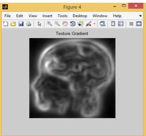
Figure 4: MRI Image output using modulated intensity gradient based segmentation.
ISSN(E): 2277-128X, ISSN(P): 2277-6451, pp. 26-32
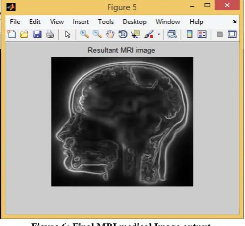
Figure 6: Final MRI medical Image output.
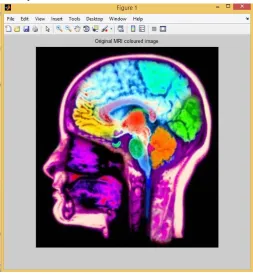
The figure 2 and 3 shows the original MRI medical image which is to be segmented and resized grey image respectively. Figure 4 and 5 shows the MRI image outputs by using modulated intensity gradient and texture gradient based segmentation respectively. Figure 6 shows resultant MRI image output of proposed system. From image outputs it can be seen that the final output MRI image is better than the other two MRI image outputs.
Table 1: Various parameters values obtained.
Table 1 shows values of PSNR, MSE and MAXERR obtained by using modulated intensity gradient based segmentation, texture gradient based segmentation and of proposed system. From the table it can be seen that the results with proposed system are better than that with other two traditional systems.
V. CONCLUSION
The objective of this work is to develop an efficient MRI medical image enhancement system so that better image can be obtained on reconstruction. In the proposed system two algorithms modulated intensity gradient and texture gradient based on image segmentation are used.
Results are derived for modulated intensity gradient based system, texture gradient based system and the proposed system. The parameters of comparison are PSNR, MSE and MAXERR. The results show that the PSNR, MSE and MAXERR values for proposed system are better than the existing system and hence it can be concluded that the proposed system performance has been enhanced. Moreover, the method is simple and computationally effective that makes it easy to implement.
REFERENCES
[1] Sajan Kor, Pramod Kumar Sethy “Interactive Image Segmentation using Color and TextureFeatures”,
ISSN(E): 2277-128X, ISSN(P): 2277-6451, pp. 26-32
[2] Priyanka Shivhare, Vinay Gupta,”Review of Image Segmentation Techniques Including Pre & Post Processing
Operations”, International Journal of Engineering and Advanced Technology (IJEAT) ISSN: 2249 – 8958, Volume-4 Issue-3, February 2015.
[3] Prabhishek Singh, Ramneet Singh Chadha, “A Novel Approach to Image Segmentation”, International Journal
of Advanced Research in Computer Science and Software Engineering Volume 3, Issue 4, April 2013.
[4] M. Joseph Prakash, Saka Kezia, I. Santhi Prabha and V. Vijaya Kumar, “A New Approach for Texture
Segmentation Using Gray Level Textons”, International Journal of Signal Processing, Image Processing and Pattern Recognition Vol. 6, No. 3, June, 2013.
[5] P.P.Acharjya1, A. Sinha2, S.Sarkar3, S.Dey4, S.Ghosh, “A new approach of watershed algorithm using
distance transform applied to image segmentation” International Journal of Innovative Research in Computer and Communication Engineering Vol. 1, Issue 2, April 2013.
[6] M. Erdt, S. Steger, G. Sakas, “A New View of Image Segmentation and Registration”, Germany Copyright ©
2012 Journal of Radiation Oncology Informatics ISSN: 1663-618X, J Radiat Oncol Inform 2012; 4:1:1-23.
[7] Anju Bala, “An Improved Watershed Image Segmentation Technique using MATLAB”, International Journal of
Scientific & Engineering Research Volume 3, Issue 6, June-2012 1 ISSN 2229-5518.
[8] D.Sasirekha, Dr.E.Chandra, “Enhanced Techniques for PDF Image Segmentation and Text Extraction”,
(IJCSIS) International Journal of Computer Science and Information Security Vol. 10, No. 9, September 2012.
[9] Ashraf A. Aly1, Safaai Bin Deris2, Nazar Zaki, “Research review for digital image segmentation techniques”,
International Journal of Computer Science & Information Technology (IJCSIT) Vol 3, No 5, Oct 2011.
[10] D. Maheswari 1, Dr. V.Radha2,”Enhanced Hybrid Compound Image Compression Algorithm Combining Block
and Layer-based Segmentation”, The International Journal of Multimedia & Its Applications (IJMA) Vol.3, No.4, November 2011.
[11] V. Dey a, Y. Zhang a, M. Zhong “A Review On Image Segmentation Techniques With Remote Sensing
Perspective”,ISPRS TC VII Symposium – 100 Years ISPRS, Vienna, Austria, July 5–7, 2010, IAPRS, Vol. XXXVIII, Part 7A.
[12] Yi Ma, Senior, Harm Derksen, “Segmentation of Multivariate Mixed Data via Lossy Data Coding and Compression”, IEEE transactions on pattern analysis and machine intelligence, vol. 29, no. 9, september 2007.
[13] Jitendra Malik, Serge Belongie, Thomas Leung∗And Jianbo Shi, “Contour and Texture Analysis for Image Segmentation”, Computer Science Division, University of California at Berkeley, Berkeley, CA 94720-1776,
USA Received December 28, 1999; Revised February 23, 2001; Accepted February 23, 2001.
[14] M. Atonini, M. Barlaud, P. Mathieu, and I. Daubechies, “Image coding using wavelet transform,” IEEE
Trans.Image Processing, vol. 1, pp. 205–220, Apr. 1992.
[15] Jha, Sonu Kumar, Purnendu Bannerjee, and Subhadeep Banik. "Random Walks based Image Segmentation Using Color Space Graphs." Procedia Technology 10 (2013).
[16] Bhargava, Neeraj, et al. "Iterative Region Merging and Object Retrieval Method Using Mean Shift Segmentation
and Flood Fill Algorithm.", Advances in Computing and Communications (ICACC), 2013 Third International Conference on. IEEE, 2013.
[17] Y. Li, J. Sun, C.-K. Tang, and H.-Y. Shum, “Lazy snapping,” in ACM Siggraph, 2004, pp. 303–308.
[18] Ning, Jifeng, et al. "Interactive image segmentation by maximal similarity based region merging." Pattern Recognition 43.2 (2010): 445-456.
[19] Felzenszwalb, Pedro F., and Daniel P. Huttenlocher. "Efficient graph-based image segmentation." International Journal of Computer Vision 59.2 (2004): 167-181.
[20] Boykov, Yuri, and Vladimir Kolmogorov. "An experimental comparison of min-cut/max-flow algorithms for energy minimization in vision." Pattern Analysis and Machine Intelligence, IEEE Transactions on 26.9 (2004): 1124-1137.
[21] Çiğla, Cevahir. "Efficient graph-based image segmentation via speeded-up turbo pixels." Image Processing (ICIP), 2010 17th IEEE International Conference on. IEEE, 2010.
[22] B.Wang and L. Zhang, “Supervised texture segmentation using wavelet transform”, Proc.of the 2003
International Conference on Neural Networks and Signal Processing, 2003, vol.2,pp. 1078-1082.
[23] Chang- Tsun Li and Roland Wilson, “Unsupervised texture segmentation using multiresolution hybrid genetic
algorithm,” in Proc. IEEE International Conference on Image Processing ICIP03 ,2003, pp. 1033– 1036. [24] T.R. Reed and J. M. H. Du Buf, “A review of recent texture segmentation, feature extraction techniques”, in
CVGIP Image Understanding , 1993, pp. 359– 372.
[25] A.K. Jain and K. Karu, “Learning texture discrimination masks”, IEEE trans. of Pattern Analysis and Machine
ISSN(E): 2277-128X, ISSN(P): 2277-6451, pp. 26-32
[26] Alp Erturk and Sarp Erturk, “Unsupervised Segmentation of Hyperspectral Images Using Modified Phase
Correlation,” IEEE geoscience and remote sensing letters, vol. 3, no. 4, october 2006.
[27] Tamas Sziranyi and Maha Shadaydeh, “Segmentation of Remote Sensing Images Using
Similarity-Measure-Based Fusion-MRF Model”, IEEE geoscience and remote sensing letters, vol. 11, no. 9, september 2014.
[28] A.K. Qin, David A. Clausi, “Multivariate Image Segmentation Using Semantic Region Growing With Adaptive
Edge Penalty,” IEEE transactions on image processing, vol. 19, no. 8, august 2010.
[29] Saiqa Khan, Arun Kulkarni, “Robust Method for Detection of Copy-Move Forgery in Digital Images,” IEEE
geoscience and remote sensing letters, 2010 IEEE.
[30] Chaobing Huang, Quan Liu, Xiaopeng Li, “Color Image Segmentation by Seeded Region Growing and Region
Merging,”, 2010 Seventh International Conference on Fuzzy Systems and Knowledge Discovery,
978-1-4244-5934-6/10/2010 IEEE.
[31] Chen Zheng, Leiguang Wang, Rongyuan Chen, and Xiaohui Chen, “Image Segmentation Using
Multiregion-Resolution MRF Model”, IEEE geoscience and remote sensing letters, vol. 10, no. 4, july 2013.
[32] Zhi-hvi Li, Meng zhang, Haibo Liv, “A fast algorithm of image segmentation based on markov random field”,
978-1-4673-4685-6112/ 2012, IEEE.
[33] Yong Xia and Rongchun Zhao, “Adaptive Segmentation of Textured Images by Using the Coupled Markov
Random Field Model”, IEEE transactions on image processing, vol. 15, no. 11, November 2006. [34] H. Narkhede, "Review of image segmentation techniques," Int. J. Sci. Mod. Eng, vol. 1, p. 28, 2013.
[35] J. F. Khan, S. M. A. Bhuiyan, and R. R. Adhami, "Image Segmentation and Shape Analysis for Road-Sign Detection," IEEE Transactions on Intelligent Transportation Systems, vol. 12, pp. 83-96, 2011.