Performance of the Exercise Test
George D. Harris and Russell D. WhiteEach year, over 1 million Americans experience a nonfatal or fatal myocardial infarction or sudden death from coronary heart disease (CHD) [1]. Unfortunately, death or myocardial infarction is the first symptom in 55% of patients with coronary artery disease [2] and is usually due to dislodgement of a plaque causing acute coronary occlusion. However, about 30% of these patients present with ischemia and have concurrent chest pain. In these individuals, exercise treadmill testing is a practical and the most commonly performed test to identify or confirm the presence of latent coronary artery disease [2]. In addition, an abnormal test has been shown to have definite predictive value. It is well known that when symptoms of typical angina are present, coronary disease can be predicted with considerable reliability. Even when there is no history of pain, there is still a strong possibility of significant coronary disease in patients with specific risk factors. Also, the reliability of the test in asymptomatic patients is improved when testing patients with a higher prevalence of the disease.
Exercise testing may also be used to measure functional capacity, assess the pa-tient’s prognosis in coronary artery disease, and evaluate the papa-tient’s treatment for hypertension, certain arrhythmias, angina, and congestive heart failure. It can be beneficial for patients who will be involved in exercise rehabilitative programs.
It may be useful for predicting mortality risk among patients who plan to start an exercise program, whose job affects public safety (airline pilot), or who have specific medical conditions (diabetes or chronic renal insufficiency).
The inherent accuracy of the test is defined by the sensitivity and specificity. The results of the test when applied to an individual depend on the prevalence of disease in the population to which the patient belongs. So, the two most important factors in the analysis of patients undergoing stress testing are the pre-test prevalence of disease and the sensitivity and specificity of the test. Various types of chest pain affect the probability of disease in each patient. By dividing the patients into one
G.D. Harris (B)
Department of Community and Family Medicine, University of Missouri-Kansas City, School of Medicine, Truman Medical Center—Lakewood, 7900 Lees Summit Rd., Kansas City, MO 64139, USA
e-mail: [email protected]
C.H. Evans, R.D. White (eds.), Exercise Testing for Primary Care and Sports Medicine
Physicians, DOI 10.1007/978-0-387-76597-6 2
C
Springer Science+Business Media, LLC 2009
Table 2.1 Pre-test probability of coronary artery disease by age, gender, and symptoms Age Gender Typical/definite
angina pectoris Atypical/probable angina pectoris Nonanginal chest pain Asymptomatic
30–39 Men Intermediate Intermediate Low Very low
Women Intermediate Very low Very low Very low
40–49 Men High Intermediate Intermediate Very low
Women Intermediate Low Low Very low
50–59 Men High Intermediate Intermediate Low
Women Intermediate Intermediate Low Very low
60–69 Men High Intermediate Intermediate Low
Women High Intermediate Intermediate Low
From Institute for Clinical Systems Improvement. Health Care Guideline: Cardiac Stress Test Sup-plement, 6th ed. 2004.
of four groups (typical angina, atypical angina, nonanginal chest pain, or no chest pain), one can predict the probability of heart disease in individuals (Table 2.1; see Chapter 5 on interpreting the exercise stress test).
The pre-test evaluation becomes very important when deciding when and how to study an asymptomatic patient. An estimated 1–2 million middle-aged men have asymptomatic but physiologically significant coronary artery obstruction, placing them at increased risk for coronary heart disease events [3,4] Unfortunately, multiple cohort studies have demonstrated that screening exercise tolerance testing identifies only a small proportion of asymptomatic persons (up to 2.7% of those screened) with severe coronary artery obstruction who may benefit from revascularization [1]. In addition, several large prospective cohort studies suggest that exercise tolerance testing can provide independent prognostic information about the risk for future
Table 2.2 Coronary artery risk factors for risk stratification Hypertension Systolic>140 mmHg Diastolic>90 mmHg Currently on antihypertensives Hyperlipidemia Serum cholesterol>200 mg% LDL cholesterol>130 mg% HDL cholesterol<35 mg% Impaired glucose intolerance Fasting blood glucose>100 mg% Cigarette smoking
Current smoker
Quit smoking 6 months or less Sedentary lifestyle
Not exercising
Exercising<minimal activity level Family history
Myocardial infarction Sudden death
coronary heart disease events (relative risk with abnormal exercise tolerance testing, 2.0–5.0). Thus, evaluation of the exercise test as a prognostic rather than a diagnostic test suggests that the prognostic value of the screening exercise test may have been underestimated.
However, when the risk for CHD is low, there is an increase in false-positive findings resulting in unnecessary further testing or patient anxiety [1]. Therefore, when deciding how to study an asymptomatic patient for coronary disease each patient must be properly evaluated by a history and physical examination as well as stratified by age, gender, symptoms (typical angina, atypical angina, nonanginal chest pain, no symptoms), and major risk factors (diabetes mellitus, hypertension, dyslipidemias, smoking) (Table 2.2).
Indications
The three major cardiopulmonary indications for performing exercise testing are (1) to diagnose the presence or absence of heart disease; (2) to determine the prognosis (risk) in assessing the severity of previously diagnosed disease; and (3) to develop a therapeutic prescription or test the adequacy of the patient’s therapy.
Since cardiovascular disease is the leading cause of death in patients with di-abetes and didi-abetes is a coronary artery disease equivalent (the risk of myocardial infarction in patients with diabetes is similar to that of patients without diabetes who have had a previous myocardial infarction), the American College of Cardiology, the American Heart Association, and the American Diabetes Association have recom-mended graded exercise testing in asymptomatic patients with diabetes who plan to begin a moderate- to high-intensity exercise program and are at increased risk for coronary heart disease (CHD) based on one or more risk factors [2, 5]. The risk factors include age>35 years; age>25 years and type 2 diabetes of>10 years du-ration or type 1 diabetes of>15 years duration; any additional risk factors for CHD; and the existence of microvascular disease (retinopathy, neuropathy, nephropathy) or peripheral arterial occlusive disease (PAOD) [6].
However, this level of evidence is based on opinion and not on well-established medical evidence [2, 5]. Furthermore, there are limitations for recommending a screening exercise treadmill testing (ETT) in asymptomatic patients with diabetes. ETT in this population can only identify a small proportion of asymptomatic persons with severe coronary artery obstruction who may benefit from revascularization [7]. The American Heart Association/American College of Cardiology and US Pre-ventive Services Task Force guidelines also acknowledge the possible value of exer-cise testing in men 45 years old and women 55 years old who plan to start vigorous exercise programs or are involved in high-risk occupations [1, 8].
The American College of Sports Medicine (ACSM) has developed recommen-dations for exercise testing prior to beginning an exercise program [9]. Patients are categorized according to low-, moderate-, and high-risk groups based on age, sex, presence of coronary artery disease risk factors, major symptoms of disease, or known heart disease [9]. A low-risk individual is an asymptomatic person (man
<45 years or woman<55 years) with no more than one risk factor. An individual at moderate risk is older (man>45 or woman>55) or has more than two risk factors. The individual at high risk has one or more symptoms/signs of CAD or has either known cardiovascular disease, pulmonary disease, or metabolic disease (diabetes mellitus, thyroid disorder, renal disease, liver disease) [9]. Their activity level is divided into moderate (3–6 METs or 40–60% VO2 max) and vigorous (>6 METs or>60% VO2 max) exercise. An individual who wants to perform moderate activity and is at high risk or wishes to perform vigorous activity levels and is at moderate or high risk needs an exercise test performed prior to starting that level of exercise [9]. Exercise testing in low-risk, asymptomatic males is usually not recommended un-less there are specific cardiac risk factors. There is no chronological age for routine screening of individuals, but the ACSM recommends that low-risk men and women (age<45 years and 55 years, respectively) be exempted from routine screening [9]. Exercise treadmill testing is indicated for evaluating a patient with chest pain, determining the prognosis and severity of disease, evaluating the effects of medical and surgical therapy, evaluating a patient after myocardial infarction for risk stratifi-cation and for the early detection of labile hypertension, and evaluating arrhythmias, congestive heart failure, and a patient’s functional capacity. It can also be used to formulate an exercise prescription or to evaluate an individual training program for an athlete.
Contraindications
Absolute Contraindications
Absolute contraindications include those situations in which the risks involved in performing the procedure outweigh any benefit [1]. Some of these patients should be referred directly to a cardiologist while others should be further evaluated prior to cardiac testing.
The absolute contraindications [10] to performing exercise testing include the following:
1. A recent significant change in the resting electrocardiograph (ECG) suggestive of significant ischemia or other recent cardiac event
2. Recent myocardial infarction (within 2 days) or other acute cardiac event 3. Unstable angina
4. Uncontrolled cardiac arrhythmias causing symptoms or hemodynamic compro-mise
5. Patients with known severe left main disease 6. Severe symptomatic aortic stenosis
7. Uncompensated congestive heart failure
8. Acute pulmonary embolus or pulmonary infarction (within 3 months) 9. Suspected or confirmed dissecting aneurysm
11. Hyperthyroidism 12. Severe anemia
13. Acute myocarditis or pericarditis 14. Uncooperative patient
15. Neuromuscular, musculoskeletal, or rheumatoid disorders that prohibit exercise or are exacerbated by exercise
Relative Contraindications
Relative contraindications include those situations in which the risks involved from performing the procedure may exceed the benefits. These patients require careful evaluation, and cardiology consultation may be indicated in some cases. Some of these conditions require correction or stabilization before testing. Relative con-traindications [10] to exercise testing include
1. Known left main artery stenosis
2. Moderately stenotic valvular heart disease
3. Electrolyte abnormalities (e.g., hypokalemia, hypomagnesemia)
4. Severe arterial hypertension (systolic greater than 200 mmHg or diastolic greater than 110 mmHg) [10]
5. Asymmetrical septal hypertrophy
6. Hypertrophic cardiomyopathy or other forms of outflow tract obstruction 7. Compensated heart failure
8. Ventricular aneurysm
9. Uncontrolled metabolic disease (e.g., diabetes mellitus, thyrotoxicosis, or myxedema)
10. Patients with high-degree atrioventricular heart block 11. Tachy- or brady-arrhythmias
12. Mental or physical impairment leading to inability to exercise adequately
Special Considerations for Exercise Testing
There are special situations in which the physician must evaluate the patient care-fully before undertaking exercise testing or consider an alternative test. An alter-native to exercise stress testing is chemical (pharmacological) stress testing, e.g., those unable to exercise on the treadmill. With either chemical or exercise stress testing one may couple imaging studies such as echocardiography or nuclear scan-ning. With chemical stress testing a specific medication, such as dobutamine, dipyri-damole, or adenosine, is administered with the patient resting supine to chemically stimulate the heart and increase the heart rate. For example, a post-cerebrovascular accident patient may be unable to exercise and may require a pharmacological stress test for evaluation.
Other special situations will usually fall into one of three groups: (1) conduction disturbances, (2) medication effects, and (3) special clinical situations.
Conduction Disturbances
Conduction disturbances involving atrioventricular (AV) blocks must be evaluated from the baseline ECG. Patients with first-degree AV blocks and some second-degree AV blocks (Mobitz type I) may undergo exercise stress testing. However, testing those with high-degree AV blocks (second-degree Mobitz type II and third-degree AV block), especially in older individuals, is not recommended [2]. With the high incidence of coronary artery disease in these persons one may see progression to or exacerbation of third-degree heart block with marked bradycardia, resulting in serious hemodynamic changes.
The presence of left bundle branch block or Wolff–Parkinson–White syndrome precludes diagnostic exercise testing for coronary artery disease. The distorted ST-segment changes seen on baseline ECGs cannot be interpreted accurately during exercise testing. Instead, one should consider nuclear imaging and chemical stress tests in these individuals [2, 5]. In contrast, those individuals with the presence of right bundle branch block can usually be studied. Because the ST-segment distortion is usually limited to leads V1–V3, one can generally diagnose ischemia based on the lateral chest leads.
Medication Effects
Many patients selected for stress testing may be under treatment for other medical problems. Medication effects may influence the exercise test and either preclude testing or alter results. It is not uncommon for the physician to see a patient who develops chest pain while under treatment for hypertension or coronary artery dis-ease with beta blockers or calcium channel blockers. Some controversy exists as to whether the beta blockers should be stopped or continued at the time of exercise testing. One may choose to test patients while receiving beta blocker therapy to test drug efficacy. If they are continued, the heart-rated response may be blunted. When the test is done for diagnosis of coronary artery disease, most clinicians stop beta blockers prior to the test, tapering short-acting agents to avoid a rebound phe-nomenon [11]. If a short-acting agent (e.g., metoprolol) is prescribed, one may hold the medication on the test day, but long-acting agents should be held 1–2 days prior to the test. Beta blockers not only will decrease the maximal heart rate achieved in a given patient but may also improve exercise capacity in cardiac patients. Some clin-icians use a stress-imaging test to diagnose coronary artery disease in those patients when beta blockers cannot be stopped. Ellestad feels these agents will not obscure ischemic ST-segment depression seen in those with epicardial coronary artery dis-ease [5].
Calcium channel blockers, in contrast, often delay the time of onset of ST-segment depression. Furthermore, the antianginal properties of these agents decrease the sensitivity of the exercise test. Specific agents, such as high-dose verapamil, may alter the heart rate response to exercise similar to that seen with beta block-ers. Excessive doses of nifedipine may produce hypotension with exercise and re-duce the exercise tolerance of the patient [12, 13]. While high doses of diltiazem
may also block the heart rate response, moderate doses are often well tolerated. Second-generation dihydropyridine agents (e.g., felodipine, nicardipine, isradipine) do not produce tachycardia, do not alter heart rate response, and extend the ischemic threshold [5].
Cardiac glycosides are known to produce ST-segment depression and inhibit the chronotropic response during exercise in both normal persons and patients with known coronary artery disease [5]. Because exercise-induced changes may persist for 14 days after digoxin is discontinued, many patients are tested while receiving the medication [14]. If marked depression (4–5 mm) occurs, ischemic heart disease is confirmed. In patients with enhancement of rest ST-segment depression, one must evaluate the degree of change that occurs to determine the presence of ischemia. Marked ST-segment changes due to ischemia will improve with nitroglycerin ad-ministration while those changes due to digitalis will not. Thus, patients receiving digitalis who experience mild ST-segment changes with exercise may need a stress-imaging study or angiography to determine whether these changes are due to drug effect.
Tricyclic antidepressants are known to alter ECGs, but there are no reports of ST-segment depression with exercise testing. However, these agents may depress left ventricular function, produce hypotension, and create increasing degrees of heart block. Thus, caution is warranted when testing persons treated with these medica-tions [15–17]. While serotonin uptake inhibitors (SSRIs) may reduce the capacity to perform prolonged exercise, no metabolic or cardiorespiratory responses to exercise have been reported and they should not affect exercise test results [18].
The common use of diuretics must be evaluated carefully. Moderate ST-segment depression may be induced if hypokalemia from diuretics is severe. Thus, serum potassium should be measured in patients receiving potassium-depleting diuretics. In contrast, angiotensin-converting enzyme inhibitors would produce neither hy-pokalemia nor significant ST-segment changes [19, 20]. During exercise testing in patients receiving angiotensin-converting enzyme inhibitors (ACEIs), the systolic blood pressure is lower during exercise than in controls with no change in heart rate response. In addition, ACEIs have been reported to reduce exercise-induced is-chemia [2, 21]. In many ways the physiology of the angiotensin II receptor blockers (ARBs) parallels the ACEIs. Hamroff et al. have shown that ARBs also improve exercise capacity and prolong the time to ischemia [22].
Special Clinical Situations
Testing hypertensive patients in clinical practice is common. However, one should not test patients with severe resting hypertension (blood pressure recordings greater than 240/130 mmHg) until their hypertension is under control [2]. In addition, labile hypertension often becomes manifest during exercise testing following a normoten-sive resting blood pressure. This hypertennormoten-sive response to exercise requires either initiation or augmentation of antihypertensive therapy.
Another special consideration in exercise testing is aortic stenosis. The pres-ence of valvular outflow obstruction is worrisome and may precipitate serious
complications including syncope, acute myocardial infarction, and lethal ventricular dysrhythmias. Thus, testing patients with severe aortic stenosis is contraindicated. Patients with moderate disease require pre-test echocardiography, careful consider-ation, and possible cardiology consultation before undergoing exercise testing.
Finally, someone who suffers from severe arthritis or marked exogenous obesity may be unable to exercise for cardiac evaluation. Pharmacologic stress testing or coronary angiography may be indicated in these individuals.
The Setting
The exercise treadmill area should be a pleasant, professional environment properly air-conditioned and regulated at a comfortable temperature and humidity. The in-dividuals in the room should be limited to only the physician, an advanced cardiac life support (ACLS) certified nurse, and the patient to provide for maximum patient safety.
The Equipment
Exercise testing equipment includes an exercise device, a monitor, an electrocardio-graphy (ECG) recorder, and resuscitative equipment. Vendors are listed in Table 2.3. The treadmill is the most common exercise device used in the United States. Most Americans are familiar with walking and are comfortable with this testing method. It allows the patient to walk, jog, or run at measured speeds and grades of incline to regulate the workload or stress to the body. Current computerized systems control these parameters while an ECG monitor records cardiovascular changes with ex-ercise. While standard testing programs have been developed, individual protocols can be designed by the physician but this is not encouraged since individual self-developed protocols may not be verified in large numbers of patients. In addition, the ramp protocol can be individualized for specific individuals.
The four features of the treadmill include the speed, grade or slope, controls, and safety features. The speed commonly begins with a warm-up rate of 0.5–1.5 mph and increases to testing speeds ranging from 2.0 to 5.0 mph. Most individuals can
Table 2.3 Equipment and supplies GE Marquette CASE Stress
System
Milwaukee, WI http://www.gehealthcare.com/ worldwide.html
Medgraphics Cardio Perfect Stress System
St. Paul, MN http://www.medgraph.com/ datasheet cardio perfect.htm Quinton Q-Stress Cardiac
Stress System
Bothell, WA www.quinton.com
Spacelabs Burdick Quest Stress Test System
Deerfield, WI www.spacelabsburdick.com Welch Allyn PCE PC-Based
ECG System
be tested within this range. For the testing of endurance or elite athletes, the speed may be increased to 7.0 mph or greater.
The treadmill incline determines the slope or grade of the treadmill. Most systems start at 0% grade and range up to 20% grade. The standard Bruce protocol begins at a grade of 10%, while protocols for elite athletes range up to 25% grade. The stage in a specific stress protocol is defined by the time interval (usually, 2 or 3 min), the speed and slope of the treadmill during that interval. By changing the speed and slope of the treadmill during a defined interval (stage), one is able to vary the workload presented to the patient.
Controls for treadmill testing are computer-assisted but can be manually over-ridden. Standard protocols with specific speed and grade intervals are available. Systems are equipped with stage-advance and stage-hold controls which permit the physician to customize a stress test to the individual patient. Safety features include front and side rails as well as emergency stop features. Most motors are quiet, powerful, and self-calibrating. An adequate running surface is necessary to accommodate tall subjects and allow them to maintain a normal stride and not affect their performance.
In contrast, most Europeans are more familiar with cycling and this testing de-vice is more common there. The two basic bicycle ergometers include manual and electric models. The manual type is inexpensive but requires mechanical adjustment of the resistance while maintaining a constant pedaling speed. Electronically braked models include a computerized system that compensates for a decrease in speed with increasing resistance or workload. These models are more expensive. The workload from the cycle ergometer is measured in kiloponds (1 W equals 6 kiloponds) and increases by 150 kiloponds for each 2 min stage of exercise.
Compared to treadmill systems ergometers require less space, less expense or cost, and produce less artifact on the ECG due to less torso movement. Some in-dividuals may have lower test results on the bicycle due to less-developed muscles required for cycling. Niederberger et al. found that bicycle testing elicits a greater stress on the cardiovascular system than does treadmill testing for specific level of oxygen uptake [23]. An individual performing on the treadmill may or will have a peak VO2 max 5–10% higher than on a cycle ergometer [24]. The greatest dis-advantage of the treadmill is the difficulty in quantifying work rate because any connections between the patient and the treadmill decrease the expected energy re-quirement for body movement at that grade and speed [24]. With the bicycle, the weight must be considered and calculated to determine the METs achieved. With the treadmill the weight occurs in both the numerator and the denominator of the calculation and is thus not considered in the final calculation.
The monitors have computer programs that can present the ECGs in a neatly arranged form. Most monitors display three simultaneous leads (e.g., II, V1, and V5) although all 12 leads can be monitored. These three leads, together with the speed, slope, stage, time, METs, and recorded blood pressure measurements, are displayed on the screen for continuous review by the physician.
The recording device is a 12-lead ECG recorder for producing a permanent record of the ECG tracing in addition to a digital record. These records include
the digitally processed ECG complexes, degree of ST-segment abnormalities, and workload accomplished together with blood pressure response, heart rate response, METs achieved, and any clinical symptoms.
While the computer-generated report may be useful, the physician must still over-read the ECG hard copy and not rely on the signal-averaged data and complexes.
Blood pressure monitoring needs to be done before, during, and after the test. The standard cuff method is still preferred even though a number of automated devices are available but not recommended [2].
Other equipment that may be utilized includes ventilation-measuring equipment to determine the oxygen uptake (VO2) during exercise. The development of newer equipment has made this less cumbersome and less costly. In addition, this can be utilized in the outpatient setting. Gas analysis enhances the accuracy of the estimated VO2 maxfrom exercise stress testing.
The Preparation
The objective should be to learn the maximum about the patient’s pathophysiolog-ical causes of exercise limitation with the greatest accuracy, with the least stress to the patient, and in the shortest timeframe [24].
Physician’s Responsibilities
The physician’s responsibilities include (1) pre-test patient evaluation and clearance; (2) selection of the proper protocol; (3) performing the test, including (a) patient preparation, (b) patient monitoring, (c) test termination; (4) recovery of the patient; and (5) interpretation of the test.
Pre-test Patient Evaluation and Clearance
Obtaining a careful history of the patient’s symptoms and past medical problems is the most important aspect of patient evaluation. Chest pain symptoms should be defined and characterized as angina, atypical angina, or atypical chest pain. One should be aware of any history or symptoms of exercise-induced syncope or near-syncope, significant left ventricular outflow obstruction, uncontrolled congestive heart failure or unstable angina as well as symptoms suggestive of viral myocarditis or pericarditis. These pathological processes may produce chest pain syndromes but are situations in which exercise testing is contraindicated. One should note any major risk factor for coronary artery disease, including a history of smoking, hyper-tension, hyperlipidemia, diabetes mellitus, obesity, as well as family history. Any family history of sudden death during exercise is particularly worrisome. Results from any previous exercise test should be reviewed and the patient queried of any problems with prior testing. It is especially important to discuss any recent interval change in the chest pain pattern or medical history before initiating exercise testing. Some patients may experience progression of atypical chest pain to unstable angina following initial scheduling of the exercise test a few days earlier.
Physical Examination
At some point before exercise testing, a pre-test cardiovascular examination should include (1) bilateral blood pressure measurements, (3) notation of pulse pressure, (3) simultaneous radial–femoral pulse evaluation, (4) carotid upstroke evaluation, and (5) careful auscultation of the second heart sound. The examiner should note any systolic murmurs suggestive of aortic stenosis or hypertrophic cardiomyopathy. One should search for any signs of uncompensated congestive heart failure or extra heart sounds.
Patient Informed Consent
The examiner should explain the test once again to the patient and then obtain written consent. Risks of the procedure are explained, and other options for eval-uation are reviewed. Safety equipment and trained personnel as described in the 2007 ACLS guidelines should be available [25]. Studies have shown that risks from submaximal testing and the testing of low-risk individuals are very minimal. In some large centers, testing is completed by properly trained health professionals in consultation with physicians [9]. It is recommended that family physicians be physically present when doing exercise testing in their office.
Selecting the Protocol
Bruce, Ellestad, Naughton, and other cardiologists have developed several treadmill protocols for inducing and detecting ECG changes consistent with myocardial is-chemia [24]. With each protocol, the measurement of performance is expressed in basal metabolic equivalents (METs). One MET equals the resting oxygen consump-tion rate of 3.5 ml O2/kg/min. The maintenance of life in the resting state (lying or sitting) requires one MET of energy. Multiples of this basal energy rate are re-quired for different activities. Most exercise testing systems automatically estimate the METs, and maximum workload attained is best expressed in these units. All test results stated in this manner can be compared with a previous or subsequent test of a given patient. With treadmill stress testing the MET level at each stage of the exercise test is the same for a given weight of the study person and requires no correction. In contrast, the patient’s weight must be factored into results when utilizing the cycle ergometer.
There are clear advantages in customizing the protocol to the individual patient to allow 8–12 min of exercise. Exercise capacity should be reported in estimated metabolic equivalents (METs) of exercise. If exercise capacity is also reported in minutes, the nature of the protocol should be specified clearly [10].
Prior to selecting the exercise protocol, one must decide whether to choose a maximal or submaximal exercise test. Most information is usually obtained from a
maximal exercise test in which a patient performs true maximum effort and reaches the point of exhaustion. In this manner a true peak maximal effort is achieved rather than a level defined from age-determined heart rate tables. If one chooses a symptom-limited maximal stress test, the patient is in control and ultimate informa-tion is obtained. If the ECG and blood pressure are normal and the patient follows the Borg Scale of perceived exertion, very little risk is posed for the patient. The use of rating of perceived exertion scales is often helpful in assessment of patient fatigue [10, 26]. One can instruct the patient in using either the Borg RPE Scale (the classic 15-grade scale) or the nonlinear 10-grade scale (the Borg CR10 Scale; Borg’s Perceived Exertion and Pain Scales, Human Kinetics, Champaign, IL) [27, 28]. The selected protocol should provide an optimal test duration of 8–12 min [5].
Symptom-limited tests are maximal tests that may be terminated easily if the patient develops a significant symptom based on either subjective or objective pa-rameters. These parameters include the patient’s appearance and breathing rate, the Borg Scale of perceived exertion, the age-predicted maximum heart rate, exercise capacity, and the blood pressure response. When choosing exercise protocols the clinician should be familiar with both an easy protocol and a more vigorous one.
The Bruce protocol is the most common protocol for exercise testing and is used in 66% of all routine clinical tests performed [7]. This protocol begins with 3 min stages of walking at 1.7 mph and 10% grade. The grade is incremented 2% every 3 min and the speed is incremented 0.8 mph every 3 min until the treadmill reaches 18% grade and 5 mph. This is a vigorous approach involving large jumps in workloads between stages reaching high exertional levels rapidly. Stage four of this protocol is frequently awkward, and it is sometimes difficult for the patient to choose between running and walking, which results in differing oxygen consumption rates. In some studies, it has been found to have a poor correlation with measured gas analysis.
There are several ways to achieve an easy protocol for less fit or elderly persons. A Naughton 2.0 or modified Bruce protocol may be chosen when the patient may require a warm-up phase or is less fit. The patient begins with 3 min of exercise at 0% grade at 1.7 mph and then increases to 3 min of 5% grade at 1.7 mph. The patient then enters the standard Bruce protocol.
Submaximal heart rate-limited tests involve terminating exercise when the pa-tient reaches some predetermined minimal heart rate. For diagnostic testing, this target heart rate is commonly 85% of the maximum predicted heart rate (MPHR). By convention, calculation of the MPHR is 220–240 beats/min minus the person’s age in years. This calculated MPHR has a standard deviation of±12 beats. The heart rate-limited tests have a lower sensitivity due to the large range in the age-predicted maximal heart rates [5]. For example, two persons of the same age may have a calculated predetermined heart rate of 170 while one may exercise maximally to 160 and the other at 182 with formal testing. This system may result in a failure to gain all of the information available in comparison with a symptom-limited exercise test. By using submaximal tests, one will usually over-test the fit, low-risk patient and under-test the unfit, high-risk patient. This can lead to false-negative results.
Submaximal tests are also used to evaluate patients’ status post-myocardial in-farction before discharge from the hospital to ensure that they can safely perform activities of daily living. Testing of these patients is usually terminated when the patient achieves 5 METs of work or approximately 65% of the maximum predicted heart rate. Often, the Naughton or USAFSAM 2.0 treadmill protocol is utilized as the submaximal test protocol.
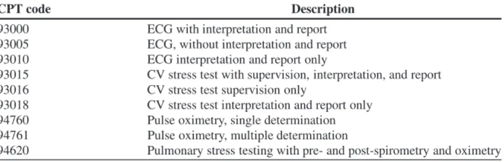
With the USAFSAM or Balke–Ware protocols, the speed remains constant while the grade is gradually increased. This is quite useful for older individuals who cannot tolerate rapid walking or running. However, these persons can tolerate an increasing grade, and, in this fashion, their workload can be increased and mea-sured. With these protocols there are smaller but equal workload changes between stages. For those patients requiring a protocol between the low-level USAFSAM and the standard Bruce protocol, a 3.3-mph modified Balke–Ware protocol might be utilized [5]. These exercise protocols are summarized in Fig. 2.1.
Incremental exercise protocols obtain continual measurements while the work rate is increased continuously (ramp protocol) or by a uniform amount each minute until the patient is limited by symptoms or the clinician feels the protocol cannot be continued safely [24].
Incremental exercise protocols are considered more physiologic and involve a continuous steady increase in workload without the abrupt, unequal changes seen in the stage protocols. The gradual increases in both speed and slope can be individu-alized to the testing situation. Typically, one estimates and then enters the maximum MET level for a given individual, the desired test duration, and the test speed. The ramp protocol then derives the increases in grade to achieve this predetermined maximum MET level at the desired finishing time. Alternatively, one may utilize
Fig. 2.1 Summary of protocols (From White RD, Evans EH [32], with permission of Primary
a ramped Bruce protocol which is designed to complete Stage IV in 12 min and has been preferred by patients [29]. Ramp protocols correlate well with measured gas analysis [5].
Some patients may benefit from proceeding directly to exercise testing coupled with echocardiography or a nuclear imaging study, such as thallium-201 or tech-netium [(techtech-netium-99m sestamibi [Cardiolite] or techtech-netium-99m tetrofosamine {Myoview}]. When choosing an imaging procedure for primary evaluation, the ex-aminer should consider the following:
1. Imaging testing greatly adds to the expense of the procedure.
2. Imaging testing may increase time for the procedure, e.g., two-phase technetium imaging.
3. One should consider radionuclide testing if there are resting ECG changes of left bundle branch block; pre-excitation syndrome, such as Wolff–Parkinson–White; marked ST-segment changes secondary to hypertension, digoxin, or quinidine; Q-wave evidence of prior large infarctions; or an inability to attain target heart rate.
4. Stress echocardiography is helpful if one suspects a wall-motion abnormality. 5. Some investigators may choose to perform cardiac catheterization directly due
to a high probability of severe coronary artery disease.
6. Some patients requiring cardiac catheterization may still benefit from exercise testing for prognostic (risk stratification analysis) or exercise prescription but not for diagnostic considerations.
The Procedure
The prediction of coronary artery disease is one of the primary functions of exercise stress testing. The reliability of the stress test depends on the magnitude and time of onset of the ST changes, on the heart rate and blood pressure response, and on the prevalence of disease in the population being studied. The heart rate response during exercise and during recovery, the maximum exercise time, and the frequency of ventricular ectopy are additional predictors of a future coronary event.
Preparing the Patient
The patient is instructed to not smoke and to eat little or no food for at least 2 h before testing. The patient should wear comfortable clothes and shoes for the test and should avoid applying body oils or lotions that might interfere with attaching the electrodes. Lastly, patients are usually reminded to take their routine medica-tions except their beta blockers or calcium channel blockers prior to exercise testing. The exception to this recommendation deals with those patients with documented CAD. These individuals do better during exercise with beta blockers, achieving a higher workload [2]. Patients taking calcium channel blockers also perform at higher
workloads even though their systolic blood pressure and heart rate decrease for a given level of exercise [2, 30].
For ECG monitoring, the skin must be prepared properly. Hair must be shaved in the appropriate locations, an abrasive rub applied to the skin to remove the su-perficial dead skin cells (which act as insulators), and any oils or other chemicals removed with an alcohol wipe. Although these steps can be time-consuming, they are very important to ensure an adequate tracing for data analysis. The goal is to re-duce the skin resistance between any two electrodes to less than 5,000 ohms. When the prep steps have been completed, the silver chloride electrodes are applied. A simple “tap test” can be done by striking the electrodes to see whether any artifact is produced. A well-prepared electrode should display no artifact. Objective testing of the electrode prep with commercial impedance meters or a simple ohmmeter is recommended.
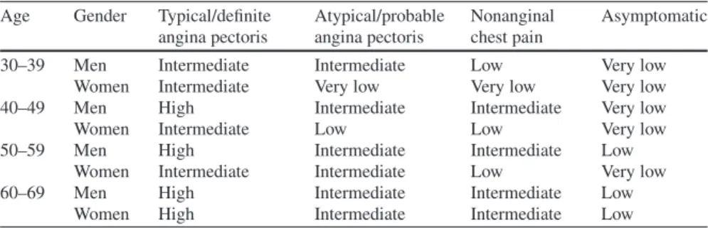
Electrodes are then placed in the standard positions for obtaining a resting 12-lead ECG. This tracing is compared with a previous tracing to determine if any recent ECG change has occurred. Next a Mason–Likar torso-mounted lead system is utilized by placing modified arm leads on the lateral front edge of the shoulders bilaterally. The modified right leg electrode (ground lead) is placed over the back-bone on the lower back while the left leg electrode (bottom of Einthoven’s triangle) is placed directly below the umbilicus [5] (Fig. 2.2).
In 1999, Michaelides et al. published an article on the use of right ventricu-lar leads to increase the sensitivity of the standard exercise test [31]. Michaelides et al. tested 275 patients referred for evaluation of angina-type symptoms. Each pa-tient underwent exercise testing with the standard leads and right ventricular leads, thallium-201 scintigraphy, and cardiac catheterization. They found that using the right ventricular leads increased the sensitivity of the exercise test to detect signif-icant coronary artery disease from 66 to 92%, creating sensitivity equal to that of thallium scanning (93%). There is some disagreement on the significance of these findings since this study has never been replicated. Some feel that right precordial leads should not be routinely used until the findings of Michaelides are validated in larger studies, while others feel using right precordial leads is of proven value both in diagnosing right ventricular infarctions and in standard exercise testing [32].
To use right precordial leads, the precordial areas of the chest on the right side that correspond to the left precordial lead of V4, V5, and V6are prepped and elec-trodes placed in these corresponding areas. The additional electrode lead wires are attached to these new leads which are labeled V4R, V5R, and V6R. Once these leads are in place, the test is performed as usual and the ECG recorder prints a 15-lead electrocardiogram at each stage and in recovery, with the right precordial leads ap-propriately labeled. This 15-lead ECG monitoring requires special equipment and programs with an increased cost.
Finally, an appropriately sized blood pressure cuff is placed snugly on the pa-tient’s arm and secured in place for serial measurements during the testing process. Although commercial automatic blood pressure manometers have been developed, none can be recommended [5]. Indications for any additional monitoring at that time can be reviewed, including pulse oximetry measurements in selected individuals
Fig. 2.2 Mason–Likar lead placement (From White RD, Evans EH [32], with permission of
Pri-mary Care Clin, with permission of Elsevier.)
with chronic obstructive pulmonary disease or in those in whom exercise-induced hypoxemia is suspected. In addition, a pre-test blood glucose measurement can be obtained and repeated at intervals in selected diabetic patients. Also, peak flow mea-surements or spirometry are done in those with suspected exercise-induced asthma (EIA), which is the most common cause of atypical chest pain associated with exer-cise in younger groups.
Monitoring the Patient
At this time, the pre-test checklist can be reviewed and final instructions given [32] (Table 2.4). One may obtain the baseline ECG tracings, blood pressure record-ings, and heart rate. These resting measurements are obtained in the supine as well as standing positions. First, a standard resting supine ECG is obtained with
Table 2.4 Pre-test checklist
1. Equipment and safety check (including defibrillator and resuscitative medications) 2. Obtain informed consent
3. Pre-test history and physical examination 4. Enter patient information into exercise test system 5. Electrode skin preparation and placement 6. Connect exercise testing monitor to electrodes
7. Place blood pressure cuff on appropriate arm and secure in place 8. Place patient supine and obtain resting blood pressure and ECG 9. Review resting 12-lead ECG for any recent changes
10. Change electrode positions to Mason–Likar torso-mounted limb lead position 11. Have patient stand and obtain blood pressure and ECG
12. Hyperventilation ECG is no longer recommended and may be omitted 13. Instruct and demonstrate appropriate treadmill walking method
a. Instruct patient to neither hold tightly onto nor grip hand rails; use light touch for balance
b. Encourage upright position and long stride length
c. Remind patient that blood pressure will be checked during each stage d. Remind patient regarding use of Borg scale
e. Remind patient to notify examiner when within one minute of maximum effort 14. Complete any other tests, e.g., fingerstick glucose, pulse oximetry, peak flow
mea-surement
15. Ask if there are any final questions prior to testing
Adapted from White RD, Evans EH: Performing the exercise test. Primary Care 21:455, 1994, with
permission of Elsevier.
the extremity leads placed on the wrists and ankles for comparison with the pa-tient’s previous standard supine tracing. With this important step the examiner can evaluate any recent ECG changes that might preclude exercise testing. These ex-tremity leads are then moved to the modified positions as described previously in the Mason–Likar lead system and a second tracing obtained. The patient is then placed in the standing position, and an ECG utilizing the modified leads is obtained. One may note a change in the R vector (usually lead aVL) with a change in the standing position. One should compare the modified standing baseline ECG, which now becomes the baseline tracing, against ECGs obtained during the exercise test. One is now ready to begin the exercise testing mode.
Exercise Testing Mode
Once the patient has completed a low-level warm-up and has adapted to the tread-mill, the chosen test protocol is begun. When the patient is comfortable, the exercise phase should be initiated and the patient checked constantly. The patient is cautioned to avoid tightly gripping the safety side or front rails. This practice alters the work-load and measured performance.
The electrocardiogram (ECG), heart rate, and blood pressure should be mon-itored and recorded during each stage of exercise and during ST-segment abnor-malities and chest pain. The patient should be monitored continuously for transient rhythm disturbances, ST-segment changes, and other electrocardiographic manifes-tations of myocardial ischemia [10].
The physician should observe the ECG monitor carefully. A 12-lead ECG tracing should be obtained during the last minute of each stage of exercise, at the point of maximum exercise testing, at 1 min in recovery, and then every 2 min during the recovery phase. The blood pressure is obtained and recorded with each ECG tracing. This includes a blood pressure reading with the patient resting supine, standing at rest, during the last minute of each stage of the exercise protocol, at the point of maximum exercise, immediately post-exercise, at 1 min in recovery and then every 2 min during the recovery phase (1, 3, 5, 7 min, etc.). During the testing procedure, the examiner should communicate frequently with and warn the patient of upcom-ing stage changes. The examiner should stop the exercise test when the patient has reached maximal effort (Borg Scale) or exhibits clinical signs to terminate. It has been found that symptom-limited testing with the Borg CR10 Scale as an aid is very important when the test is used to assess functional capacity.
Terminating the Test
The patient should be informed that he/she is in charge and can stop the test at any time. Otherwise, test termination requires clinical evaluation and judgment con-cerning the status of the patient. Absolute indications to terminate exercise testing include the following:
1. Acute myocardial infarction or suspicion of myocardial infarction 2. Onset of progressive angina or anginal equivalents
3. Exertional hypotension – decrease of systolic blood pressure (20 mmHg) with increasing workload or a decrease below the baseline standing systolic blood pressure prior to test, accompanied by signs or symptoms indicating poor left ventricular function and poor cardiac output
4. Serious dysrhythmias, e.g., ventricular tachycardia
5. Signs of poor perfusion (pallor, cyanosis, nausea or cold, clammy skin)
6. Central nervous system symptoms (ataxia, vertigo, visual or gait problems, and confusion)
7. Failure of increasing heart rate response with increasing workload 8. Technical problems with monitoring the ECG or equipment failure 9. Patient requests to stop
Relative indications to terminate exercise testing include the following:
1. Pronounced ECG changes from baseline, including more than 2 mV of horizontal or downsloping ST-segment depression or 2 mV of ST-segment elevation 2. Progressive or increasing chest pain
4. Wheezing
5. Leg cramps or intermittent claudication
6. Hypertensive response (systolic blood pressure greater than 250 mmHg or dias-tolic blood pressure greater than 115 mmHg) [5]
7. Less serious dysrhythmias such as supraventricular tachycardia
8. Exercise-induced bundle branch block that cannot be distinguished from ventric-ular tachycardia
Recovery of the Patient
During the recovery phase, the patient should be placed immediately in the supine position or allowed a “cool-down walk” and then placed in a chair. The examiner should auscultate the patient immediately for any abnormal heart findings such as a new-onset heart murmur or third heart sound. In addition, one should auscultate the lungs for any evidence of exercise-induced bronchospasm, which might be a cause of chest pain complaints. During the recovery phase the blood pressure and ECG recordings should be obtained at 1 min and then every 2 min. The heart rate recording at 1 min post-graded exercise and the 3 min systolic BP:peak BP ratio have been correlated with overall mortality and prognosis [33]. The patient is observed carefully until he/she is stable and any observed ST-segment changes have returned to baseline. This usually requires 8–10 min. It is important to watch carefully for any “recovery-only” ST-segment depression [34].
Some debate exists as to whether the patient should be immediately placed supine or allowed a cool-down walk. The consensus opinion is that a cool-down walk will delay “recovery-only” ST-segment depression, and thus the patient should be mon-itored in recovery for at least 10 min with this method. If, instead, the patient is immediately placed supine after maximal exercise and the legs elevated, recovery monitoring can be limited to 6–8 min. Maximal test sensitivity is achieved with the patient supine post-exercise. If any ST-segment depression persists in the recovery period, one may consider administration of sublingual nitroglycerin. If nitroglycerin is given, one should place the patient supine and monitor the blood pressure and other vital signs until the ST-segment changes have resolved.
Once the patient is stable, the ECG monitoring equipment can be removed, the information interpreted, the patient informed of the results, and a formal dictation of the procedure completed.
Hazards and Complications of Exercise Testing
Serious complications include acute myocardial infarction, ventricular fibrillation, or death. To deal with these possible complications, one must be trained in car-diopulmonary resuscitation (CPR) as well as advanced cardiac life support (ACLS) protocols. ACLS equipment, including proper medications and a defibrillator, should be available at all times. The most important safety precaution is careful pre-test pa-tient evaluation and selection of the proper protocol in light of the contraindications.
Table 2.5 CPT codes
CPT code Description
93000 ECG with interpretation and report 93005 ECG, without interpretation and report 93010 ECG interpretation and report only
93015 CV stress test with supervision, interpretation, and report
93016 CV stress test supervision only
93018 CV stress test interpretation and report only 94760 Pulse oximetry, single determination 94761 Pulse oximetry, multiple determination
94620 Pulmonary stress testing with pre- and post-spirometry and oximetry
The Report
Current computer treadmill systems produce tabular and graphical analysis of the results of the study. ECG, heart rate, and blood pressure data provide the information leading to pathophysiologic diagnosis. The report should include a brief summary of relevant clinical information, medications, specific exercise-related complaints, a brief description of the methods and procedure, and a narrative analysis and in-terpretation. In addition, recommendations regarding the results are included [24]. CPT codes are listed in Table 2.5.
Summary
With proper training, exercise testing is a useful procedure for the primary care physician in the outpatient as well as the inpatient setting. By careful pre-test eval-uation, one is able to study patients safely, obtain both diagnostic and prognostic information concerning the risk of cardiovascular disease, formulate appropriate treatment plans, and develop therapeutic prescriptions for exercise.
References
1. Fowler-Brown A, Pignone M, Pletcher M, et al. Exercise tolerance testing to screen for coro-nary heart disease: A systematic review for the technical support for the U.S. preventive ser-vices task force. Ann Intern Med. 2004;140:W9–W24.
2. Ellestad MH. Stress testing: Principles and practice, 5th ed. Oxford University Press. 2003. 3. Thaulow E, Erikssen J, Sandvik L, Erikssen G, Jorgensen L, Cohn PF. Initial clinical
pre-sentation of cardiac disease in asymptomatic men with silent myocardial ischemia and an-giographically documented coronary artery disease (the Oslo Ischemia Study). Am J Cardiol. 1993;72:629–633.
4. Institute for clinical systems improvement. Health Care Guideline: Cardiac Stress Test Sup-plement, 6th ed. 2004.
5. Froelicher VF, Myers JN. Exercise and the Heart, 5th ed. Philadelphia, Elsevier, Inc., 2006, 15.
6. Diabetes mellitus and exercise. ADA standards of medical care in diabetes—2007. Diabetes Care. 2007;30:S4–S41.
7. Stuart RJ, Ellestad MH. National survey of exercise stress testing facilities. Chest. 1980;77:94–97.
8. Gibbons RJ, Balady GJ, Bricker JT, et al. American College of Cardiology/American Heart Association Task Force on Practice Guidelines (Committee to Update the 1997 Exercise Test-ing Guidelines). ACC/AHA 2002 guideline update for exercise testTest-ing: summary article: A report of the American College of Cardiology/American Heart Association Task Force on Practice Guidelines (Committee to Update the 1997 Exercise Testing Guidelines). Circulation. 2002;106:1883–1892.
9. American College of Sports Medicine. Guidelines for Exercise Testing and Prescription, 6th ed. Baltimore, Lippincott Williams & Wilkins, 2000, 26.
10. ACC/AHA 2002 Guideline Update for Exercise Testing. A report of the American College of Cardiology/American Heart Association Task Force on Practice Guidelines (Committee on Exercise Testing). Circulation J Am Coll Cardiol. 2002.
11. Fletcher GF, Froelicher VF, Hartley LH, Haskell WL, Polluck ML. Special report of exercise standards: A statement for health professionals from the American Heart Association. Circu-lation. 1990;82:2286–2322.
12. De Ponti C, De Biase AM, Cataldo G, et al. Effects of nifedipine, acebutolol, and their association on exercise tolerance in patients with effort angina. Cardiology. 1981;68: 195–199.
13. Fox KM, Deanfield J, Jonathan A, et al. The dose-response effects of nifedipine on ST- seg-ment changes in exercise testing: Preliminary studies. Cardiology. 1981;68:209–212. 14. Sundqvist K, Atterhog JH, Jogestrand T. Effect of digoxin on the electrocardiogram at rest and
during exercise in healthy subjects. Am J Cardiol. 1986;57:661–665.
15. Gibbons RJ, Balady GJ, Beasley JW, et al. ACC/AHA guidelines for exercise testing: A report of the American College of Cardiology/ American Heart Association Task Force on Practice Guidelines (Committee on Exercise Testing). J Am Coll Cardiol. 1997;30:260–315. 16. Smith RB, Rusbatch BJ. Amitriptyline and heart block. Br Med J. 1967;3:311.
17. Vohra J, Burrows GD, Sloman F. Assessment of cardiovascular side effects of therapeutic doses of tricyclic anti-depressant drugs. Aust NZ J Med. 1975;5:7–11.
18. Wilson WM, Maughan RJ. Evidence for a possible role of 5-hydroxytryptamine in the gen-esis of fatigue in man: Administration of paroxetine, a 5-HT re-uptake inhibitor, reduces the capacity to perform prolonged exercise. Exp Physiol. 1992;77:921–924.
19. Fagard R, Amery A, Reybrouck T, et al. Effects of angiotensin antagonism on hemodynamics, renin, and catecholamines during exercise. J Appl Physiol. 1977;43:440–444.
20. Pickering TG, Case DB, Sullivan PA, et al. Comparison of antihypertensive and hormonal effects of captopril and propranolol at rest and during exercise. Am J Cardiol. 1982;49: 1566–1568.
21. Dickstein, K, et al. Comparison of the effects of losartan and enalapril on clinical status and exercise performance in patients with moderate or severe chronic heart failure. J Am Coll Cardiol. 1995;26:438.
22. Hamroff, G, et al. Addition of angiotensin II receptor blockade to maximal angiotensin-converting enzyme inhibition improves exercise capacity in patients with severe congestive heart failure. Circulation. 1999;99:990.
23. Niederberger M, Bruce RA, Kusumi F, et al. Disparities in ventilatory and circulatory re-sponses to bicycle and treadmill exercise. Br Heart J. 1974;36:377.
24. Wasserman K, Hansen JE, Sue DY, Stringer WW, Whipp BJ. Principles of Exercise Testing and Interpretation, 4th ed. Lippincott Williams & Wilkins. 2005.
25. ACC/AHA Guideline Revision. ACC/AHA 2007 Guidelines for the Management of Patients with Unstable Angina/Non–ST-Elevation Myocardial Infarction—Executive Summary. J Am Coll Cardiol. 2007;50:652–726.
26. Borg GA. Psychophysical bases of perceived exertion. Med Sci Sports Exerc. 1982;14: 377–381.
27. Borg G. Perceived exertion as an indicator of somatic stress. Scand J Rehabil Med. 1970;23:92–93.
28. Borg G, Holmgren A, Lindblad I. Quantitative evaluation of chest pain. Acta Med Scand. 1981;644:43–45.
29. Will PM, Walter JD. Exercise testing: Improving performance with a ramped Bruce protocol. Am Heart J. 1999;138:1033–1037.
30. Rice, KR, et al. Effects of nifedipine on myocardial perfusion during exercise in chronic stable angina pectoris. Am J Cardiol. 1990;65:1097.
31. Michaelides AP, Psomadake ZD, Divaveris PE, et al. Improved detection of coronary artery disease by exercise electrocardiography with the use of right precordial leads. N Engl J Med. 1999;340:340–345.
32. White RD, Evans EH. Performing the exercise test. Primary Care. 2001;28(1):44.
33. Cole CR, Blackstone EH, Pashkow FJ, et al. Heart-rate recovery immediately after exercise as a predictor of mortality. N Engl J Med. 1999;341:1351–1357.
34. Lachterman B, Lehmann KG, Abrahamson D, Froelicher VF: “Recovery only” ST segment depression and the predictive accuracy of the exercise test. Ann Intern Med. 1990;112:11–16.

![Fig. 2.1 Summary of protocols (From White RD, Evans EH [32], with permission of Primary Care Clin, with permission of Elsevier.)](https://thumb-us.123doks.com/thumbv2/123dok_us/8995919.2384549/13.659.87.576.571.846/summary-protocols-white-evans-permission-primary-permission-elsevier.webp)
![Fig. 2.2 Mason–Likar lead placement (From White RD, Evans EH [32], with permission of Pri- Pri-mary Care Clin, with permission of Elsevier.)](https://thumb-us.123doks.com/thumbv2/123dok_us/8995919.2384549/16.659.85.576.163.556/mason-likar-placement-white-evans-permission-permission-elsevier.webp)