Journal of Athletic Training 2016;51(8):629–636 doi: 10.4085/1062-6050-51.10.01
Óby the National Athletic Trainers’ Association, Inc
www.natajournals.org original research
Muscle Activation During Landing Before and
After Fatigue in Individuals With or Without Chronic
Ankle Instability
Kathryn A. Webster, PhD, ATC*; Brian G. Pietrosimone, PhD, ATC
†
;
Phillip A. Gribble, PhD, ATC, FNATA
‡
*Department of Physical Therapy and Athletic Training, Boston University, MA; †Department of Exercise Science and Sport Science, University of North Carolina, Chapel Hill; ‡College of Health Sciences, University of Kentucky, Lexington
Context: Ankle instability is a common condition in physi-cally active individuals. It often occurs during a jump landing or lateral motion, particularly when participants are fatigued.
Objective: To compare muscle activation during a lateral hop prefatigue and postfatigue in individuals with or without chronic ankle instability (CAI).
Design: Cross-sectional study.
Setting: Sports medicine research laboratory.
Patients or Other Participants: A total of 32 physically active participants volunteered for the study. Sixteen partici-pants with CAI (8 men, 8 women; age¼20.50 6 2.00 years, height¼172.25610.87 cm, mass¼69.136 13.31 kg) were matched with 16 control participants without CAI (8 men, 8 women; age¼22.0063.30 years, height¼170.5069.94 cm, mass ¼ 69.63 6 14.82 kg) by age, height, mass, sex, and affected side.
Intervention(s): Electromyography of the tibialis anterior, peroneus longus, gluteus medius, and gluteus maximus was measured before and after a functional fatigue protocol.
Main Outcome Measure(s): Activation of 4 lower extremity muscles was measured 200 milliseconds before and after landing from a lateral hop.
Results: We observed no interactions. The group main effects for the peroneus longus demonstrated higher muscle activation in the CAI group (52.89% 6 11.36%) than in the control group (41.12%611.36%) just before landing the lateral hop (F1,30¼8.58,P¼.01), with a strong effect size (d¼1.01). The gluteus maximus also demonstrated higher muscle activa-tion in the CAI group (45.55% 6 12.08%) than in the control group (36.81% 6 12.08%) just before landing the lateral hop (F1,30¼4.19,P¼.049), with a moderate effect size (d¼0.71). We observed a main effect for fatigue for the tibialis anterior, with postfatigue activation higher than prefatigue activation (F1,30¼7.45, P¼.01). No differences were present between groups for the gluteus medius.
Conclusions: Our results support the presence of a centralized feed-forward neuromuscular alteration in patients with CAI, not only in the ankle-joint muscles but also in the proximal hip muscles. These results may have implications for rehabilitation programs in these patients.
Key Words: ankle injuries, hip, proprioception, postural control, feed-forward mechanism
Key Points
Just before landing a lateral hop, activation values in the peroneus longus and gluteus maximus muscles were
higher in participants with chronic ankle instability than in control participants.
After repeated ankle trauma, proximal and distal neuromuscular alterations may result from a centralized feed-forward mechanism to prepare the lower extremity for landing without injury.
More research involving functional activity with perturbation is needed to help investigators address neuromuscular
deficits in patients with chronic ankle instability.
A
nkle sprains are one of the most common injuries in activity, and reinjury rates as high as 73% have been reported.1 When a substantial lateral anklesprain occurs, a common outcome is repeated giving way of the ankle during activities. Termedchronic ankle instability (CAI), it is characterized by residual lateral instability2
categorized as (1) mechanical instability related to anatomical changes in tissues surrounding the ankle, (2) functional or perceived instability related to neuromuscular changes,2 or (3) recurrent sprains in which a patient
experiences repeated inversion injury with activity.
Indi-viduals with CAI may be subcategorized into 1 of these categories or a combination of the 3.3–5
Much still needs to be understood about why patients continue to experience residual effects after an ankle sprain. Numerous authors6–10 have proposed altered peripheral
neuromuscular control due to ankle injury. Specifically, impairments are related to proprioception, neuromuscular control, or strength, leading to deficits2,3 not only at the
ankle joint but of the entire lower kinetic chain.11–13
Wyke14 proposed that damaged mechanoreceptors in a
disruption could, in turn, create changes in the movement of the entire lower extremity.15Caulfield and Garrett16further
substantiated these changes, reporting differences in the kinematics of both the ankle and the knee before jump landing. Researchers13,17,18 have proposed that residual
symptoms in individuals with CAI not only include changes at the ankle due to poor afferent feedback information but also a neuromuscular reorganization to a centralized feed-forward mechanism during dynamic tasks to avoid reinjury. Beckman and Buchanan11found changes in the firing of the
hip muscles in individuals with CAI compared with healthy controls. Proximal neuromuscular control at the hip, specifically the gluteus medius (Gmed) and gluteus maximus (Gmax), contributes to the positioning of the lower extremity19–21 and may be affected in individuals
with CAI. Continuing to investigate the possibility of these changes in those with CAI is important to help us understand the best means of implementing interventions that may prevent subsequent injury.
Another factor possibly contributing to CAI is muscle fatigue, which has been demonstrated to alter motor control,22 joint position sense,23 and muscle response
during activity.24These alterations due to fatigue can lead
to improper positioning of the lower extremity and difficulty responding quickly to changes in movements, which can predispose patients to injuries.22,25In addition,
moderate and major injuries more commonly occur at the end of athletic practices and games.26 Physically active
patients often perform many of the mechanisms of ankle sprain, namely changing direction and jump landing; therefore, fatigue may be an additional factor leading to CAI that has not been studied extensively in patients with the condition.
Researchers24,27investigating the effects of fatigue on the
lower extremity have found decreased muscle activation and increased postural sway after fatiguing protocols. To our knowledge, no one has examined whether fatigue is associated with altered electromyography (EMG) activity in the hip and ankle muscles in this patient population during a functional task. Examining the prefatigue and postfatigue activation of the proximal muscles of the Gmed and Gmax and the distal muscles of the tibialis anterior (TA) and peroneus longus (PL) during a lateral hop may identify mechanisms that can lead to ankle sprain over time. Acquiring data from the hip and ankle muscles during this activity may contribute to a better understanding of the mechanism of ankle injury and aid in designing rehabili-tation protocols to prevent recurrent sprains. Therefore, the purpose of our study was to examine the prefatigue and postfatigue EMG activity of the TA, PL, Gmed, and Gmax in individuals with or without CAI before and during the landing of a lateral hop.
METHODS
We implemented a cross-sectional, repeated-measures design to examine prefatigue and postfatigue EMG of the hip and ankle musculature in participants with or without CAI during a lateral hop.
Participants
A total of 32 individuals volunteered for the study. Sixteen participants with CAI (8 men, 8 women; age ¼
20.50 62.00 years, height¼172.25 610.87 cm, mass¼ 69.13 6 13.31 kg) were matched with 16 volunteers without CAI serving as control participants (8 men, 8 women; age¼22.0063.30 years, height¼170.5069.94 cm, mass¼69.63 6 14.82 kg) by age, height, mass, sex, and affected side. Participants reported to the sports medicine research laboratory for a single session.
The participants with CAI presented with a history of ankle injury resulting in an antalgic gait or pain for 24 hours or longer and 2 or more self-reported episodes of the ankle giving way in the 6 months before the study. These participants needed to score 90% or less on the Foot and Ankle Disability Index (FADI; mean¼82.0%616.19%) or 80% or less on the FADI-Sport28 (mean¼64.19% 6
20.36%) for inclusion in the study. Control-group participants did not have previous ankle sprains and had completed the FADI (mean ¼100% 6 0%) and FADI-Sport (mean ¼ 100% 6 0%). These tools helped to establish that the participants had similar levels of instability during activities of daily living (FADI) and sport activity (FADI-Sport). Control participants were assigned an‘‘injured’’limb, which was compared with the injured side of their matched participants in the CAI group.
Neither group had a previous lower extremity fracture or surgery or any vestibular changes, including concussions, within the 6 months before the study, and they reported beingphysically active, which was defined as participating in 20 to 30 minutes of cardiovascular activity at least 3 times each week. To verify the level of physical activity, all participants completed the Participation Activity Readiness Questionnaire and answerednoto all questions. All patients completed the Tegner Activity Scale to document weekly physical activity; all answered level 5 or above, which indicated they were minimally involved in recreational activity on uneven ground at least twice weekly. On the basis of a previous study,29 a power-analysis calculation30
suggested that 15 participants in each group would be sufficient (power ¼ 0.80, P , .05). All participants provided written informed consent, and the study was approved by the University of Toledo Institutional Review Board (No. 106670).
Equipment
Procedures
We tested the injured limb of the CAI group and the matched limb of the control group. We shaved the skin superficial to the 4 muscles when necessary and lightly abraded and cleaned it with alcohol. The electrodes were positioned in the direction of the muscle fibers that were being measured, which was consistent with established protocols.31 For the hopping portion, an investigator
(K.A.W.) wrapped the TA and PL electrodes and leads with nonadhesive tape (Powerflex; Andover Healthcare, Inc, Salisbury, MA) to avoid movement both prefatigue and postfatigue.
At prefatigue, we measured the length of participants’ fibulas and marked this distance with 1 piece of tape on the floor and 1 piece on the force platform. We then instructed individuals to use the test limb to perform 5 lateral hops onto and off of the force platform a distance equal to the length of their fibula and over a barrier that was 5 cm high. The subsequent fatigue protocol32 consisted of 535-m
cone drills involving combinations of forward sprints, lateral shuffles, pivoting, and backward running. Next, participants completed 30 2-footed lateral hops over a 10-cm barrier, followed by 3 successive step-ups onto and 2-footed hop-downs from boxes measuring 30, 38, and 46 cm high (Figure). Participants used the floor-mat–activated timing device to time the fatigue trials. They had 20 seconds between fatigue bouts to return to the starting position.
We instructed the participants to repeat the protocol until fatigue, which was defined as (1) 50% increase in their fastest time to complete the course, (2) inability to repeatedly clear the 10-cm barrier on the lateral jumps, (3) inability to step onto the plyometric box, or (4)
unwillingness to continue. The average number of com-pleted fatigue protocols was 8.4. Two participants ended the protocol due to reason 1; 1, due to reason 2; 1, due to reason 3; and all other participants, due to reason 4. Immediately postfatigue, participants completed the same 5 1-footed lateral hops as during the pretest while EMG data were collected.
Data Processing
The EMG signals from the TA, PL, Gmed, and Gmax were collected at 1000 Hz for the periods of prelanding (200 milliseconds pre-IC to IC) and postlanding (IC until 200 milliseconds post-IC), filtered using a fourth-order band-pass filter with cutoffs at frequencies of 10 Hz and 500 Hz, smoothed using a 50-millisecond root mean square algorithm, and full-wave rectified. The prefatigue and postfatigue data were normalized to the mean peak EMG amplitude of the corresponding prefatigue and postfatigue 5 hopping trials for each participant.29
Statistical Analysis
Means and standard deviations of the normalized percentage of average peak muscle activation were used for analysis. Dependent variables were amplitude of the TA, PL, Gmed, and Gmax prelanding and postlanding. For each dependent variable, we performed a separate group-by-fatigue repeated-measures analysis of variance. Effect sizes were calculated using the Cohen d with 95% confidence intervals (CIs) and interpreted as small (0.2), medium (0.5), or large (0.8).33 Data were analyzed using
SPSS (version 15.0; SPSS Inc, Chicago, IL). We set the a level a priori at .05.
RESULTS
Our results for the prelanding and postlanding compari-sons are presented in Tables 1 through 4. We noted no interactions. However, during prelanding postfatigue, the PL had notably higher muscle-activation levels in the CAI group (57.71% 6 25.43%) than in the control group (41.29% 6
11.63%). Activation levels for the Gmax were also higher in the CAI group (50.55%623.98%) than in the control group (36.77%611.36%). Although not different, the effect sizes for the PL (P¼.25, d¼0.81) and Gmax (P¼.10, d¼0.72) were moderate to strong (Table 1).
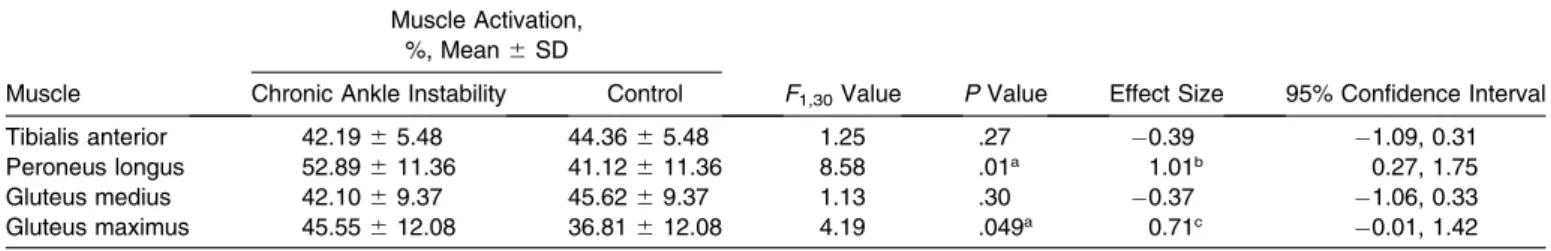
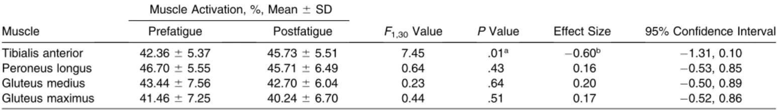
The group main effects for the PL demonstrated higher muscle activation in the CAI group (52.89% 6 11.36%) than in the control group (41.12% 6 11.36%) just before landing the lateral hop, with a strong effect size (P¼.01, d ¼ 1.01; Table 2). The Gmax also demonstrated higher muscle activation in the CAI group (45.55% 6 12.08%) than in the control group (36.81% 6 12.08%) just before landing the lateral hop, with a moderate effect size (P¼ .049, d¼0.71; Table 2). We observed a main effect for fatigue of the TA, which demonstrated higher activation postfatigue than prefatigue (Table 3). No differences were demonstrated in group or fatigue for the Gmed (Table 3). The only difference for the postlanding phase was in the TA, which demonstrated higher activation postfatigue than prefatigue across groups (Table 4).
DISCUSSION
Activation of the PL and Gmax was higher in the CAI than in the control group during the prelanding phase of a lateral hop. When we introduced functional fatigue, we also observed differences with moderate to strong effect sizes in
the postfatigue results for the PL and Gmax, demonstrating clinical importance.
The higher muscle-activation values in the CAI group just before landing the lateral hop may help to support the theory suggested by Delahunt et al,13 who proposed a
centralized feed-forward mechanism to explain changes seen in patients with CAI. Authors of several subsequent studies2,9,13,16,18,29,34 have attributed findings to this theory
of the feed-forward mechanism. The theory is based on the premise that, due to injury, the normal reaction pattern of the muscles that protect the ankle in healthy patients is too slow to prevent injury positions in patients with CAI; therefore, a centralized feed-forward neural adaptation is implemented to protect the ankle from injury both proximally and distally. We instructed participants to hop laterally and return to a specific location. In concept, this activity would allow them to attempt to implement any protective neuromuscular control necessary to stabilize during the task, which is often associated with an ankle-sprain mechanism. These muscles may have been activated to a greater extent in the CAI group to help place the lower limb in a more protected position prelanding.35
Distal Alterations
When considering how these changes may appear in the distal segment of the lower kinetic chain, researchers have found that patients with CAI demonstrate changes at the ankle compared with healthy participants when preparing for the foot to contact the ground.9,16,17,36 Similar to our
findings for jump-landing preparation, Gutierrez et al9
reported that patients with CAI demonstrated increased PL activity when preparing to land from a drop jump on a supinating surface. Whereas we used a lateral-hop landing, Table 1. Muscle-Activation Prelanding Interaction Results
Muscle Group
Muscle Activation, %, Mean6SD
F1,30
Value P Value
Postfatigue Between-Groups Comparison
Effect Size
95% Confidence Interval Prefatigue Postfatigue
Tibialis anterior Chronic ankle instability 40.2263.90 44.1566.53 0.01 .94 0.27 0.95, 0.44 Control 42.5066.86 46.2268.35
Peroneus longus Chronic ankle instability 48.0767.70 57.71625.43 1.35 .25 0.81a 0.09, 1.53
Control 40.96613.72 41.29611.63
Gluteus medius Chronic ankle instability 42.1766.53 42.0267.38 0.34 .56 0.27 0.97, 0.42 Control 46.53613.81 44.70611.41
Gluteus maximus Chronic ankle instability 40.5564.83 50.55623.98 2.84 .10 0.72b 0.00, 1.43
Control 36.84611.93 36.77611.36
aIndicates strong effect size. bIndicates moderate effect size.
Table 2. Muscle-Activation Prelanding Results by Group
Muscle
Muscle Activation, %, Mean6SD
F1,30Value PValue Effect Size 95% Confidence Interval
Chronic Ankle Instability Control
Tibialis anterior 42.1965.48 44.3665.48 1.25 .27 0.39 1.09, 0.31 Peroneus longus 52.89611.36 41.12611.36 8.58 .01a 1.01b 0.27, 1.75
Gluteus medius 42.1069.37 45.6269.37 1.13 .30 0.37 1.06, 0.33 Gluteus maximus 45.55612.08 36.81612.08 4.19 .049a 0.71c 0.01, 1.42 aIndicates difference.
they9 instructed patients to land on a surface that allowed
the ankle to supinate. Both circumstances are common mechanisms for ankle injury, so it is not surprising that similar findings would be demonstrated when patients anticipated a challenge at landing. The theory of a centralized feed-forward mechanism could explain a strategy to protect the joint when landing by attempting to place the ankle in a more stable position, as evidenced by higher activation of the PL in this case. Levin et al36also
recently found changes in EMG firing of the PL, but the changes were in the contralateral limb of patients with CAI. Their results showed that patients with CAI had higher levels of contralateral PL activity just before jump landing than did healthy control participants. The authors also proposed that centralized feed-forward mechanisms were responsible for these changes. In another study, Caulfield and Garrett16instructed participants to perform a drop jump
and noted earlier ground reaction force peaks in patients with CAI than in control participants. These authors also attributed this alteration to the feed-forward response mechanism of the neuromuscular system compensating for previous ankle injury by modifying ankle-joint landing. These data contribute to the premise that patients with CAI demonstrate different motor-behavior patterns than control participants when preparing their ankles for landing. Further prospective studies are necessary to determine whether this is an adapted response due to injury or whether patients had these differences before injury, which would suggest a predisposition to CAI. Caulfield and Garrett16did
not collect kinematic data to determine whether the foot actually was positioned differently in patients with CAI than in healthy controls.
Delahunt et al17obtained kinematic data for a lateral hop
of 30 cm and reported that participants with CAI displayed a less everted position of the ankle than did control participants. Caulfield and Garrett37 noted the same
positioning just before foot contact during gait. This observation seems counterintuitive given the findings of the studies discussed earlier with higher PL activity prelanding, theoretically placing the foot in a more everted position. The increased PL activation in our CAI group may
suggest the neuromuscular system is attempting to evert the ankle to avoid an inversion motion during the lateral hop landing but is unable to move the foot into a more everted position.
Another protective tactic is a more dorsiflexed position, which allows for a more stable, close-packed orientation of the ankle joint. Researchers37have found that patients with
CAI demonstrate increased dorsiflexion before landing from a jump compared with healthy controls. The higher postfatigue activation of the TA just before landing was observed across groups and may indicate that participants with or without CAI both attempt to increase ankle stability to handle landing. Whereas the TA is the muscle most responsible for ankle dorsiflexion, it also has a medially located insertion, allowing it to contribute to midfoot inversion. Activation of the PL possibly prevents the TA from placing the foot in a more inverted position but still allows for more dorsiflexion.
Proximal Alterations
Participants with CAI demonstrated differences not only in the lower leg and ankle but also in the proximal musculature compared with healthy controls. One of the aims of our study was to investigate the role of the proximal musculature in a landing task for participants with or without CAI. The Gmed and Gmax are involved in positioning the femur, which subsequently affects position-ing of the ankle through the kinetic chain. Landposition-ing from a lateral hop requires control of hip flexion and rotation, in part by the Gmed and Gmax.20The increased Gmax activity
of the CAI group observed prelanding may suggest an effort to position the lower extremity more under the center of mass, thereby creating slight hip extension. The Gmax also may be more highly activated as it prepares to limit internal femoral rotation at landing, which would put greater stress on the ankle to stabilize than if the femur was positioned properly under the body. These explanations are speculative, given that we did not quantify kinematic patterns in this study.
Table 3. Muscle-Activation Prelanding Results by Time
Muscle
Muscle Activation, %, Mean6SD
F1,30Value PValue Effect Size 95% Confidence Interval
Prefatigue Postfatigue
Tibialis anterior 41.3663.95 45.1965.30 8.61 .01a 0.80b 1.52,0.80
Peroneus longus 44.5267.87 49.50613.98 1.55 .22 0.43 1.13, 0.27 Gluteus medius 44.3567.64 43.3666.79 0.48 .50 0.13 0.56, 0.83 Gluteus maximus 38.7066.44 43.66613.27 2.76 .11 0.45 1.17, 0.24
aIndicates difference. bIndicates strong effect size.
Table 4. Muscle-Activation Postlanding Results for Fatigue
Muscle
Muscle Activation, %, Mean6SD
F1,30Value PValue Effect Size 95% Confidence Interval
Prefatigue Postfatigue
Tibialis anterior 42.3665.37 45.7365.51 7.45 .01a 0.60b 1.31, 0.10
Peroneus longus 46.7065.55 45.7166.49 0.64 .43 0.16 0.53, 0.85 Gluteus medius 43.4467.56 42.7066.04 0.23 .64 0.20 0.50, 0.89 Gluteus maximus 41.4667.25 40.2466.70 0.44 .51 0.17 0.52, 0.86
aIndicates difference.
Whereas we are not aware of other researchers who reported Gmax activation during a lateral hop in patients with CAI compared with healthy controls, investigators have observed changes in proximal activity in this patient population. Rios et al38 addressed the activation of the
proximal muscles during another functional task, kicking a soccer ball, in individuals with or without CAI. Increased activation in the proximal muscles for the CAI group was present compared with the control group. They attributed these findings to an attempt by the proximal portion of the leg to maintain stability, which may protect the previously injured ankle.38 Our results confirmed these findings of
increased Gmax activity in patients with CAI prelanding, which may suggest a feed-forward mechanism to help maintain stability during the lateral jump landing. Van Deun et al39 found that patients with CAI demonstrated
later onset times for the hip than did control participants when transitioning from a double- to a single-legged stance. Whereas Bullock-Saxton et al12observed participants in an
open chain position, they also noted changes in Gmax activation in patients with CAI compared with control participants, as evidenced by delayed activation during prone hip extension. Continued research is needed to determine whether these muscle-activation patterns are contributing to the movement patterns at the ankle in patients with CAI in conjunction with kinematic variables. Although it would seem that more muscle changes might be demonstrated in the Gmed due to the frontal-plane movement of the lateral hop, we did not observe differences in this muscle. The altered neuromuscular system of participants with CAI may defer to the larger, more powerful Gmax muscle. Overall, researchers have demon-strated neuromuscular changes in the activation timing of the Gmed and Gmax in participants with CAI compared with healthy participants,11,12,38,39 but more research is
necessary to investigate patterns of feed-forward neuro-muscular control demonstrated in the proximal joints during dynamic movements.
Fatigue
We selected our fatigue protocol to simulate the demands of physical activity—including elements of sprinting, cutting, and lateral shuffles and an extended period of lateral hopping—with the intention of targeting and fatiguing the lower extremity. Evidence22,23,26 has pointed
toward greater injury risk and decreased neuromuscular control in fatigued participants, which may result in greater changes in muscle activation when comparing CAI and control participants.
Whereas we observed no interactions for fatigue (Table 1), the effect sizes for both the PL and Gmax were strong and moderate, respectively, with the CAI group demon-strating higher activation than the control group for both muscles in the postfatigue state. The effect sizes indicated some clinical importance of these comparisons. As muscles fatigue, contractile capability decreases, resulting in additional motor recruitment and increased frequency of firing.40The increase in EMG amplitude during fatigue may
represent the recruitment of more motor units as the force-producing capabilities of type II fibers are diminished.41
Increased amplitude in the EMG signal of a fatigued participant with CAI can also be explained by the reduced
conduction velocity of the muscle action potential seen with fatigue, which widens the pulse and increases the area under the curve, resulting in a larger mean amplitude of the rectified EMG signal.42The results are varied as to whether
fatigue is controlled centrally or peripherally or by a combination of the two.43 Researchers43 have determined
that feeling fatigued, as in this study, is a complex process in which the body integrates sensory information to determine its ability to maintain homeostasis. Regardless of the cause, the clinically important changes in muscle activation in the CAI group may imply that rehabilitation programs should include functional exercise during a fatigued state to help overcome this deficit. More specifically, they could also demonstrate that the PL and Gmax should be targeted with more repetitions during therapeutic exercise in patients recovering from ankle instability to help avoid early fatigue when returning to activity.
Postlanding
After ground contact, greater TA activation was observed postfatigue than prefatigue across groups (Table 4). The TA may fatigue fastest and be most involved in landing, leading to higher activation postlanding for all participants, so clinicians do not need to focus on postlanding techniques related to the TA in patients with CAI. The lack of differences for all other muscles during the postlanding phase suggested that the CAI and control groups managed the jump landing similarly over ground contact and weight acceptance despite the altered neuromuscular control systems of participants with CAI, perhaps due to the planned nature of the task.
Limitations
Clinical Implications
Our findings have several clinical implications. The neuromuscular changes that participants with CAI demon-strated while performing a common functional athletic task indicate that alterations after ankle sprain occurred not only distally at the ankle but also proximally at the hip. Clinicians treating patients with CAI may not only need to focus on exercises at the ankle but also may need to consider the entire lower extremity chain and specifically gluteal muscle firing. Finally, functional fatigue may need to be incorporated into rehabilitation protocols because neuromuscular alterations were demonstrated in effect sizes in participants with CAI postfatigue.
CONCLUSIONS
During the prelanding phase of a lateral hop, higher activation values were observed in both the PL and Gmax muscles of participants with CAI than in control partici-pants. These proximal and distal neuromuscular alterations may result from a centralized feed-forward mechanism developed after repeated ankle injuries that attempts to prepare the lower extremity for an injury-free landing. Moderate to strong effect sizes suggested that these values increased in the CAI group after completing a functional fatigue protocol.
Whereas researchers continue to show alterations in the proximal and distal musculature during dynamic tasks in individuals with CAI, more research using functional activity with perturbation is necessary to find more consistent results in these participants, so that more concrete conclusions can be drawn to address the neuromuscular deficits in this pathologic condition.
REFERENCES
1. Yeung MS, Chan KM, So CH, Yuan WY. An epidemiological survey on ankle sprain.Br J Sports Med. 1994;28(2):112–116.
2. Hertel J. Functional anatomy, pathomechanics, and pathophysiology of lateral ankle instability.J Athl Train. 2002;37(4):364–375. 3. Hiller CE, Kilbreath SL, Refshauge KM. Chronic ankle instability:
evolution of the model.J Athl Train. 2011;46(2):133–141. 4. Gribble PA, Delahunt E, Bleakley CM, et al. Selection criteria for
patients with chronic ankle instability in controlled research: a position statement of the International Ankle Consortium. J Athl Train. 2014;49(1):121–127.
5. Kaminski TW, Hertel J, Amendola N, et al. National Athletic Trainers’ Association position statement: conservative management and prevention of ankle sprains in athletes.J Athl Train. 2013;48(4): 528–545.
6. Gribble PA, Hertel J, Denegar CR, Buckley WE. The effects of fatigue and chronic ankle instability on dynamic postural control.J Athl Train. 2004;39(4):321–329.
7. Hertel J. Functional instability following lateral ankle sprain.Sports Med. 2000;29(5):361–371.
8. Fernandes N, Allison GT, Hopper D. Peroneal latency in normal and injured ankles at varying angles of perturbation.Clin Orthop Relat Res. 2000;375:193–201.
9. Gutierrez GM, Knight CA, Swanik CB, et al. Examining neuromus-cular control during landings on a supinating platform in persons with and without ankle instability. Am J Sports Med. 2012;40(1): 193–201.
10. Konradsen L. Factors contributing to chronic ankle instability: kinesthesia and joint position sense.J Athl Train. 2002;37(4):381– 385.
11. Beckman SM, Buchanan TS. Ankle inversion injury and hypermo-bility: effect on hip and ankle muscle electromyography onset latency.Arch Phys Med Rehabil. 1995;76(12):1138–1143.
12. Bullock-Saxton JE, Janda V, Bullock MI. The influence of ankle sprain injury on muscle activation during hip extension.Int J Sports Med. 1994;15(6):330–334.
13. Delahunt E, Monaghan K, Caulfield B. Altered neuromuscular control and ankle joint kinematics during walking in subjects with functional instability of the ankle joint. Am J Sports Med. 2006; 34(12):1970–1976.
14. Wyke B. The neurology of joints.Ann R Coll Surg Engl. 1967;41(1): 25–50.
15. Bullock-Saxton JE. Local sensation changes and altered hip muscle function following severe ankle sprain.Phys Ther. 1994;74(1):17–31. 16. Caulfield B, Garrett M. Changes in ground reaction force during jump landing in subjects with functional instability of the ankle joint.
Clin Biomech (Bristol, Avon). 2004;19(5):617–621.
17. Delahunt E, Monaghan K, Caulfield B. Ankle function during hopping in subjects with functional instability of the ankle joint.
Scand J Med Sci Sports. 2007;17(6):641–648.
18. Gribble PA, Robinson RH. Alterations in knee kinematics and dynamic stability associated with chronic ankle instability. J Athl Train. 2009;44(4):350–355.
19. Ayotte NW, Steets DM, Keenan G, Greenway EH. Electromyo-graphical analysis of selected lower extremity muscles during 5 unilateral weight-bearing exercises. J Orthop Sports Phys Ther. 2007;37(2):48–55.
20. Winter DA. Sagittal plane balance and posture in human walking.
IEEE Eng Med Biol Mag. 1987;6(3):8–11.
21. Riegger-Krugh C, Keysor JJ. Skeletal malalignments of the lower quarter: correlated and compensatory motions and postures.J Orthop Sports Phys Ther. 1996;23(2):164–170.
22. Chappell JD, Herman DC, Knight BS, Kirkendall DT, Garrett WE, Yu B. Effect of fatigue on knee kinetics and kinematics in stop-jump tasks.Am J Sports Med. 2005;33(7):1022–1029.
23. Miura K, Ishibashi Y, Tsuda E, Okamura Y, Otsuka H, Toh S. The effect of local and general fatigue on knee proprioception.
Arthroscopy. 2004;20(4):414–418.
24. Wojtys EM, Wylie BB, Huston LJ. The effects of muscle fatigue on neuromuscular function and anterior tibial translation in healthy knees.Am J Sports Med. 1996;24(5):615–621.
25. Borotikar BS, Newcomer R, Koppes R, McLean SG. Combined effects of fatigue and decision making on female lower limb landing postures: central and peripheral contributions to ACL injury risk.
Clin Biomech (Bristol, Avon). 2008;23(1):81–92.
26. Ostenberg A, Roos H. Injury risk factors in female European football: a prospective study of 123 players during one season.Scand J Med Sci Sports. 2000;10(5):279–285.
27. Yaggie J, McGregor S. Effects of isokinetic ankle fatigue on maintenance of balance and postural limits.Arch Phys Med Rehabil. 2002;83(2):224–228.
28. Hale SA, Hertel J. Reliability and sensitivity of the Foot and Ankle Disability Index in subjects with chronic ankle instability. J Athl Train. 2005;40(1):35–40.
29. Delahunt E, Monaghan K, Caulfield B. Changes in lower limb kinematics, kinetics, and muscle activity in subjects with functional instability of the ankle joint during a single leg drop jump.J Orthop Res. 2006;24(10):1991–2000.
30. Simple Interactive Statistical Analysis. SISA Web site. http://www. quantitativeskills.com/sisa/calculations/samsize.htm. Accessed Sep-tember 20, 2009.
31. Basmajian JV, ed. Biofeedback: Principles and Practice for Clinicians. 2nd ed. Baltimore, MD: Williams & Wilkins; 1983. 32. Douex AT, Kaminski TW. Comparison of fatigue effects between
33. Cohen J.Statistical Power Analysis for Behavioral Sciences. 2nd ed. Hillsdale, NJ: Lawrence Erlbaum Associates; 1988:59.
34. Caulfield BM, Crammond T, O’Sullivan A, Reynolds S, Ward T. Altered ankle-muscle activation during jump landing in participants with functional instability of the ankle joint.J Sport Rehabil. 2004; 13(3):189–200.
35. Santos MJ, Kanekar N, Aruin AS. The role of anticipatory postural adjustments in compensatory control of posture: 1, Electromyo-graphic analysis.J Electromyogr Kinesiol. 2010;20(3):388–397. 36. Levin O, Vanwanseele B, Thijsen JR, Helsen WF, Staes FF, Duysens
J. Proactive and reactive neuromuscular control in subjects with chronic ankle instability: evidence from a pilot study on landing.Gait Posture. 2015;41(1):106–111.
37. Caulfield BM, Garrett M. Functional instability of the ankle: differences in patterns of ankle and knee movement prior to and post landing in a single leg jump.Int J Sports Med. 2002;23(1):64– 68.
38. Rios JL, Gorges AL, dos Santos MJ. Individuals with chronic ankle instability compensate for their ankle deficits using proximal
musculature to maintain reduced postural sway while kicking a ball.
Hum Mov Sci. 2015;43:33–44.
39. Van Deun S, Staes FF, Stappaerts KH, Janssens L, Levin O, Peers KK. Relationship of chronic ankle instability to muscle activation patterns during the transition from double-leg to single-leg stance.
Am J Sports Med. 2007;35(2):274–281.
40. Kamen G, Caldwell GE. Physiology and interpretation of the electromyogram.J Clin Neurophysiol. 1996;13(5):366–384. 41. Nilsson J, Tesch P, Thorstensson A. Fatigue and EMG of repeated
fast voluntary contractions in man.Acta Physiol Scand. 1977;101(2): 194–198.
42. Winter DA.Biomechanics and Motor Control of Human Movement. 2nd ed. New York, NY: Wiley-Interscience Publication; 1990:210. 43. St Clair Gibson A, Noakes TD. Evidence for complex system
integration and dynamic neural regulation of skeletal muscle recruitment during exercise in humans. Br J Sports Med. 2004; 38(6):797–806.