INFLUENCE OF CORE NEUROMUSCULAR CONTROL ON PELVIC POSITION DURING AN OVERHEAD SQUAT
John Bonney
A thesis submitted to the faculty of the University of North Carolina at Chapel Hill in partial fulfillment of the requirements for the degree of Exercise and Sports Science with a
Specialization in Athletic Training in the Department of Exercise and Sports Science
Chapel Hill 2014
iii ABSTRACT
John Bonney: Influence of Core Neuromuscular Control on Pelvic Position during Overhead Squat
(Under the direction of Darin Padua)
iv
TABLE OF CONTENTS
LIST OF FIGURES ... vii
CHAPTER 1 ... 1
INTRODUCTION ... 1
1.1 Variables ... 6
1.2 Hypotheses ... 7
1.3 Operational Definitions ... 8
1.4 Assumptions ... 10
1.5 Delimitations ... 10
1.6 Limitations ... 10
CHAPTER II ... 12
LITERATURE REVIEW ... 12
Introduction ... 12
Epidemiology ... 12
Potential Neuromuscular Risk Factors for ACL Injury ... 15
LPHC Contribution to Lower Extremity Neuromuscular Deficits ... 16
Trunk Motion ... 17
Sagittal Plane Trunk Motion ... 17
v
Frontal and Transverse Plane Knee and Hip Motion... 19
Range of Motion ... 20
LPHC Assessment Techniques ... 23
Double Leg Lowering Test (DLLT) ... 25
McGill’s Endurance Protocols ... 25
Prone Plank ... 26
Davies Test ... 26
Video Analysis ... 26
Functional Movement Screen ... 27
Sudden Force Release Apparatuses ... 27
Overhead Squat Assessment ... 27
Proposed Assessment Techniques ... 28
CHAPTER III ... 30
Experimental Design ... 30
Inclusion and Exclusion Criteria ... 30
Instrumentation ... 31
Procedures ... 32
Range of Motion Assessment ... 33
Electromyography (EMG) ... 34
Kinematics Assessment ... 36
vi
Data Processing and Reduction... 38
CHAPTER 4 ... 40
RESULTS ... 40
CHAPTER 5 ... 44
DISCUSSION ... 44
vii
LIST OF FIGURES
1: Thomas Test Hip Flexor and Rectus Femoris ROM Assessment ... 51
2: 90/90 Hamstring ROM Assessment ... 51
3: Prone Gastrocnemius ROM Assessment ... 52
4: Prone Soleus ROM Assessment ... 52
5: Weight Bearing Soleus ROM Assessment ... 53
6: Lat Length Test ROM Assessment ... 54
7: DLLT Assessment ... 55
1 CHAPTER 1 INTRODUCTION
Between 1999 and 2008 over 6 million knee injuries were treated in emergency rooms, with 49% related to sport or recreation, and 15-24 year olds showing the highest injury rate (Gage, McIlvain, Collins, Fields, & Comstock, 2012). Knee injuries make up a significant percentage of injuries suffered by college athletes, and of those injuries, anterior cruciate ligament (ACL) injury is one of the most devastating an athlete can suffer. ACL injury rates are estimated at 80,000 to 250,000 a year, with 50% of these injuries occurring in young athletes ages 15-25(Griffin et al., 2006).
2
Lumbo-pelvic hip complex (LPHC) neuromuscular control has gained increased attention as an important factor associated with those proposed neuromuscular risk factors for ACL injury (Bell, Padua, & Clark, 2008; Ingram, Fields, Yard, & Comstock, 2008; Leetun, Ireland, Willson, Ballantyne, & Davis, 2004; Noehren, Hamill, & Davis, 2012; Padua, Bell, & Clark, 2012; B. T. Zazulak, Hewett, Reeves, Goldberg, & Cholewicki, 2007). The LPHC is defined anatomically as the abdominals, paraspinals and gluteals, pelvic floor musculature, hip flexors, abductors, and rotators, diaphragm, and hamstrings. Numerous research studies have demonstrated that movement of the LPHC can alter knee kinematics and kinetics, indicating that deficits in LPHC neuromuscular control could increase the risk of knee injury (Jamison, Pan, & Chaudhari, 2012; Popovich & Kulig, 2012; Shirey et al., 2012). For
example, Blackburn et al. (2008) demonstrated that an increased trunk flexion angle during a drop landing task was associated with increased hip and knee flexion angles which decreased the forces contributing to ACL loading. Kulas et al. (2012) also demonstrated that increased forward trunk lean during a single leg squat decreased ACL loading. In a separate study by Kulas et al. (2010), increasing trunk load by 10% of body mass resulted in an increase in anterior tibial shear forces and hamstring force dependent on landing posture. In general, these studies demonstrated those who landed in a more erect or extended posture experienced greater forces during functional landing tasks.
3
increase ACL load and risk of injury. Frank et al. (2013) conducted a study looking at the LPHC influence on triplanar knee moments. They found that a greater internal knee varus moment was associated with a greater internal hip adduction moment and trunk rotation displacement away from the stance leg during a side step cutting task. Shirey et al. (2012) found that increased activation of the abdominal musculature during a single leg squat decreased lateral hip displacement. Zazulak et al. (2007) found that increased trunk displacement during a sudden force release task predicted knee injury risk as well, specifically in females. Specifically, those who showed an increased lateral displacement following the release of a counterweighted isometric hold were more likely to suffer a knee injury.
With so much research demonstrating the impact the LPHC has on knee kinematics and kinetics, it has become increasingly clear that there is a detrimental impact on knee motion in individuals with poor LPHC neuromuscular strength and control. One component in particular that has not been studied in depth is the effect anterior pelvic tilt (APT) has on the knee.
4
relationship between increased APT and lower extremity biomechanics, such as hip internal rotation and adduction.
Several underlying muscle imbalances are proposed to explain the presence of increased APT. Specifically, range of motion deficits in the musculature could contribute to the aforementioned risk factors of excessive APT and femoral internal rotation and
adduction. A tight iliopsoas pulling the lumbar spine anteriorly, coupled with a tight erector spinae group bowing the lumbar spine anteriorly could increase lumbar lordosis (LL), while a tight rectus femoris could tilt the pelvis anteriorly, causing femoral internal rotation and knee valgus (Neumann, 2010). The lattisimus dorsi originates from the thoracolumbar fascia, and when tight, can contribute to lumbar lordosis when the arms are forward flexed such as in an overhead squat assessment (OSA) (Neumann, 2010) or when jumping for a rebound in basketball or a block in volleyball. This occurs because the vector of force created by the lats is superior and anterior to the lumbar spine, causing the spine to be pulled in that direction. If these muscles are not lengthened to a functional point, then in theory dynamic motion, such as increased APT, may be altered.
5
increased activity of the ipsilateral erector spinae, which again, could contribute to APT. The combination of these muscle imbalances could lead to altered force couples acting on the pelvis in the sagittal plane, thus facilitating increased APT and subsequently altering lower extremity biomechanics associated with ACL injury (Mayer, Verna, Manini, Mooney, & Graves, 2002).
Assessing the LPHC to determine which, if any, of the aforementioned variables are present is an important task that the clinician faces. Several different means have been used to quantify core stability, such as McGill’s endurance protocols, the prone plank, the Davies or closed kinetic chain upper extremity stability test, the functional movement screen, sudden force release apparatuses, and video analysis. Among all assessment tools, the double leg lowering test (DLLT) has been shown to be a very reliable predictor of lumbo-pelvic motor control (Krause, Youdas, Hollman, & Smith, 2005). The time required to administer this test is not excessive, but the patient must be trained to posteriorly tilt the pelvis while lowering the legs, and requires two clinicians if a sphygmomanometer is not used. If the
sphygmomanometer is used, the clinician must be adept at its utilization while also monitoring the participant’s leg angle. Although this test challenges the abdominals
effectively (Shields & Heiss, 1997), the body should be positioned in supine, therefore it is not very functional and may not be as applicable to the athletic population. However, because of its reliability, it will be the assessment tool of choice for LPHC neuromuscular control in this study.
6
al., 2012). It is functional in nature, requires very little equipment or set up, takes very little time, and can be used to assess multiple factors related to dynamic movement based on the relationship between tight/overactive and weak/underactive muscles (Neumann, 2010). However, there has been little research on the validity of the OSA in relation to the LPHC and its strength and neuromuscular control capabilities. To validate the OSA, muscle activity, ROM, and neuromuscular control and strength need to be compared between individuals with excessive APT and those without to determine if there are significant differences between the two.
If the OSA was found to be a valid assessor of LPHC neuromuscular control and strength, it would give further credence to its use as an assessment technique, and provide the clinician with a quick and easy tool to utilize when attempting to assess the LPHC in the sagittal plane for ACL risk factors.
In summation, the purpose of this study is to validate the OSA as a tool to measure APT. Differences between groups of individuals with and without significant APT will be quantified by observing LPHC motion during an OSA (lumbar lordosis), LPHC
neuromuscular control during a DLLT, flexibility, EMG activity of trunk and hip flexors and extensors, and trunk and lower extremity kinematics. To date, there is little research looking at the relationship between anterior pelvic tilt and lower extremity kinematics. However, based on what research there is, screening for sagittal plane LPHC neuromuscular control is warranted in ACL injury prevention, and a quick, easy, reliable, and valid tool is needed.
1.1 Variables
7 LPHC neuromuscular control score
o DLLT (in degrees) Joint Ranges of Motions
o Hip Flexors o Knee Extensors o Knee Flexors o Lattisimus Dorsi o Triceps Surae Kinematics
o Anterior Pelvic Tilt (Peak)
o Hip Internal Rotation (Peak and displacement) o Hip Adduction(Peak and displacement)
EMG
o Erector Spinae
Iliocostalis, longissimus o Gluteus Maximus
o Hamstrings
Semitendinosus Biceps Femoris o Rectus Femoris
8
RQ1: Were there differences in neuromuscular control characteristics in participants with varying levels of APT during an overhead squat?
o HA1: Sagittal plane APT angle would be positively correlated with hip adduction and hip
internal rotation angle.
o HO1: Sagittal plane APT angle will not be positively correlated with hip adduction and
hip internal rotation angle.
o HA2: Sagittal plane APT angle will be positively correlated with rectus femoris and
erector spinae activity, and negatively correlated with gluteus maximus and medial and lateral hamstrings.
o HO2: Sagittal plane APT angle will not be positively correlated with rectus
femoris and erector spinae activity, and will not be negatively correlated with gluteus maximus, medial and lateral hamstrings activity.
o HA3: Sagittal plane APT angle will be negatively correlated with rectus femoris,
iliopsoas, triceps surae and lattisimus dorsi ROM, and positively correlated with hamstring ROM.
o HO3: Sagittal plane APT angle will not be negatively correlated with rectus femoris,
iliopsoas, triceps surae and lattisimus dorsi ROM, and will not be positively correlated with hamstring ROM.
o HA4: Sagittal plane pelvic angle during the OSA will be positively correlated with DLLT.
o HO4: Sagittal plane pelvic angle during the OSA will be positively correlated with DLLT.
9
LPHC – the definition provided by previous study (Bliss & Teeple, 2005) will be used to build a case for the Lumbo-Pelvic Hip Complex (LPHC) as the preferred term. The definition is as follows:
“Anatomically, the core is the musculature that surrounds the lumbopelvic region and includes the abdominals anteriorly, the paraspinals and gluteals posteriorly, the pelvic floor musculature inferiorly, the hip abductors and rotators laterally, and diaphragm superiorly.”
In addition, the hip flexors and extensors should be added to this definition, as they play a significant role in pelvic position.
Poor Lumbar Lordosis - Defined relative to parallel longitudinal lines through the trunk and shank in reference to a grid. A distinct curvature of the lumbar spine in reference to a line drawn from C7 to S1 will be denoted as poor.
Good Lumbar Lordosis - Defined relative to parallel longitudinal lines through the trunk and shank in reference to a grid. Little to no curvature of the lumbar spine in reference to a line drawn from C7 to S1 will be denoted as good.
Good Pelvic Control – The pelvis maintains a neutral position with little to no anterior tilt visually determined in reference to the lumbar spine and kinematically determined via the electromagnetic motion capture system.
Poor Pelvic Control – The pelvis tilts anteriorly and is visually determined in reference to the lumbar spine and kinematically determined via the electromagnetic motion capture system.
Assessment Technique – A test designed to assess the degree of core stability present in an individual. The DLLT will be used.
10
test for the latissimus dorsi, and weight bearing, straight, and bent knee measurements of ankle dorsiflexion from 90 degrees.
Muscle Activation – EMG activity of the, erector spinae, gluteus maximus, rectus femoris, rectus abdominis, and medial and lateral hamstrings, which is normalized to EMG activity of maximal voluntary isometric contraction of the designated muscle group.
Physically Active – Participating in physical activity for at least 30 minutes a day 3 times a week.
1.4 Assumptions
o The instruments used to collect kinematic and EMG data are reliable. o The core stability test is a reliable and valid measure of LPHC stability and
neuromuscular control.
1.5 Delimitations
o 18 males and 20 females will be selected from a physically active population.
o Individuals recruited for the study are Division I athletes or physically active students. o No history of injury in the last 6 months or current pain in the shoulder, low back, or
lower extremity.
o No history of shoulder, back, abdominal, hip, or knee surgery. o All participants will be between the ages of 18 and 25.
1.6 Limitations
o Unable to control current training status
11
o The result of this study will not apply to individuals with LPHC or lower extremity pathologies.
12 CHAPTER II LITERATURE REVIEW Introduction
The purpose of this literature review is to gain an understanding of the Lumbo-Pelvic Hip Complex (LPHC), specifically current definitions, relation to Anterior Cruciate
Ligament (ACL) injury, and assessment techniques. The information gathered will be used to make a case for the need of a simple, timely, reliable, and valid assessment tool that
clinicians may use when screening athletes for ACL injury risk. The Overhead Squat Assessment (OSA) has become a valid and reliable tool in assessing faulty movement patterns in participant’s trunk and lower extremities (Bell et al., 2008; Butler et al., 2010; Macrum et al., 2012; Padua et al., 2012). However, there has been little, if any, research on the validity of the OSA in relation to the LPHC and its strength and neuromuscular control capabilities in the sagittal plane, specifically in regards to Anterior Pelvic Tilt (APT). If the OSA was found to be a valid assessor of LPHC neuromuscular control and strength, it would give further credence to its use as an assessment technique, and provide the clinician with a quick and easy tool to utilize when screening for ACL injury risk.
Epidemiology
13
suffered by college athletes, and of these injuries, ACL injury is one of the most devastating an athlete can suffer. ACL injury rates are estimated at 80,000 to 250,000 a year, with 50% of these injuries occurring in young athletes ages 15-25(Griffin et al., 2006). Females suffer ACL injury at a rate two to eight times higher than males (Toth & Cordasco, 2001). Over a 13 year period, Agel et al. (2005) tracked ACL injury rates in males and females in NCAA basketball and soccer and found that females had a higher injury rate than males in these two sports. Hootman et al. (2007) found that over a 16 year period, out of 5000 ACL injuries suffered by collegiate athletes, female gymnastics had the highest rate of ACL injury, and along with female soccer and basketball, comprised the top four sports for ACL injury including football. Other data has shown that males have a higher overall injury rate,
accounting for 57-69% of ACL injuries, but this is for a more diverse population and is based off of surgical databases and insurance records (Csintalan, Inacio, & Funahashi, 2008;
Gianotti, Marshall, Hume, & Bunt, 2009; Granan, Bahr, Steindal, Furnes, & Engebretsen, 2008). In addition to the increased rate of ACL injury among females, the re-injury rate is also high. Using the Swedish National ACL Register, Ahlden et al. (2012) found that, out of all female soccer players 15-18 years old who underwent ACL reconstruction, 22% suffered another ACL injury.
14
physician, were significantly related to the prevalence of osteoarthritis, especially as a result of injuries suffered playing soccer and ice hockey (Thelin et al., 2006).
In addition to the physical harm, ACL injury causes emotional disturbances as well. The immobilization, extensive rehabilitation, and removal from sport and the accustomed lifestyle an athlete is familiar with all contribute to depressive manifestations. In a
comparison of emotional responses to concussion and ACL injury, Mainwaring et al. (2010) found that ACL injured patients suffered significantly longer from depression. Whereas the concussed patients showed similar depressive states to the ACL patients close the to the injury, as time progressed depression levels remained higher for ACL injured patients, while depression levels decreased for the concussed, and even ceased, long before the ACL
patients.
Another major consideration with respect to ACL injury is the financial impact. The cost of an ACL reconstruction can be upwards of approximately $9,000 for the surgery alone, to say nothing of the rehabilitation and future health care costs. Even when treated
conservatively, the cost is still well over $2000 per incident (Farshad et al., 2011). At an estimated rate of 200,000 injuries in the U.S. alone each year, the cost of this injury is substantial to say the least (Brophy, Wright, & Matava, 2009).
15
this literature review, neuromuscular risk factors will be addressed, as this is related to the direct purpose of the study, and are perhaps the easiest to modify.
Potential Neuromuscular Risk Factors for ACL Injury
Multiple kinematic risk factors have been proposed for ACL injury, particularly in females, including greater knee valgus (Ford, Myer, & Hewett, 2003; Malinzak, Colby, Kirkendall, Yu, & Garrett, 2001; McLean, Huang, Su, & Van Den Bogert, 2004); lesser knee and hip flexion angle (Blackburn & Padua, 2008, 2009; Chappell et al., 2007; McLean, Lipfert, & van den Bogert, 2004; Walsh et al., 2012); greater hip adduction(Frank et al., 2013; Padua et al., 2012) greater hip internal rotation (Ireland, 2002; Pollard, Sigward, & Powers, 2007); greater proximal tibia anterior shear force (Chappell et al., 2002), and delayed co-contraction of the hamstrings and quadriceps (Chappell et al., 2007).
One of the most researched kinematic risk factors is knee valgus (Besier et al., 2001; Brazen, Todd, Ambegaonkar, Wunderlich, & Peterson, 2010; Cashman, 2012; Chappell et al., 2007; Chappell et al., 2002; Dempsey et al., 2012; Ford et al., 2003; McLean, Huang, & van den Bogert, 2005; McLean, Lipfert, et al., 2004; Padua et al., 2012; Pantano, White, Gilchrist, & Leddy, 2005; Pollard et al., 2007) Knee valgus loads the ACL beyond capacity primarily while the knee is in a partially or fully extended position, and with the additional component of knee rotation, can apply enough load beyond the normal capabilities of the ACL to cause a rupture (Besier et al., 2001). Several studies have shown that in vivo, the ACL is tightest at full extension (Iwahashi et al., 2008; Jordan et al., 2007; Yoo et al.,
16
ACL tension must also be considered. The tibia externally rotates in relation to the femur as full knee extension is reached in an open kinetic chain, providing an added stress to the ACL. In a closed kinetic chain, increased femoral internal rotation accomplishes the same thing (Neumann, 2010). Because of the orientation and attachment of the ACL, anterior tibial translation puts a direct load on the ACL. This may be caused by planting and cutting, anterior force produced by the quadriceps as extension is neared, and/or delayed co-contraction of the hamstrings and quadriceps. Although computer simulations suggest that anterior tibial shear force alone will not rupture an ACL (McLean, Huang, et al., 2004), in conjunction with knee valgus, hip internal rotation, and a latency in hamstring activation, the ACL can easily reach a failing point.
LPHC Contribution to Lower Extremity Neuromuscular Deficits
The core has been described a multitude of ways in the literature. For the purpose of this literature review, the definition provided by Bliss and Teeple (Bliss & Teeple, 2005) will be used to build a case for the Lumbo-Pelvic Hip Complex (LPHC) as the preferred term. The definition is as follows:
“Anatomically, the core is the musculature that surrounds the lumbopelvic region and includes the abdominals anteriorly, the paraspinals and gluteals posteriorly, the pelvic floor musculature inferiorly, the hip abductors and rotators laterally, and diaphragm superiorly.”
17
static and dynamic stability. All these muscles must work together as functional agonists and antagonists to maintain proper musculoskeletal alignment and force couples during
movement (Patla, 2003). The term core can lead to vague and imprecise notions when
describing this area of the body, and invariably narrows the scope of the discussion. It is with this purpose that LPHC will be used throughout the remainder of this literature review.
Trunk Motion
Lumbo-pelvic hip complex (LPHC) neuromuscular control has been shown to have a significant effect on hip and knee kinematics and kinetics, which may be associated with knee injury if it is not functionally sound (Blackburn & Padua, 2008; Kulas et al., 2012). If the trunk is not held in an advantageous position during dynamic activity, the forces acting on the lower extremities can increase beyond the limits of ACL tissue resilience. An ever expanding amount of research has shown a relationship between the LPHC neuromuscular control hip and knee kinematics and injury risk. The majority of this research has focused on frontal plane risk factors, including knee valgus and hip adduction, but less so on sagittal plane risk factors. Numerous studies are outlined in the following paragraphs to give a more comprehensive picture of this area of study, and are organized by kinematic motions that were of primary interest of this research study.
Sagittal Plane Trunk Motion
18
Kulas et al. (2012) also showed that forward trunk lean during a single leg squat decreased ACL load. In another study, Kulas et al. (2010) concluded that trunk loads increased knee anterior sheer and muscle forces, specifically when the trunk extensors were dominant and the trunk flexors less so. This could theoretically cause a more erect landing posture. The hamstrings also showed greater activation with a flexion dominant strategy, which would help to stabilize the knee and prevent further anterior shear force.
Shirey et al. found that core activation during a single leg squat decreased hip frontal plane motion. After assessing the core neuromuscular control of 14 participants, each subject completed a single leg squat with and without the core engaged. Performance on the single leg squat as measured by medial hip displacement and knee flexion increased significantly, lending credence to the idea that the core does indeed play a significant role in lower extremity kinematics. (Shirey et al., 2012).
Frontal Plane Trunk Motion
Trunk positioning in frontal plane has also been shown as a risk factor for injury, with Dempsey et al. (2012) finding that torso lateral flexion and torso rotation were significantly correlated with peak knee valgus moment. As the trunk is moved away from the body’s center of mass, the lower extremities are put into compromising situations which are difficult to recover from. If the athlete does not have the capabilities to correct this position before landing the risk of injury is increased.
19
and total angular excursion in all planes of motion. In other words, weak gluteus medius and maximus muscles allowed for greater trunk motion and affected knee kinematics adversely.
Jamison et al. (2012) conducted research that suggested torso lean may increase ACL load and risk of injury. Specifically, as trunk lateral flexion occurs, the hip is adducted and the foot abducted without proper hip abductor activation, contributing to the aforementioned position of no return.
Leetun et al. (2004) looked at LPHC factors contributing to injury and found that male and female athletes who suffered injury had significantly less hip external rotation and hip abduction strength than the uninjured athletes. Additionally, females displayed decreased side bridge endurance.
Zazulak et al. (2007) found that deficits in trunk neuromuscular control predicted injury risk as well, particularly in females. By utilizing a sudden force release apparatus to measure the core musculatures ability to activate quickly and control trunk displacement, 277 collegiate athletes were assessed. 25 injuries were recorded over a 3 year period, with those who had poor trunk activation suffering more knee, ligament, and ACL injuries than
uninjured athletes.
Frontal and Transverse Plane Knee and Hip Motion
Padua et al. (2012) looked at muscle activation patterns for healthy participants during a double leg squat task and found that the hip adductors, gastrocnemius, and tibialis anterior had increased activation levels in those displaying medial knee displacement. The tight adductors could in theory lead to inhibited hip abductors, and thus compound the issue.
20
must be undertaken. A relationship between anterior pelvic tilt and femoral internal rotation has been suggested (Duval et al., 2010; Hruska, 1998; Ireland, 2002), and a study by
McKeon et al. (2009) showed significantly more anterior pelvic tilt in women compared to men, which could be a contributing factor to the increased ACL injury rates in women. Anterior pelvic tilt is associated with increased lumbar lordosis, femoral internal rotation, tibial external rotation, and foot pronation (Duval et al., 2010; Hruska, 1998; Ireland, 2002; Joseph et al., 2008; Khamis & Yizhar, 2007; Pinto et al., 2008). These all contribute to the “position of no return” in which video analysis has shown to be the most common position for non-contact ACL injury. In short, anterior pelvic tilt may cause a host of lower extremity kinematic changes.
Range of Motion
21
musculature has on kinematics (Bell et al., 2008; Macrum et al., 2012; Padua et al., 2012). If these muscles are not lengthened to a functional point, dynamic motion will be altered to a potentially injurious point.
Electromyography
Assessing the level of activation for the erector spinae, gluteus maximus, rectus femoris, and medial and lateral hamstrings through electromyography (EMG) will allow for possible differentiation between contributing factors to anterior pelvic tilt.If the longissimus and rectus femoris muscles become tight they can lead to inhibited antagonists, specifically the gluteus maximus, hamstrings, and rectus abdominus, which further contribute to anterior pelvic tilt (Neumann, 2010; Workman, Docherty, Parfrey, & Behm, 2008)
If the erector spinae and rectus femoris muscles are found to be overactive and the gluteus maximus, rectus abdominis, and hamstring muscles are found to be underactive in participants with an increased anterior pelvic tilt, the connection could be made that a weak LPHC (specifically in the sagittal plane) is a risk factor for ACL injury. Mayer et al. (Mayer et al., 2002) looked at EMG activity for the erector spinae, gluteus maximus, and hamstrings during a Roman chair back extension exercise. It was found that femoral internal rotation caused an increase in trunk extensor activity, theoretically because the gluteus maximus was put at a
biomechanical disadvantage as an extensor. It could then be theorized that an athlete with a weak gluteus maximus would tend to display an increased anterior pelvic tilt. This could in turn contribute to femoral internal rotation and the rest of the
22
EMG analysis during the OSA itself would shed further light on the activation levels of the hip and spinal flexors and extensors during dynamic motion. Clark et al. (Clark, Manini, Mayer, Ploutz-Snyder, & Graves, 2002) looked at activation patterns during a trunk extension exercise regimen that was based on different percentages of maximum voluntary isometric contraction. It was discovered that at varying intensities the spinal extensors and hip extensors had varying degrees of activation. Although this can be used to make a general statement about the role the muscles play, it cannot be extrapolated to functional movement. In order to accomplish this, valid and reliable assessment techniques for the LPHC must be utilized. The Overhead Squat Assessment (OSA) has become a reliable tool in assessing faulty lower extremity movement patterns in participants (Bell et al., 2008; Butler et al., 2010; Macrum et al., 2012; Padua et al., 2012). It is functional in nature, requires very little equipment or set up, takes very little time, and can be used to assess multiple factors related to dynamic movement based on the relationship between tight/overactive and
weak/underactive muscles. However there has been little, if any, research on the validity of the OSA in relation to the LPHC and its strength and neuromuscular control capabilities. If the OSA was found to be a valid assessor of LPHC neuromuscular control and strength, it would give further credence to its use as an assessment technique, and provide the clinician with a quick and easy tool to utilize.
While there are clearly multiple factors to take into account, anterior pelvic tilt is one that has garnered little, if any, interest. The ability to quickly and accurately assess this with a simple clinical tool could help identify those individuals at risk and allow appropriate
23
is affected by LPHC neuromuscular control, and flexibility, and whether an overhead squat assessment (OSA) can be a valid tool to assess this potential risk factor.
LPHC Assessment Techniques
Assessing the level of activation for the erector spinae, gluteus maximus, and medial and lateral hamstrings through electromyography (EMG) will allow for possible differentiation between contributing factors to APT. If the erector spinae muscles are found to be overactive and the gluteus and hamstring muscles are found to be underactive in participants with an increased APT, the connection could be made that a weak LPHC (specifically in the sagittal plane) is a risk factor for ACL injury. Mayer et al. (Mayer et al., 2002) looked at EMG activity for the erector spinae, gluteus maximus, and hamstrings during a Roman chair back extension exercise. It was found that femoral internal rotation caused an increase in trunk extensor activity, theoretically because the gluteus maximus was put at a
biomechanical disadvantage as an extensor. It could then be theorized that an athlete with a weak gluteus maximus would tend to display an increased APT. This could in turn lead to an increased APT, which would then contribute to femoral internal rotation and the rest of the precipitating factors associated with the position of no return.
24
different percentages of maximum voluntary isometric contraction. It was discovered that at varying intensities the spinal extensors and hip extensors had varying degrees of activation. Although this can be used to make a general statement about the role the muscles play, it cannot be extrapolated to functional movement. In order to accomplish this, valid and reliable assessment techniques for the LPHC must be utilized. The Overhead Squat
Assessment (OSA) has become a reliable tool in assessing faulty lower extremity movement patterns in participants (Bell et al., 2008; Butler et al., 2010; Macrum et al., 2012; Padua et al., 2012). It is functional in nature, requires very little equipment or set up, takes very little time, and can be used to assess multiple factors related to dynamic movement based on the relationship between tight/overactive and weak/underactive muscles. However there has been little, if any, research on the validity of the OSA in relation to the LPHC and its strength and neuromuscular control capabilities. If the OSA was found to be a valid assessor of LPHC neuromuscular control and strength, it would give further credence to its use as an
assessment technique, and provide the clinician with a quick and easy tool to utilize.
While there are clearly multiple factors to take into account, anterior pelvic tilt is one that has garnered little, if any, interest. The ability to quickly and accurately assess this with a simple clinical tool could help identify those individuals at risk and allow appropriate
interventions to be applied. With this in mind, this study will look at how anterior pelvic tilt is affected by LPHC neuromuscular control, and flexibility, and whether a dynamic overhead squat assessment (OSA) can be a valid tool to assess this potential risk factor.
The current favored LPHC assessment tools are the double leg lowering test,
25
apparatuses. There are reasons for and against the use of each, which are briefly discussed below.
Double Leg Lowering Test (DLLT)
The DLLT has been shown to be a very reliable predictor of lumbo-pelvic motor control(Krause et al., 2005). It is similar in execution to the Sahrmann core stability test (Aggarwal, Kumar, Madan, & Kumar, 2011), but is simpler and requires fewer steps. Krause et al (2005) found an intratester reliability of 98% with the DLLT. The drawbacks with this test are the time, tools, and precision involved with its execution. In order to accurately use the test, one must have a method for measuring the angle of the legs. Multiple options have been employed, including digital goniometers, handheld goniometers, and angle charts set up on the opposite side of the participant from the clinician. The question of how to determine the level of LL which is cause for cancelation of the test is an issue as well. Some research has suggested that pelvic tilt occurs immediately upon lowering of the legs, and therefore needs to be assessed with more than just palpation of the lumbar spine by the
clinician(Zannotti, Bohannon, Tiberio, Dewberry, & Murray, 2002). To accomplish this, a pressure cuff must be used, adding yet another confounding element to the testing procedure. In addition, if a participant has inflexible hamstrings the starting position of the legs will be less than 90o and variable between patients.
McGill’s Endurance Protocols
26
level of LPH neuromuscular control a participant may have in the same manner the DLLT does. Although the side bridge is quickly administered, the flexion and extension tests require more setup and planning, and all the tests are isometric contractions timed to fatigue and therefore non-functional.
Prone Plank
One of the most common tests of LPHC endurance is the prone plank. Despite its popularity as an assessment tool, very little research has been done on its reliability. There are multiple start positions and multiple deviations from that position that can confound the results. In order to control these problems, an objective measure of termination such as the one used by Cowley et al. (2009) must be employed. Again, this is time consuming and requires extra equipment and training in its use.
Davies Test
The Davies Test, or closed kinetic chain upper extremity stability test (Goldbeck & Davies, 2000) looks at a more dynamic assessment of the LPHC, and incorporates the shoulders. Although primarily a test for upper body functional stability, the test has been used to assess the LPHC because of the high stabilization demand on the LPHC during its execution as a result of only having three points of contact with the ground in a non-linear relationship.
Video Analysis
27
start point to objectively give an end time for the test. It was shown to be very reliable, but clearly was much more involved than the tests mentioned previously.
Functional Movement Screen
The Functional Movement Screen has been used to assess LPHC stability(Butler et al., 2010; Minick et al., 2010; Onate et al., 2012; Smith, Chimera, Wright, & Warren, 2012; Teyhen et al., 2012) via the rotary stability test and the trunk stability pushup. While these assessments are purported to provide information on functional asymmetries that may lead to injury, they have not been validated as tests for LPHC stability in any research to this point. Further research is needed before the FMS can be validated as a tool to assess LPHC stability.
Sudden Force Release Apparatuses
Sudden force release apparatuses have been used to assess neuromuscular control in the trunk specifically (B. T. Zazulak et al., 2007). Participants that demonstrated delayed firing patterns in their trunk were found to be at higher risk for knee injury. Although this assessment technique shows promise, it is a sophisticated laboratory tool, and does not allow the hip musculature to play any part in the assessment.
Overhead Squat Assessment
28
2007), no studies were found validating its use to assess the sagittal plane motor control of the LPHC.
In theory, excessive APT would be associated with weak glutes, hamstrings, rectus abdominis, and the deeper muscles of the LPHC as discussed earlier. Possible overactive muscles would include the hip flexors, erector spinae, and latissimus dorsi. Therefore it would seem plausible to say that a participant with a weak LPHC would display APT. This basic theoretical model would allow the OSA to be used as an assessment tool.
A drawback to using this technique would be the lack of assessment for the lateral musculature of the trunk, particularly the internal and external obliques, and quadratus lumborum (QL). The QL has been shown to play an important role in spinal stability, primarily in the frontal plane (S. McGill, Juker, & Kropf, 1996; S. M. McGill et al., 1999), but is out of the scope of this study.
Proposed Assessment Techniques
There are numerous assessment techniques available for evaluation of the LPHC, and there is minimal literature to link the different tests together in terms of their validity
(Aggarwal et al., 2011). In addition, many of these tests require more time and effort than many clinicians have to devote to the assessment, and therefore are less likely to be used. By assessing APT during an OSA, and comparing the results with those of a battery of tests designed to encompass multiple aspects in the sagittal plane musculature (McGill’s
29
In summation, the purpose of this study is to validate the OSA as a tool to measure APT. Differences between groups of individuals with and without significant APT will be quantified by observing LPHC motion during a OSA, LPHC neuromuscular control and endurance, flexibility, EMG activity of trunk and hip extensors, and trunk and lower
30 CHAPTER III
METHODS Experimental Design
A correlational study design was used to investigate relationships between LPHC neuromuscular control and boney characteristics in individuals with varying degrees of APT. During the testing session, each participant underwent a series of tests, including a navicular drop assessment, femoral anteversion assessment, ROM assessment of the lower extremity and lattisimus dorsi, three-dimensional motion analysis of APT, and EMG assessment of the erector spinae, gluteus maximus, rectus femoris, rectus abdominis, and medial and lateral hamstrings during an OSA, and the DLLT test.
Participants
To achieve a power of 0.80 for the study, a sample of 40 participants were chosen based on research by, Ford et al. (2003), McLean et al. (2005), and Pantano et al. (2005) and the study conducted in our laboratory. 18 male and 20 female participants with varying degrees of APT were selected for the study. Participants were selected from a convenience sample of Division I athletes taken from all available sports and the physically active (3 times a week for at least 30 minutes) student population.
Inclusion and Exclusion Criteria
31
o Athletes who played sports at the Division I level or physically active individuals who were active at least 3 times a week for 30 minutes. Participants were excluded if they meet at least one of the following:
o History of low back, abdominal, hip, knee, ankle or shoulder injuries
(ligament sprains, muscle strains, and any chronic pain) within 6 months prior to the testing session.
o
Instrumentation
Electromagnetic motion capture system (MotionStar, Ascension Technology
Corporation, Burlington, VT. ) integrated with extended range transmitter (Ascension Technology Corporation, Burlington, VT) and 5 Ascension Flock of Birds electrical magnetic trackers will be used to sample Lumbar, hip, and knee kinematics during the task at a sampling frequency of 100 Hz (Blackburn & Padua, 2009).
Surface electromyography (Delsys, Inc., Boston, MA; amplification factor = 1,000
(20 – 450 Hz); CMRR @ 60 Hz > 80 dB; input impedance > 1015//0.2 //pF) will be used to record muscle activity of the rectus femoris, gluteus maximus, medial and lateral hamstrings, and erector spinae at 1000 Hz (Blackburn & Padua, 2009; Padua et al., 2012)
A nonconductive force plate (4060-08, Bertec Corporation, Columbus, Ohio, USA) will be used to sample ground reaction forces using a sampling frequency of 1000 Hz (Blackburn & Padua, 2009; Kulas et al., 2010).
32
Participants performed the DLLT on a 1.5cm thick dense foam exercise pad. Procedures
The participants reported for one testing session. The primary investigator provided an overview of the study, had participants complete a consent form, and provided an
opportunity for participants to ask questions. Once consenting to participate in the study, participants were asked to remove their shoes and fitted with compression shorts and a sports bra for females, and compression shorts for males. Each participant’s height and weight was recorded. The participant completed a five minute stationary cycle warmup before being taught how to perform an overhead squat. While in front of a black background, the
participant stood with feet shoulder width apart, toes pointed straight ahead, and arms fully flexed overhead. The participant was instructed to squat as if they were sitting down in a chair and was encouraged to reach 90o of knee flexion by lightly touching a box with their buttocks. Each participant performed 5 overhead squats to the beat of a metronome set at approximately 25 squat cycles/minute (scm) or 80 bpm to control the velocity of
performance across the participants (Boling, Padua, Blackburn, Petschauer, & Hirth, 2006). Boney Alignment Assessment
The navicular drop test was used to classify participants as having normal or
33
The femoral anteversion test, or Craig’s test, was used to assess the degree of femoral anteversion in each participant. The participant lay prone with the knee flexed to 90o. The clinician then palpated the greater trochanter while rotating the femur internally and externally. When the trochanter was at its most lateral aspect the angle of the tibia from vertical was measured using a digital inclinometer zeroed at vertical and aligned with the shank (Gross, 1995).
Range of Motion Assessment
Each participant was assessed for hip flexor, rectus femoris, hamstring, triceps surae, and latissimus dorsi ROM. An average of three trials wasrecorded as the passive range of motion in degrees.
The hip flexor and rectus femoris were assessed using the Thomas test. Each
participant stood with his/her gluteal crease on the edge of a table, pulled one knee as close to their chest as possible, and lay back supine allowing one leg to slowly extend as the clinician palpated the anterior superior iliac spine to ensure lumbar lordosis remained minimal
(Harvey, 1998). A digital inclinometer wwas zeroed along the horizontal world axis and aligned with the femur at the midpoint between the ASIS and the patella, and the angle recorded. The inclinometer was thenplaced on the anterior edge of the mid-tibia and the angle recorded (Ferber, Kendall, & McElroy, 2010). (Figure 1)
34
primary investigator placed the digital inclinometer on the anterior edge of the mid-tibia and record the angle as measured from 0o at vertical (Fasen et al., 2009). (Figure 2)
To assess range of motion for the triceps surae, each participant wasinstructed to lay supine with the heels off the edge of the table. A goniometer aligned with the fibula and centered at the distal malleolus was set parallel to the 5th metatarsal and all measurements taken from 90 o as the participant was passively dorsiflexed (Figure 3). The participant lay prone with the knee bent to 90o as determined by the goniometer, with the fixed arm parallel to the femur and the movement arm parallel with the fibula, and the same measurement was taken as with the supine assessment (Figure 4). The weight bearing dorsiflexion range of motion assessment required the participants to stand with their second toe on a line bisecting their heel, and attempt to touch the knee to another vertical line on the wall in front of them. They gradually slid their foot away from the wall until they could barely touch the wall with their knee. At that point the digital inclinometer was placed at approximately the midway point of the anterior tibia and the result recorded (Rabin & Kozol, 2012). (Figure 5)
For latissimus dorsi range of motion, each of the participants was instructed to stand with their feet far enough from a wall so that the lower back could be flattened against it. The participant was instructed to actively forward flex the shoulders so that the thumbs were in the sagittal plane and to attempt to touch the wall behind them. The clinician palpated the lumbar spine and asked the participant to stop in the position at which he/she could no longer maintain wall contact so that the angle could be recorded. A digital inclinometer zeroed at vertical and attached to a straight edge was aligned with the participant’s humerus. (Borstad & Briggs, 2010; Rose, 2006) . (Figure 6)
35
Electrode placement sites for GMAX, medial and lateral hamstrings, rectus femoris, and erector spinae were shaved if needed, abraded with a coarse sponge, and cleaned with isopropyl alcohol and allowed to dry before electrode placement. Electrodes were placed parallel with the muscle fibers, with the reference electrodes on the bilateral acromion processes. The electrodes were fixated to each muscle using a double-sided adhesive skin interface (Delsys, Inc., Boston, MA) and secured using surgical tape and non-woven retention tape (Hartmann USA, Inc., Rock Hill, SC). An active contraction through the muscles full ROM was used to confirm EMG placement for all muscles tested. Activity of each muscle was recorded during maximal voluntary isometric contraction (MVIC) and OHA described below.
MVIC was collected post-assessment using the techniques described by Bolgla et al. (Bolgla & Uhl, 2007). Participants generated maximum force over a two second period and held it for five seconds while the clinician provided strong verbal encouragement.
For the rectus femoris, the participant was positioned supine with the knee in slight flexion. The electrodes were positioned half way between the anterior superior iliac spine and the superior pole of the patella. The participant extended the knee against manual resistance to confirm electrode placement and then MVIC was collected at the end of the session.
Erector spinae electrode placement was broken into the illiocostalis and longissimus muscles. For the illiocostalis, the participant lay prone in a slightly flexed position and the electrodes were placed approximately one finger width medial from the line between the posterior superior iliac spines to the lowest point of the lower rib at the L2 level. The
36
collected at the end of the session. For the longissimus, electrodes were placed 2 finger widths lateral from the spinous process of L1. The participant again lifted the trunk to confirm EMG placemen and then MVIC was be collected at the end of the session.
For GMAX, electrodes were placed halfway on the line between the sacral vertebrae and the greater trochanter. From a prone position, the participant lifted the leg with the knee flexed to 90o against manual resistance to confirm EMG placement and then MVIC was collected at the end of the session.
Hamstring electrode placement was broken into the biceps femoris and semitendinosus muscles. For the biceps femoris, electrodes were placed halfway between the ischial tuberosity and the lateral epicondyle of the tibia. From a prone position, the participant attempted to flex the knee at 90 degrees against manual resistance to confirm EMG placement. For the semitendinosus, electrodes were placed halfway between the ischial tuberosity and the medial epicondyle of the tibia. From a prone position, the participant attempted to flex the knee at 90 degrees against manual resistance to confirm EMG placement and then MVIC was collected at the end of the testing session. All electrode placements were determined through guidelines set forth by the Surface ElectroMyoGraphy for the Non-Invasive Assessment of Muscles organized by the Biomedical Health and Research Program (BIOMED II) of the European Union(Freriks)..
Wires and sensors were secured to the skin with non-woven retention tape and surgical tape to prevent motion artifact. All wires were held together by Velcro ties to prevent excess noise in the EMG signal.
37
All kinematic sensors were applied using double sided tape, non-woven retention tape, prewrap, and athletic tape to achieve secure fixation. A segment linkage model was used, with the spinal sensors placed on the T2 and T10 spinous process, and spinal column
landmarks digitized at T12-L1 and C7-T1. The sacral sensor was placed directly over the S1 spinous process and both ASIS were digitized. The femoral sensor was placed at
approximately the midpoint of the right shank, at the space between the vastus lateralus and biceps femoris on the midpoint of the right lateral thigh. The shank sensor was placed on the midpoint of the medial tibia and the foot sensor was placed on the dorsum of the foot over the navicular. A right hand coordinate system was used to establish a world and segment axis system in which the positive direction for the x-axis was anterior, the y-axis was medial, and the z-axis was superior (Blackburn & Padua, 2008, 2009; Padua et al., 2012).
Each participant performed 5 overhead squats to the beat of a metronome set at approximately 25 scm. The participants stood with feet shoulder width apart, toes pointed straight ahead, and arms fully flexed overhead. The participants were instructed to squat as if they were sitting down in a chair and were encouraged to reach 90o of knee flexion as
determined by a box placed behind the participant which they touched lightly each time. Joint angle displacement wasmeasured starting at the first movement of the participant’s knee to the peak flexion angle during the decent phase.
DLLT
38
“swallow your stomach,” and tactile cueing through palpation of the lower abdominals just medial to the anterior superior iliac spines. Patients were then instructed on how to
posteriorly tilt the pelvis and flatten the lumbar spine against the table through demonstration and verbal cueing including “tuck your tailbone,” and “roll your hips back.” Next, the
primary investigator placed one hand under the lumbar spine and palpated the spinous processes. The participants legs were passively raised as close to 90o as possible, and then actively lowered during a 10 second countdown. As soon as the lumbar spine came off the primary investigators finger tips, the research assistant held the legs, and the angle was measured using a digital inclinometer zeroed at horizontal taped to a straight edge and aligned with the femur (Krause et al., 2005; Stanton, Reaburn, & Humphries, 2004). (Figure 7)
Data Processing and Reduction
Data was synchronized using the Motion Monitor software for kinematics, and EMG. Kinematic data was low pass filtered at 10 Hz with a 4th order, zero phase lag Butterworth filter (Blackburn & Padua, 2009).
39
EMG was bandpass filtered (20-350Hz) and notch filtered (59.5-60.5Hz) using a 4th order Butterworth filter. EMG was smoothed using a 25ms root mean square sliding window function. Mean EMG amplitude of each muscle during the OSA was normalized to mean amplitude of each muscle during MVIC (% MVIC). Range of motion assessments were averaged over 3 trials, and DLLT scores were in displacement in degrees from horizontal.
Statistical Analysis
40 CHAPTER 4
RESULTS
APT has been proposed as a possible risk factor for ACL injury, although there is little
research to support this . The purpose of this study was to determine what neuromuscular and bony
characteristics are associated with APT and if APT could be determined via lumbar lordosis during an
overhead squat. Subject’s flexibility, bony characteristics, muscle activity, and trunk and lower
extremity kinematics were used to look for correlations with APT.
Twenty females (age=20.35 yrs ± 0.88, height=166.96 ± 6.88, weight=62.51 ± 9.91) and 18
males (age=21.00 yrs ± 1.37, height=177.54 ± 7.23, weight=75.29 ± 8.66) participated in the study
(Table 1). EMG data were eliminated once for the rectus femoris, erector spinae, illiocostalis, and
medial hamstrings, and 3 times for the biceps femoris because of values outside the normal range.
We observed significant correlations between anterior pelvic tilt and EMG of the gluteus
maximus (r(38)=0.40, p=0.01)(Figure 8) and the longissimus (r(37)=0.33, p=0.05)(Figure 9). We did
not observe any significant correlations between anterior pelvic tilt and any of the other EMG values,
range of motion, boney alignment, kinematics, or double leg lowering test.
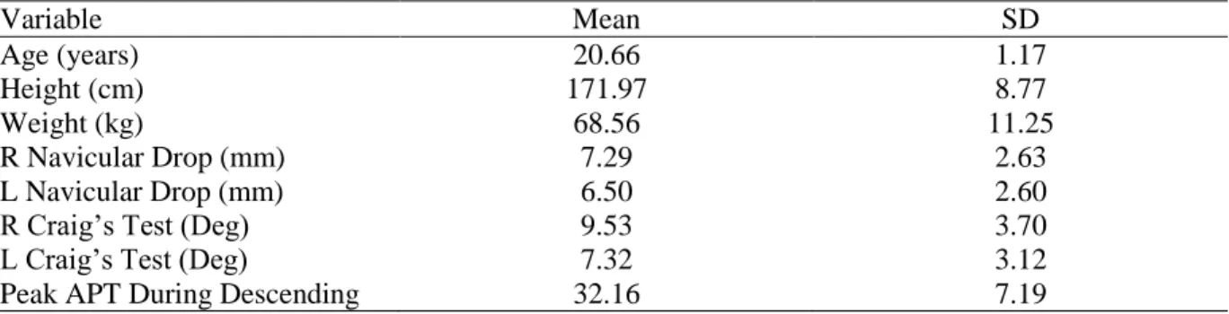
Table 1 – Age, height, weight, sex, navicular drop, Craig’s test, and amount of peak APT during descending phase.
Variable Mean SD
Age (years) 20.66 1.17
Height (cm) 171.97 8.77
Weight (kg) 68.56 11.25
R Navicular Drop (mm) 7.29 2.63
L Navicular Drop (mm) 6.50 2.60
R Craig’s Test (Deg) 9.53 3.70
L Craig’s Test (Deg) 7.32 3.12
41 Phase (Deg)
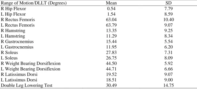
Table 2 – ROM, DLLT
Range of Motion/DLLT (Degrees) Mean SD
R Hip Flexor 0.54 7.79
L Hip Flexor 1.54 8.59
R Rectus Femoris 63.04 10.40
L Rectus Femoris 63.79 9.07
R Hamstring 13.35 9.25
L Hamstring 11.29 8.34
R Gastrocnemius 15.44 5.54
L Gastrocnemius 11.95 6.20
R Soleus 27.83 7.31
L Soleus 26.75 8.09
R Weight Bearing Dorsiflexion 44.50 5.92
L Weight Bearing Dorsiflexion 44.71 6.66
R Latissimus Dorsi 19.52 9.07
L Latissimus Dorsi 18.51 9.00
Double Leg Lowering Test 30.49 14.75
Table 3 – EMG during descending phase of OSA
EMG Mean SD
Rectus Femoris 44.32 24.60
Lateral Hamstring 9.88 5.03
Medial Hamstring 8.43 6.04
Gluteus Maximus 9.49 8.55
Illiocostalis 30.57 20.67
Longissimus 35.01 12.78
Table 4 – Joint angles during descending phase of OSA
Descending Phase Peak Value Descending Phase Displacement
Mean SD Mean SD
Hip Adduction -0.35 4.85 1.12 3.00
Hip Abduction -9.47 7.04 -8.00 4.95
Hip Internal Rotation 17.45 15.98 11.36 9.53
42
Variable R-Value P-Value
R Navicular Drop (mm) -.11 .51
L Navicular Drop (mm) -.07 .68
R Craig’s Test (Deg) .20 .23
L Craig’s Test (Deg) .21 .21
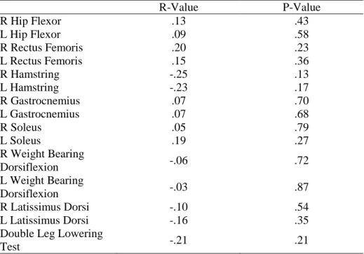
Table 6 – Correlations between Peak APT and . ROM
R-Value P-Value
R Hip Flexor .13 .43
L Hip Flexor .09 .58
R Rectus Femoris .20 .23
L Rectus Femoris .15 .36
R Hamstring -.25 .13
L Hamstring -.23 .17
R Gastrocnemius .07 .70
L Gastrocnemius .07 .68
R Soleus .05 .79
L Soleus .19 .27
R Weight Bearing
Dorsiflexion -.06 .72
L Weight Bearing
Dorsiflexion -.03 .87
R Latissimus Dorsi -.10 .54
L Latissimus Dorsi -.16 .35
Double Leg Lowering
Test -.21 .21
Table 7 – Correlations between APT and. EMG
EMG R-Value P-Value
Rectus Femoris .15 .36
Lateral Hamstring .03 .86
Medial Hamstring .16 .34
Gluteus Maximus .40 .01*
Illiocostalis .21 .22
Longissimus .33 .05*
Table 8 – Correlations between APT and hip kinematics
Peak Displacement
R-value P-Value R-Value P-Value
Hip Front Descending
-.07 .69 -.05 .76
Hip Trans Descending
.28 0.09 .20 .23
Hip Trans Ascending
43
Table 9 – Correlations between APT and. Rectus Femoris ROM in Females
Peak Descending Phase Peak Ascending Phase
R-Value P-Value R-Value P-Value
R Rectus Femoris .39 .09 .470 0.04*
44 CHAPTER 5 DISCUSSION
Our research showed a significant positive correlation between increased activity in the
gluteus maximus and longissimus muscles and anterior pelvic tilt. This is contrary to our original
prediction for the gluteus maximus but consistent with that for the longissimus. It does not appear as
though bony characteristics, range of motion, the double leg lowering test, kinematics, or other
muscle activity are associated with anterior pelvic tilt during the descending phase of an OSA in this
particular group of individuals. Because increased gluteus maximus and erector spinae activity are
not considered risk factors for ACL injury, anterior pelvic tilt during the OSA does not appear to be a
relevant risk factor to screen for. Although our results showed only two significant associations
between these factors during the descending phase of the squat, we did notice a trend toward
significance in the correlation between anterior pelvic tilt and peak hip internal and external motion.
Based on this information we decided to look at the correlations during the ascending phase of the
overhead squat assessment, and this time the correlations became significant. (Table 8)
Integrate
These findings moderately support the previous findings and theories of authors who reported
associations between anterior pelvic tilt and femoral internal rotation (Duval et al., 2010; Hruska,
1998; Ireland, 2002). These authors also suggested that anterior pelvic tilt could be caused by
pronation or femoral anteversion but our results did not support those theories.
Previous literature has suggested and our hypothesis predicted, increased activity of the rectus
femoris and erector spinae paired with decreased gluteus maximus and hamstring activity could
45
et al. (2002) in regards to increased erector spinae activity being associated with femoral internal
rotation. In Mayer’s study, internally rotating the participant’s legs during a back extension exercise
increase lumbar extensor activity by 18% compared to external rotation. Ours results showed a
correlation between longissimus activity and hip internal rotation (r(38)=0.33, p=0.04), although only
during the ascending phase of the overhead squat assessment, which was not part of our hypotheses.
In addition, what is presented as common knowledge in Neumann (2010) with regards to
agonist/antagonist interactions and muscular inhibitions, did not play out in exactly the same fashion
in our study, with the increased longissimus activation being the only expected significant
relationship found with anterior pelvic tilt.
Range of motion showed no significant associations with anterior pelvic tilt in the group as a
whole, but when the males and females were separated, there was a significant positive correlation
between increased anterior pelvic tilt and increased rectus femoris range of motion, which is opposite
of what we expected to find. (Table 9) In theory as the rectus femoris is shortened, it should draw the
pelvis into a more anteriorly rotated position, but in our study increased anterior pelvic tilt was
associated with those females who had a greater range of motion, suggesting that potentially different
neuromuscular factors could contribute to increase APT in males and females.
It does not appear as though anterior pelvic tilt is associated with tight triceps surae either.
Bell et al. (2008) and Macrum et al. (2012) found correlations between decreased ankle dorsiflexion
and medial knee displacement, which in theory could increase anterior pelvic tilt, but our results
showed no such association. Padua et al. (2012) found increased activity of the gastrocnemius and
tibialis anterior in participants displaying medial knee displacement during an overhead squat,
suggesting that this would be associated with decreased ankle range of motion, but once again there
was no apparent consequence up the kinetic chain to the pelvis in our results as we had theorized
46
The latissimus dorsi does not appear to play any role in influencing anterior pelvic tilt, as
none of our results showed any significance. This again goes against our theory that decreased range
of motion in the latissimus dorsi could lead to an increased lordotic curve and therefor increased
anterior pelvic tilt.
The double leg lowering test showed no significant correlations with anterior pelvic tilt
despite the theory that weak abdominals would allow for a greater degree of anterior pelvic tilt along
with increased activity in the erector spinae.
The lack of significance between the navicular drop test and anterior pelvic tilt could
potentially be the result of the fact that our sample on average did not have a clinically relevant drop.
Without excessive navicular drop to cause changes up the kinetic chain it would stand to reason that
nothing out of the ordinary would be observed. Similarly, in regards to femoral anteversion, our
sample did not have one participant who displayed excessive anteversion. On the contrary, the
average for our sample was retroverted.
Interpret
The two significant findings in this study both related to EMG activity, specifically the
gluteus maximus and longissimus muscles. It is important to keep in mind that increased activity
does not necessarily mean that the muscle is also creating more force. As stated before, the increased
longissimus activity was predicted by our hypothesis, but the gluteus maximus activity was opposite
of what we had anticipated. This could potentially be due to the fact that as the pelvis tilts anteriorly,
the gluteus maximus is lengthened eccentrically, thereby eliciting the stretch reflex, which leads to
increase GMAX activity. It could also mean that the gluteus maximus was weak in our participants
and therefore must work harder during the overhead squat assessment. Because of the increased
activity of the gluteus maximus, the hamstrings may not need to do as much work, and most likely
functions more as a stabilizer at the knee, having no real effect on pelvic tilt. The lack of correlation
47
laterally of the three erector spinae muscles and therefore would contribute least to an increase in the
lordotic curve. Finally, the lack of correlation between rectus femoris activation level and anterior
pelvic tilt could be attributed to the fact that because the gluteus maximus was so active it reciprocally
inhibited the rectus femoris.
The lack of correlations between anterior pelvic tilt and range of motion could be explained
by several instances. On average our participants had tight iliopsoas’ and tight rectus femoris (except
when compared to values given by Harvey (1998)), which in theory should increase anterior pelvic
tilt. However, if there is a certain point in passive hip range of motion where inflexibility no longer
has an effect on anterior pelvic tilt, or there is a point beyond the average range of motion into flexion
than what our participants displayed, this could potentially explain the lack of correlations. The fact
that our subjects operated primarily in the midrange of motion at minimal load during the overhead
squat assessment could also provide an explanation. If the muscles aren’t being used to achieve end
range of motion at any given joint and there is not sufficient load to cause a challenge, any deviations
from the norm would be unlikely to show up. All other range of motion values were, on average,
within normal limits, so again, the lack of correlations could be the result of a lack of non-normative
participants.
The double leg lowering test did not show any significant correlations for perhaps two
reasons. In order to achieve the posterior rotation of the pelvis during an overhead squat assessment,
the rectus abdominis would have to overcome the reciprocal inhibition caused by the increased
activation of the erector spinae. Because none of the subjects were consciously recruiting the
abdominals during the overhead squat assessment and there was minimal load, it wouldn’t necessarily
matter how well they performed in the much more difficult double leg lowering test, as the difficulty
of the two tasks are disparate. On the other hand, it could be that since our participants were all
physically active individuals, they may have been quite adept at controlling their pelvic tilt. In a
48
occurred almost immediately upon lowering of the legs and continued to increase as the legs were
lowered. Although we did not assess joint kinematics during DLLT, many of the participants were
able to maintain consistent pressure on the clinician’s hand until reaching the failing point. In
addition, Zannotti had participants hold onto the table above their heads in a shoulder flexed position,
potentially contributing to increased lumbar lordosis and pelvic tilt, while our participants kept their
arms at their sides.
Implications
Based on our results, several things are worth noting. First of all, it does not appear as though
the LPHC muscles function together as is commonly thought. The increase in gluteus maximus
activity in those with increased anterior pelvic tilt raises the question of just how much reciprocal
inhibition and agonist antagonist interactions come in to play at lower levels of physical activity.
While we tend to view these relationships as all-encompassing scenarios, perhaps they only take
effect when a certain stress threshold is reached, for example during actual sport participation or
weightlifting.
Second, if increased activity in a muscle is taken to mean that muscle is also in a shortened
position, the erector spinae should be stretched to minimize its effect on anterior pelvic tilt, as this
could theoretically decrease ACL injury risk. This seems counter-intuitive to the gluteus maximus
though, as it is not a commonly thought of as being inflexible, therefore the potential that the muscle
is weak and operating at near maximal capacity must be acknowledged.
Thirdly, in order for many risk factors to become apparent, fatigue or increased load may
need to be introduced to any assessment technique. Faulty movement patterns may not be apparent
during an overhead squat assessment, but may become blatantly obvious if weight is added or a
jumping task is used. The fitness level of our participants may have been a large part of the reason