Effects of HSV-1 Infection on Disparate Cell Signaling Pathways
Devon Arch Gregory
A dissertation submitted to the faculty of the University of North Carolina at Chapel Hill in partial fulfillment of the requirements for the degree of Doctor of Philosophy in the
Curriculum of Genetics and Molecular Biology.
Chapel Hill 2007
Approved by
Abstract
DEVON ARCH GREGORY: Effects of HSV-1 infection on disparate cell signaling pathways.
(Under direction of Steven L. Bachenheimer)
Herpes simplex virus (HSV) commandeers cellular machinery in order to replicate. Two pathways which are utilized by the virus are the NF B pathway and the DNA double strand break (DSB) response. The NF B pathway is activated in a sustained manner following infection. This activation leads to a suppression of apoptosis in certain cell types and may also assist in viral gene expression. The most common route of activation for NF B is through the IKK complex and I B. This dissertation examines the NF B pathway
upstream of I B, characterizing the role of IKK and IKK , the two kinase subunits of the IKK complex. Both were found to be necessary for NF B activation, though at different junctions in the pathway. Potential upstream effectors of the IKK complex are also characterized. PKR, NIK and Tak1 are kinases that activate the IKK complex following various normal cellular stimuli. None play a role in activation following HSV infection however, suggesting a unique method of activation by HSV.
The cellular DSB response normally recognizes breaks in cellular DNA in order to repair the damage and initiate cell cycle arrest or apoptosis to maintain the integrity of the cell and organism. This dissertation examines the machinery involved in the initial detection and signaling of the DSB response following HSV infection. HSV activates the DSB
Acknowledgements
Table of Contents
List of Tables... vii
List of Figures... viii
List of Abbreviations... ix
Chapter I. INTRODUCTION... 1
HSV-1... 2
NF B... 6
NF B and viruses... 8
Double Strand Break Response... 11
Viruses and the DNA DSB response... 13
Materials and Methods... 17
II. CHARACTERIZATION OF NF B ACTIVATION FOLLOWING HSV INFECTION... 22
Differential roles of IKK and IKK in NF B activation... 23
NF B activation in PKR-/- MEFs... 26
NF B activation in the presence of dominant negative TAK1 and NIK... 27
Significance of Results... 28
The requirement of de novo gene expression for
the DSB response and Mre11 loss... 38
DSB response activation and Mre11 loss are dependent upon a late viral event... 39
Importance of the DSB response for HSV replication... 40
Significance of results... 41
IV. CHARACTERIZATION OF MRE11 LOSS FOLLOWING HSV INFECTION... 54
Mre11 loss is independent of ICP0, the proteasome and lysosome... 54
Mre11 loss is cell type specific... 56
Analysis of Late mutants for Mre11 loss... 56
Camptothecin causes Mre11 loss... 58
Significance of Results... 60
V. CONCLUSIONS... 69
NF B activation... 69
DBS response activation... 73
Mre11 loss... 74
List of Tables
Table
List of Figures
Figure
2.1 IKK is the primary kinase for I B loss and p65 nuclear translocation... 31 2.2 KK and IKK are necessary for NF B
DNA binding activity following infection... 32 2.3 PKR is not required for NF B activation following HSV infection... 34 2.4 Nik and Tak1 are not necessary for NF B activation... 35 3.1 The DSB response is activated and Mre11 lost
following HSV infection... 45 3.2 Mre11 and Nbs1 localize to sites of viral DNA replication... 46-48 3.3 Activation of the DSB response and Mre11 loss
following infection require de novo gene expression... 50 3.4 The DSB response activation and Mre11 loss require viral DNA
replication... 51 3.5 ATM activation is not required for Mre11 loss,
nor late gene expression... 52 4.1 Mre11 loss does not require ICP0 or
List of Abbreviations
ActD Actinomycin D
AT Ataxia Telangiectasia
ATCC American Type Culture Collection A-TLD Ataxia Telangiectasia-Like Disorder ATM Ataxia Telangiectasia Mutated ATMi KU-55933, ATM inhibitor ATP Adenosine tri-phosphate
ChIP Chromatin IP
CPT Camptothecin
CycloX Cycloheximide
DMEM Dulbecco's Minimal Essential Medium
DN Dominant Negative
DNA Deoxyribonucleic Acid
DNA-PKcs DNA dependent Protein Kinase catalytic subunit
DNS Data Not Shown
DSB Double Strand Break
E Early
EBV Ebstein-Barr Virus
eIF2 eukaryotic translation Initiation Factor 2 EMSA Electrophoretic Mobility Shift Assay
EtOH Ethanol
gB, gC, ... Glycoprotein B, Glycoprotein C, ... HCMV Human Cytomegalovirus
HIV Human Immunodeficiency Virus HFK Human Foreskin Keratinocyte hpi hours post infection
HR Homologous Recombination HSV Herpes Simplex Virus
HSV-1 Herpes Simplex Virus Type 1 HSV-2 Herpes Simplex Virus Type 2 ICP Infected Cell Protein
IE Immediate Early
IF Immunofluorescence microscopy
I B Inhibitor of B
IKK Inhibitor of B Kinase
IL Interleukin
IP Immunoprecipitation
KSHV Kaposi Sarcoma-associated Herpes Virus
L Late
LTR Long Terminal Repeat LPS Lipopolysaccharide MEF Mouse Embryo Fibroblast MOI Multiplicity of Infection
mRNA messenger RNA
NBS Nijmegen Break Syndrome NF B Nuclear Factor B
NHEJ Non-homologous End Joining PAA Phosphonoacetic acid
PAGE Polyacrylamide Gel Electophoresis PBS Phosphate Buffered Saline
PCR Polymerase Chain Reaction pfu plaque forming units PGA1 Prostaglandin A1
PKR double-stranded RNA-depending Protein Kinase RNA Ribonucleic Acid
SDS Sodium Dodecyl sulfate siRNA small interfering RNA
ss single-strand
SV40 Simian Virus 40
TBST Tris Buffered Saline with Tween-20 TNF Tumor Necrosis Factor
UL Unique Long
UNC University of North Carolina
Us Unique Small
VP16 Virion associated Protein 16
CHAPTER 1
INTRODUCTION
Herpes simplex virus type 1 (HSV-1) is estimated by the CDC to infect up to 90% of the US population. It is closely related to herpes simplex virus type 2, sharing a homologous genome with 40% sequence identity. Normally, HSV-1 causes oral fever blisters or cold sores, while HSV-2 is associated with genital lesions; however, the viruses are capable of infecting other sites and producing other pathologies. Most common of these are keratitis from ocular infections that can lead to blindness and herpetic whitlow from infection of one’s hands or fingers, which is common among health care professionals. The most severe pathologies occur when HSV enters the central nervous system, causing encephalitis that can result in death, and when HSV is contracted by neonates.
reoccurrences are normally mild in comparison to the initial infection and are sometimes even asymptomatic.
Currently there are effective drugs to control HSV, both for initial infection and recurrences; however, there is no method to remove latent virus and resistances to these drugs can develop. There are a number of vaccines against HSV and more in development, however to date none are efficacious enough to be practical. Obviously, by studying the mechanisms of infection of HSV, we hope to develop additional treatments and perhaps eventually even a cure. Better understanding viral infection can also help us better understand the workings of the host as well as help us in developing tools for research and treatment of other diseases.
HSV-1
HSV-1 is one of the eight known human herpesviruses and is the prototypical alphaherpesvirus. The other two known human alphaherpesviruses are HSV-2 and varicella-zoster virus. The betaherpesviruses include human cytomegalovirus (HCMV) and human herpesviruses 6 and 7. The human gammaherpesviruses are Epstein-Barr virus (EBV) and Kaposi sarcoma-associated herpes virus or human herpes virus 8 (KSHV). HSV-1 has a linear double stranded DNA genome of roughly 152kb, which encodes over 80 genes. In the virion, the genome is surrounded by an icosahedral capsid. Surrounding the capsid is the tegument layer, comprised of many proteins encoded by the virus. The envelope, comprised of a lipid bilayer embedded with glycoproteins surrounds the tegument and capsid.
number of receptors can interact with gD, including the TNF receptor family member HVEM, immunoglobulin family members HveB (nectin-2), HveC (nectin-1) and HIgB, and 3-O-sulfated heparin sulfate (21, 43, 100, 138). The above has been characterized for cell surface mediated entry, though HSV can also enter cells through an endocytic route (102). Regardless, the tegument and nucleocapsid are eventually released into the cytoplasm. The capsid is then transported via the microtubule network to the nucleus in order to release the viral DNA through a nuclear pore (122, 139).
112, 120). ICP27 also activates several cellular signal transduction pathways (54, 96, 109). ICP47 is the only IE gene that does not function to regulate gene expression. Instead it blocks peptide loading onto transporter of antigen presentation (TAP) complexes in order to promote immune evasion (39, 59).
Dependent upon IE gene products, expression of the second class of HSV-1 genes occurs. These E genes encode components of the viral DNA replication machinery, including the viral DNA polymerase UL30, single stranded DNA binding protein ICP8 (also called UL29), the processivity factor UL42, origin-binding protein UL9 and the helicase-primase complex consisting of UL5, UL8 and UL52. These localize to so-called prereplicative sites, likely previous ND10 sites, where viral DNA replication is thought to be initiated. Active DNA replication expands these sites, forming replication compartments that fill most of the host nucleus. HSV DNA replication initiates dependent upon UL9, where bi-directional theta replication occurs. At some point however, DNA replication is thought to switch to a rolling circle and/or recombination dependent mechanism (165). The resultant viral DNA is a complex multi-branched concatameric structure.
NF B
All human pathogens must contend with their host's immune response, and HSV is no exception. Central to many of these barriers is nuclear factor Kappa B (NF B). NF B is a family of transcription factors that can positively and negatively regulate a number of genes involved in proliferation, apoptosis, inflammation, stress responses, innate and adaptive immune response, oncogenesis and development, all of which contain B elements, to which NF B binds, (44, 67, 105, 107). The exact function of NF B when activated is highly dependent upon the state of other signaling pathways, other transcription factors and the stimuli responsible for activating NF B (57, 113). The interplay between all these cellular components can determine whether apoptosis or an inflammatory response is initiated.
There are five different NF B subunits that can dimerize to form a functional transcriptional factor; p65 (relA), RelB, c-Rel, p50 and p52. All the subunits contain a Rel-homology domain which is necessary for DNA binding and dimer formation. Both p50 and p52 can form homodimers or dimerize with any other subunit. The other three subunits only dimerize with p50 or p52 and also contain transactivation domains. Dimers of p50 and p52 can still activate gene expression by interaction with the transcriptional co-activator BCL-3 (10).
signaling cascades that can trigger NF B activation. Normally however, these signaling pathways converge at a signaling bottleneck, the IKK complex. After this point, NF B activation again is somewhat varied, though there is one pathway that is most frequently observed across many cell types, known as the classical or canonical pathway. Because of its relevance here and the complexity of the many other routes of activation, it will be the only one described.
The classical pathway signals through the IKK complex consisting of inhibitor of B kinase (IKK ), IKK and IKK . IKK and IKK are kinases that target NF B subunits and a class of regulator proteins known as Inhibitor of B (I B). There are 5 I B proteins; I B , I B , I B , I B and BCL-3. In the classical pathway, I B binds to a p65/p50 heterodimer and masks p65’s nuclear localization sequence. Phosphorylation of I B by IKK leads to its poly-ubiquitination and proteasome dependent degradation. Once released from I B NF B localizes to the nucleus. It is further regulated by post-translational modifications. Phosphorylation of serine 276 results in a switch of p65’s interaction from histone deacetylases such as HDAC to histone acetyl-transferases such as CBP/p300, promoting gene transactivation. Serine 529 and 536 also play a role in promoting transactivation, while serine 311 is important for DNA binding. Acetylation at a number of sites also promotes NF B transactivation (reviewed in (57, 113, 132, 161)).
the IKK complex and TAK1, which leads to activation of both (136). Once IKK has been activated, NF B signaling proceeds as described above.
NF B and viruses
NF B is very significant for microbial pathogens. Its role in the immune response is a important hurdle to overcome, and many viruses and bacteria have developed mechanisms to interfere with NF B activation (46, 62). NF B activation can also serve to benefit viruses however, and several viruses activate and exploit the pathway (61). Perhaps the most well known case is HIV-1. HIV-1 contains B elements in its LTR that, in conjunction with other transcription elements, are necessary for efficient transcription. HIV-1 can cause NF B activation through several mechanisms. HIV-1 binding to its receptor, CD4, which can signal to the NF B pathway when activated, causes induction of NF B. HIV-1 Tat induces NF B activation through engagement of cellular signaling cascades involving p56ick, while Vpr and Nef induce IL-8 and IL-2 signaling respectively, causing NF B activation. And finally, activation of the immune response by HIV-1 also results in NF B activation.
HSV’s ability to activate NF B was first observed in studies looking at HSV-1 activation of HIV provirus (47). The activation is primarily of p65/p50 heterodimers necessary for efficient HSV replication (109, 130). More recently, two phases of NF B activation have been reported. The initial activation is likely triggered by HSV engagement of receptors capable of signaling to NF B, and is transient in duration (4, 98, 155). The second activation of NF B is dependent upon I B turnover and is sustained for the length of infection (109). Though ICP4 (vi13) and ICP27 (n59r) deficient mutants are both impaired for NF B DNA biding, n12, a ICP4 mutant, shows only partial impairment of I B loss while d27-1, a ICP27 null mutant is unable to cause I B loss (55, 109). Also, an HSV mutant expressing only ICP27, ICP22 and ICP47 (d100) causes I B degradation by 24 hpi. ICP27’s activity that leads to NF B activation was mapped to a domain within residues 12 to 63 and is also in part dependent upon activation of JNK and p38 signaling pathways, as pharmacological inhibitors of JNK and p38 also reduce I B loss (55).
knocked out for p65, p50 or both also do not undergo apoptosis following wt or d120 (a ICP4 deleted mutant) infection, while the latter causes apoptosis in other cell types (148).
The conclusions from the latter experiments are problematic however. R5104 varies from wildtype HSV by several changes, and it is unclear how these may effect both the induction and inhibition of apoptosis separate from NF B activity (16). It is difficult to interpret the results from the MEFs because while they do not undergo apoptosis following wt infection, as one might expect if NF B was necessary to prevent apoptosis following infection, they also do not undergo apoptosis following d120 infection. Infection with d120 does not activate NF B and thus should cause apoptosis regardless of NF B's presence in the cell. This result suggests that these cell lines have gained a function that either prevents induction of apoptosis due to HSV infection or inhibits apoptosis independent of NF B. Because these cell lines are sensitive to TNF or sorbitol apoptosis induction, the latter is unlikely (148).
Currently, the pathways leading to NF B activation are poorly understood and have been part of my dissertation research. IKK is activated following infection, and treatment of infected cells with prostaglandin A1(PGA1) inhibited both IKK and NF B activation (3). In
The reason for this discrepancy is unclear. I have also characterized two other potential upstream kinases, TAK1 and NIK, and shown that they are not relevant for HSV activation of NF B.
Double Strand Break Response
Many types of DNA damage threaten the genomic integrity of a cell. DNA DSBs are one of the more dangerous types of damage, resulting from ionizing radiation, genotoxic chemicals, collapsed replication forks (often due to pre-existing damage in the DNA) and purposeful DNA cleavage for cellular processes, such as V(D)J recombination. If not repaired promptly and properly, DSBs can result in loss of large portions of chromosomes and in translocations and large deletions, potentially causing cell death or deregulation, a worse fate for the organism if cancer results. To avoid these fates, cells have a complex system in place to quickly detect DSB and respond to them.
ATM also phosphorylates 53BP1 and BRCA1, which act as additional scaffolding and are important to recruit and aid in activation of some of ATM’s other targets, such as p53 and the Chk proteins (38, 41, 94, 99).
In mammalian cells, repair normally occurs via non-homologous end joining (NHEJ), resulting in two broken DNA ends being ligated together regardless of sequence. This non-specificity can lead to small deletions or insertions. For this process of repair, the heterodimeric Ku (Ku70/86) recognizes the DNA ends while the actual rejoining of the ends occurs via the Xrcc4/LigaseIV complex aided by polµ(63, 92, 108, 158). Though we know these components are necessary for NHEJ, the full details of their interactions are as yet unknown. Another protein that is sometimes involved in NHEJ is the catalytic subunit of DNA dependent protein kinase (DNA-PKcs). DNA-PKcs seems to be necessary for a subset of NHEJ that does not involve the MRN complex.
form a duplex with the complimentary strand from the broken DNA, all promoted by Rad51 and Rad54. Strands exchange between the homologous DNA occurs and the crossed DNA strands are resolved by being cut and rejoined to the appropriate new dsDNA molecule.
In addition to facilitating repair, ATM also activates cell cycle checkpoints and in extreme cases, apoptosis. ATM phosphorylates ATR, Chk2, Chk1 and p53, all of which can inhibit cell cycle progression (23, 66, 68). ATR can also be activated through interactions with the MRN complex and is a potent activator of Chk1 (94). Chk1 and Chk2 target Cdc25 proteins to inactivate them (42). Normally Cdc25 proteins serve to positively regulate Cdk activity, necessary for cell cycle progression, by removing inhibitory phosphorylations (56, 169). p53 can also inactivate Cdk proteins through its transcriptional regulation of p21, which directly binds to Cdk/cyclin complexes and prevents ATP binding (124). Apoptosis can also be induced by p53, though this requires either additional signals or differential activation as this only occurs in cases of irreparable damage (90).
Viruses and the DNA DSB response
Many previous reports have found interactions between viruses and host cell DNA DSB response machinery. In some instances, cellular DSB machinery is detrimental to a virus, and as such the virus in question has mechanisms to prevent the actions of the cellular response machinery. In other cases, viruses are able to harness the cellular machinery for their own advantage. Whether the cellular machinery is harmful or beneficial for a virus depends on the life cycle of the virus, and in some instances the machinery can be both.
activity that is important in the DNA damage response (76). More recent reports have elucidated a role for SV40 large T antigen in hindering the initial stages of the DNA damage response. Study of cardiomyocytes transformed with SV40 large T antigen found that Mre11 co-immunoprecipitated with SV40 large T antigen and that transformed cell lines had greatly reduced Mre11 protein, though not mRNA. Nbs1 and Rad50 in contrast seemed not to have decreased protein levels (28). In fibroblasts, the presence of SV40 large T antigen was noted to cause a decrease in Mre11 foci formation following IR treatment (166). Unfortunately, none of these studies ventured into actual SV40 infection, so the benefit these actions of large T antigen have on SV40 replication remain in the realm of speculation.
One study of interesting note did find a role for SV40 large T antigen in the context of infection. Wu and colleagues discovered that SV40 large T antigen directly binds to Nbs1. This binding prevents Nbs1, and presumably the entire MRN complex, from preventing refiring of origins of replication that lead to erroneous duplication during S phase. SV40 relies entirely on host DNA replication machinery to duplicate its genome, so loss of replication control allows the SV40 genome to undergo exponential replication (119, 129, 173). Though it is unknown whether this function of the MRN complex is tied to a DNA damage response, these results show an interesting interaction between a virus and DNA damage response machinery.
E1B55K/E4orf6 (31). This late loss of the MRN complex prevents detection of DSB breaks and activation of ATM. For the virus, this failure of the DNA damage response allows its linear progeny genomes to escape concatamerization, which would prevent their packaging. Earlier in infection however, E4orf3 binds the MRN complex and sequesters it to foci throughout the nucleus. This activity of E4orf3 is necessary for viral DNA replication to occur (73).
Recently Kudoh and colleagues characterized components of the DNA damage response during the EBV lytic cycle (95). They found that ATM was activated and phosphorylated Chk2, p53, H2AX and Nbs1. They also observed that activated ATM and MRN complex colocalized with replicating EBV DNA. Interestingly, EBV BZLF-1, a major transcriptional transactivator, counters the down stream effects of cell cycle checkpoint induction by ATM by binding and inhibiting p53 and by activating promoters of the cell cycle, such as E2F-1, cyclin E and Cdc25A (80, 106). These finding indicate that activation of the cell cycle checkpoint by the damage response would be detrimental to EBV replication. However, whether other components such as the MRN complex have any affect on EBV replication is not yet known.
protein, ICP8, which is present at sites of DNA replication, has also been shown to interact with DNA-PKcs, Rad50, Ku, Mre11, RPA and BRCA1 (84, 137, 164). Similar to EBV, HSV-1 also causes activation of many DSB response factors, in this case Nbs1, RPA, ATM, Chk2 and 53BP1 (85). These activation events appear to occur by the MRN complex’s activation of ATM, independent of active viral DNA replication. The importance of this activation is underlined by a report by Lilley et al. that demonstrates the inability of virus to replicate in cells deficient for the MRN complex or ATM (137). The importance of ATM has been disputed however, as Shirata et al. reported that while cells from individual with AT or NBS are defective in supporting viral infection, specific knockdown of ATM with siRNA had no effect on viral replication (154). Interestingly, cells deficient in Ku actually facilitate viral replication better than wild type (wt) cells (163). In contrast, HSV inhibits ATR, and thus Chk1, activation (165).
As of yet, we do not know if the DSB response machinery might have an effect on late events, such as processing and packaging of replicated DNA.
Materials and Methods
Cells: Spontaneously immortalized mouse embryo fibroblasts (MEFs) derived from normal mice or mice with targeted deletions in IKK , or IKK (172) were obtained form Tal Kafri (UNC-Chapel Hill). MEFs from normal mice or mice with a targeted deletion of the double-stranded RNA-activated protein kinase PKR (37) were obtained from Bryan Williams (Cleveland Clinic). Diploid human embryonic lung fibroblasts (HELs) and HeLa cells were obtained from ATCC. The above cells were maintained in Dulbecco’s Minimal Essential Medium (DMEM) supplemented with 10% fetal bovine calf serum, 100U/ml penicillin, 1% streptomycin, and 1% L-glutamine. Hep2 cells obtained from ATCC were maintained in Minimal Essential Medium supplemented as for DMEM. CV-1 cells were obtained from Saul Silverstein (Saul Silverstein) and grown in DMEM supplemented with 5% newborn calf serum, 100U/ml penicillin, 1% streptomycin, and 1% L-glutamine. Telomerase immortalized human foreskin keratinocytes (HFKs) were a gift from Shannon Kenney (University of Wisconsin-Madison) and Nancy Raab-Traub (UNC-Chapel Hill) with permission from A. J. Klingelhutz who derived the cell line as previously described (104). HFKs were maintained in Keratinocyte-SFM from Gibco supplemented with EGF and bovine pituitary extract.
Pittsburgh); UL17 mutant UL17 (Joel Baines, Cornel Univ.); UL25 and UL28 mutants kUL25 and GCB (Fred Homa, Univ. of Pittsburgh); UL15, UL32 and UL6 mutants hr81-1, hr64 and hr74 (Sandy Weller, Univ. of Conneticut) and UL26 mutants dZ and H61E (Prashant Desai, John Hopkins Univ.).
Antibodies: Rabbit polyclonal antibodies against IKK 470, sc-7607), IKK (H-744, sc-7218), I B- (C-21, sc-371), PARP (H-250, sc-7150) were purchased from Santa Cruz. Polyclonal antibody against p65 (100-4165) was purchased from Rockland. Monoclonal antibodies against Rad50 (13B3) and Mre11 (12D7) and polyclonal antibody against Mre11 (GTX30294) were purchased from Genetex, Inc. Monoclonal antibody against Nbs1 (100-222) and polyclonal antibodies against phospho-Nbs1 (100-284) and phospho-ATM (100-307A2) were obtained from Novus Biologicals. Monoclonal antibody DNA-PKcs (05-423) was purchased from Upstate. Monoclonal antibody against ICP0 (H1A027-100) was purchased from Virusys. Monoclonal antibodies against ICP4 (1101) and ICP27 (H1113) were purchased from Rumbaugh-Goodwin Institute. Polyclonal antibody against ICP8 (3-83) was a gift from David Knipe. Polyclonal antibody against gC (R47) was a gift from Gary Cohen and Roselyn Eisenberg. Polyclonal antibody against VP16 was purchased from Clontech. Goat anti-rabbit and anti-mouse HRP-conjugated secondary antibodies were purchased from Amersham Biosciences
of herpesvirus DNA polymerase)), 10µg/mL cycloheximide, 5 µg/mL Actinomycin D, 5mM Caffeine, 10µM KU-55933 or, NH4Cl or MG132 (Sigma) at the indicated concentrations.
Monolayers were harvested by washing with cold PBS and then scraping directly into 1X SDS sample buffer [3.85mM Tris-base (pH 6.8), 9.1% -mercaptoethanol, 1.82% SDS, 4.6% glycerol, and 0.023% bromophenol blue (in 100% EtOH)] and boiling for 5’. In the case of HELs, media from monolayers was collected and the monolayers were treated with trypsin until the cells detached. The cells were then suspended in PBS, combined with the previously collected media and were spun down and washed once with PBS before addition of 1X SDS sample buffer and boiling as above. Fractionated cytoplasmic and nuclear extracts were prepared as described previously (60, 104, 109). Briefly, cells were collected by trypsinization, spun through a cushion of bovine calf serum, washed twice in PBS, resuspended in 3 packed cell volumes of CE buffer (10 mM HEPES, pH 7.8, 1 mM EDTA, 60mM KCl, 1 mM PMSF, 0.1% NP-40, 25% glycerol, 0.4mM NaF, 0.4 mM Na3VO4, 10µM
pepstatin, and Complete Protease Inhibitor Cocktail [Roche]) and incubated on ice for 10’. Following a 10 sec spin in a bench-top microcentrifuge, the supernatant was collected and the nuclear pellet resuspended in CW buffer (CE buffer without NP40 or glycerol), and repelleted. Nuclei were resuspended in 2 packed cell volumes of NE buffer (20mM Tris-HCl, pH 8.0, 420 mM NaCl, 1.5 mM MgCl2, 0.2 mM EDTA, 0.5 mM PMSF, 25% glycerol, and
phosphatase and protease inhibitors as described above). After incubation on ice for 10’, nuclei were pelleted at 60,000 rpm at 4oC for 20’ in a Beckman TLA100.3 rotor and the nuclear supernatant recovered.
Life Sciences). Ponceau S (Sigma) was used to visualize total protein and insure no discrepancies in protein equivalences. Membranes were blocked in 5% milk in TBST (150mM NaCl, 20mM Tris [pH 7.6], 0.05% Tween 20) then washed and probed with appropriate primary antibodies, followed by further washing and probing with the appropriate secondary antibody, all in TBST. After final washing in TBST, signal was detected using Perkin-Elmer Western Lightening substrate. Multiple exposures were made on Kodak BMR film to insure the linear range was obtained. Images were scanned and processed using Adobe Photoshop.
Electrophoretic mobility shift assay (EMSA): Preparation of nuclear extracts and mobility shift assays for NF B were performed as described previously (50). Nuclear extract were incubated with a radiolabeled probe containing a B-binding site TGGGGATTCCCCA in buffer containing 10mM Tris-HCl (pH 7.9), 50mM NaCl, 0.5mM EDTA, 10% glycerol, 1 mM DTT, and 2µg poly (dIdC)·(poly dIdC). Following 20’
incubation at room temperature, aliquots were fractionated at 4oC on non-denaturing 4% polyacrylamide gels prepared in 0.25X TBE [1X TBE is 10mM Tris base (pH 8.3), 9mM boric acid, and 2mM EDTA]. Gels were then placed on 3MM paper, dried under vacuum and heat, and exposed to BMR film at –70oC.
medium was aspirated from the wells and plaques stained with 0.8% crystal violet in 50% ethanol.
Immunofluorescence: Cells were seeded on glass coverslips and allowed to grow until confluent. The monolayers were infected at low multiplicity (MOI of .1 or .01). At the indicated times, cells were washed with PBS, then fixed with 5% paraformaldehyde in PBS for 10 minutes followed by permeabilization with 1% Triton X-100 in PBS. The cells were then incubated in blocking buffer (2.5% normal goat serum, 2.5% normal horse serum and 1% gelatin in PBS) for at least 30 minutes, probed with the indicated primary antibody, and then washed and probed with either Texas red conjugated anti-rabbit or FITC conjugated anti-mouse antibody (Santa Cruz), all in blocking buffer. Primary antibodies were omitted for control cells. Following final washing with PBS, the coverslips were mounted and allowed to set before visualization with an Olympus FV500 confocal laser scanning microscope.
Real time PCR: Cell monolayers were infected at an MOI of 5 as above and then harvested using Promega Wizard SV Genomic DNA Purification System to isolate cellular and viral DNA. Primers against c-Myc (TCAAGAGGTGCCACGTCTCC,
TCTTGGCAGCAGGATAGTCCTT) and UL19 (AAACATCGCTGCCTGGAG,
GGGGCGCTTAAACTGTACG) were designed by Dirk Dittmer. DNA and primers diluted 1:200 were mixed with SyberGreen and run on an ABI prism 7000 for 40 cycles at 95o denaturing for 30" and 55oannealing for 1'. 7000 systems SDS software was used to collect cycle data. Fold viral DNA copy was calculated based on dCtof UL19 normalized to the dCt
CHAPTER 2
CHARACTERIZATION OF NF B ACTIVATION FOLLOWING HSV INFECTION
[A portion of the results presented here were published in Gregory et al., 2004 (50)]
At the outset of our research presented here, it had long been known that NF B was activated by HSV infection, but little was known regarding the cause or reason for NF B activation. A report by Amici et al had recently demonstrated the activation of the IKK complex following HSV infection by kinase assay of immunoprecipitated IKK complexes (109). To test whether IKK activity was causing NF B activation following HSV infection, they used PGA1, a prostaglandin, to inhibit IKK activity and assess for NF B activation by I B turnover, NF B gel shift activity and NF B reporter activity. In all cases, PGA1 treatment caused loss of IKK activity and failure of NF B activation. They concluded from this that IKK was behaving normally as part of the canonical NF B pathway, phosphorylating I B and causing its degradation. They also demonstrated, as have others, that loss of NF B activation greatly reduced virus yield, concluding it to be a necessary component of HSV infection (125).
They did assay for toxicity of PGA1 in their HSV system and claimed no affect. However, their results do show a marked reduction in ICP4 protein levels, which has been shown to contribute to NF B activation, as well as a general failure of HSV to create a normal [35S] methionine-labeled polypeptide profile (159). Due to these results, the contribution of IKK to NF B activation and HSV infection remained unclear.
To clarify the role of the IKK complex in NF B during HSV infection and the involvement of its common upstream effectors, we sought to use a genetic approach. We obtained wild type (wt) mouse embryo fibroblasts (MEFs) or MEFs knocked out for either IKK or IKK . These cell lines were characterized for their ability to facilitate HSV infection and NF B activation. Our results indicated that IKK is indeed necessary for NF B activation, with each kinase subunit uniquely contributing to infection. We also sought to determine if PKR, NIK or TAK1, known upstream kinases of IKK, contributed to NF B activation. MEFs knocked out for functional PKR and cells transiently transfected with dominant negative NIK or TAK1 constructs were characterized for NF B activation following infection.
Differential roles of IKK and IKK in NF B activation
control for nuclear extracts. No IKK proteins were detected in the nuclear fractions (Data not shown (DNS)). Similarly, PARP was used as our nuclear loading control and cytoplasmic purity control and was also not detected in our cytoplasmic fractions (DNS). I B is the IKK complex’s immediate down stream target and is degraded after IKK dependent phosphorylation. I B was lost in wt and IKK -/- MEFS following both HSV infection and TNF treatment. IKK -/- MEFs however did not lose I B . Translocation of p65 from the cytoplasm to the nucleus followed a pattern similar to I B loss, as expected. Both wt and IKK -/- MEF lysates had an increase in nuclear p65 upon HSV infection or TNF treatment. Both also had increased total p65 following TNF treatment, possibly due to Sp1 dependent activation of the p65 gene (109). One difference between these two cell lines was the portion of p65 translocated. While wt MEFs translocated the majority of p65 to the nucleus, a significant portion was still detectable in the cytoplasm. This was not the case for IKK -/-, where nearly all the p65 was translocated to the nucleus. This difference may be due to the apparent smaller pool of total endogenous p65 in the IKK -/- cells. The IKK -/- MEFs had some translocation of p65 to the nucleus following HSV infection or TNF treatment, though much reduced in comparison to the proportions translocated in the other two cell lines, which was consistent with the failure to turnover I B in the IKK -/- MEFs. These results suggest that IKK is the dominant subunit responsible for phosphorylating I B and causing NF B nuclear translocation following HSV infection and may indicate that some upstream signaling components to IKK following infection are the same as those involved in TNF or IL signaling, which also primarily signal through IKK .
-/-and IKK -/- lines had reduced late protein abundance however. The true late gC was nearly undetectable in both knockout lines, while the leaky late VP16 was reduced by roughly 5 fold in both lines. The failure to produce late proteins suggests either a failure to properly replicate the viral genome or disruption of viral late gene transactivation.
To assess NF B DNA binding ability, another marker of activation, the nuclear lysates were subjected to electrophoretic mobility shift assay (EMSA). NF B complexes were detected in all cell lines following either HSV infection or TNF treatment (Fig 2.2). However, the level of a slower mobility complex in both IKK -/- and IKK -/- lines was greatly reduced, slightly more so in the IKK -/- MEFs than the IKK -/- MEFs. Previously, p65 and p50 were reported to form the dominant NF B dimer activated by HSV (11, 45, 74, 175). In order to determine if that was the case in our system, we used antibodies to supershift the complexes. Antibody against E2F2 was used as an irrelevant control and did not shift either complex. Antibody against p65 shifted the more abundant slower mobility complex and antibody against p50 shifted both the abundant slow mobility complex and a less abundant fast mobility complex. This pattern suggests that HSV primarily activates p65/p50 heterdimers and to a lesser extent p50 homodimers in these MEFs. Some complexes appeared to not be shifted following incubation with the p65 or p50 antibodies. The make up of these complexes is unclear; possibly they are other NF B dimers or simply p65/50 or p50/p50 dimers that were not bound by antibody.
at 16 and 24 hpi respectively than wt MEFs. The IKK -/- yields were 16.3 and 28.5 fold less than wt. These results are consistent with the Western blot and EMSA data, showing a greater contribution of IKK to NF B activation. However, the large difference in yield is inconsistent with the similar deficiencies in late protein levels in the two cell lines and may indicate further deficiencies of the IKK -/- cells.
NF B activation in PKR-/- MEFs
As active IKK is clearly necessary for NF B activation and viral replication, we sought to determine what was leading to IKK activation. The double-stranded RNA dependent protein kinase (PKR) is activated by a number of physiological stresses and can cause NF B activation through Trafs, NIK and IKK (20). PKR is activated following HSV infection, likely due to substantial symmetrical transcription following viral DNA replication (175). One of the primary targets of PKR is eIF-2 , which, once phosphorylated by PKR, inhibits translation. Normally HSV antagonizes PKR via the late protein ICP34.5, which promotes dephosphorylation of eIF-2 . However, because PKR is not itself inhibited, it may still activate NF B. To determine if this was the case we characterized HSV activation of NF B in wt MEFs or MEFs lacking functional PKR (PKR+/+and PKRo/orespectively).
and TNF treatment induced more NF B binding, though primarily of the p50/p50 complex. TNF induction of p65/p50 complex was reduced in comparison to the wild type cell, consistent with previous reports (27). HSV induction of the p65/p50 complex in PKRo/ocells was unaffected, suggesting that PKR is not necessary for HSV activation of NF B. Western blotting of protein equivalent samples supported this, as I B was lost following infection in both cell types. The amount of ICP4 however, was greater in the PKRo/ocell line.
NF B activation in the presence of dominant negative TAK1 and NIK
Significance of Results
HSV activation of NF B is essential for viral replication (64, 81-83, 150, 151). The results here help further our understanding of how NF B is activated by HSV. Both IKK and IKK appear to have distinct necessary functions for HSV NF B activation. PKR does not appear to be necessary for NF B activation, though it is also activated by HSV infection. Likewise, NIK and TAK1 activity is not necessary for NF B activation following infection.
The IKK subunits are both necessary for efficient replication though for different reasons. Loss of I B and thus translocation of p65 to the nucleus were almost wholly dependent upon IKK , while mostly independent of IKK . There was still some observable p65 translocation and I B loss in the absence of IKK , likely due to compensating activity of IKK . Both subunits are critical for NF B DNA binding, and thus likely for transactivation ability. These roles are the same as those reported in the canonical NF B pathway following TNF induction in MEFs. IKK was shown to be critical for I B phosphorylation and degradation, and NF B translocation, DNA binding and gene transactivation, while IKK had a minimal role in I B phosphorylation and degradation and NF B degradation, but was also important for NF B DNA binding and gene transactivation (149). Studies utilizing siRNA knockdown of the IKK subunits in HeLa cells also demonstrated that IKK was the more important of the two subunits, but that the two IKKs had a greater redundancy than the MEF studies indicated (109). Using siRNA knockdown would also benefit our understanding of HSV NF B activation in more biologically relevant human cell lines.
IKK cells respectively, and appears to be important for late stages of infection. While there was no deficiency in IE and E protein levels, late protein levels were greatly reduced. This result is different from that seen previously following infection of I B -SR transfected C33 cells, where there was no observable change in viral proteins, including gC, despite a ~5 fold reduction in yield and inhibition of I B loss and NF B DNA binding (50). However, repetition of the DN-I B experiment in HEp-2 cells resulted in a similar decrease in gC, suggesting that the difference in results is due to cell type differences (55). This result, along with the results from the MEFs, indicates NF B serves an important function for viral DNA replication and/or late gene expression.
If the PKRo/o cells have an altered NF B pathway, any results regarding HSV NF B activation are suspect. To more clearly determine if PKR is involved in HSV NF B activation a different approach, such as siRNA or a specific PKR inhibitor, will have to be used.
Figure 2.1
Figure 2.2
Table 2.1
Figure 2.3
Figure 2.4
CHAPTER 3
ACTIVATION OF THE DNA DSB RESPONSE FOLLOWING HSV INFECTION
Recently, many studies have begun to characterize the cellular the DSB response following infection with HSV, the results of which were detailed in Chapter 1. For the following part of my dissertation research I studied the DSB response in two biologically relevant cell lines; hTERT immortalized human keratinocyes (HFKs) and normal human lung fibroblasts (HELs). I initially characterized the status of the machinery most upstream in the DSB response: the MRN complex and ATM. Upon discovering that the DSB response was activated and that Mre11 was lost following infection, I further characterized the requirements for each. The latter is the focus of Chapter 4. Here I will describe my findings regarding the activation of the cellular DSB pathway.
The DSB response and Mre11 Loss Following HSV-1 Infection
Between 2 and 4 hpi I also observed activation of the DSB response proteins ATM and Nbs1. Others have reported that such activation resulted from viral DNA replication (85). Activated Chk2, however, was detected at 2 hpi. Chk2 activation preceding detectable activation of other DSB response components following HSV-1 infection was also reported by Lilley et al. and possibly indicates a limited amount of viral DNA replication at this earlier time (85, 137, 164). I also observed loss of DNA-PKcs following infection in HFKs. The loss began as early as 1hpi, though complete loss did not occur until after 2 hpi. Total levels of Rad50 and Nbs1 did not significantly change following infection in HFKs, similar to previous reports of other cell types (85, 137, 164). Essentially identical results were obtained using HEL cells (DNS).
Mre11 levels were also reported to be unchanged following HSV-1 infection (85). However, I observed a sharp decline in Mre11 levels between 2 and 4 hpi in HFKs and HELs. This loss was slightly delayed when compared to DNA-PKcs loss. Due to the temporal resolution of this experiment, I was unable to determine whether Mre11 loss coincided with, or was preceded by, viral DNA replication. However, as Mre11 is necessary for efficient viral DNA replication, the latter explanation is more likely (85, 137, 154, 164).
throughout the nucleus with nucleolus exclusion in uninfected cells. Early during infection, prior to detection of ICP8 or when ICP8 was diffuse, Mre11 and Nbs1 localization was indistinguishable from uninfected cells (Fig 3.2 A&B). However, once ICP8 became punctuate or globular, indicating localization to prereplicative sites and then replication compartments, these proteins became enriched co-locally with ICP8, indicating their presence at prereplicative sites and in replication compartments. Mre11 levels did not appear to change during these times.
When I allowed the infection to proceed for 24h or 48h, I detected cells with bright ICP8 staining throughout the nucleus but with little or no Mre11 staining (Fig 3.2C). In contrast, Nbs1 staining persisted at these late times in cells with bright ICP8 staining. Results from quantification of cellular staining patterns are shown in Table 3.1. These data support the Western blot results in Figure 3.1 in suggesting Mre11 is lost following viral DNA replication. The discrepancy in times at which Mre11 is lost is likely due to the differences in MOI.
The requirement of de novo gene expression for the DSB response and Mre11 loss
either drug prevented viral gene expression, as indicated by the lack of detectable ICP0. Some gC was detected following drug treatment, likely from input virions. No Nbs1 activation was observed in the presence of either drug, indicating that input genomes are not sufficient to cause detectable activation of the DSB response. Also, drug treatment did result in roughly 50% reduction of Mre11 and Nbs1 levels, in both infected and uninfected cells. However, HSV-1 reduced Mre11 levels to 10% of the untreated mock control, indicating that loss was due to more than protein instability. These data also indicate that de novo viral gene expression is needed for full Mre11 loss, consistent with the observed kinetics in Fig 3.1. DSB response activation and Mre11 loss are dependent upon a late viral event
Our initial Western blot analysis (Fig. 3.1) suggested that the action of either early or late viral functions were the effectors of DSB response activation and Mre11 loss. In order to differentiate between early and late functions, I used various mutants to halt infection at specific stages of gene expression. Mutants which fail to express either ICP4 (n12) or ICP27 (d27) are deficient in early and late gene expression. Infecting either HFKs or HELs with these viruses did not result in DSB response activation or Mre11 loss (Fig 3.4). The amount of ICP8 was reduced in n12 infection as expected, while ICP8 levels in d27 infection were equivalent to wt. I did detect gC in both infections, though the minimal amounts are likely the contribution of input virions. No ICP4 or ICP27 was detected in the lysates of cells infected with their respective mutants, confirming their genotype. These data indicate that DSB response activation and Mre11 loss require ICP4 and ICP27, as well as the expression of early or late proteins dependent upon ICP4 and ICP27.
presence of PAA, an inhibitor of the viral DNA polymerase. These conditions prevent viral DNA replication and thus prevent true-late gene expression. HFKs or HELs infected with the two mutants exhibited gC amounts above that seen after n12 infection, though reduced in comparison to wt infection, while PAA treatment resulted in gC levels equivalent to that seen with n12. IE and E gene expression was largely unaffected as demonstrated by ICP4, ICP0, ICP27 and ICP8 levels. I did not observe Nbs1 activation under conditions which prevented viral DNA replication, which is in contrast to previous reports that observed activation of ATM and Nbs1 following HSV infection in the presence of PAA or acyclovir (another inhibitor of viral DNA replication), though not following HP66 (pol-) infection (85). I did not observe Mre11 loss under these conditions, suggesting that viral DNA replication and/or L gene expression are necessary for its loss.
Importance of the DSB response for HSV replication
was much more effective. Also, while caffeine greatly reduced gC levels, ATMi had no effect. Neither prevented Mre11 loss, indicating that the loss was not dependent upon events downstream of ATM in the DSB response. Caffeine treatment also resulted in a ~30% reduction in viral genome copy number and a 10 to 38 fold reduction in virus yield while ATMi treatment had no effect on genome copy number and only caused up to a 4 fold reduction in yield. These results suggest that caffeine’s effects on HSV infection are largely due to activities other than ATM inhibition, and that while ATM activation does contribute to HSV replication, it is not essential.
Significance of results
The DSB response is activated following HSV infection. Input virus alone was insufficient to trigger this activation and the activation required viral DNA replication and/or late gene expression. While activation of this pathway did aid virus replication to some degree, it was not necessary for replication. These observations are mostly in keeping with those reported by others (137).
and Weller also saw inhibition of the DSB response following PAA treatment and also saw no activation by HP66 (pol-) infection (154). These reports, and that of Taylor and Knipe (85, 137), also demonstrated that DSB components localize to sites of viral DNA replication, and ATM and Nbs1 at these sites are activated. Taken together with my results, these reports indicate that viral DNA replication triggers activation of the DSB response.
One discrepancy between our results and those of others is the effect of PAA on inhibition of the DNA damage response. I used a relatively high MOI in my experiments, which, according to Lilley et al. and Shirata et al. (164), should have been refractory to PAA’s effects on Nbs1 activation. One likely reason that PAA, and also acyclovir, do not always inhibit activation of the DSB response is that, depending upon the exact conditions, these drugs allow some limited DNA replication. There were cell type and virus strain differences between our study and others, which might account for different extents of viral replication following drug treatment. Viral DNA replication was never directly assayed, though I used gC as a surrogate and Wilkinson and Weller used IF to visualize replication compartment formation (85, 137, 154, 164). In the future, a direct assay of viral DNA replication, such as quantitative PCR, will be able to determine if it explains the different results.
caffeine or expressing adenovirus E1B and E4orf6 proteins, which respectively are inhibited for ATM activation or have the MRN complex degraded, also had over a log reduction in yield. Mre11 deficient cells were also impaired for viral DNA replication. These results are called into question by the report by Shirata et al. (137). They also noted a deficiency in viral replication at the level of viral E protein in cells from AT or NBS (Nbs1 deficient) patients. However, if the DSB response primarily plays a role in aiding viral DNA replication, it should only cause reductions in L protein levels and not IE or E protein levels. They also went on to show that siRNA knockdown of ATM in 293T cells had no effect on virus yield. It is unclear what differences there might be in the cell types to account for the discrepant results. It is possible that SV40 large T antigen is disrupting the normal DSB response in 293T cells, though Shirata et al. demonstrated that it was intact following irradiation (58).
without ATM activity, its absence results in defective genomes which are unable to contribute to infectious progeny virus.
The results with ATM do not, however, provide insights into what role the MRN complex, which is upstream of ATM, might play in HSV replication. Because recombination is likely used by HSV to replicate its genome, and the MRN complex is normally necessary for such mechanism, it is very likely that the MRN complex is necessary for viral DNA replication, as suggested by the A-TLD and adenovirus protein expressing cells (85). Because the results from the ATM deficient AT cells calls into question the results from A-TLD cell lines and because of the possible multiple effects of adenovirus E1B and E4orf6 that cause MRN degradation, further studies are needed to elucidate the role of MRN in HSV infection.
Figure 3.1
Figure 3.2C
Table 3.1
Cells w/o Mre11 24hpi 48hpi Uninfected 14.7%
(n=129)
22.7% (n=79)
Infected 48.1%
(n=27)
70.8% (n=24)
Figure 3.3
Figure 3.3. Activation of the DSB response and Mre11 loss following infection require de novo gene expression. HELs were infected in the presence or absence of Actinomycin D
Figure 3.4
Figure 3.5
Table 3.2
CHAPTER 4
CHARACTERIZATION OF MRE11 LOSS FOLLOWING HSV INFECTION
As presented in Chapter 3, Mre11 is lost following HSV infection of HFKs and HELs. Adenovirus also causes loss of Mre11 in order to protect linear progeny genomes from recognition by the MRN complex and subsequent repair, which would result in their concatermizaration and/or circularization (18, 34, 89, 106). Though HSV DNA replication mechanisms do not result in linear genome accumulation, the replicated DNA must be processed into genome length units in order to be properly packaged into progeny virions. I wondered if HSV might be also be inactivating the MRN complex by degrading Mre11 in order to protect efficient processing and packaging of viral DNA. To test this model, I set out to characterize the mechanism of Mre11 loss following infection. In Chapter 3 I demonstrated that Mre11 loss requires de novo protein synthesis and is not caused by inherit instability of the protein. I have also shown that Mre11 loss requires viral DNA replication and occurs independent of ATM activity.
Mre11 loss is independent of ICP0, the proteasome and lysosome
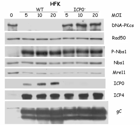
Mre11 loss, I infected HFKs with either wild type HSV-1 or the ICP0 mutant 7134 at various MOIs. At 16 hpi, the cells were harvested and lysates analyzed by Western blot (Fig 4.1A).
At low MOI infections, ICP0 mutants are deficient in expression of other viral genes (36). Here I saw no difference in gene expression between wt HSV-1 and 7134, regardless of MOI, indicating that all the MOIs used were above the threshold of this deficiency. As expected, 7134 failed to express ICP0 and thus did not cause DNA-PKcs degradation. However, the absence of ICP0 had no discernable effect upon either activation of Nbs1 or Mre11 loss. I observed similar results with HELs (DNS). These data indicate that ICP0 is not required for Mre11 loss following HSV-1 infection.
The possibility remained that Mre11 was being degraded by the proteasome due to the actions of other viral or cellular proteins. To determine if Mre11 loss was mediated by the proteasome, I infected HFKs with wt HSV-1 in the presence or absence of MG132, an inhibitor of the 26S proteasome. Cells were harvested at 8 hpi and lysates analyzed by Western blot (Fig 4.1B). Treatment with MG132 protected DNA-PKcs from degradation and had only a minor impact on viral protein levels. Activation of Nbs1 and viral protein levels were slightly reduced by treatment with MG132. This was expected, since MG132 was previously reported to hinder the progression of HSV infection (85, 137, 164). Mre11 levels were minimally affected by MG132 treatment. While some Mre11 was retained following MG132 treatment, comparison with DNA-PKcs protection clearly indicated that Mre11 loss was largely due to mechanisms other than proteasomal degradation.
harvested at 8 hpi and lysates subjected to Western blotting (Fig 4.1C). I observed no change in Mre11 loss with either 10 or 20 mM NH4Cl, an inhibitor of lysosomal activity. These
concentrations were sufficient to prevent productive infection when present at the time of infection (DNS). These data indicate that Mre11 loss is not mediated by the lysosome. Mre11 loss is cell type specific
Others have reported that Mre11 is not lost following HSV infection of Vero, HeLa or HFF cells (1, 8, 117, 126, 131, 133, 134). To determine if I could observe Mre11 loss in cell types other than HFKs and HELs, I infected HeLa, HEp-2 and CV-1 cells with various HSV strains. HeLa and HEp-2 cells were infected at MOIs of 5 or 10 respectively, with wild type KOS or 17+, and ICP0 mutants 7134, d99 or d22, and harvested at 16 and 20 hpi (Fig 4.2 A & B). CV-1 cells were infected with KOS or 7134 at various MOIs and harvested at 20 hpi (Fig 4.2 C).
Western blotting of lysates from HeLa or HEp-2 cells showed infection with all strains tested resulted in decreased Mre11 levels, while other cellular proteins were largely unaffected. This was not the case with CV-1 cells. Initially I observed no change in Mre11 levels in whole cell lysates from CV-1 cells (DNS). To insure Mre11 was not being inactivated by mislocalization, I prepared fractionated lysates from CV-1 cells infected at various MOIs at 20 hpi. All the MRN components largely localized to the nucleus, with no relocalization or reduction of levels. These results indicate that Mre11 loss is a cell type dependent phenotype, though not a virus strain dependent phenotype.
Analysis of Late mutants for Mre11 loss
their ability to cause Mre11 loss. Because any benefit to HSV replication due to Mre11 loss is likely to be due to preventing detection and subsequent repair of free DNA ends at late times of infection, I felt it likely that Mre11 was being lost in order to protect the efficacy of processing and packaging of progeny genomes, which results in generation of free DNA ends. Thus, I chose to test mutants that have been characterized as deficient in proper processing and packaging of progeny genomes. Mutant viruses individually deleted for UL6 (hr74), UL15 (hr81-1), UL17 (dUL17), UL25 (kUL25), UL28 (GCB) and UL32 (hr64) were tested for Mre11 loss following infection (101). UL6 encodes the portal protein through which DNA enters the capsid (2, 174). UL15 and UL28 gene products are components of the terminase (97, 145). The UL25 gene product is necessary for stabilizing encapsidated DNA and late stages of capsid maturation (75, 153). UL17 and UL32 expression is necessary for correct localization of preformed capsids to sites of DNA packaging (30).
I infected HFKs (Fig. 4.3A) and HELs (DNS) with these mutants and harvested at 8hpi to test for Mre11 loss. I noted that the UL15 and UL17 mutants consistently, and UL6 mutant occasionally, showed less Mre11 loss than wt virus, as seen in Fig. 4.3A. However, further analysis of the lysates revealed reduced levels of selected viral proteins. This defect has been previously seen for these mutants and contributed to a high complementation index when titers were determined on complimenting cell lines (personal communication, S. Weller & J. Baines). These mutants also exhibited reduced activation of Nbs1, indicating inefficient viral DNA replication. As such I could not determine whether the failure to cause Mre11 loss was due to the specific loss of the viral genes or due to the general replication defect.
protein levels in the mutant infected cells were equal to or in excess of that of wt virus at an MOI of 20, and Nbs1 activation was also equivalent to wt infected cells. However, under these conditions I also no longer saw any defect in Mre11 loss. This result would indicate that UL15, UL6 and UL17 products are unnecessary for Mre11 loss.
Another late function that could affect Mre11 loss is the viral protease encoded by UL26, which conceivably could directly target Mre11. To determine if UL26 was necessary for Mre11 loss, I infected HFK cells with UL26dZ, a mutant with a deletion of the entire UL26 orf, or UL26H61E, a point mutant disabling the protealytic activity of the viral protease (Fig. 4.3C). At an MOI of 20, these mutants had no defect in Mre11 loss or viral protein accumulation. This result suggests that the viral protease does not contribute to loss of Mre11.
Camptothecin causes Mre11 loss
If MRN detection and engagement of DSBs were sufficient to cause Mre11 loss, I would expect this to occur outside the context of HSV infection. To test if this was the case, I treated cells with camptothecin (CPT), which results in the generation of free DNA ends and activation of the DSB response in S-phase cells due to topoisomerase I inhibition. Because CPT primarily functions in S-phase, I serum starved confluent HEL cultures and then released them by passaging in media with serum. After allowing 20 hours for the cells to re-enter the cell cycle, which has previously been shown to result in an enriched S-phase population, cells were treated with CPT and/or ActD (85, 137, 164). ActD was used to control for differences in protein level due to differential cell growth. After 4 hours treatment cells were harvested and lysates subjected to Western blotting (Fig 4.4). Rad50 and tubulin levels were reduced equally in all cells treated with ActD, with or without CPT, in comparison to untreated cells. ActD treatment also resulted in reduced Mre11 levels, though combined ActD and CPT treatment caused further reduction. CPT treatment alone resulted in a further reduction of all protein levels compared to ActD treatment. A possible cause for this might be a greater induction of apoptosis in these cells, though no cytopathology was observable (DNS).
release from serum starvation. As such, a portion of the cells were unaffected by CPT treatment, thus reducing the observable loss.
Significance of Results
Mre11 loss following HSV infection would be expected to inhibit the ability of the cellular DSB response machinery to recocognize and respond to DSBs. How this loss might affect HSV replication, if it does at all, is unclear. Here, I have shown that Mre11 loss following HSV infection is a late event that is dependent upon viral DNA replication and perhaps late gene expression. Unlike many other cellular proteins that are lost following HSV infection, Mre11 loss is not caused by ICP0 activity, nor does it depend upon the proteasome or lysosome, though it is possible that NH4Cl does not fully inhibit the
protealytic activity of lysosomes. As these general degradation mechanisms appear not to be involved in Mre11 loss, the actions of specific proteases is left as the most plausible explanation for the loss. Another possibility is that Mre11 is being displaced from the MRN complex, resulting in its instability and degradation. This is unlikely however, as it would still depend upon Mre11 being degraded by the proteasome or lysosome.
My results suggest that either viral DNA replication or the function of late genes trigger Mre11 loss following HSV infection. While I have shown a number of late gene functions to be unnecessary for Mre11 loss, it remains possible that there is a functional redundancy amongst those I tested or that a function I have not tested is responsible for Mre11 loss. I expect this not to be the case however, as Mre11 loss is observable in the absence of late protein expression, which leaves viral DNA replication as the likely trigger for loss. To further confirm this, late genes could be expressed from a cosmid library to determine if any are sufficient to cause Mre11 loss.
If viral DNA replication does trigger Mre11 loss, the mechanisms of how this occurs is unclear. It is possible that viral DNA replication allows an IE or E protein to either directly or indirectly target Mre11, leading to Mre11’s degradation. However, I have already demonstrated that the most likely candidate, ICP0, is not necessary for Mre11 loss. No other IE or E proteins have characterized functions that would suggest they would be involved in protein loss. Another possible mechanism would involve a cellular protein being triggered by viral DNA replication to cause Mre11 loss. As it is known that viral DNA replication does trigger some cellular stress pathways, such as the DSB response and PKR, this is a reasonable possibility (85).
is simply a by-product of DSB response activation, it may be that loss does not contribute to viral replication. Given that Mre11 is likely important for viral DNA replication, its loss may in fact be detrimental (48, 114). Even so, the observed loss of Mre11 following CPT treatment may represent a novel negative regulatory mechanism for the DSB response.
Further experiments are needed to identify the exact mechanism of Mre11 loss and determine if loss following HSV infection and CPT treatment are in fact the same. Use of specific and broad range protease inhibitors might identify the relevant protease and thus also allow the significance, if any, of Mre11 loss to viral infection to be determined. Otherwise, tagging of Mre11 and either pull down experiments or live cell microscopy tracking may provide clues to the fate of Mre11 through identification of interacting proteins or observation of aberrant localization.
Figure 4.1. Mre11 loss does not require ICP0 or the activities of the proteasome or lysosome. A. HFKs were infected with wt or a ICP0 deficient virus at the indicated MOIs and harvested at 8 hpi. Whole cell lysates were assayed for cellular and viral proteins by Western blot. B & C HFKs were infected with wt virus and were overlayed with media with or without MG132 or NH4Cl at the indicated concentrations at 1 hpi. Lysates were assayed
Figure 4.1B
Figure 4.2A
Figure 4.2C
Figure 4.3
Figure 4.4
CHAPTER 5
CONCLUSIONS
Viruses are efficient pathogens that are absolutely dependent upon a host cell to complete their life cycle. To facilitate viral replication, host cell machinery is co-opted by the virus to carry out necessary tasks in replication. In this dissertation I have characterized two instances in which HSV interacts with host cell signaling machinery to benefit its own replication. First, I have demonstrated the independent importance of the two kinase components to the IKK complex, IKK and IKK , in the activation of the NF B pathway following HSV infection. Second, I have demonstrated activation of the DSB response and the loss of Mre11, a component of the DSB response, following HSV infection. Here I will discuss the relevance of these findings to our knowledge of HSV life cycle and consider future research in these areas.
NF B activation
IKK has been shown to be important for NF B binding to a subset of its responsive promoters, likely due to phosphorylating p65 and or p50 (113). IKK can also function as a derepressor by targeting transcriptional repressors SMRT and HDAC3 for removal from NF B target genes and by phosphorylating histone H3 (123). To determine if IKK has similar functions during HSV infection several experiments need to be performed. If IKK does directly target p65 or p50 during infection, this could be revealed by comparision of phosphorylated residues detected by Western blot of lysates from cells with or without functional IKK . Also, cells expressing p65 with alanine substitutions mutations at sites targeted by IKK could be infected to determine if they replicate the IKK null phenotype seen in Chapter 2. Conversely, glutamic acid substitutions, which can mimic phosphorylation at these residues, could be used to assay complementation of NF B activation and HSV replication defects in IKK -/- cells.
Determining if IKK also has a role in activation of p65/p50 by phosphorylation following infection will be more difficult, as the defect in I B turnover and p65 translocation must be bypassed to determine if any such phosphorylations are important for DNA binding or transactivation. Some p65 translocation is seen in IKK -/- cells, and thus it may be possible to directly detect phosphorylation sites by Western blot. Sites that are targeted in wt, but not IKK -/-, cells would determine which residues, if any, are targeted by IKK following HSV infection. Alanine replacements could again be tested to determine if any of the IKK targeted sites are important. Glutamic acid replacement would be useful if I B degradation can be affected without IKK activation and activation of other pathways that potentially lead to p65 or p50 phosphorylation.
The observation that IKK -/- cells had up to a 28 fold reduction in yield and IKK -/-cells had only up to a 6 fold reduction while both failed to activate NF B and allow for substantial late gene expression suggests that IKK has additional roles outside of NF B activation. In addition to the NF B subunits, IKK has been shown to phosphorylate Bcl10, IRS1, Dok1, 14-3-3 and FOXO3a (109). Currently there are no reports of activation of these targets following infection. Assaying for such activations could potentially reveal additional roles for IKK in HSV infection.