DEFINING THE STRUCTURAL CUES WITHIN HEPARAN SULFATE THAT DIRECT HEPARANASE CLEAVAGE
Sherket Breshon Peterson
“A dissertation submitted to the faculty of the University of North Carolina at Chapel Hill in partial fulfillment of the requirements for the degree of Doctor of Philosophy in the School of Pharmacy (Chemical Biology and Medicinal Chemistry).”
Chapel Hill
2012
Approved by:
Jian Liu, PhD
Michael Jarstfer, PhD
Stephen Frye, PhD
K.H. Lee, PhD
ii ABSTRACT
SHERKET PETERSON: Defining the Structural Cues within Heparan Sulfate that Direct Heparanase Cleavage
(Under the direction of Jian Liu)
Heparanase is a glucuronidase that cleaves heparan sulfate in a variety of cells and
tissues. The depolymerization of HS by heparanase results in smaller fragments of variable
sizes that bind and modulate the functions of a multitude of proteins, including growth
factors and their receptors, chemokines, enzymes, and extracellular matrix proteins.
Identification of precise structural features within HS that direct heparanase cleavage has
been hindered by the heterogeneity of substrates, the lack of techniques to synthesize
structurally defined oligosaccharides or polysaccharides with site-specific sulfation, and a
lack of large quantities of pure heparanase. Here, we report how advances in the
methodology for synthesizing substrates with defined sulfation types has allowed us to
identify specific sulfation types as well as residues that may direct heparanase cleavage.
Utilizing chemo-enzymatically synthesized polysaccharide substrates with unique sulfation
patterns, we were able to dissect that heparanase cleaves the linkage between a GlcUA unit
and an N-sulfo glucosamine unit carrying either a 3-O-sulfo or a 6-O-sulfo group. In
addition, heparanase cleaves the linkage of a GlcUA unit and N-sulfo glucosamine unit with a 2-O-sulfated GlcUA residue, not a 2-O-sulfated IdoUA residue, in proximity. We also discovered that the polysaccharide with repeating disaccharide units of IdoUA2S-GlcNS
iii
oligosaccharides coupled with ESI-MS analysis and large quantities of a pure enzyme
provided the opportunity to examine the substrate specificity of heparanase in greater detail.
We determined that heparanase utilizes the GlcNAc6S/GlcNS6S at the nonreducing end to
direct the cleavage pattern. Moreover, heparanase depolymerizes the oligosaccharides
utilizing a non-processive method. Our data reveals for the first time that heparanase is
regulated by two main factors; the primary factor is heparanase recognition of different
sulfation types with its non-processive mode of action having a secondary role. These
findings advance the understanding of the substrate specificity of heparanase and reveal a
unique control mechanism for the action of heparanase. Moreover, our result advances the
understanding of the regulation of HS biosynthesis and the intimate relationship between HS
iv
DEDICATION
To my mom, Beverly Ann Peterson, who sacrificed all she had to provide a wonderful life for me and my siblings (Enoy Norman, Kadren Peterson, and Jason Peterson) and for her support, love, and motivation during my graduate career. To my sister, Enoy Norman,
your words of encouragement provided me with the strength to never give up and understand that there was a greater purpose for my life.
To the love of my life, Donald Quincy Goggans, for his love, motivation, and support during my graduate career. Your strength and determination is an inspiration to me daily.
I thank you for helping me to smile through the challenges of graduate school and challenging me to be a better person. Without you, this experience would have been a
v
ACKNOWLEDEMENTS
I would definitely like to thank my adviser, Dr. Jian Liu, for allowing me the space and intellectual freedom to grow into an independent scientist. Dr. Liu has provided me with exceptional training, a valuable graduate experience, and a wealth of knowledge that I can utilize throughout my career. He has always challenged me to be stand strong in what I believe and think outside the box. My research has allowed both of us to grow which I believe is rare to a mentor-mentee relationship. Dr. Liu’s has provided me with a lifetime of advice and support and I will be forever grateful to have been one of his students.
Additionally, I would like to thank my lab colleagues (former and current) Dr. Courtney Jones Law, Dr. Tanya Burch, Dr. Heather Horne, Dr. Renpeng Liu, Dr. Miao Chen, Dr. Yongmei Xu, Dr. Juzheng Sheng, Dr. Xianxuan Zhou, Kasemsiri Chandarojiti, Ryan Bullis, Tim O’Leary, Susan Woody, Po-Hung Hsieh, Troung Pham, and Elizabeth Pempe. In particular I would like to thank Dr. Tanya Burch, Dr. Courtney Jones Law, and Dr. Yongmei Xu for taking me under your wing when I first joined the lab and always being available to answer questions. To Kasemsiri Chandarojiti, I would like to thank you for your support and always lending a listening ear when I needed one. Last but not least I would like to thank Dr. Courtney Jones Law, Dr. Heather Horne, Dr. Yongmei Xu, Ryan Bullis, Kasemsiri Chandarojiti and the rest of the Liu lab for their support and laughs throughout my graduate career. They have made these years a wonderful experience.
vi
TABLE OF CONTENTS
LIST OF TABLES ... xi
LIST OF FIGURES ... xii
LIST OF ABBREVIATIONS ... xv
CHAPTER I: INTRODUCTION ... 1
Section 1: Structure of heparan sulfate ... 1
Section 2: Biosynthesis and regulation of HS ... 3
Biosynthesis of HS ... 3
Regulation of the sulfation patterns in HS ... 5
Physiological functions of HS ... 6
Section 3: Biological function and potential therapeutic application of HS ... 10
Heparin in malignancies ... 10
Heparin as an anti-inflammatory therapy ... 12
Section 4: HS degradation and heparanase molecular properties ... 14
Heparanase history ... 15
Proheparanase and heparanase ... 16
Heparanase mechanism of action ... 19
vii
Heparanase 2... 22
Section 5: Heparanase involvement in physiology and disease ... 23
Heparanase role in normal development ... 23
Heparanase in pathophysiological processes ... 25
Nonenzymatic effects exhibited by heparanase ... 29
Regulation of heparanase ... 31
Section 6: Inhibition of heparanase ... 34
Heparin and heparanase inhibition ... 34
Heparanase inhibitors that function as substrate mimetics ... 35
Silencing of the heparanase gene ... 38
Anti-heparanase peptides and antibodies ... 39
Heparanase inhibition by small molecules ... 40
Other inhibitory strategies ... 40
Section 7: Structural recognition of heparanase ... 41
Section 8: Statement of problem ... 43
CHAPTER II: MATERIALS AND METHODS ... 46
Section 1: Cloning, expression, and purification of heparanase ... 46
Preparation of heparanase mammalian expression plasmid ... 46
Expression of recombinant heparanase in CHO cells ... 46
Preparation of heparanase yeast expression plasmid with His-tag ... 47
Expression of recombinant heparanase in K. Lactis cells ... 49
viii
Expression of GS3 construct in Sf9 cells ... 51
Purification of recombinant GS3 construct ... 52
Section 2: Preparation of heparanase substrates ... 52
Preparation of metabolically 35S-labeled HS from CHO cells ... 52
Preparation of 35S-labeled HS polysaccharide substrates ... 53
Preparation of oligosaccharide substrates ... 54
Section 3: Analytical procedures for heparanase substrates ... 56
Synthetic polysaccharides ... 56
High resolution DEAE-HPLC analysis ... 57
Mass spectrometry analysis ... 58
Separation of oligosaccharide by gel permeation chromatography ... 58
Section 4: Heparanase activity assays ... 58
Assaying for heparanase enzymatic activity using polysaccharides ... 58
Inhibition of the activity of heparanase ... 59
Assay to probe heparanase enzymatic activity ... 59
Assaying for heparanase enzymatic activity using oligosaccharides ... 60
CHAPTER III: UNRAVELING THE SPECIFICITY OF HEPARANASE UTILIZING SYNTHETIC SUBSTRATES ... 61
Section 1: Introduction ... 61
Section 2: Expression of heparanase in CHO cells ... 62
ix
with different sulfation patterns ... 64
Section 4: Contribution of each sulfation
type on heparanase cleavage... 66
The contribution of sulfation on the glucosamine
residue to the cleavage by heparanase... 66
Effects of 2-O-sulfation on the susceptibility
of heparanase digestion ... 69
Section 5: Evidence of substrate cleavage by heparanase ... 70
Heparanase cleavages the linkage that contains
nonsulfated glucuronic acid ... 70
Section 6: Polysacchride 7 (-IdoUA2S-GlcNS- repeating unit)
inhibits heparanase activity ... 73
Section 7: Conclusion ... 75
CHAPTER IV: DIVERSE APPROACHES TO EXPRESSING RECOMBINANT HEPARANASE IN VARIOUS
EXPRESSION SYSTEMS ... 80
Section 1: Introduction ... 80
Section 2: Expression of heparanase in mammalian
expression system (CHO cells) ... 82
Section 3: Expression of heparanase in bacteria expression system
is not a realistic option ... 83
Section 4: Expression and purification of heparanase from a
yeast expression system (K. Lactis cells) ... 84
Section 5: Expression of heparanase in Sf9 cells ... 88
Section 6: Assay to probe purified heparanase to determine
purification efficiency ... 92
Section 7: Purification of GS3 heparanase from
serum containing media ... 95
x
Serum-free media ... 96
Section 9: Conclusion ... 99
CHAPTER V: STRUCTURAL CUES WITHIN HS THAT DIRECT HEPARANASE CLEAVAGE... 100
Section 1: Introduction ... 100
Section 2: Preparation of structurally defined oligosaccharide substrates ... 101
Section 3: Minimum size for a heparanase substrate ... 106
Section 4: The role of NS6S/NAc6S in directing heparanase cleavage ... 111
Section 5: Site preference by heparanase cleavage ... 118
Section 6: Conclusion ... 123
CHAPTER VI: CONCLUSION ... 126
APPENDIX FIGURES ... 128
APPENDIX TABLES ... 130
xi
LIST OF TABLES
1. Summary of the modification to the synthetic polysaccharides
and the results of their heparanase degradation……….…65
2. Analysis of oligosaccharide substrates………105
xii
LIST OF FIGURES
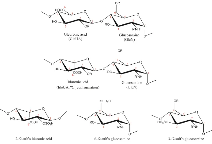
1. Structures of disaccharide repeating units of HS
and commonly found monosaccharides ... 2
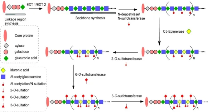
2. Biosynthesis of HS ... 4
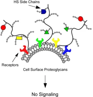
3. HS regulates cellular signaling ... 14
4. Heparanase processing forms ... 17
5. Three-dimensional model of heparanase ... 18
6. Mechanism of action of heparanase based on the retention mechanism of a β-glycosidase ... 20
7. The steps of heparanase cellular processing ... 22
8. HS degradation by heparanase ... 26
9. C-terminal domain of heparanase ... 30
10.Contradictory reports of heparanase specificity in literature ... 42
11.Optimized method for the preparation of yeast expression plasmid with His-tag ... 49
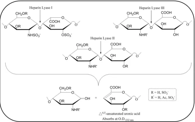
12.Enzymatic digestion by lyases I, II, and III ... 56
13.Nitrous acid degradation ... 57
14.Preparation and characterization of recombinant heparanase ... 63
15.Size of the heparanase cleaved HS ... 64
16.Scheme for the synthesis of polysaccharide substrates ... 65
xiii
of the synthesized substrates ... 66
18.GPC-HPLC profiles of polysaccharide substrates with or without heparanase digestion ... 67
19.GPC-HPLC profiles of substrate 7 (synthesized with 2-OST WT) with or without heparanase digestion ... 69
20.Determination of the heparanase cleavage sites within substrates 4 and 5 ... 71
21.Determination of the heparanase cleavage sites within substate 6 with 35S-labeled 2-O-sulfation ... 72
22.Determination of the heparanase cleavage sites within substrate 6 with unlabeled 2-O-sulfation ... 73
23.Inhibition of the activity of heparanase ... 75
24. Proposed heparanase substrate recognition sites ... 78
25.Known glycosylation sites of heparanase ... 83
26.Issues with heparanase activation in a cell-free system ... 85
27.Artificial TEV linker to regulate heparanase proteolytic activation ... 86
28.Expression of heparanase in K. Lactis cells... 87
29.Homology model of proheparanase and α-L-arabinofuranosidase ... 89
30.His-tag GS3 construct lacks the need for proteolytic activation ... 90
31.GPC profile of 35S-labeled 6-O-sulfo heparosan with or without GS3 heparanase cleavage ... 91
32.Homology model of GS3 construct and α-L-arabinofuranosidase ... 92
33.FPLC-HPLC profile of GS3 heparanase... 97
xiv
35.Scheme for the synthesis of oligosaccharide substrates ... 102
36.ESI-MS profiles from structural Analysis of the synthesized substrates ... 103
37.DEAE-HPLC profiles from structural analysis of the synthesized substrates ... 104
38. Analysis of heparanase-digested Nona-3 ... 107
39.Analysis of heparanase-digested Hepta-2 ... 108
40.Analysis of heparanase-digested Penta-1... 111
41. Analysis of heparanase-digested Nona-4 ... 112
42.Analysis of heparanase-digested Nona-5 ... 113
43.Analysis of heparanase-digested Nona-6 ... 114
44.Analysis of heparanase-digested Nona-7 ... 115
45.Analysis of heparanase-digested Nona-8 ... 116
46. Schematic representation of heparanase cleavage toward different nonasaccharide substrates ... 117
47.DEAE-HPLC chromatogram of 35S-labeled Nona-3 and Nona-7 digested with heparanase at different times ... 120
48.Partial digestion of unlabeled Nona-3 ... 121
xv
LIST OF ABBREVIATIONS
2-OST 2-O-sulfotransferase
3-OST 3-O-sulfotransferase
6-OST 6-O-sulfotransferase
C5-Epi C5-Epimerase
CHO Chinese hamster ovary cells
ECM Extracellular matrix
ER Endoplasmic reticulum
GAG Glycosaminoglycan
GlcN Glucosamine
GlcNAc N-acetyl glucosamine
GlcNAc6S 6-O-sulfo N-acetylated glucosamine
GlcNH2 N-unsubstituted glucosamine
GlcNS N-sulfo glucosamine
xvi
GlcNS6S 6-O-sulfo N-sulfo glucosamine
GlcUA Glucuronic acid
GlcUA2S 2-O-sulfo glucuronic acid
HS Heparan sulfate
HSPG Heparan sulfate proteoglycans
IdoUA Iduronic acid
IdoUA2S 2-O-sulfo iduronic acid
LMWH Low molecular weight heparin
NDST N-deacetylase/N-sulfotransferase
NDST-2 N-deacetylase/N-sulfotransferase isoform 2
Chapter I
INTRODUCTION
Section 1. Structure of heparan sulfate
Heparan sulfate is a ubiquitous macromolecule present in animal tissues [1]. It belongs to the
glycosaminoglycan (GAG) family of natural products. In addition to HS, the family consists
of chondroitin sulfates, keratan sulfate and hyaluronic acid. The members of this class of
macromolecules differ in the structures of the disaccharide repeating units as well as the
number of sulfo groups on the polysaccharide, with the exception of hyaluronic acid, which
contains no sulfo groups. HS contains a disaccharide-repeating unit of glucosamine (GlcN)
and glucuronic/iduronic acid (GlcUA/IdoUA) residues with various sulfo groups (Figure 1).
The majority of GlcN residues are modified with either N-acetyl or N-sulfo groups. However, 1–7% of GlcN in HS is present in N-unsubstituted glucosamine (GlcNH2) [2]. HS is the most extensively studied GAG, demonstrating a variety of biological functions that range from
regulating blood coagulation [3]and assisting viral infections [4] to regulating cell growth
[5].Due to the diverse functions of HS and heparin, the possibility of exploiting its
anticancer, anti-inflammatory and antiviral activities, in addition to its anticoagulant activity,
is generating considerable interest.
Heparin, a special form of HS, is a commonly used anticoagulant drug. HS is
2
mechanism. Heparin, a highly sulfated version of HS, contains 2.6 sulfo groups per
disaccharide on average, while its HS counterpart contains about 0.6. Also, some 80% of the
hexauronic acid units of heparin are IdoUA, while only about 20% of hexauronic acid units
of most HS are IdoUA. Furthermore, in vivo, heparin is only found in intracellular vesicles of
connective tissue mast cells, while HS is present in a variety of cell types.
Figure 1. Structures of disaccharide repeating units of HS and commonly found monosaccharides.
Sulfation (R = -SO3) at Carbon 6 (known as 6-O-sulfo glucosamine) of glucosamine is common. Sulfation at
Carbon-2 of iduronic acid (known as IdoUA2S) is common. Sulfation at Carbon-3 of glucosamine (known as 3-O-sulfo glucosamine) is rare. Both N-acetyl (R’ = acetyl, GlcNAc) and N-sulfo glucosamine (R’ = -SO3,
GlcNS) are common. N-unsubstituted glucosamine (R’ = -H, GlcNH2) is a low abundance component.
Although the fine structure of heparin differs significantly from that of HS, the global
structures of these polysaccharides are surprisingly comparable. Similarly, heparin and HS
are biosynthesized as HS proteoglycans (HSPG), comprising a core protein covalently
3
proteins, HSPG includes syndecans, serglycin, glypicans, agrin and perlecans. The major cell
surface HSPGs are the syndecans and glypicans,[6, 7] and the major ECM HSPGs are
perlecan and agrin [8]. The core protein carrying two or three HS chains can be anchored to
the cell membrane via the transmembrane domain, such as syndecan, or via the
glycosylphosphatidylinositol (GPI)-linked motif, such as glypican. The core protein
determines the location of HS in a specific tissue and may play an important role in
enhancing HS effects. In addition, the core protein is directly involved in growth factor
binding and can participate in cellular signal transduction [9]. The biological functions of
HSPG are predominantly determined by the HS side chains that can bind a variety of protein
ligands and thereby influence many biological processes. HS ability to regulate a variety of
functions is a result of its structural heterogeneity via sequential enzymatic modifications.
Section 2. Biosynthesis and regulation of HS
Biosynthesis of HS
The biosynthesis of HS occurs mainly in the Golgi apparatus and involves a complex
set of specialized enzymes (Figure 2) [10]. Biosynthesis of the HS polysaccharide chains
takes place in three phases: chain initiation, polymerization, and polymer modification. Chain
initiation begins with the stepwise addition of a xylose residue, two galactose residues and a
GlcUA residue [11]. This tetrasaccharide linkage region is attached to specific serine
residues of the core protein containing a structure of
β-xylose-(1,4)-β-galactose-(1,3)-β-galactose-(1,3)-β-glucuronic acid. HS is then synthesized as a linear copolymer of GlcUA
4
of polymer modifications are then carried out by numerous biosynthetic enzymes, including
glucosaminyl N-deacetylase/N-sulfotransferase [12], which converts the GlcNAc unit to a GlcNS unit. A specific isoform of N-deacetylase/N-sulfotransferase (NDST-2), highly expressed in mast cells, is proposed to be involved in synthesizing heparin, but not HS [13].
After N-sulfation, C5-epimerase (C5-Epi) converts a GlcUA unit to an IdoUA unit. The
resulting polysaccharide is further modified by 2-O-sulfotransferase (2-OST), 6-O -sulfotransferases (6-OST) and 3-O-sulfotransferases (3-OST) to introduce the 2-O-sulfo group to IdoUA/GlcUA and the 6-O-sulfo and 3-O-sulfo groups to the GlcN unit [14]. The enzymatic reactions do not continue to completion, thus resulting in substantial sequence
diversity within HS [7].
5
Regulation of the sulfation patterns in HS
HS structural complexity is regulated by the HS biosynthetic enzymes. Since HS
biosynthesis is not a template-driven process, the substrate specificities of the biosynthetic
enzymes dictate the structures of the final HS product. These biosynthetic enzymes exhibit
their substrate specificities at two different levels: the regioselectivity of the residue at the
modification site (monosaccharide level) and the sulfation patterns around the modification
site (oligosaccharide level). The regioselectivity at the modification site for a given HS
sulfotransferase is nearly exclusive [15-18]. For example, 6-OST recognizes a GlcNS or
GlcNAc residue and specifically transfers the sulfo group to the 6-OH position to form
GlcNS6S [or 6-O-sulfo N-acetylated glucosamine (GlcNAc6S)].
HS structure is also regulated by distribution of GlcNS residues. The synthesis of
GlcNS by NDST is the first step of a series of biosynthetic modifications. It is known that the
presence of the GlcNS residue is critical for all subsequent modifications during the
biosynthesis of HS. In other words, a cluster of GlcNS residues determines the location of
other O-sulfations and epimerization of GlcUA. These clusters form so-called ‘‘sulfated domains of HS,’’ which dictate the majority of their functions [19]. Results from cell-based
assays demonstrated that cells lacking NDST-1 or NDST-4 synthesize undersulfated HS [20,
21]. Carlsson and colleagues demonstrated that NDST-1 has the ability to form clusters of
GlcNS residues, suggesting its role in the distribution of GlcNS to the overall structure of the
final HS [22]. Targeted gene deletion of NDSTs also point to the physiological significance
of GlcNS in the biosynthesis of HS. For example, NDST-1 null mice display dramatically
reduced amounts of sulfated HS and die shortly after birth [23]. Studies on the conditional
6
regulates L-selectin- and chemokine-mediated neutrophil trafficking [24]. In contrast, mice
deficient in NDST-2 are viable but show a lack of heparin synthesis in mast cells [25]. The
distinct phenotypes of NDST-1 and -2 knockout mice strongly suggest that unique GlcNS
distribution directs the synthesis of HS with specific physiological functions.
Another regulation of different sulfation patterns is via different HS biosynthetic
enzyme isoforms. It has been hypothesized that cells regulate expression levels of different
isoforms in order to synthesize the HS with defined saccharide sequences to achieve unique
biological functions [26]. HS biosynthetic enzymes are present in multiple isoforms, with the
exception of 2-OST and C5-Epi. NDST is present in four different isoforms, 6-OST is
present in three and 3-OST in seven. In addition, these isoforms have slightly different
substrate specificities and unique tissue expression patterns [20, 27-29]. 3-OST isoforms are
essential for HS synthesis with a variety of saccharide sequences that exhibit different
functions [30]. For instance, 3-OST-1 sulfates HS to synthesize a product with anticoagulant
activity, while 3-OST-3 sulfates HS to synthesize a product that serves as an entry receptor
for herpes simplex virus (HSV). 3-OST-5 can synthesize HS with either anticoagulant
activity or an HS that can serve as an entry receptor for HSV. Various isoforms of 6-OST are
important for building the HS that bind to FGFs and FGFRs, although the substrate
specificities among 6-OSTs are not experimentally distinguishable [31, 32].
Physiological functions of HS
One method to study the structure and function of HS that carry different sulfation
patterns in vivo is to conduct gene knockout experiments in various organisms of HS
7
these enzymes in numerous processes. The results from knockout experiments should be
explained with caution because the lack of sulfation in these knockout organisms can be
compensated by an increase in other sulfation types as the case demonstrated in Drosophila
[33].
The importance of Exostosin genes (EXT1 and EXT2), which encode
glycosyltransferases or HS polymerases for the synthesis of HS backbone, has been shown in
several knockout mouse studies. Specifically, EXT1 and EXT2 enzymes function as a
hetero-oligomeric complex to polymerize the HS backbone [34]. The phenotype expressed by EXT
null mice are embryonic lethal due to the shorter HS chains linked to the core protein [35].
Subsequently, Strickens et al. observed similar results in an Ext2 null mouse model [35]. While a complete loss of both EXT genes can be lethal, partial loss in either isoform results
in bone exostosis, which is a genetically heterogeneous human disease characterized by bony
outgrowths near the ends of the long bones [35, 36].
Dramatic effects on the HS sulfotransferases and C5-Epi were also observed for
enzymes defective in modification steps. NDST is a bifunctional enzyme that removes the
acetyl group from GlcNAc residues and transfers a sulfo group to the deacetylated GlcN [37,
38]. Silencing of NDST alters the HS structure owing to changes in reduced
C5-epimerization and O-sulfations [39]. Mice lacking NDST-1 exhibit severe defects such as abnormal brain and face development, including cerebral hypoplasia, lack of olfactory bulbs,
eye defects, and axon guidance errors [40]. In addition, NDST-1 has also been shown to be
essential for normal respiratory function [23]. The phenotypes observed in NDST-1-null
mice are thought to be a result of NDST-1’s ability to modify signaling in hedgehog, FGF,
8
essential. This reasoning is based on studies of NDST-2 null mice in which the only
abnormality seen was in connective tissue type mast cells [39]. Further, NDST-3 -knockout
mice are fertile and exhibit only minor hematological and behavioral phenotypes [42].
Therefore it is believed that other NDST genes can compensate for all isoforms except for
the loss of NDST-1.
C5-Epi converts D-GlcUA to L-IdoUA units [43, 44]. Li et al. observed that the HS
isolated from C5-Epi knockout mice lacked the IdoUA unit and thus a change from the
predominantly sulfated disaccharide unit IdoUA2S-GlcNS to GlcUA-GlcNS6S. The lack of
C5-Epi also produced mice with a lethal immature lung phenotype, abundant skeletal
abnormalities, and kidney agenesis. It is interesting to note that the phenotypes displayed by
C5-Epi knockout mice are similar to those observed in 2-OST null mice.
HS 2-OST catalyzes the transfer of a sulfo group to the C2 position of GlcUA or
preferentially IdoUA to form 2-O-sulfo glucuronic acid (GlcUA2S) and 2-O-sulfo iduronic acid (IdoUA2S), respectively [45, 46]. The 2-OST-knockout mice demonstrated a complete
failure of kidney development and several skeletal abnormalities [47, 48]. However, in
contrast to C5-Epi-null mice, the loss of 2-O-sulfation in mutants is compensated for, in terms of charge, by a 13% increase in 6S and a 9% increase in NS in NS sulfated domains
[49]. Furthermore, the 2-OST-null mice survive until birth, unlike C5 epimerase mutants, but
die soon after due to kidney agenesis [46]. In Caenorhabditis elegans, 2-OST has been
determined to be essential for axon migration/guidance and nervous system development [50,
9
HS 6-OST catalyzes the transfer of a sulfo group to the C6 position of GlcNS to form
GlcNS6S,[52] which has been shown to have functional consequences for several proteins.
Mice deficient in 6-OST-1 had growth retardation, aberrant eye and lung morphogenesis, and
impaired placenta function and died at or soon after birth [53]. Habuchi et al. observed that 6-O-sulfated residues participate in Wnt2 recognition and that HS 6-OST-1 is considerably less important than NDST-1 in controlling lung development and function. In spite of this
finding, more studies are needed to resolve the individual roles of 6-OST isoforms.
HS 3-OST transfers a sulfo group to the 3-OH position of the glucosamine unit to
form 3-O-sulfo N-sulfo glucosamine (GlcNS3S). The 3-O-sulfation is essential for binding to gD [54] and FGF-7 and its receptor [55]. The 3-O-sulfation also plays a critical role in
binding to AT, exhibiting the anticoagulant activity [18]. The essential role of 3-O-sulfation for synthesizing anticoagulant HS has led to the hypothesis that 3-OST-1 null mice would
demonstrate a procoagulant phenotype and reduced antithrombin binding HS [56]. However,
mice lacking 3-OST-1 do not exhibit a procoagulant phenotype. These results can be
explained by the fact that other isoforms of 3-OST, such as 3-OST-5, may take the place of
3-OST-1 to synthesize a low level of anticoagulant HS [57]. These mice displayed a range of
unanticipated abnormalities like spontaneous eye degeneration, aberrant cardiovascular
response to anesthesia, reduced fertility, postnatal death, and intrauterine growth retardation.
The levels of 3-O-sulfation, the products modified by 3-OST, are typically low in the HS isolated from natural sources [58]. There are numerous 3-OST isoforms depending on the
organisms. It has been widely speculated that 3-O-sulfation participates in very specific biological processes in addition to regulating the blood coagulation. In particular, the roles of
10
technique, Kamimura and colleagues concluded that knockout Hs3stB, which is the 3-OST-3
gene in humans and mouse, resulted in neurogenic phenotype due to the effect on Notch
signaling pathway in Drosophila [59]. In addition, the expression of seven 3-O -sulfotransferase isoforms exhibit distinct patterns at various developmental stages in
zebrafish [60].
Section 3. Biological function and potential therapeutic application of HS
HS is expressed ubiquitously on the cell surface and in the extracellular matrix
(ECM), where it acts as an important biological mediator of various cellular events. HS
interacts with matrix components such as fibronectin, laminin, and collagen to maintain the
structural integrity of the ECM. In addition, HS sequesters cytokines and growth factors to
the ECM [7, 61, 62]. Consequently, HS has roles in regulating angiogenesis, metastasis,
inflammation, and wound healing. Heparin, a highly sulfated version of HS, is commonly
used to treat thrombotic disorders. Furthermore, biochemical and clinical evidence suggest
that heparin also has anticancer and anti-inflammatory activities. Hence, the diverse
functions of HS and heparin as well as it therapeutic potential has attracted considerable
interest.
Heparin in malignancies
Thromboembolism is a frequent complication of malignancies; often, spontaneous
thrombosis is an indication of occult cancer [63-65]. Tumor cells release procoagulants,
including cysteine proteases and tissue factors that directly activate factors X and VII. The
11
leading to thrombin production and thrombosis. This interaction also causes the release of
tumor necrosis factor, interleukin-1, and interleukin-6. These mediators initiate endothelial
damage and create a thrombogenic vascular in surface [66]. Heparin’s anticoagulant
properties are useful against thrombosis in cancer. Briefly, heparin may directly influence
tumor cell behavior by interacting with integrins to interfere with tumor cellular adhesion and
binding to growth factors [67]. In addition, heparin prevents cancer cell interaction with
platelets and endothelial cells by inhibiting selectin binding [68]. Low molecular weight
heparins (LMWH) are suggested to regulate angiogenesis by inhibiting matrix-degrading
enzymes [69, 70]and have been utilized in prophylaxis and treatment of thromboembolic
disease in patients with malignancy.
Clinical evidence for heparin use as an antimetastatic and antiangiogenesis agent is
less conclusive and shows dose-limiting anticoagulant side effects [62]. A systemic review
by Tagalakis and colleagues revealed heparins (predominantly LMWH) may improve the
survival of patients with small cell lung cancer and advanced malignancy with more
favorable prognoses. More research is necessary to elucidate which type of cancer and stage
would benefit most from heparin therapy [71]. Currently, the American Society of Clinical
Oncology and American College of Chest Physicians do not recommend anticoagulants to
improve survival in patients who have cancer but are not at risk for venous thromboembolism
[72]. However, the Society encourages patient participation in clinical trials designed to
evaluate anticoagulant therapy as an adjunct to standard anticancer therapy [73].
Although heparins have been shown to be advantageous in cancer patients, the
anticoagulant properties of heparin and heparin-like compounds limit the antitumor
12
antiangiogenic and anti-metastatic properties, a novel anticancer agent could possibly be
developed. However, patients may still require anticoagulation as a result of cancer and
chemotherapy hypercoagulable effects. The development of new antiangiogenic and
anti-metastatic compounds that are structurally or functionally related to non-anticoagulant
heparin could serve as a promising cancer therapy.
Heparin as an anti-inflammatory therapy
A hallmark of chronic inflammatory diseases, such as rheumatoid arthritis, asthma,
inflammatory bowel disease, and psoriasis, is the unwarranted infiltration and accumulation
of inflammatory cells into affected organs. These events can lead to tissue damage and
cellular remodeling. Heparin has been shown to reduce and even inhibit leukocyte
accumulation in inflamed skin [74], brain [75], lung [76], and hyper-responsive bronchial
tissue in animal models. Furthermore, its anti-inflammatory effect has been shown to be
distinct from its anticoagulant properties [12, 77]. In addition, a study by Ahmed et al. in the New England Journal of Medicine showed that heparin could reduce inflammatory cell
activation and accumulation in lung tissue and thereby improve respiratory function. Inhaled
heparin has provided evidence of efficacy in exercise-induced asthma treatment. Partial
thromboplastin times were tested at baseline and following a test dose of inhaled heparin in
12 patients. These patients were then pretreated with 1000 U/kg heparin, 20 mg of cromolyn
sodium, or placebo followed by an exercise challenge. Heparin did not alter the pH or the
bronchorestrictor response to histamine but did prevent exercise-induced asthma [78]. It has
been hypothesized that LMWH may also attenuate exercise induced bronchoconstriction.
13
revealed enoxaparin prevents exercise-induced bronchoconstriction in a dose-dependent
manner independent of its effect on plasma Xa activity. However, enoxaparin did not modify
the bronchoconstrictor activity of methylcholine [79].
Nevertheless, topical administration of heparin may be effective for limiting
inflammatory events in allergic rhinitis. In a study with 10 patients, the effect of 4 ml of
intranasal unfractionated heparin at 3750 U/ml was compared with placebo in a double-blind,
randomized trial. Patients treated with unfractionated heparin had reduced symptom scores,
eosinophil counts, and eosinophilic cationic protein levels in the nasal lavage following
allergen challenge [80].
Heparin has been evaluated in small clinical trials and individual cases as a treatment
for inflammatory bowel disease [81, 82]. The beneficial effects of heparin in inflammatory
bowel disease include attenuation of the prothrombotic effects of the disease, prevention of
thrombotic occlusion and subsequent intestinal infarction, enhancement of endothelial barrier
function, reduction of inflammatory cytokines, and inhibition of neutrophil diapedesis in
intestinal mucosa [83]. In addition, heparin has been reported to be effective in case reports
of Crohn’s disease, but this evidence has been limited. In three uncontrolled studies with 39
patients with severe, steroid-resistant ulcerative colitis and 4 patients with Crohn’s disease,
28 of these patients experienced remission following heparin administration [84]. A recent
Cochrane review recommended against the use of unfractionated heparin and LMWH in the
treatment of active ulcerative colitis because of lack of evidence and risk of bleeding with
heparin administration [85]. However, Chande and colleagues felt trials of unfractionated
14
Section 4. HS degradation and heparanase molecular properties
HS is a highly sulfated polysaccharide that is expressed ubiquitously on the cell
surface and in the ECM. HS occurs as a proteoglycan in which two or three HS chains are
attached to a core protein. These HS chains possess various types of modifications (discussed
in Section 2) that allow HS to bind a variety of ligands and regulate an array of functions,
such as angiogenesis, tumor metastasis, developmental processes, and blood coagulation.
Under normal physiological conditions, HS interacts with fibronectin, laminin, and collagen
to help maintain the structural integrity of the ECM and participates in cell and
cell-ECM interactions. Furthermore, HS has the ability to bind a multitude of proteins (i.e.,
growth factors, chemokines, lipoproteins, enzymes, etc.) and act as a storage depot by
preventing these proteins from binding to their respective receptors (Figure 3).
15
In turn, this allows HS to regulate cellular signaling as well as accessibility, function, and
mode of action of its binding partners. It is because of the important and multifaceted roles
of HS in cell and tissue physiology that allows the cleavage of HS to have such a significant
effect within the body [86]. Therefore, enzymatic degradation of HS is expected to
profoundly affect fundamental biological processes, such as pregnancy, morphogenesis,
inflammation, neurite outgrowth, angiogenesis, and cancer metastasis [87].
Heparanase history
Heparanase is an endo-β-glucuronidase that cleaves HS in a variety of normal and
malignant cells and tissues, such as placenta, platelet, skin, fibroblast, melanoma, lymphoma,
carcinoma, hepatocytes, Chinese hamster ovary (CHO) cells, keratinocytes, neutrophils,
macrophages, T and B lymphocytes, mast, and endothelial cells [1, 86, 88-96]. The journey
for identifying and characterizing heparanase has been long and challenging. The first
mention of heparanase in literature occurred in 1975 when Ogren and Lindahl [97] reported
the cleavage of heparin by an enzyme isolated from the mastocytomas of tumor bearing
mice. Due to the lack of a simple and reliable assay to quantify heparanase activity,
molecular probes, antibodies, and a low abundance of heparanase in cells, the cDNA of
heparanase remained unidentified for another two decades. In 1999, cloning of the
heparanase gene was reported by several groups [89, 91, 94, 96] from human and other
species. In the recent years, progress in the field has led to the discovery of new information
which have expanded our understanding of heparanase as well as its function and
16
Proheparanase and heparanase
It is now known that human heparanase cDNA contains an open reading frame of
1629 bp that encodes for a 543 amino acids [96]. Sequence analysis revealed that human
heparanase is highly conserved, with similar sequences found in chicken [98], rat [99],
mouse [100], cow [101, 102], Spalax or subterranean mole rat [103], mollusks, and zebra fish . Surprisingly, human heparanase has the highest homology with bovine and Spalax
heparanase with a homology percentage of 86.1% and 84.5%, respectively [87]. However,
the gene has not been identified in Drosophilia melanogaster or Caenorhabditis elegans,
which suggest that HS cleavage in these organisms, may be carried out by a different
enzyme.
Heparanase has a similar structure to that of a cysteine protease. To date, heparanase
is known to exist in three forms: a pre-proheparanase, proheparanase, and heparanase (Figure
4). Heparanase is synthesized in the cell as an inactive precursor composed of 4 distinct
domains: an N-terminal domain of variable size, a large subunit, a small subunit, and a linker
region between the large and small domains. The inactive precursor of heparanase, known as
pre-proheparanase, is initially synthesized as a latent 65kDa precursor. When the N-terminal
signal peptide (Met1-Ala35) [96] of the pre-proheparanase is removed, the new form of
heparanase becomes known as proheparanase. However, activation of heparanase cannot be
induced until it undergoes further processing by cathepsin L before it can exist in its mature
form [104]. Cathepsin L is a lysosomal cysteine proteinase that plays a major role in
intracellular protein catabolism. Proheparanase must undergo proteolytic activation by
17
Figure 4. Heparanase processing forms. Heparanase is synthesized as a 65kDa precursor (pre-proheparanase) with 6 N-glycosylation sites (blue trophies). Pre-proheparanase becomes proheparanase when the signal peptide is removed. After proteolytic activation by cathepsin L, the linker region is removed and the 50 kDa and 8 kDa subunit noncovalently associate to form mature heparanase.
Removal of the linker region allows the 50 kDa (Lys158-Ile543) and 8 kDa (Gln36-Glu109)
heterodimer to noncovalently associate in an active conformation known as heparanase
[105]. This structural complexity tightly regulates heparanase activity. The detailed
processing of heparanase will be discussed in a later section entitled “Heparanase cellular
processing.”
To date, there is no published crystal structure of heparanase or any of the members
of other enzymes of Carbohydrate Active enZYmes (CAZy) family 79. CAZy is a database
that describes the families of structurally-related catalytic and carbohydrate-binding
18
(http://www.cazy.org/). Multiple-sequence alignment approaches in combination with
secondary structure predictions indicate that heparanase contains sequences that are
homologous to family 10, 39, and 51 of the clan A glycosyl hydrolases [90]. Several sources
[90, 106] including a predicted three dimensional structure of active [106] heparanase
revealed that heparanase is organized into two structural domains: a catalytic domain with
(α/β)8 triosphosphate isomerase (TIM)-barrel fold followed by a β-sandwich C-terminal
domain (Figure 5A and 5B). The C-terminal domain is composed of 8 β-strands, one of which is contributed by the 8 kDa subunit (yellow), arranged in two sheets from the 50 kDa
subunit (blue and orange), which are connected by an unstructured, flexible loop (arrow)
(Figure 5C). The C-terminal domain has been shown to be responsible for the heparanase secretion and the nonenzymatic functions of heparanase, such as activation of the Akt
signaling pathway [106]. The detailed functions of the C-terminal domain will be discussed below in “Nonenzymatic effects exhibited by heparanase.”
Figure 5. Three-dimensional model of heparanase. The model, including the 8-kDa (yellow) and 50-kDa (gray) protein subunits, amino acids critical for heparanase catalysis (Glu225 and Glu343; red), and heparin binding regions (Lys158-Asp171 and Gln270-Lys280;cyan and green, respectively), is shown in the left and middle panels. A more detailed structure of the C-domain is shown in the right panel. The model illustrates eight β-strands, one of which is contributed by the 8-kDa subunit (yellow), arranged in two sheets
19
Heparanase mechanism of action
Currently, there is no crystal structure of heparanase or any of the members of other
enzymes of CAZy family 79. Therefore, sequence comparisons had to be utilized to indicate
similarities between heparanase and family 10, 39, and 51 of the clan A glycosyl hydrolases
[90] in order to investigate the enzymatic mechanism of heparanase using 3-D models. From
this data, it has been inferred that heparanase uses a β-retaining hydrolytic mechanism
(Figure 6). This mechanism involves two carboxylates: Glu343 acts as a nucleophile and
Glu225 as an acid/base. Heparanase operates through a two-step mechanism, with each step
resulting in inversion, for a net retention of stereochemistry [107]. During the first step, the
nucleophile (deprotonated Glu) attacks the anomeric center of the GlcUA, resulting in the
formation of a glycosyl enzyme intermediate, with acidic assistance provided by the acidic
carboxylate. In the second step, the now deprotonated acidic carboxylate acts as a base and
assists a nucleophilic water to hydrolyze the glycosyl enzyme intermediate, giving the final
20
Figure 6. Mechanism of action of heparanase based on the retention mechanism of a β-glycosidase. Glu343 acts as a nucleophile to form the glycosyl-enzyme intermediate. The enzyme is then displaced from the
glycosyl-enzyme intermediate by now deprotonated acidic carboxylate acts as a base (Glu225) and water in a second step. The second inversion restores the original configuration.
Heparanase cellular processing
Heparanase proteolytic activation is extensively regulated by its complex processing
(Figure 7). The process of heparanase proteolytic activation begins when pre-pro-heparanase
is first targeted to the endoplasmic reticulum (ER) by the signal peptide (Figure 7, step 1).
This signal peptide is then cleaved by a signal peptidase and pro-heparanase is shuttled to the
Golgi apparatus (Figure 7, step 2). The pro-form of heparanase subsequently secreted via
21
interacts with cell membrane heparan sulfate proteoglycans (HSPGs) (such as syndecan
family members) (Figure 7, step 4). Binding of heparanase to the HSPG is followed by rapid
endocytosis of the heparanase-HSPG complex (Figure 7, step 5) that appears to accumulate
in endocytic vesicles such as endosomes (Figure 7, step 6). Conversion of endosomes to
lysosomes results in a change to a more acidic pH and as a result heparanase processing and
activation to its mature form (Figure 7, step 7). Heparanase exhibits maximal
endo-glycosidase activity between pH 5.0 to 6.0, and is inactivated at pH > 8.0 [108]. At
physiological pH, little enzymatic activity is detected, but non-enzymatic functions of
heparanase may still be preserved. These non-enzymatic functions will be discussed in a later
section entitled “Non-Enzymatic effects exhibited by heparanase.”
Based on the predicted amino acid sequence, the 50 kDa subunit of human
heparanase contains 6 putative N-glycosylation sites at asparagine residue 162, 178, 200, 217, 238, and 459 [109]. In a study by Simizu et al., Asn459 was mutated and the amounts of heparanase in the Golgi were reduced compared to WT heparanase. This indicated that
heparanase glycosylations may affect secretion and ER-to-Golgi transport. However, it is not
clear if it accelerated retrograde trafficking from Golgi to ER therefore reducing the amount
of protein in the Golgi. Although glycosylation was shown to be not required for enzymatic
activity, it has not been shown if the glycosylation sites could affect proper folding or the
22
Figure 7. The steps to heparanase cellular processing. Pre-proheparanase is targeted to the ER by the SP (step 1). After SP cleavage, proheparanase is shuttled to the Golgi (step 2) and then it is secreted via vesicles that bud from the Golgi (step 3). Proheparanase rapidly binds to syndecans (step 4) and is endocytosed into the cell (step 5). Heparanase is stored in endosome (step 6) until it is converted into a lysosome producing active heparanase (step 7).
Heparanase 2
To date, heparanase or heparanase-1 is the only known mammalian enzyme capable
of degrading HS, despite earlier reports of the existence of several HS-degrading enzymes. In
2000, a heparanase homolog was reported by Mckenzie et al. using the heparanase-1 amino acid sequence in a BLAST search of a proprietary human expressed sequence tags (EST)
23
heparanase-1 but lacked the ability to degrade HS [110]. As a result of alternative splicing,
heparanase-2 is predicted to encode three proteins (Hpa2a, Hpa2b, Hpa2c) that range in size
from 48 to 60 kDa [111]. Until 2010, there was no data reported about the functions of
heparanase-2. A study by Levy-Adam et al. reported that similar to heparanase, heparanase-2 is secreted and exhibits a high affinity toward heparin and HS. However, heparanase-2c
(Hpa2c) does not need to be internalized for activation like heparanase, instead once it binds
to syndecans it remains on the cell surface for relatively a long period of time [112].
Interestingly, Hpa2c inhibits heparanase and its expression correlates with reduced tumor
metastasis in head and neck cancer patients. More research is needed to uncover the
specificity of heparanase-2 as well as its mechanism in cancer inhibition.
Section 5. Heparanase involvement in physiology and disease
Heparanase role in normal development
Heparanase is known to play a fundamental role in regulating several
pathophysiological processes such as tissue remodeling, inflammation, angiogenesis,
metastasis, and tumor progression. Consequently, there has been a considerable amount of
studies exploring the role of heparanase in various disease states. However, less seems to be
known about heparanase and its role in normal physiology. It is known that heparanase
exhibits limited expression in human tissues. Heparanase mRNA is expressed in the
placenta, lymphoid organs, keratinocytes, and blood platelets [87, 113-115]. Interestingly, in
the mole rat (Spalax), heparanase is highly expressed in a diverse group of tissues, such as the liver, heart, brain, and eye. It is believed that this abundant expression of heparanase
24
increased density of blood vessels observed in comparison to mammals that reside above
ground [103].
In 2004, homozygous transgenic mice overexpressing heparanase in all tissues were
generated by Zcharia et al. to characterize heparanase in normal development and tissue remodeling. In comparison to the control mice, the mice that had an overexpression of
heparanase demonstrated a profound decrease in the size of the HS chains as expected.
Unexpectedly, the mice appeared normal, were fertile, and exhibited a normal life span
[116]. Furthermore, overexpression of heparanase resulted in defects in kidney function,
reduced food consumption and body weight, increase in implanted embryos, altered
phenotype of mammary glands, and accelerated hair growth. Collectively, the
characterization of the heparanase overexpressing mouse model revealed the involvement of
heparanase in embryonic implantation, food consumption, tissue morphogenesis, hair growth
and vascularization [116]. The enzyme also plays a role in wound repair, HS-turnover, and
immune surveillance [117, 118].
Five years later, Zcharia et al. also developed a mouse model with targeted disruption of the heparanase gene to further elucidate the biological significance of HS and heparanase.
Unlike mice with HS biosynthetic enzyme disruption that either die in the early neonatal
period [39, 48], exhibit renal agenesis [47], or changes in the cytoskeleton or nervous system
[36], the heparanase knockout were fertile, exhibited a normal life span, and did not show
any prominent pathological changes [119]. The lack of major abnormalities was surprising
and attributed to a marked elevation in matrix metalloproteinases (MMP), in particular,
MMP-2 and MMP-14 in the KO liver and kidney. Moreover, a decrease in the expression of
MDA-25
MB-231 cells. Thus, this data suggests a co-regulation of heparanase and MMPs. Therefore,
Zcharia et al. propose that MMPs (2 and 14) which exert some the effects elicited by
heparanase (over branching of the mammary glands, enhanced angiogenic response) that the
MMPs can compensate for heparanase, in spite of their different enzymatic substrates [119].
Heparanase in pathophysiological processes
Heparanase cleaves HS, which is expressed in a variety of cells and tissues. The
degradation by heparanase results in smaller HS fragments ranging in size from 10 to 20
sugar units, and these fragments modulate the functions of growth factors and growth factor
receptors [120]. Therefore, heparanase plays a fundamental role in regulating several
pathophysiological processes such as tissue remodeling, inflammation, angiogenesis,
metastasis, and tumor progression. Consequently, there is considerable interest in the
development of heparanase modulators for the treatment of inflammatory diseases [121],
neurodegenerative diseases [122], metabolic diseases [123], and cancer [105]. The
contribution of heparanase to tumor formation, invasion, and metastasis has been extensively
studied. The level of heparanase is up-regulated in a number of primary tumors such as head
and neck, pancreatic carcinoma, hepatocellular carcinoma, and several cultured human tumor
cell lines [124-128]. The overexpression of heparanase has been correlated to an invasive
phenotype in experimental animals [105]. Furthermore, heparanase has been linked to
tumorigenesis in a wide array of cancers, such as breast, prostate, and colon [96, 129-138].
Thus, there is considerable interest to find inhibitors of heparanase for anticancer drug
development. Therefore, I will concentrate on heparanase role in tumor progression and
26
Figure 8. HS degradation by heparanase. Heparanase cleavage of HS leads to the release of active molecules bound HS fragments that are able to bind to their respective receptors and induce cellular signaling.
Angiogenesis is defined as the growth of new capillary blood vessels within the body
and it is essential for healing and reproduction [139]. This complex process is tightly
regulated by a delicate balance between growth and inhibitory factors. A critical early event
in angiogenesis is the degradation of the subendothelial basement membrane, followed by
endothelial cell migration toward the angiogenic stimulus [105]. Hence, heparanase may
facilitate endothelial cell invasion and sprouting by degrading HS in the basement
membrane. Briefly, when heparanase degrades HS from the proteoglycan core it releases HS
HS-27
growth factor bound fragments are now able to bind to their respective receptors and this
creates HS-growth factor-receptor complexes that can lead to the activation of intracellular
pathways (Figure 8). Thus, heparanase degradation causes cellular invasion, motility, and
angiogenesis.
FGFs and FGFRs play critical roles in the control of many fundamental cellular
processes, such as cell proliferation, differentiation, and migration [140-142]. In particular,
basic fibroblast growth factor (bFGF) is a heparin-binding mitogenic protein that enhances
proliferation of a wide variety of cells. Heparanase expressed by platelets, tumor, and
inflammatory cells release active bFGF from the ECM and basement membrane as a
complex with HS fragments [143]. These bFGF-HS complexes released by heparanase
potentiate bFGF receptor binding and dimerization which is needed to induce signaling [143,
144]. Furthermore, heparanase overexpression in U87 glioma [145], HT-29 colon carcinoma
[146], MCF7 [147], human myeloma cells [148], and MDA-MB-231 breast carcinoma cells
[149] correlated with enhanced xenograft tumor growth and vascularization [87]. Moreover,
the overexpression of heparanase in a variety of malignant tumor cells resulted in a 3-to-6
fold increase in VEGF protein and mRNA levels [150]. Thus, given the number of biological
mediators that interact with HS, heparanase appears to regulate the activity of an array of
active molecules that act cooperatively or synergistically to promote neovascularization.
Collectively, this information reveals that heparanase elicits an indirect angiogenic response
by releasing HS-growth factor fragments.
Metastasis is a multistep process that involves tumor cell escape from the primary
tumor and migration to new locations not adjacent to the primary tumor. Cell dissemination
28
body cavities are spaces surrounding the organs [151]. Cell invasion and metastasis involves
ECM degradation by the sequential action of several enzymes such as MMPs, serine and
cysteine proteases, and endo-glycosidases. One of HS most important roles is to interact with
fibronectin, laminin, collagen, and growth factors to help maintain the structural integrity of
the ECM. Therefore, cleavage of HS by heparanase can result in disassembly of the
subendothelial ECM and extravasation of blood-borne cells. Thus, heparanase
overexpression in tumor cells has been correlated with an invasive phenotype in experimental
animals [105]. Moreover, elevated levels of heparanase were detected in the urine samples of
some patients with aggressive metastatic disease [152] and high levels of heparanase were
detected in the sera of metastatic tumor bearing animals and cancer patients [92]. Mouse
melanoma (B16-BL6) tumor cell lines possess high levels of endogenous heparanase and are
known to have high metastatic potential [153]. It was shown by Edovitsky et al., that B16-BL6 cells transfected with anti-heparanase siRNA resulted in reduced heparanase expression
and enzymatic activity. Also, lung colonization in an experimental mouse model was
significantly reduced >90% by anti-heparanase siRNA in comparison to control cells. Thus,
the association of reduced heparanase levels and altered metastatic properties in cells
transfected with siRNA indicates that heparanase is fundamentally involved in cancer
progression. Interestingly, patients with heparanase-positive tumors display a dramatically
higher rate of local and distant metastasis compared to patients with heparanase-negative
malignancies. Subsequently, the postoperative survival was inversely correlated with
heparanase expression [148, 154-157]. Taken together, these data indicate a direct
29
Non-enzymatic effects exhibited by heparanase
Until recently, the involvement of heparanase in cancer was associated with HS
degradation, which lead to cellular invasion and the release of biologically active molecules
bound to HS that elicit angiogenic responses. Recent studies have revealed that heparanase
exhibits non-enzymatic functions associated with signal transduction. This conclusion was
drawn from data that showed that inactive heparanase (due to a double point mutation in the
active site of residue Glu225 and Glu343) exerts non-enzymatic signaling, independent of HS
cleavage or whether it was overexpressed in transfected cells or exogenously added [150,
158, 159]. In particular, enzymatically inactive heparanase was shown to enhance Akt
signaling and stimulate PI3K- and p38-dependent endothelial cell migration and invasion
[160]. It also promoted VEGF expression via the Src pathway [150]. Also, cell surface
expression of inactive heparanase was shown to elicit a firm cell adhesion [158]. At the time
of the above studies (between 2003-2006) it was still unknown if a particular heparanase
domain was responsible for this non-enzymatic function. It wasn’t until 2009 that it was
discovered that heparanase non-enzymatic function was a result of the C-terminal domain and the TIM-barrel fold was responsible for the enzymatic functions of heparanase.
The discovery of the C-terminal domain arose from a 3D prediction of a
constitutively active heparanase variant [106]. In this enzyme, the 6 kDa linker segment was
replaced with 3 glycine-serine repeats (GS3), resulting in a constitutively active enzyme
[161]. Thus, the GS3 construct eliminated the strict requirement of protein secretion for
proteolytic activation needed by endogenous heparanase. Fux et al. illustrated two things with their 3D predicted structure: 1) the TIM barrel fold which was previously anticipated for
30
TIM-barrel fold. Interestingly, cells transfected with the TIM-barrel construct (amino acids
36-417) failed to display heparanase enzymatic activity. This suggested that the C-terminal domain was required for heparanase activity and possibly needed to stabilize the TIM-barrel
structure [106]. Notably, the C-terminal domain does not only affect the activity and secretion of heparanase, but is also responsible for the signaling function. While the
TIM-barrel construct alone yielded no Akt activation in comparison to controls, the overexpressed
C-terminal domain (amino acids 413-543) stimulated Akt phosphorylation.
Figure 9. C-terminal domain of heparanase. The model illustrates eight β-strands, one of which is
contributed by the 8-kDa subunit (yellow), arranged in two sheets (blue and orange), which are connected by an unstructured, flexible loop (arrow).
It is important to note that the C-terminal domain construct lacked the 8 kDa segment (amino acid 36-55) which contributes one beta strand to the β-sandwich structure of the C-terminal domain (Figure 9) [106]. However, Akt phosphorylation was increased from 3-fold by the
C-terminal domain alone to 8-fold with the addition of the 8 kDa segment liked to the C -terminal domain in comparison to the controls. This conclusion further validates the
31
cells seemed markedly enlarged compared with xenografts produced by heparanase
transfected cells, possibly through enhanced angiogenesis [106]. The proangiogenic effect is
supported by the marked induction of Akt phosphorylation in the primary endothelial cells.
Their xenograft models illustrated that in some tumor systems (glioma), heparanase
facilitates primary tumor progression regardless of its enzymatic activity [106].
Regulation of heparanase
Heparanase has a role in a number of normal and pathophysiological functions and
therefore, must be tightly regulated to prevent potential chaos within the body. Some obvious
regulators of heparanase activity are seen in the processing and activation of heparanase,
such as local pH, cellular localization, secretion, and proteolytic activation by cathepsin L.
Briefly, in order for pre-proheparanase to become mature heparanase, it must undergo a
complex process as discussed in “Heparanase structure” and “Heparanase cellular
processing.” Heparanase must not only for-go secretion to be engulfed back into the cell but
it must also be localized to perinuclear acidic endosomal and lysosomal compartments [98]
to have the 6 kDa linker proteolytically cleaved by cathepsin L. This yields mature
heparanase that has restricted activity at physiological pH but displays maximum activity
between pH 5.0-6.0. These regulatory factors help to control the balance needed to prevent
chaotic heparanase activity.
Heparanase is also regulated by extensive promoter methylation. In the human
genome there are methylated cytosine bases followed by guanine bases known as “CpG
islands.” CpG islands are associated with the 5’ region of most housekeeping and many
32
differentiation, and the inactivation of X chromosomes and imprinted genes. Cytosine
hypermethylation of the heparanase promoter is associated with inactivation of the
heparanase gene. For example, two tumor cell lines (JAR (human choriocarcinoma) and C-6
(rat glioma)) that were found to express low-level heparanase activity were found to have
extensively methylated CpG islands. However, when these cells where treated with
demethylating agents (5-azacytidine, 5-aza-2’-deoxycytidine) it resulted in the increase of
heparanase mRNA, protein, and enzymatic activity [163]. This data indicated that the level of
heparanase expression is inversely related to the methylation status of the heparanase
promoter. Furthermore, hypomethylation of the CpG island in the heparanase promoter plays
an important role in activating the heparanase gene in various tumor cell lines [163, 164].
Protein 53 or p53 is a tumor suppressor protein that plays a role in apoptosis, genomic
stability, and inhibition of angiogenesis by regulating the cell cycle [165-167]. This
“guardian of the genome” regulates a wide variety of cellular promoters and its mutation is
found in 40-50% of human cancers [168]. Under normal physiological conditions, p53
suppresses the heparanase gene. In a study by Baraz et al., they demonstrated that wild-type (WT) p53 binds to the heparanase promoter and inhibits its activity, whereas, the mutant p53
variants failed to exert an inhibitory effect and activated heparanase promoter activity.
Moreover, elimination or inhibition of p53 in several cell types resulted in a significant
increase in heparanase gene expression and enzymatic activity. The regulation of heparanase
by p53 provides a possible explanation for the frequent increase of heparanase levels
observed in cancer after p53 mutation [169].
Also, heparanase is regulated by cytokines tumor necrosis factor-alpha (TNF-α) and
33
inflammation, cause necrosis of some cancers and promote the growth of other types of
tumor cells, etc [170]. IFN-γ is a cytokine that is critical for immunity against viral and
intracellular bacterial infections and tumor control [171]. Both of these cytokines were found
to induce heparanase mRNA expression 2-3 fold in cultured in endothelial cells and
upregulated its enzymatic activity [172]. In addition, TNF-α and IFN-γ were associated with
local heparanase induction upon elicitation of delayed-type hypersensitivity (DTH) reaction
in the mouse ear. Interestingly, addition of anti-heparanase siRNA or a heparanase inhibitor
halted the DTH inflammatory response [172].
Estrogen is a female sex hormone and one of the main driving forces in breast
tumorigenesis. In a study by Elkin et al., four putative estrogen response elements were identified in the heparanase promoter region. Estrogen was shown to induce heparanase
mRNA transcription in only estrogen receptor-positive breast cancer cells. This result was
confirmed when they discovered that the transcription of a luciferase reporter gene driven by
the heparanase promoter was significantly increased in estrogen receptor-positive cells after
estrogen treatment [173]. Furthermore, the estrogen effect on heparanase mRNA levels was
abolished in the presence of pure anti-estrogen ICI 182780, indicating that the classic
estrogen receptor pathway is involved in transcriptional activation of heparanase. In vivo,
exposure to estrogen augmented levels of heparanase protein in MCF-7 cells embedded in
Matrigel plugs and correlated with increased plug vascularization. Collectively, this study
shows how estrogen can induce heparanase overexpression and thus, promote breast
carcinoma development and progression [173].
Recently, heparanase has been shown to have a role in diabetes, thus it is makes sense