CHARACTERIZING THE MOLECULAR MECHANISMS OF BLOOD PRESSURE REGULATION BY GRAF3, A SMOOTH MUSCLE-SPECIFIC RHO-GAP
Rachel Ann Dee
A dissertation submitted to the faculty at the University of North Carolina at Chapel Hill in partial fulfillment of the requirements for the degree of Doctor of Philosophy in the Department
of Pathology and Laboratory Medicine in the School of Medicine.
Chapel Hill 2019
Approved by: Joan Taylor
ABSTRACT
Rachel Ann Dee: Characterizing the molecular mechanisms of blood pressure regulation by GRAF3, a smooth muscle-specific Rho-GAP
(Under the direction of Joan Taylor)
Although hypertension (HTN) is a major risk factor for stroke, myocardial infarction, and kidney failure and contributes to over 350,000 deaths annually in the United States, we know surprisingly little about its development or the mechanisms by which it promotes cardiovascular disease (CVD). Blood pressure (BP) is complex and tightly regulated by the integrated control of multiple organs including the brain, heart, kidneys, GI tract, endocrine and vascular systems. A key feature of HTN is increased peripheral vascular resistance which is controlled primarily by vascular smooth muscle cell (SMC) contractility. We identified a SMC-specific RhoA-GAP (termed GRAF3) that is critical for limiting RhoA dependent SMC contractility and for
controlling BP homeostasis. The work presented below aimed to assess the therapeutic potential of GRAF3 and to identify its mechanisms of biochemical regulation.
Excitingly, to this end, we found that SMC overexpression of GRAF3 resulted in a
and decreased RhoA activity, indicating the importance of this site for GRAF3 functionality. Moreover, we showed that Src and FAK kinases phosphorylate GRAF3 at Y376 as part of a mechanotransduction feedback loop.
During this project, we discovered the potential importance of another seemingly SM-specific RhoGAP, GRAF2. We set out to discover whether the role of GRAF2 overlaps that of GRAF3 and found that not only does GRAF2 depletion lead to an increase in basal BP, but it also upregulates SMC gene expression and may play a role in arterial stiffening, another feature common to CVD. Collectively, the work presented in this dissertation may aid in the
ACKNOWLEDGEMENTS
There are so many people that have been instrumental to my Ph.D. training. First and foremost, I want to thank my PI and mentor, Joan Taylor, for being the best role model I could have asked for at UNC. Joan’s excitement for our science is infectious and I walked out of every meeting with her more energized than when I walked in. Thank you for helping me stay on track whenever I felt lost and for pushing me to improve my weaknesses without ever making me feel bad about them. The last year of my Ph.D. has been really tough for me, but you were always in my corner and never stopped having faith in me. Thank you for always putting the human before the science. I was thankful from the start to have joined your lab, but every year I grew more thankful than the last. You have been influential not only to my development as a scientist, but also to my development as a supervisor. Whenever I was in a situation where I didn’t quite know what to do, I’d stop and ask myself, “What would Joan do?”, and there would be the answer. There are no words strong enough to express my gratitude and appreciation.
I feel very lucky to have a 2nd (unofficial) PI, Chris Mack, to mentor me through graduate
conference you attended, just because you thought I’d like it—make a huge difference and set you apart from other mentors.
I would also like to thank the additional members of my committee, Bill Arendshorst, Anthony Viera, and David Williams, for their vital input and suggestions to make my project the best that it could be. Thank you for making me feel like my committee meetings were just friendly conversations.
I would like to thank all the past and present members of the Taylor/Mack labs, especially Xue Bai, Qiang Zhu, Zachary Opheim, and Matthew Combs. Whether we talked about science, politics, pop-culture, or life, I have always enjoyed your company and comradery. I genuinely feel invested in one another’s success and will always cherish how we help bring each other up to the next level. I’m so thankful to have shared an office with you all and that we never had to choose someone to move to the “other room”. Thank you to Kevin Mangum, for your encouraging nature and for always taking the time to talk and give me sound advice.
I’d like to give a very special extended thank you to Xue Bai, for the tremendous amount of mentoring she gave me. I wouldn’t be half the scientist I am without your teaching and teamwork. Thank you for setting the foundation of this project and for your scientific
contributions to Chapters 2 and 3 of this dissertation. Thank you for always being patient with me and letting me ask you “one more question” several times over.
This work wouldn’t have been possible without the help of several outstanding people at UNC Core Facilities. Pablo Ariel, Victoria (Vicky) Madden , and Kristen White at the
Not only for your surgical expertise, but for your riddles and stimulating conversation, too. Thank you as well, to Dale Cowley and the Animal Models core and Brenda Temple at the Structural Bioinformatics Core.
I would be remiss to not give thanks to the American Heart Association, the National Institutes of Health, the UNC Department of Pathology and Laboratory Medicine, and the UNC Graduate School Royster Society of Fellows for funding me and our research.
A huge thank you is in order to all the wonderful staff in the Training Initiatives in Biological and Biomedical Sciences (TIBBS) office, for all the activity planning, skill building workshops, and career preparation seminars they do. This unique program is one of the reasons I chose UNC for graduate school. I’d like to especially thank Rebekah (Beka) Layton, Director of TIBBS, for being my career coach and life coach and for always holding me accountable.
Last but not least, thank you to all the friends and family without whom this would never have been possible. Grad school can be very stressful, and you all are the real reason I made it through. To my “Garter Girls”, “Run Squad”, and “Bachelorette Gang”: This is just the
TABLE OF CONTENTS
LIST OF FIGURES ...xii
LIST OF TABLES ... xiii
LIST OF ABBREVIATIONS ... xiv
CHAPTER 1: INTRODUCTION ... 1
1.1 A brief overview of hypertension ... 2
1.2 Impact of the RhoA pathway on blood pressure homeostasis ... 3
1.2.1 RhoA and arteriole tone ... 5
1.2.2 RhoA and kidney function ... 7
1.2.3 RhoA in the central and peripheral nervous system ... 9
1.2.4 RhoA in the myocardium ... 10
1.3. Targeting the RhoA pathway for therapeutic blood pressure control ... 11
1.3.1 Targeting Rho Kinase ... 13
1.3.2 Targeting RhoA directly ... 18
1.3.2a GTP-binding inhibitors ... 19
1.3.2b Selective non-covalent RhoA modifiers ... 20
1.3.3 RhoA GEFs and blood pressure control ... 21
1.3.3a Targeting RhoGEFs ... 24
1.3.3b Targeting the RhoA-GEF interface ... 24
1.3.4a GRAF3 and Hypertension ... 28
1.3.4b Druggability of RhoGAPs ... 31
1.4 Conclusions ... 32
1.5 Objectives ... 33
1.6 Figures... 34
CHAPTER 2: STRETCH ACTIVATION OF SRC AND FAK KINASES PHOSPHORYLATE GRAF3 AT Y376 IN VASCULAR SMOOTH MUSCLE CELLS TO PROMOTE RELAXATION AND LOWER BLOOD PRESSURE ... 40
2.1 Introduction ... 40
2.2 Results ... 42
2.2.1 Smooth muscle-specific GRAF3 overexpression decreases systolic blood pressure ... 42
2.2.2 GRAF3 displays autoinhibition of its GAP domain by its own BAR-PH domain ... 43
2.2.3 GRAF3 is phosphorylated at Y376 by Src and FAK kinases in vitro... 45
2.2.4 Phosphorylation of GRAF3 at Y376 increases GAP activity and decreases RhoA activity in smooth muscle cells ... 47
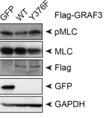
2.2.5 GRAF3 phosphorylation at Y376 prevents phosphorylation of myosin light chain in smooth muscle cells ... 47
2.3 Discussion ... 48
2.4 Materials And Methods... 51
2.5 Figures... 58
2.6 Supplemental Figures ... 65
CHAPTER 3: CONTRIBUTION OF GRAF2 TO BLOOD PRESSURE REGULATION AND ARTERIAL STIFFNESS ... 67
3.1 Introduction ... 67
3.2 Results ... 69
3.2.2 GRAF2 depletion elevates blood pressure ... 69
2.2.3 GRAF2 expression is increased in mice with HTN ... 70
3.2.4 GRAF2 depletion causes an increase in expression of SMC contractile genes and collagens, increasing vessel stiffness ... 70
3.3 Discussion ... 71
3.4 Materials and Methods ... 72
3.5 Figures... 77
CHAPTER 4: CONCLUSIONS AND FUTURE DIRECTIONS ... 83
4.1 Summary of work and future directions ... 83
4.2 Concluding remarks ... 90
4.4 Figures... 91
APPENDIX A: ANALYSIS OF GRAF3 GENOTYPE AND BP IN HUMAN POPULATIONS ... 96
APPENDIX B: TABLE OF CLINICAL COHORT CHARACTERISTICS ... 97
LIST OF FIGURES
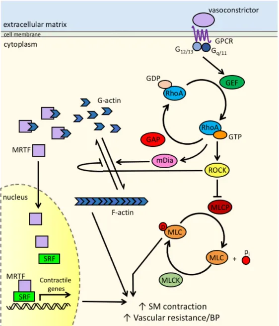
Figure 1.1 RhoA pathway in smooth muscle cells ... 34
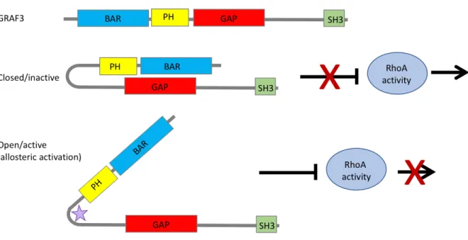
Figure 1.2 Domain Structure and therapeutic strategy for targeting the SMC-specific RhoGAP GRAF3 ... 38
Figure 1.3 Therapeutic potential of the RhoA pathway to treat HTN ... 39
Figure 2.1 SMC-specific GRAF3RQ overexpression decreases basal systolic blood pressure and maintains decrease through L-NAME dependent hypertension ... 58
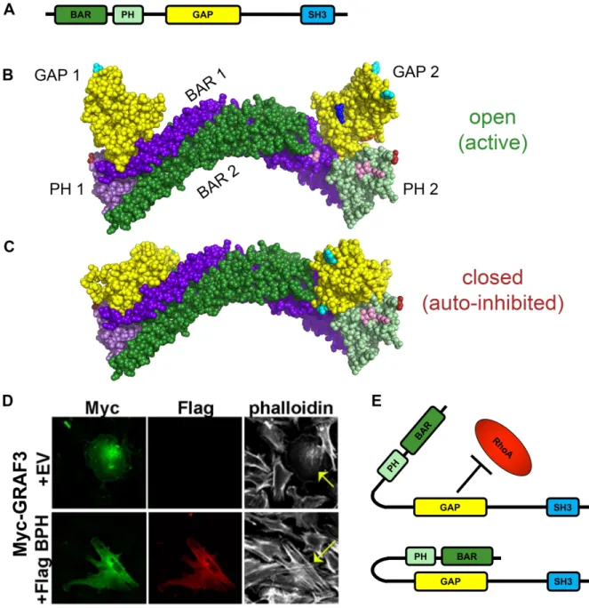
Figure 2.2 BAR-PH mediated autoinhibition of GRAF3 ... 59
Figure 2.3 Src and FAK kinases phosphorylate GRAF3 at Y376 in vitro ... 61
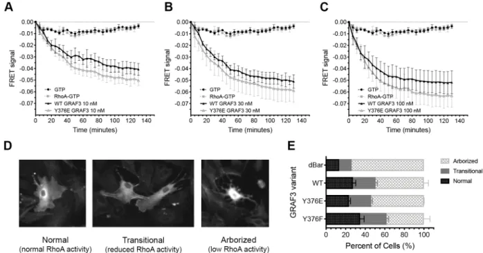
Figure 2.4 Phosphorylation of GRAF3 at Y376 increases GAP activity in vitro and decreases RhoA activity in SMC ... 62
Figure 2.5 GRAF3 phosphorylation at Y376 decreases pMLC in SMC ... 63
Figure 2.6 Working model of GRAF3 activation ... 64
Supplemental Figure S2.1 SMC-specific GRAF3RQ overexpression has no effect on diastolic BP, MAP, or HR ... 65
Supplemental Figure S2.2 Phosphorylation of GRAF3 by Src and FAK occurs at Y376 (predominant) and Y792 (to a lesser degree) ... 66
Figure 3.1 Smooth muscle specificity of GRAF2 and GRAF3 gene expression in humans ... 78
Figure 3.2 Generation of GRAF2 depleted mice ... 79
Figure 3.3 Novel GRAF2 deficient mouse model ... 80
Figure 3.4 GRAF2 deficiency leads to increased basal blood pressure ... 81
Figure 3.5 GRAF2 regulates SMC phenotypes ... 82
Figure 4.1 Post translational modifications of the GRAF family ... 92
Figure 4.2 Phosphorylation of GRAF3 at S152 by p38 delta affects RhoA signaling ... 93
Figure 4.3 Working model of GRAF3 phosphorylation at S152 ... 94
LIST OF TABLES
Table 1.1 ROCK inhibitors currently in clinical trials ... 35
Table 1.2 Domain Structure of RhoGEF proteins associated with blood pressure ... 36
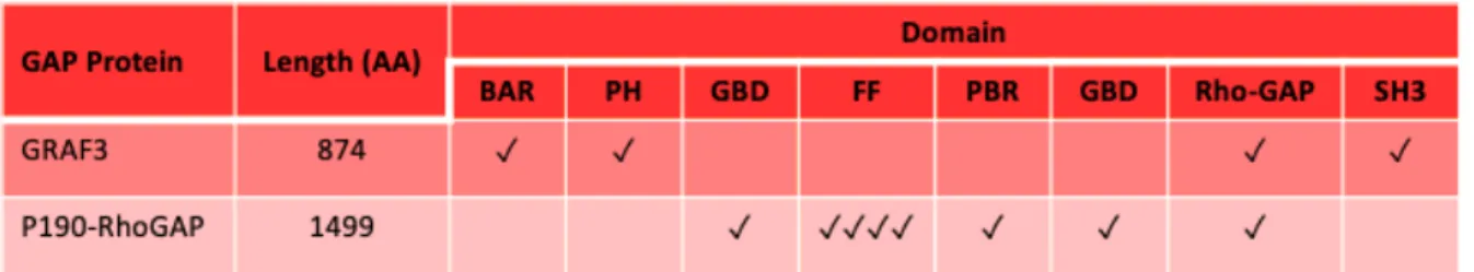
Table 1.3 Domain Structure of RhoGAP proteins associated with blood pressure. ... 37
LIST OF ABBREVIATIONS
ACE angiotensin converting enzyme AII angiotensin II
BAR Bin/amphiphysin/Rvs BFA Brefeldin A
BP blood pressure
CNS central nervous system CVD cardiovascular disease DOCA deoxycorticosterone-acetate ENaCs epithelial sodium channels ERM ezrin-radixin-moesin ET1 endothelin-1
FAK focal adhesion kinase GAP GTPase activating protein GDP guanosine diphosphate
GEF guanine nucleotide exchange factor GPCR G protein-coupled receptor
GRAF GTPase Regulator Associated with Focal adhesion kinase GTP guanosine triphosphate
GWAS genome-wide association studies HTN hypertension
L-NAME L-NG-Nitroarginine methyl ester
MD macula densa
mDia mammalian Diaphanous MLC myosin light chain
MLCK myosin light chain kinase MLCP myosin light chain phosphatase
MRTF-A myocardin related transcription factor-A MRTF-B myocardin related transcription factor-B NHE3 sodium-hydrogen exchanger
NTS nucleus tractus solitarii
PAH pulmonary arterial hypertension PE phenylephrine
PH pleckstrin homology
pMLC phosphorylated myosin light chain RaAoSMC rat aortic smooth muscle cells
ROCK Rho Kinase or Rho-associated coiled-coil domain containing protein kinases S1P sphingosine-1-phosphate
SH3 SRC Homology 3
SHR spontaneously hypertensive rat
SM smooth muscle
CHAPTER 1: INTRODUCTION1
Although hypertension (HTN) is a major risk factor for stroke, myocardial infarction, and kidney failure and contributes to over 350,000 deaths annually in the United States1, we know
surprisingly little about its development or the mechanisms by which it promotes cardiovascular disease (CVD). A number of antihypertensive drugs are available, but regimens are usually chosen empirically and multiple drugs that target different organ systems are frequently required for effective treatment. One reason for these difficulties is that blood pressure (BP) is a complex trait that is regulated by many organ systems and a large number of humoral factors. Thus, a better understanding of the molecular and genetic mechanisms that control BP under normal and pathologic conditions should lead to novel drug targets and/or to personalized therapies that are more effective and less toxic. Recent advances suggest that RhoA signaling plays a role in the development of human HTN. The focus of this introduction will be 1) to provide a very brief overview of HTN, 2) to highlight the mechanisms underlying RhoA-dependent regulation of BP, 3) to summarize the current data on the strategies and efficacy of targeting this pathway in hypertensive patients, and 4) to state the objectives of this dissertation.
1 This chapter contains text and figures previously published in the following review articles:
1.1 A brief overview of hypertension
Hypertension is a major cardiovascular risk factor that significantly increases the incidence of stroke, myocardial infarction, heart failure, retinopathy, and kidney disease2.
Although HTN is one of the most modifiable cardiovascular risk factors, the number of
individuals with HTN is increasing world-wide. Further amplifying the importance of HTN, the American Heart Association has recently revised its definition of Stage 1 HTN to include individuals with systolic BP between 130 and 139 mmHg or diastolic BP between 80 and 89 mmHg. This change was prompted by studies demonstrating beneficial effects of lowering BP below the 120/80 mmHg threshold3-5 and effectively increased the number of Americans
categorized as hypertensive from 32% to 46%5. It is also becoming clear that many people
suffer from masked HTN (normal readings in the clinic, but hypertensive outside the clinic) and non-dipping HTN (steady BP through the day but no decrease in BP at night)6-8, suggesting that
more intensive BP monitoring would identify additional at-risk individuals9.
A number of relatively inexpensive first-line therapies are available to treat HTN including diuretics, angiotensin-converting enzyme (ACE) inhibitors, angiotensin II (AII) receptor blockers, and calcium channel blockers. However, these drugs are usually prescribed empirically and are often ineffective. Indeed, over 50% of adults who are being treated for HTN still do not have their BP under control10. Although treatment can be improved by multidrug
regimens that target different BP control mechanisms, 13% of treated patients have
drug-resistant HTN and remain hypertensive even after taking three 3 or more medications, or require 4 medications for adequate BP control11, 12. Taking multiple BP medications also increases the
poor patient compliance contribute to the difficulties in treating HTN, our lack of understanding of the etiology of HTN is also a major factor.
Many of the difficulties of treating HTN stem from the fact that BP is an extremely complex trait regulated by many organ systems. Although the major determinants of BP are cardiac output and systemic vascular resistance, BP homeostasis requires proper regulation of heart and vasculature function by the autonomic nervous system, kidneys, and endocrine organs. The fact that these systems are tightly integrated by many feedback loops further complicates our understanding of the development of HTN and its treatment. Nearly all heritable genetic
mutations that cause HTN affect kidney function and/or salt balance, but these variants only explain about 10% of HTN cases. More recent genome wide association studies (GWAS) have identified many genetic loci that correlate with relatively small differences in BP between populations. However, because most of these variations are within or near genes with no known connection to BP regulation, our understanding of how they affect the development of HTN is limited. A number of the genes identified by GWAS are highly expressed in endothelial and smooth muscle cells (SMCs), highlighting the importance of the vasculature as a major regulator of BP and as a target for potential therapies13-16.
1.2 Impact of the RhoA pathway on blood pressure homeostasis
member of this subfamily and has been shown to regulate a variety of cellular processes
including (but not limited to) actin and microtubule dynamics, cell force, cell shape and polarity, endocytosis, exocytosis, cell adhesion and migration, proliferation, and differentiation17, 18.
Like all GTPases, RhoA is regulated by guanosine triphosphate (GTP) binding and cycles between the active GTP-bound form and the inactive guanosine diphosphate (GDP)-bound form and this cycle is under the direct control of three groups of regulatory protein; guanine
dissociation inhibitors (GDIs) sequester RhoA into an inactive cytoplasmic fraction, guanine nucleotide exchange factors (GEFs) activate RhoA by facilitating exchange of GDP for GTP, and GTPase activating proteins (GAPs) promote RhoA’s intrinsic GTPase activity to hydrolyze GTP to GDP and efficiently turn off (or limit) RhoA-dependent signaling. When GTP-bound, RhoA interacts with a variety of effector molecules that mediate its varied functions including the Rho-associated coiled-coil domain containing protein kinases (ROCK I and II), the
diaphanous-related formins (mDia1 and mDia2), protein kinase N, citron kinase, rhophilin, and the rhotekins I and II, among other enzymes17.
Recent advancements suggest that RhoA signaling in the vasculature is a particularly attractive target for therapeutic intervention in the treatment of HTN. With respect to regulation of BP, Rho kinases are arguably the most important effectors as evidenced by the findings that increased ROCK activity has been observed in spontaneously hypertensive rats and some hypertensive patient populations19, 20 and that ROCK inhibitors like Y-27632, Fasudil, and
SAR407899 have been shown to reduce BP in hypertensive animal models and patients21.
Extensive studies have shown RhoA signaling enhances Ca2+-dependent, myosin-based force
Moreover, human genetic studies have identified BP-associated variants in several additional Rho-related genes further implicating this pathway in human HTN23.
Although Rho signaling components are relatively strongly expressed in vascular SMCs, nearly all, with the exception of the RhoGAP GRAF3, are expressed in many other tissues. Thus, when evaluating Rho signaling molecules as targets of anti-HTN therapy, it is important to consider the potential impact of modulating Rho-signaling in other organ systems. Interestingly, with respect to BP regulation, studies using pre-clinical models indicate that attenuating RhoA signaling in the vasculature, kidney, myocardium, and CNS could all lead to the desired outcome of lowering BP.
1.2.1 RhoA and arteriole tone
Vascular resistance is a major determinant of BP and is controlled, in large part, by smooth muscle cell contraction within small peripheral arterioles24-28. Mechanistically,
excitation-contraction coupling in SMCs is mediated by the Ca2+-dependent activation of myosin
light chain kinase (MLCK), and SMC tension is directly proportional to myosin light chain (MLC) phosphorylation at S19 as this enables myosin’s molecular interaction with actin29, 30
(Figure 1.1). Interestingly, besides promoting an inositol triphosphate-mediated increase in intracellular Ca2+, many circulating GPCR-coupled contractile agonists including AII,
norepinephrine, endothelin-1 (ET1), and sphingosine-1-phosphate (S1P) also stimulate RhoA activity in SMCs and in intact arteries which further enhances Ca2+-dependent SMC
contractility19, 20, 22. Active RhoA leads to ROCK-dependent inhibition of myosin phosphatase
and results in elevated MLCK activity and enhanced sensitization to Ca2+ 19, 31-33. Importantly,
RhoA-dependent pathways are involved in the increased vascular resistance associated with hypertension19-22, 34.
Studies in genetically engineered mice revealed that germline deletion of the
Rho-specific GEF, LARG, significantly attenuated salt-induced HTN35, while SMC-specific knockout
of the related GEF, p115RhoGEF, inhibited the development of HTN in response to AII20. In
addition, we recently showed that depletion of the SMC-selective, Rho-specific GAP, GRAF3 (ArhGAP42) in mice leads to basal HTN, increased pressor responses to AII, ET1, and
phenylephrine (PE), and elevated deoxycorticosterone-acetate (DOCA)-salt induced HTN36-38.
RhoA regulates several effector molecules that impact SMC contractility (Figure 1.1). Direct phosphorylation of MLC by ROCKs I and II promotes actin-myosin crossbridge cycling as does ROCK dependent inhibition of MLC phosphatase (MLCP). RhoA activity is also critical for de novo formation of actin filaments and formation of focal adhesions that are required for myosin-dependent force development and transmission, respectively. The Rho effectors mDia 1 and 2 are the most potent regulators of actin filament formation as these proteins function to directly catalyze actin polymerization in cooperation with the actin binding protein, profilin. ROCKs also inhibit actin de-polymerization by phosphorylating and activating LIM-kinase 1 and 2 (on Thr 508 or 505 respectively), which in turn, phosphorylate and inhibit the actin filament severing protein, cofilin39-42. Finally, ROCK-dependent phosphorylation of ezrin-radixin-moesin
(ERM) proteins promotes their tethering to integral plasma membrane proteins effectively stabilizing actin filaments and increasing force transmission43.
within the promoters of nearly all SMC contractile genes (including SM myosin heavy chain, SM22, calponin, and SM α-actin)44. SRF activity is modulated by transcription cofactors of the
myocardin family45-49 and two such co-factors, myocardin transcription factor A and B
(MRTF-A and MRTF-B) mediate strong trans-activation of SMC contractile genes50, 51. We have
previously demonstrated that RhoA promotes SMC contractile gene expression through actin polymerization-dependent regulation of MRTF-A and MRTF-B nuclear localization44, 51-54.
Cytoplasmic monomeric G-actin is abundant when RhoA activity is low (for example in SMC under low tension55), and under these conditions, G-actin binds to MRTF and masks an
N-terminal nuclear localization sequence, resulting in cytoplasmic sequestration of these SRF co-factors. Upon RhoA activation, G-actin is recruited into growing F-actin filaments and MRTF-G-actin association decreases. As a consequence, MRTF nuclear localization sequence is un-masked, and MRTF accumulates in the nucleus and promotes SRF-dependent gene expression56.
Moreover, elevated RhoA in endothelial cells impairs endothelial cell-mediated vasorelaxation as it decreases availability of the potent vasodilator, nitric oxide by reducing both eNOS expression and activity57-61. Thus, signaling through RhoA in small arteriolar SMC enhances
Ca2+ sensitivity, promotes actin remodeling and induces expression of contractile proteins and
these responses are necessary for maintaining sustained SMC contractility and elevated vessel tone.
1.2.2 RhoA and kidney function
peripheral vascular resistance. In most vascular beds, arteriolar tone is controlled by autonomic innervation and circulating hormones. However, in pre-glomerular afferent arterioles, increased kidney perfusion (manifesting as increased renal BP) stimulates SMC contraction through the tubuloglomerular feedback and myogenic responses (see 62 for review). The former mechanism
is mediated by increased glomerular filtration and NaCl delivery from the loop of Henle to the macula densa (MD), a cluster of epithelial cells located at the junction between the distal convoluted tubule and the end of the thick ascending limb and adjacent to the abluminal SMCs of the afferent arterioles. Increased NaCl uptake by MD cells results in secretion of ATP and adenosine which stimulate afferent arteriole SMC contraction via P2Y4/P2Y6 and A2 GPCRs, respectively63-68. The myogenic response is mediated by the activation of stretch-sensitive cation
channels. Together these mechanisms stabilize renal blood flow to protect the sensitive
glomerular capillaries from flow-induced trauma. Importantly, afferent arterioles express RhoA, ROCK I and II68, and several studies have convincingly demonstrated that the Rho/Rho kinase
pathway influences both of these feedback mechanisms in response to increased kidney perfusion63-67. The requirement of RhoA is likely due, at least in part, to its necessity for
P2Y4/P2Y6 and A2 receptor-dependent contractility. Indeed, ATP (via P2Y4/Y6) and
adenosine (via A2) stimulate RhoA activity in SMC and their pressor responses were prevented by pretreatment with the Rho-kinase inhibitor, Y-2763268.
ENaCs was significantly increased by expression of wildtype or constitutively active RhoA (G14V) and suppressed by expression of dominant negative RhoA (T19N). The changes in current correlated with alterations in the density of ENaCs at the PM70 and mechanistic studies
determined that RhoA signaling was essential for intracellular vesicle mediated transport of ENaCs to the apical cell surface71, 72. RhoA signaling also regulates the activity and subcellular
localization of NHE3, a key regulator of sodium absorption in the proximal convoluted tubule. NHE3 associates with ezrin and cortical actin filaments at the plasma membrane and treatment with either the RhoA inhibitor, diarrheal toxin toxin B, or Y-27632 disrupted these interactions and promoted the internalization of NHE3 to sub-membrane compartments73, 74. Moreover,
Nishiki et al. showed that spontaneously hypertensive rats exhibited elevated NHE3 activity and an exaggerated level of Na+ reabsorption when compared to normotensive controls and that Na+
reabsorption was normalized by treatment of the hypertensive animals with Y2763275.
1.2.3 RhoA in the central and peripheral nervous system
The central nervous system (CNS) constantly assesses pressure levels in the vasculature and makes necessary signaling adjustments to prevent BP variability. The main mechanism by which the CNS monitors BP is through a rapid negative feedback loop termed the baroreceptor reflex. Baroreceptors are sensory neurons located primarily in the aortic arch and carotid sinuses that continuously respond to pressure-induced stretching of the vessels in which they reside. Impulses from baroreceptors are relayed via glossopharyngeal and vagus nerves to the nucleus tractus solitarii (NTS) in the brainstem76, which in turn relays the signal to the rostral
ventrolateral medulla77 and increases or decreases parasympathetic and sympathetic stimulation
been shown to be dependent on RhoA/Rho-kinase signaling. Rho-kinase inhibitors
microinjected directly into the NTS or infection of this structure with an adenovirus expressing a dominant-inhibitory form of Rho-kinase reduces sympathetic nerve activity, heart rate, and BP in normotensive rats and these effects are even more pronounced in spontaneously hypertensive rats78, 79. Moreover, infusing the ROCK inhibitor, Y27632, into the neural cistern attenuated the
BP increase that resulted from AII infusion into the same area of the brainstem80.
Recent studies indicate that the RhoA pathway may also regulate neurotransmitter release from perivascular nerves. Some studies in cells and invertebrate model systems indicate that inhibition of Rho/Rho kinase signaling in motor neurons antagonized the secretion of
parasympathetic relaxation factors (including acetylcholine) and promoted the secretion of sympathetic contractile agonists (including dopamine)81, 82. Yamaguchi et al. found that Gα
12/13
-mediated activation of RhoA/ROCK inhibited Ca2+ dependent exocytosis81. In support of these
studies, an activating mutation in ArhGEF10, a RhoGEF highly expressed in the peripheral nervous system, was identified in patients who exhibited slowed nerve conduction velocities83, 84.
Thus, it is possible that RhoA's ability to block neurotransmitter release in peripheral nerves could affect vascular tone and BP but such outcomes may limit the therapeutic efficacy of RhoA/ROCK inhibitors as future anti-hypertensive therapies.
1.2.4 RhoA in the myocardium
Several studies have shown that RhoA signaling has direct effects on cardiac function that increase cardiac output and BP. Transgenic mice that overexpressed either GDIa or
contractility observed in vivo85, 86. Vlasblom et al. showed that treatment of neonatal ventricular
cardiomyocytes with Y27632 reduced the expression and activity of the sarcoplasmic reticulum Ca2+ ATPase, SERCA2a, thereby limiting the amount available for Ca2+ -induced Ca2+ release
in the next cardiac cycle87. In addition, RhoA-dependent pathways have been shown to be
critical for phosphorylation and sensitization of cardiac troponin T complex to intracellular Ca2+
levels88. Moreover, while not initially thought to be a major mechanism for modulating cardiac
contractility, it is becoming clear that cardiac MLC phosphorylation can enhance muscle contractility by increasing Ca2+ sensitivity 89and that MLC phosphatase is a target for Rho
kinase-dependent inhibition in the myocardium (like in SMC). Indeed, Lauriol et al. showed that cardiac-restricted deletion of RhoA led to decreased contractility and this effect was correlated with decreased MLC activity90. Other similarities between RhoA signaling in cardiomyocytes
and SMC include the ability of RhoA-mediated signals to promote differentiation/maturation by promoting the expression of contractile genes91.
1.3. Targeting the RhoA pathway for therapeutic blood pressure control
In agreement with the pre-clinical animal studies highlighted above, several lines of evidence strongly implicate RhoA signaling in the development of human HTN. First, increased Rho-kinase activity has been observed in some hypertensive patient populations, reviewed below. Rho kinase inhibitors have been successful in reducing systemic HTN in these cases, although current formulations exhibit relatively short-term effects23, 92, 93. Second, an autosomal
reduced ubiquitin-mediated RhoA degradation in vascular SMCs, and increased BP94, 95. Third,
many GWAS conducted over the past decade have identified common BP-associated genetic variations in coding and non-coding regions within or near genes linked to the RhoA signaling cascade. For example, in one study that used HTN as a dichotomous trait, two of the eight BP-associated loci were located in RhoA-related genes. One was within RhoBTB1, which functions with the aforementioned Cullin-3 complex to maintain low RhoA levels94, 96, while the other was
within rhotekin-2 (RTKN2), a RhoA effector with unknown function. Two separate GWAS identified a BP-associated locus within PLEKHA7 (Plekstrin Homology domain containing family A member 7)97, 98 which interacts with the junctional proteins cingulin and paracingulin to
regulate several Rho family GTPases, including RhoA in the heart and kidneys99. Importantly,
PLEKHA7 was subsequently shown to be required for the development of salt-induced HTN in mice100. Moreover, a few variants in ROCK II have been associated with the regulation of BP.
Of particular interest was the identification of a common nonsynonymous ROCK II variant 431N (versus 431T) that was associated with an increase in BP in twins69, 101-104. This result was
supported by Liao et al., who showed that the 431N variant had increased kinase activity and was associated with enhanced arterial stiffening, a vascular property strongly associated with HTN69, 101-104. This group identified a second variation in the ROCK II 3'UTR (rs9789060) that
was also associated with increased stiffening and went on to show it affected ROCK II
expression by interfering with miR-1183-dependent degradation of ROCK II mRNA levels. It is important to note that a third study failed to find an association between rs9789060 and BP101. In
another study on 586 normotensive and 607 hypertensive Caucasians, Rankinen et al. identified a minor allele locus within the ROCK II gene that lowered the risk of HTN by 85%. 103. Finally,
endpoints identified a novel BP associated locus within the Rho-specific GAP,
GRAF3/ArhGAP4215, 16, 105, 106. Recent causality studies from our group demonstrated that
GRAF3 is selectively expressed in SMC and is required for BP homeostasis in mice36-38.
Collectively, these studies reveal that the RhoA signaling axis may provide tractable targets for the treatment of human HTN and related cardiovascular sequela. Indeed, some commonly used anti-hypertensives (i.e. ACE inhibitors, AII blockers, and statins) likely function by interfering with RhoA signaling, supporting the clinical utility of inhibiting this pathway22, 107-109. Nonetheless, despite the importance of the RhoA pathway in the pathogenesis of HTN and
several other debilitating diseases including amyotrophic lateral sclerosis, mental retardation, hepatocellular, lung, and colorectal carcinomas110-112, surprisingly few treatments are available to
directly target this pathway. In fact, of the nearly 300,000 ongoing clinical trials, only a handful involve compounds that target RhoA signaling components (clinicaltrials.gov). This could be due to the fact that many members of the RhoA pathway (with exception of Rho kinase) have traditionally been regarded as “undruggable”. However, as described below, significant advancements in high resolution crystal structures, structure-function analyses and drug development technology are beginning to overcome these challenges and provide hope for the development of new therapies to target this critical pathway.
1.3.1 Targeting Rho Kinase
The serine/threonine kinases, ROCK I and ROCK II, are the best studied RhoA effectors and have been implicated in a variety of diseases including HTN23. Since the development of
inhibitors are the furthest along in regard to clinical testing. Because ROCK I and II share 60% identity overall, 90% identity within the kinase domain identity, and 100% identity within the ATP binding pocket113 they share many substrates and promote many of the same downstream
cell functions93. Although ROCK I and ROCK II expression can vary somewhat between
tissues, both of these kinases are widely expressed113, 114. Differences in subcellular localization
have been noted with ROCK I localizing more readily to microtubule-organizing centers115 and
catenin/E-cadherin containing complexes at the plasma membrane116 and ROCK II to vimentin 117 and actin fibers118114, 119. Isoform-specific inhibitors are being developed that could have
differential effects depending upon the disease treated or end-point measured23, 120-123
(clinicaltrials.gov).
While over 30 common downstream targets of ROCK have been identified (Adducin, Diaphanous [mDia], LIM kinase [LIMK], NHE1, MARCKS, NF-L, CRMP2, FAK, c-Jun N-terminal kinase [JNK], MLC, MLCK, MLCP, ezrin/radixin/moesin [ERM], rhophilin, rhotekin, citron kinase, and Tau)23, 93, 119, 124, the most pertinent in regard to SM contraction and BP
regulation is the myosin binding subunit (MYPT-1) of the MLCP complex. ROCK-dependent phosphorylation of MYPT-1 at T696, T853, and S854 inhibits MLCP activity resulting in increased MLC phosphorylation and hence increased contraction114, 125. ROCK has also been
shown to directly phosphorylate MLC at S19.
As reviewed more thoroughly elsewhere23, 93, over 170 ROCK inhibitors are in various
neurodegenerative diseases (Alzheimer’s disease, Parkinson’s, Huntington disease, and
amyotrophic lateral sclerosis [ALS]), cardiovascular disease (systemic HTN, pulmonary arterial HTN [PAH], spastic and stable angina, atherosclerosis, Raynaud syndrome), asthma, glaucoma, autoimmune diseases, cancer, erectile dysfunction, and kidney disease. Although the results of these studies and clinical trials provide important information on ROCK and its role in disease progression, we will focus our discussion on the major ROCK inhibitors that have been most commonly used to treat vascular diseases.
Fasudil (also known as HA-1077) was the first ROCK inhibitor described and was also the first to be tested clinically. This isoquinoline-based drug is classified as a class I ROCK inhibitor because it reversibly competes with ATP binding to the ROCK kinase domain. Fasudil inhibits both isoforms of ROCK with an IC50 of 1µM while hydroxyfasudil, the major active
metabolite, is slightly more potent with an IC50 of ~0.7µM126. Like most other ROCK inhibitors
of this class fasudil also inhibits other members of the broad AGC kinase family including PKA and PKC, albeit with less potency (IC50 of 5µM and 37µM, respectively)126, 127. Fasudil was
shown to reduce BP by attenuating the Rho-mediated inhibition of MLCP in SMC126, by
increasing endothelial nitric oxide synthase expression128, and by reducing circulating ACE and
AII levels129. Fasudil also inhibited pulmonary artery SMC proliferation, a process important for
vessel stiffening, by a mechanism that likely involved downstream inhibition of c-Jun N-terminal kinase (JNK) and ERK-dependent activation of c-jun and c-fos expression130.
treatment with oral fasudil reduced stable effort-induced angina and improved exercise tolerance with no major adverse effects132, 133. In a rat model, moderate doses of fasudil decreased PAH
while higher doses also decreased mean systemic arterial pressure134. By 2011, intravenous and
inhaled fasudil were approved to treat PAH135, 136, and a new extended release formulation of
fasudil, AT-877ER, was shown to reduce PAH in patients after three months of use137. Fasudil
also decreased forearm vascular resistance more dramatically in hypertensive patients than in normotensive controls21. Fasudil has been shown to have beneficial effects on kidney function in
diabetic rats138, suggesting that it might be useful for treating diabetic patients who are frequently
hypertensive and have kidney disease. Ongoing phase III clinical trials are also assessing whether fasudil is an effective treatment for Reynaud’s syndrome and carotid stenosis
(clinicaltrials.gov). Fasudil has been approved in China, but not in the United States or Europe. Several fasudil derivatives that are more potent and specific inhibitors of ROCK have been developed and are being used to treat glaucoma and ocular HTN. Ripasudil (Glanatec®), approved in Japan in 2014, affects the trabecular meshwork in the eye and reduces intraocular pressure by facilitating the outflow of aqueous humor through Schlemm’s canal. The most common side effect of ripasudil treatment is conjunctival hyperemia, which subsides over time or with discontinued use139. Netarsudil (Rhopressa ®) has similar effects and indications, and in
2017, became the first ROCK inhibitor approved by the United States140, 141. Netarsudil also
decreased the amount of aqueous humour produced, a feature attributed to additional effects of netarsudil as a norepinephrine transport inhibitor140, 141.
(SHR), DOCA-salt treated rats, and rats made hypertensive by clipping of the renal artery142.
Although mesenteric and cerebral arteries in SHR or DOCA/salt treated rats were more
responsive to Y-27632 than those in normotensive rats143, 144, Y-27632 likely affected HTN by
multiple mechanisms. For example, Y-27632 also reversed the decrease in renal sodium excretion observed in the SHR model75, most likely by affecting the activity and location of
Na+/H+ exchanger, NHE373, 74. In addition, local infusion of Y-27632 into the nucleus tractus
solitarius of the brainstem caused a reduction in BP, heart rate, and renal sympathetic nerve activity, and the magnitude of these effects was greater in the SHR model78, 79. Subsequent
studies by the same group demonstrated that fasudil had similar effects78, 79. Although Y-27632's
poor potency and kinase selectivity limit its use clinically, Y-27632 derivatives with better pharmacologic properties are being developed and tested.
SAR407899 is a promising relatively new isoquinoline-based class I ROCK inhibitor that is significantly more potent than older generation drugs (IC50 between 122 and 280 nM)145. In
vitro studies demonstrated that SAR407899 inhibited myosin phosphatase phosphorylation, stress fiber formation, cell proliferation, and monocyte chemotaxis145. SAR407899
dose-dependently lowered BP in SHR, stroke-prone SHR, L-NG-Nitroarginine methyl ester
(L-NAME), and DOCA-salt rat models, and its effects in some models was superior to ACE inhibitors and calcium channel blockers145. SAR407899 inhibited pressor responses to PE, AII,
and vasopressin in rats more strongly than Fasudil or Y-27632, and it inhibited ET1-induced vasoconstriction of renal arteries isolated from diabetic rats more strongly than Y-27632146. In
and microvascular coronary artery disease and it may prove useful for treatment of erectile dysfunction in diabetic and hypertensive patients where the eNO system is impaired147.
Although ROCK inhibition has been shown to reduce BP and vascular resistance in many models, there are concerns about the suitability of ROCK inhibitors as viable long-term
treatments for systemic HTN. Although most ROCK inhibitors seem to be fairly well-tolerated, the ubiquitous nature of ROCK I and ROCK II expression coupled with the relative lack of specificity of most ROCK inhibitors (especially the class I drugs) makes potentially unknown side-effects a significant drawback. While local delivery strategies can sometimes mitigate this concern (i.e. to the eye or specific vascular beds)93, 148, systemic hypotension can be a serious
problem for patients being treated in this manner. Another potential problem is that the systemic BP lowering effects of ROCK inhibitors frequently decrease after 7-10 days of chronic
treatment93. Moreover, ROCK inhibitors did not affect systolic BP in some long-term studies of
salt-sensitive hypertensive Dahl rats69, 75. The precise causes of this tolerance or inactivity are
unknown, but likely involve compensation by the many feedback pathways that regulate BP. Finally many ROCK inhibitors have short half-lives, which is not ideal for the treatment of a chronic disease like HTN149.
1.3.2 Targeting RhoA directly
Numerous failed attempts to identify small molecule inhibitors of H-Ras have led to the concept that small GTPases per se do not make good drug targets due to their globular structure and lack of surface moieties required for high affinity binding of small molecules110, 150, 151.
153. Moreover, in the case of HTN, direct targeting could provide an added benefit over ROCK
inhibitors, as such an approach would block additional downstream pathways implicated in SMC contractility (i.e. mDia1 and mDia2, MRTF, etc.).
1.3.2a GTP-binding inhibitors
Although direct targeting of the GTP binding site of RhoA (or related small GTPases) is challenging due to its high affinity for GTP (in the pico to nano-molar range) and the high concentration of GTP in cells (~0.5 mM), some studies support the validity of this approach. Indeed, using an in silico virtual docking approach followed by surface-plasmon resonance validation of synthesized chemicals, Deng et al. identified lead compounds that inhibited Rho-GTP binding in a dose-dependent fashion with IC50 values ranging from 1.24–2.05 µM154. After
further structural modifications to increase water solubility, one compound ((E)-3-(3
-(ethyl(quinolin-2-yl)amino)phenyl)acrylic acid) was shown to both attenuate PE-induced contraction in thoracic aorta rings ex vivo and to reduce cerebral vasospasm in a subarachnoid hemorrhage model in rats155. While the in vivo efficacy was similar to that of fasudil, this second
generation inhibitor still exhibited relatively low potency (IC50 71 µM in the contractile assay).
Future studies will be necessary to determine whether these or related compounds exhibit specificity for RhoA versus other Rho family GTPases and other GTP-binding proteins.
are active in cell based assays156-158. The subsequent application of this technology to screen the
Prestwick Chemical Library of off patent and FDA approved drugs for inhibitors of eight Ras-related GTPases (but not RhoA, B, or C) led to the identification of R-enantiomers of naproxen and ketorolac (approved NSAIDs) as GTP binding inhibitors of Rac1 and Cdc42159. Although
not biochemically confirmed, in silico docking analyses predicted that these drugs bind to an allosteric site near the GTP binding site that alters Mg2+ ion coordination and results in
stabilizing the GTPase in its GDP-bound form. To our knowledge, this approach has not been used successfully to identify RhoA specific inhibitors, but these results provide strong proof-of-concept for this approach.
1.3.2b Selective non-covalent RhoA modifiers
The potential for identifying allosteric modifiers to inhibit RhoA has been demonstrated by the fact that a number of bacterial toxins are highly potent (though non-selective) inhibitors of Rho family members. For example, Histophilus somni and Vibrio parahemolyticus produce toxins that inhibit Rho proteins by promoting the covalent attachment of an AMP molecule to tyrosine 34, while toxin B produced by Clostridium difficile induces glucosylation of nearby threonine 37. These residues lie within the regulatory switch-I domain of Rho family members and addition of these bulky modifications in this domain inactivate the Rho GTPases by multiple mechanisms that include inhibition of GTPase cycling (by blocking GEF and GAP association), inhibition of cytosol-membrane cycling (by blocking Rho GDI interactions) and inhibition of Rho effector coupling160, 161. Similarly, Clostridium botulinum exoenzyme C3 transferase toxin
pharmacological tools to completely block Rho-dependent signaling pathways in cells and in pre-clinical animal models. However, their potential as therapeutic agents is limited because of their difficult delivery, their non-specific actions, and their sometimes covalent and irreversible effects110, 163. Nonetheless, the future exploitation of derivatives or mimetics of such enzymes
could lead to the development of new inhibitors with tremendous clinical utility. Finally, Rho family inactivation can be achieved by blocking plasma membrane
targeting. For example, Rho GTPases are isoprenylated on carboxy-terminal Cysteine residues (within a so-called CAAX box) and this modification is important for membrane targeting and activation as evidenced by the fact that proteolytic cleavage of this site by Yersinia spp.-derived toxins effectively block Rho, Rac and CdC42 activation161. Likewise, as RhoA is
geranylgeranylated at this site, geranylgeranyl-transferase inhibitors and HMG-CoA-reductase inhibitors (which block both cholesterol and isoprenyl biogenesis) block RhoA membrane association and activation. In fact the clinical utility of this approach is highlighted by the fact that HMG-CoA reductase inhibitors such as simvastatin and atorvastatin, used to treat high cholesterol, also have anti-hypertensive properties109 and their BP lowering effects have been
attributed to their ability to block RhoA signaling108, 109, 164. However, the BP lowering effects of
statins are modest (2 mmHg decrease systolic BP) and these isoprenoid pathway inhibitors display poor selectivity for individual Rho GTPases. Thus, further advancements in drug development are needed to realize the full potential of this approach.
1.3.3 RhoA GEFs and blood pressure control
these have been implicated in regulating SMC differentiation and/or contractility. This list includes 3 members of the RGS-GEF subfamily (P115-RhoGEF, PDZ-RhoGEF, LARG), p63RhoGEF and lymphoid blast crisis (Lbc)20, 165-168. Each of these RhoGEFs contains a Dbl
homology (DH) domain (also known as the RhoGEF domain) followed by a pleckstrin homology (PH) domain (Table 1.2). The DH domain serves as both the catalytic site and the major binding interface for RhoA, while the PH domain facilitates membrane binding and
cooperates with DH domains to fully activate RhoA. Other common domains include a regulator of G-protein signaling (RGS) domain that binds heterotrimeric G proteins, or the density 95, disk large, zona occludes-1 (PDZ) domain that binds to specific transmembrane receptors (including the Lysophosphatidic Acid (LPA) receptor among others169. Importantly, these 5 GEFs are all
highly expressed in conductance and resistance arteries of rats, mice, and humans20, 165-168
(Genotype-Tissue Expression [GTEx] Portal, accessed on 07/12/2018).
The regulator of G-protein signaling (RGS) family of Rho GEFs (LARG, p115RhoGEF, and PDZRhoGEF)170 has received a lot of attention in the BP field because these proteins are
activated by Gα12 and Gα13- coupled receptors which transduce signals mediated by major
contractile agonists that include AII, PE, ET1, and thromboxane A2171. P115RhoGEF (p115) is
the critical GEF that mediates AII-dependent RhoA activity in SMC and small arterioles, and importantly, Guilluy et al. showed that SM-specific deletion of p115 rendered mice resistant to AII-dependent HTN35. However, AII–dependent activation of RhoA in SMC has also been
linked to activation of LARG172, PDZ-RhoGEF165, 167, and p63RhoGAP173 and inhibition of
p190RhoGAP174 (see below), suggesting significant overlap between these pathways. For
example, Ying et al. showed that Ca2+/PYK2-dependent activation of PDZ-RhoGEF was
which is highly expressed in arterial SM, was shown to be important for the early phase of AII-dependent vessel contractility173 and for maximal pressor response to other vasoconstrictors such
as PE and ET1 that act through Gα q/11175. Interestingly, p115 mutant mice exhibited normal
pressor responses to ET1 and PE, but did not respond fully to AII and had a partial reduction in DOCA/ induced HTN. On the other hand, LARG knockout mice were fully resistant to salt-induced HTN20, which is consistent with subsequent findings that LARG regulates RhoA activity
and SMC contractility in response to mechanical forces—which are known to be applied to small vessels in this volume overload model176. While not yet confirmed in pre-clinical animal
models, the non-RGS Rho GEF termed lymphoid blast crisis (Lbc) is necessary for serotonin-dependent activation of RhoA and contractility in vascular SMC177. Thus, specific
vasoconstrictors can lead to activation of distinct but overlapping sets of RhoGEFs to enable the fine-tuning of vessel tone and BP homeostasis.
With respect to pharmacological treatments, these findings indicate that targeting a critical SM GEF might provide greater therapeutic benefit than targeting RhoA or Rho kinase, because it could lead to a more modest (and cell type restricted) reduction of RhoA activity and result in fewer side effects. Moreover, the use of validated inhibitors could provide
1.3.3a Targeting RhoGEFs
GEF protein-dependent nucleotide exchange involves a multi-step reaction. Upon binding to the GDP-bound form of the GTPase, GEFs facilitate release of GDP resulting in the formation of a nucleotide-free GTPase–GEF transition complex. The reaction then terminates with the binding of GTP (which is in a 10:1 molar excess in the cell) and dissociation of the GEF from the now active GTPase. The implication of this multi-step process for drug discovery is that these transition-binding intermediates tend to have the characteristics of druggable hotspots (i.e. exposed hydrophobic surfaces, unpaired polar groups, deeply curved surfaces, etc.) while the unbound, inactive proteins do not178. Thus, it may be possible to identify small molecules
that specifically target the GEF-GTPase interface (i.e. inhibit GEF binding or stabilize the GEF-GTPase transition state) or directly interfere with GEF activity.
1.3.3b Targeting the RhoA-GEF interface
Facilitated by high resolution crystal structures and sophisticated in silico screening, some recent drug discovery approaches support the possibility of targeting the RhoA-GEF interface. For example, Shang et al. virtually screened 4 million compounds for their ability to pack into the Rho-GEF binding surface groove of RhoA. One of the drugs identified (termed Rhosin) contains two aromatic rings tethered by a linker which wraps around RhoA AA58 (a critical Tryptophan that is essential for GEF binding) and prevents RhoA from interacting with LARG, DBL, LBC, p115RhoGEF and PDZ RhoGEF without interfering with RhoGAP,
RhoGDI, or RhoA effector binding151. Rhosin reversibly inhibited serum-induced RhoA, RhoB
and RhoC activity, but did not inhibit other Rho GTPases (Rac1 or Ccd42)151. Rhosin also
including MLC and PAK phosphorylation and stress fiber and focal adhesion formation151.
Rhosin suppressed invasion and mammary sphere formation in breast cancer cells151, and also
mitigated the acquisition of drug resistance in cancer stem cells179. The ability of Rhosin to
impact either BP homeostasis (or cancer growth) has not yet been tested in animal models. Since Rhosin exhibits a relatively low binding affinity, (Kd ~0.4 µM for RhoA binding in vitro) the
identification of derivatives with better pharmacologic properties will be needed before Rhosin compounds can be tested clinically151. Moreover, since Rhosin binds to RhoA and prevents all
GEFs from binding to this site, this drug is likely to have more off-target effects than one that could interfere with specific GEF complexes.
The first proof-of-concept for targeting a specific GEF-GTPase interface was a series of elegant studies that unveiled the mechanism by which the fungal toxin, Brefeldin A (BFA), inhibited Arf1-dependent trafficking of proteins from the endoplasmic reticulum to the Golgi. Biochemical studies revealed that BFA selectively blocked the ability of the Arf1GEF, Sec7, from catalyzing GDP release. Importantly co-crystallization studies revealed that BFA binds to a hydrophobic pocket that does not exist in Arf1GDP but is created upon Sec7 binding. Since this
energetically unfavorable hydrophobic pocket drives the conformational changes necessary for nucleotide dissociation, BFA binding effectively locks the complex in a GDP bound
conformation180-182. In spite of high sequence homology among the Arf1GEFs, only the Sec7
complex is targeted by BFA, suggesting that ‘interfacial’ small molecule inhibitors could be identified that specifically target RhoA-GEF interactions.
These studies have inspired a new line of investigation that seeks to exploit novel pockets at small GTPase-GEF interfaces. The most productive approach so far used NMR-based
molecule inhibitors of kRas and its GEF, Sos1. These screens identified compounds that either block Sos1 binding to kRAS or that bind to a site adjacent to the functionally important switch I/II regions of KRas and (like BFA) block SOS1-dependent nucleotide exchange. A similar approach was used to identify a ligand that binds to the cavity adjacent to the switch II region of RhoA and inhibits the interaction between RhoAGDP and the LARG DH domain183. Small
molecules have also been identified that inhibit TrioGEF's interaction with Rac1 and RhoG and Cdc42's interaction with the GEF, intersectin-1184. Clearly much work needs to be done to
realize the potential for such ligands. The inhibitors identified exhibit very low potency (in the high micromolar range) and their ability to block additional GEFs has not yet been evaluated. In fact, in silico evaluation of aforementioned SOS1 inhibitor suggests that the hydrogen bonds formed by this ligand in the Ras-SOS1 complex would likely be conserved in the complex of Ras and another cognate GEF, RasGRF1183. While challenging, further medicinal chemistry and
molecular dynamics approaches should facilitate our ability to target RhoA's interaction with specific GEFs.
1.3.3c Targeting GEF activity
Another viable approach is to directly target the activity of specific RhoGEFs before they interact with the small GTPases. Using a high-throughput screen, Shang et al. identified a
compound, Y16, that binds LARG at the junction between the DH and PH domains with a Kd of
~80nM185. Y16 prevented LARG from interacting with RhoA in vitro and reversibly attenuated
serum-induced RhoA activity in NIH 3T3 cells and mammary sphere formation MCF7 cells with little to no toxicity185. A separate fluorescent ligand-based screen of ten thousand compounds
their mechanisms of action have not yet been determined186. Again, to date, many of these lead
compounds exhibit low potency, but theoretically, combination therapy strategies which pair Y16 with some of these compounds and/or their derivatives may lead to a highly efficacious approach to limit LARG-mediated signaling.
Finally, since many GEFs are regulated by protein-protein interactions and
post-translational modifications, it may be possible to identify drugs that block these mechanisms. In an excellent example of this approach, cAMP is a potent activator of the Rap1 GEFs, Exchange Proteins directly Activated by cAMP (EPAC1 and 2) and high throughput screens to identify small molecules that displace cAMP binding led to the discovery of several inhibitors or partial agonists for these GEFs187-189. Such studies support the concept that identification of small
molecules that prevent physiological activation of specific GEFs could prove therapeutically useful. Along these lines, several studies have shown that RGSRhoGEFs are regulated by phosphorylation. Guilluy et al. demonstrated that AII-dependent activation of p115GEF was mediated by phosphorylation of the PH domain (Tyr738) by Janus tyrosine kinase22. Importantly,
phosphorylation mimetic and deficient variants of Tyr738 elevated and reduced p115's GEF activity, respectively. PDZRhoGEF and LARG are also activated by tyrosine phosphorylation. Focal adhesion kinase (FAK) and its related family member, proline-rich tyrosine kinase 2 (PYK2) phosphorylate and activate PDZRhoGEF167, 190, while FAK, Tec and the
serine-threonine kinase, p90 ribosomal kinase-2 each phosphorylate and activate LARG167, 170, 190, 191.
1.3.4 RhoA GAPs and blood pressure control
Although GEFs have classically been considered to be the major regulators of RhoGTPase activity, increasing evidence suggest the GAPs are also critically important. As their name implies, GAPs enhance the intrinsic GTPase activity of the Rho GTPases by several orders of magnitude thus decreasing the length of time that GTPases are in the active form192-194.
The 66 RhoGAPs in the human genome comprise a broad and diverse family that can be further subdivided based upon the presence of a variety of functional domains195 (Table 1.3). By
mediating interactions with different membrane and protein components these domains are critical for the selective function of the GAP proteins and the dynamic inhibition of small GTPase signaling. Several Rho-selective GAPs including p190ARhoGAP, ArhGAP1, Myr5, GRAF1, and GRAF3 have been shown to inhibit RhoA activity in cultured vascular SMC and could be potential targets for therapeutic interventions that affect vascular function and BP36, 196.
However, GRAF3 is the only RhoGAP that has been directly linked to the regulation of SM contractility and BP homeostasis15, 16, 36, 105, 106.
1.3.4a GRAF3 and Hypertension
We originally identified the founding member of the GRAF (GTPase Regulator
Associated with FAK) family by screening an embryonic λgt11 expression library for proteins that interacted with the carboxyl-terminal domain of FAK197-199. This family which is now
striated muscle (cardiac and skeletal), and our studies in GRAF1-depleted Xenopus and mice revealed that GRAF1-dependent inhibition of RhoA activity promoted mammalian muscle growth by facilitating myoblast fusion and injury repair199-202. GRAF2 is more ubiquitously
expressed203 and could partially compensate for the loss of GRAF1 during myotube formation,
supporting at least some functional redundancy within this family201. Evolutionarily, GRAF3 is
the youngest family member and is the most recently annotated. Importantly, we found that GRAF3 was highly and selectively expressed in SMC with particularly high expression in resistance vessels36 suggesting that its expression levels might be a major determinant of RhoA
activity, SM contraction, and vessel tone.
Over the last several years, studies from our laboratory confirmed that GRAF3 is a SMC-selective Rho GAP protein that imparts tight control of BP homeostasis by modulating vascular resistance. Specifically, we showed that mice with gene-trap-mediated reductions in GRAF3 levels exhibited significant basal HTN and elevated pressor responses to AII, ET1, PE, and DOCA salt36. Notably, the hypertensive phenotype in this model was fully reversed by treatment
with ROCK inhibitors or by Cre-mediated re-expression of GRAF3 in SMC, strongly supporting the contention that GRAF3 control of SMC RhoA activity is necessary for homeostatic control of basal and pressor-induced BP. Mechanistically, our data supported a model wherein GRAF3 controls BP homeostasis by limiting RhoA-dependent MLC phosphorylation and blunting acute Ca2+-mediated SMC contractility in resistance vessels. Moreover, depletion of endogenous
GRAF3 from vascular SMC enhanced MRTF-A nuclear accumulation, and stimulated
subjected to cyclic stretch and in isolated portal vein segments subjected to static stretch and we showed that these effects were mediated via the RhoA/MRTF/SRF pathway37. Since similar
physical forces are known to be increased in the vessel wall under hypertensive conditions55, we
postulated that GRAF3 might serve as a transcriptionally mediated negative feedback loop of the RhoA signaling axis. In support of this possibility, arterial GRAF3 mRNA levels were
significantly increased in mice made hypertensive by L-NAME or DOCA-salt regimens36. In
fact, taking advantage of an elegant mouse model (of aldosteronism) developed by the Smithies lab in which plasma volumes of mice range from ∼50% below normal to ∼50% above normal204,
we showed that GRAF3 expression increased in parallel to and was strongly correlated with plasma volume (r2=0.94)37. Taken together, these findings suggested that GRAF3 serves as a
mechanical strain-sensitive rheostat that acts to prevent excessive feed-forward activation of the RhoA signaling axis in order to control SMC tone and BP.
Of clinical importance, three separate GWAS studies identified a blood-pressure associated allele within the GRAF3 locus. Notably, our recently published follow-up studies revealed that GRAF3 mRNA levels were approximately 3-fold higher in arteries from individuals homozygous for the protective allele and our human genetic data from
well-characterized untreated patients confirmed that the protective allele was associated with a 5mm Hg decrease in BP15, 16, 38, 106 (Appendix A and Appendix B). We went on to identify a novel
mechanism for the BP-associated locus which mapped to the first intron of the GRAF3 gene38.
strongly supports our hypothesis that this variation reduces BP by inhibiting RhoA-dependent constriction of resistance vessels. Our data add to a growing body of evidence that common noncoding variants alter cardiovascular risk by altering transcription factor binding and gene expression and support previous studies implicating RhoA signaling in the regulation of BP homeostasis in mice. Moreover, when coupled with the remarkable SMC-selective expression pattern of GRAF3, these studies provide strong evidence for GRAF3 as a novel therapeutic target for HTN.
1.3.4b Druggability of RhoGAPs
To date, targeting GAPs for therapeutic advances has been largely overlooked. This is due, in part, because GAPs have little to no effect on promoting GTP hydrolysis of oncogenic Ras and Rho mutants, and therefore would not be good targets for cancer treatment205. GAPs
have also been considered less attractive targets for anti-hypertensive therapies because the drug would need to enhance GAP activity and it is traditionally thought to be more difficult to develop small molecule activators than small molecule inhibitors. Nonetheless, due to the multi-domain nature and the varied physiological regulation of RhoGAP proteins, there are several possibilities for allosteric modulation of GAP activity. Indeed, the activities of several RhoGAPs including the GRAFs, OPHN1, β-chimerin, DLC1, and p50 Rho GAP are regulated by intramolecular auto-inhibition. For GRAF1 and similarly structured Oligophrenin and ASAP1, the BAR and PH domains physically associate with the GAP domain to sterically inhibit its function206, 207.
results could lead to the development of GRAF3 activators that target the BAR-PH/GAP interface that should be useful for reducing arterial tone.
1.4 Conclusions
In conclusion, the search for new approaches to control high BP remains dramatically important for reducing global health burden, and targeting RhoA-mediated SM contractility is a promising avenue (Figure 1.3). Indeed, strong evidence from pre-clinical animal models
indicate that modulating the activity of SM Rho-GEFs, Rho-GAPs, or ROCK has a major impact on systemic BP homeostasis. While new advances in drug development have led to potent and specific ROCK inhibitors that can be safely used in patients, whether any of these compounds exhibit the necessary selectivity and pharmacological profiles required for BP management in patients requires further study. Although efforts to target GEF and GAPs or the RhoA binding interface for these enzymes are lagging behind, recent advances in drug discovery indicate that it will likely soon be possible to engineer clinically-relevant small molecule regulators of these enzymes that could be effective anti-hypertensive therapies. To ensure success in this regard, we will need to continue to gain a better understanding of the mechanisms that regulate these
enzymes. Moreover, based on the fact that BP is a highly variable trait among individuals, a better understanding of the genetic mechanisms regulating this disease is critical for a more personalized treatment plan for patients. Given that genetic variations in both upstream
activators and downstream mediators of RhoA have been linked to BP regulation, screening for such variants could potentially be used to tailor more effective individualized treatment
1.5 Objectives
Due to the smooth-muscle specificity of GRAF3 and the regulation of RhoA signaling that GRAF3 exhibits in vitro and in vivo, the first major objective of this dissertation was to assess the therapeutic potential of GRAF3 activation/overexpression to reduce basal blood pressure or combat HTN. The second objective of this work was to identify the biochemical mechanisms that regulate GRAF3 in SMCs. Expanding our understanding of how GRAF3 functions endogenously is pertinent for the screening and design of small molecule activators. More recently, our lab has discovered that in humans, GRAF2 is more highly expressed in SMCs than GRAF3 is. The third and fourth objectives of this dissertation, therefore, respectively, were to better characterize the role of GRAF2 in BP homeostasis and to evaluate the degree of

![Table 1.2 Domain Structure of RhoGEF proteins associated with blood pressure. The RhoGEF domain (also known as the Dbl homology [DH] domain), is the catalytically active domain of RhoGEFs and major binding site for RhoA GTPase](https://thumb-us.123doks.com/thumbv2/123dok_us/8324370.2207034/51.918.172.746.120.310/domain-structure-proteins-associated-pressure-homology-catalytically-rhogefs.webp)