CHEMICAL MODULATION OF AAV TRAFFICKING
Garrett Edward Berry
A dissertation submitted to the faculty at the University of North Carolina at Chapel Hill in partial fulfillment of the requirements for the degree of Doctor of Philosophy in the Curriculum
in Genetics and Molecular Biology in the School of Medicine.
Chapel Hill 2016
Approved by: Aravind Asokan Ronald Swanstrom Tal Kafri
© 2016
ABSTRACT
Garrett Berry: Chemical Modulation of Adeno-associated virus trafficking (Under the direction of Aravind Asokan)
Adeno-associated virus is widely studied due to the promise it holds as a gene therapy vector. Gene therapy broadly describes strategies in which genetic material is introduced into a target cell in an effort to treat or cure disease. However, even with AAV being used as a gene delivery vector in over 100 clinical trials to date, there is still much unknown about the biology of the vector. Further understanding of the trafficking of the vector through the host cell will contribute to the safety and efficacy of the inevitable clinical trials and therapies that are to come. In this dissertation, we utilized small molecules to dissect and modulate the trafficking of AAV vectors.
Firstly, we utilized numerous small molecules to dissect the potential role of several cellular degradation mechanisms in the AAV infectious pathway. We identified the ERAD inhibitor Eeyarestatin I (EerI) as a molecule that augments AAV transduction. EerI increased transduction by approximately 10-fold in a serotype, cell type, and genome type independent manner. Additionally, EerI and the proteasome inhibitor MG132 acted in distinct ways to augment AAV transduction. Further, EerI modulated the intracellular trafficking of AAV by redirecting AAV to enlarged Rab7/LAMP1 positive vesicles. This EerI-mediated redirection of AAV protected capsids from proteasomal degradation, thereby increasing the nuclear
Next, we utilized ionomycin and BAPTA-AM to modulate the intracellular calcium environment and determined that intracellular calcium concentration influences AAV transduction. Ionomycin increases intracellular calcium concentration, and decreases
transduction by approximately 10-fold. Ionomycin acts to block transduction at or before AAV nuclear entry. BAPTA-AM decreases intracellular calcium concentration, and increases
transduction by approximately 10 to 100-fold in vitro and in vivo. BAPTA-AM likely acts at multiple steps in the AAV pathway to increase transduction. However, we identified that BAPTA-AM increased RNA transcription from the AAV vector genome, thereby increasing transgene protein levels.
To my parents Wayne and Paula &
ACKNOWLEDGEMENTS
First, I would like to thank my advisor, mentor, and friend, Aravind Asokan. His
direction throughout my graduate career has played a pivotal role in not only the scientist I have become, but also the person I have grown into. I would also like to thank my committee
members Ron Swanstrom, Tal Kafri, Cary Moody, and Mike Emanuele, for their direction and their time throughout the pursuit of my degree at UNC. Their thoughtful insight into every detail of my graduate work has made this all possible. Further, I’d like to thank both the Biological and Biomedical Sciences program and staff for accepting me into their outstanding program, as well as the Curriculum in Genetics and Molecular Biology program and staff, for their support throughout my graduate studies.
Next, I would like to thank the past and present members of the Asokan Lab, who have meant more to my experience in the Asokan Lab than they can possibly realize. Firstly, Erin Borchardt and Giridhar Murlidharan, who joined the lab at the same time and took this sometime tumultuous graduate school journey with me while providing friendship and camaraderie
Lab its vibrant on enjoyable atmosphere. Finally, I’d like to thank the myriad of undergraduate students that have populated the lab throughout my graduate career. Sarah Jones and Andrew Troupes were instrumental in providing both laughs and the training necessary for me to perform animal experiments. Lavanya Rao, Travis Corriher, and Robert Edmiston each brought a unique flavor and humor to the lab environment, especially Lavanya, who always laughed at my terrible jokes. Leonidas Vandoros, Ryan Fogg, Becca Reardon, Kelly Klinc, Kelsey Ford, Danny Oh and Lindsay Wells were also wonderful to be around while providing helpful experimental support when it was needed the most. Last, but certainly not least, I’d like to thank Dasean Nardone-White, who was a hard-working and enjoyable person to be around, and whose dedication saved me from countless late nights in the lab.
In addition, I’d like to thank all of the individuals at Michigan State University who helped me begin my career as a scientist. I’d like to especially thank Gabriel Hamer, Edward Walker, and Steven van Nocker for each taking me under their wing and teaching me what it means to be a researcher.
I’d also like to thank my family, who have been nothing but supportive throughout my educational career, but especially during the pursuit of my Ph.D. My parents, Wayne and Paula, have never stopped believing in me, and they continue to provide moral support when it is
needed the most. My sister Kira and my brother Colton have also been a big part of my pursuit of knowledge, providing encouragement along the way.
TABLE OF CONTENTS
LIST OF TABLES ... x
LIST OF FIGURES ... xi
LIST OF ABBREVIATIONS ... xiii
CHAPTER 1: Introduction ... 1
1.1 Parvoviruses ... 1
1.2 Adeno-associated Virus and Gene Therapy ... 2
1.3 Biology of Adeno-associated Virus ... 4
1.4 Trafficking of Adeno-associated virus ... 7
1.5 AAV Vectorology ... 14
1.6 Synthetic rAAV Strains ... 15
1.7 Strategies to Augment rAAV Transduction ... 19
CHAPTER 2: Chemical modulation of endocytic sorting augments adeno-associated viral transduction ... 25
2.1 Overview ... 25
2.2 Introduction ... 26
2.3 Materials and Methods ... 28
2.4 Results ... 31
CHAPTER 3: Modulation of intracellular calcium influences recombinant
AAV transduction ... 51
3.1 Overview ... 51
3.2 Introduction ... 52
3.3 Materials and Methods ... 54
3.4 Results ... 59
3.5 Discussion ... 63
CHAPTER 4: Conclusions and future directions ... 80
4.1: Summary ... 80
4.2: Modulation of AAV trafficking with Eeyarestatin I ... 81
4.3: Modulation of AAV transduction with intracellular calcium modulators ... 83
4.4: Clinical Implications ... 84
4.5: Final Remarks ... 86
APPENDIX: Analysis of the VP1 unique region of various natural AAV serotypes ... 87
A.1: Overview ... 87
A.2: Introduction ... 88
A.3: Materials and Methods ... 89
A.4: Results and Discussion ... 92
LIST OF TABLES
LIST OF FIGURES
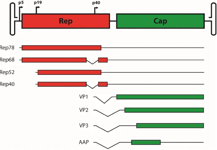
Figure 1: Schematic representation of the wildtype AAV genome ... 23
Figure 2: Model of the intracellular trafficking pathway of AAV... 24
Figure 3: The ubiquitin proteasome system, but not deubiquitinases, impact AAV transduction ... 39
Figure 4: Modulation of autophagy does not impact AAV transduction... 40
Figure 5: The ERAD inhibitor EerI, but not Kif, increases AAV transduction... 41
Figure 6: EerI increases vector transduction in a dose dependent manner. ... 42
Figure 7: EerI increases vector transduction in a vector dose and serotype independent manner. ... 43
Figure 8: EerI increases vector transduction in a cell type and genome type independent manner. ... 44
Figure 9: EerI redirects AAV particles from a perinuclear pattern to a dispersed cytosolic punctate pattern. ... 45
Figure 10: AAV particles accumulate within enlarged Rab7+ and Lamp1+ vesicles upon treatment with EerI. ... 46
Figure 11: EerI does not alter binding or internalization of AAV. ... 47
Figure 12: EerI and MG132 increase AAV transduction through distinct, yet cumulative mechanisms. ... 48
Figure 13: Schematic outlining a potential approach to enhance transduction by redirecting the vesicular trafficking of AAV particles. ... 49
Figure 14: Intracellular calcium inversely affects AAV transduction in HeLa cells. ... 69
Figure 15: Intracellular calcium inversely affects AAV transduction in MB114 cells. ... 70
Figure 16: AAV transduction increase by BAPTA-AM is dependent on the presence of calcium in the extracellular environment. ... 71
Figure 18: Intracellular calcium alters nuclear accumulation of AAV when
increased by ionomycin. ... 73
Figure 19: Time of intracellular calcium perturbation differentially affects AAV transduction. ... 74
Figure 20: Intracellular calcium alters AAV transduction independent of vector genome or vector dose. ... 75
Figure 21: Intracellular calcium alters AAV transduction by altered transcript levels. ... 76
Figure 22: BAPTA-AM increases AAV1 transduction in mice injected via ICV route. ... 77
Figure 23: The effect of intracellular calcium concentration on receptor-mediated endocytosis and fluid phase uptake. ... 78
Figure 24: Intracellular calcium concentration does not affect proteasome activity. ... 79
Figure 25: Alignment of the VP1 region of AAV serotypes 1-9. ... 97
Figure 26: Structural characterization of the AAV1 P mutants. ... 98
Figure 27: Grafting of other VP1u regions onto AAV1 alters transduction of both HeLa cells and MB114 cells. ... 99
Figure 28: AAV1 P mutants differentially transduce mouse muscle tissue. ... 100
LIST OF ABBREVIATIONS
AAP assembly activating protein AAV Adeno-associated virus
AAVR Adeno-associated virus receptor
AAVS1 Adeno-associated virus integration site 1 AIDS acquired immune deficiency syndrome Arf1 ADP-ribosylation factor 1
BBB blood-brain barrier
BR basic region
CBA chicken-beta actin
CBh chicken-beta actin hybrid
CLIC/GEEC clathrin-independent carriers/GPI-enriched endocytic compartments
CMV cytomegalovirus
CNS central nervous system co-IP co-immunoprecipitation
CPV canine parvovirus
CRISPR clustered regularly interspaced short palindromic repeats
CTB cholera toxin B
DMEM Dulbecco’s modified eagle medium
DMSO dimethyl sulfoxide
DUB deubiquitinase
EBOV ebloa virus
EIPA ethylisopropyl amiloride
ER endoplasmic reticulum
ERAD endoplasmic reticulum associated degradation fLuc firefly luciferase
FGFR1 fibroblast growth factor receptor 1 FPV feline panleukopenia virus
GABA gamma-aminobutyric acid
GFAP glial fibrillary acidic protein GFP green fluorescent protein GPI glycosylphosphatidylinositol
GRAF1 GTPase regulator associated with focal adhesion kinase 1
HBoV human bocavirus
HCC hepatocellular carcinoma
HGFR hepatocyte growth factor receptor
HPV human papilloma virus
Hsp90 heat shock protein 90
HSPG heparan sulfate proteoglycan HSV herpes simplex virus
ICRAC calcium release-activated channel
ICV intracerebroventricular
ITR inverted terminal repeat
Kif kifunensine
MMTV-LTR mouse mammary tumor virus long terminal repeat
MRN Mre11, Rad50, Nbs1 complex
MTOC microtubule organizing center MVM minute virus of mice
Nab neutralizing antibody
NLS nuclear localization signal
NMDA N-methyl-D-aspartate
NPC nuclear pore complex
NS non-structural
ORF open reading frame
PDGFR platelet-derived growth factor receptor PI3K phosphoinositide 3-kinase
PKC protein kinase C
PLA2 phospholipase A2
PLC phospholipase C
qPCR quantitative polymerase chain reaction rAAV recombinant Adeno-associated virus
RT-qPCR reverse transcription quantitative polymerase chain reaction scAAV self-complementary Adeno-associated virus
SEM standard error of the mean ssDNA single stranded DNA
STB Shiga toxin B
TBG thyroxine binding globulin
TGN trans-Golgi network
UTR untranslated region
UV ultraviolet
VA viral associated
VACV vaccinia virus
VCP valosin-containing protein
vg vector genomes
VP viral protein
VP1u VP1 unique region
VP1/2 VP1 VP2 shared region
CHAPTER 1: Introduction1
1.1 Parvoviruses
Parvoviruses are a family of non-enveloped single-stranded DNA (ssDNA) viruses. These viruses are very small, usually ~25 nm in diameter, and package similarly small genomes, generally between 4 and 6 kb in length, that are capped by terminal repeats that form hairpin structures that can be either symmetrical or asymmetrical (1). The capsids of these viruses are made of 60 viral protein subunits, termed VPs, as icosahedral capsid structures that exhibit T=1 symmetry. Parvoviruses typically have at least two VP subunits, although some parvoviruses have been shown to have up to 5 VP subunits, which are named in numerical order of decreasing molecular size (i.e. VP1, VP2 etc.) (2). These VP subunits are generally the result of alternate splicing, alternate start codons, or both. In addition, the VP1 subunit of parvoviruses have been demonstrated to have a domain that acts as a broad spectrum phospholipase A2 (PLA2) enzyme, a domain that is required for productive infection. The genome of most Parvoviridae has only two open reading frames (ORFs), termed rep and cap. The rep ORF generally contains one or more nonstructural (NS) genes that are required for DNA replication, virion assembly, and DNA packaging. The cap ORF contains the structural genes, VPs described above, which assemble to make up the viral capsid.
1 This chapter includes the original publication: Berry, G.E., Asokan, A. Cellular transduction mechanisms of
Parvoviridae are divided into two subfamilies: Densoviridae, which infect invertebrates, specifically insects, and Parvovirinae, which infect vertebrates (3). Members of the Parvovirinae subfamily infect a broad range of mammalian hosts, ranging from rodents up to humans. These viruses utilize various host cell surface glycan receptors for cell attachment, with sialic acid and heparin sulfate being among the more commonly used glycans. However, the secondary
glycoprotein receptors used by Parvovirinae vary more widely, ranging from transferrin receptor to several different growth hormone receptors. Some of the better studied Parvovirinae include minute virus of mice (MVM), human parvovirus B19, canine parvovirus (CPV), feline
panleukopenia virus (FPV), Adeno-associated virus (AAV) and more recently, human bocavirus (HBoV). Several mammalian parvoviruses have been demonstrated to be associated with various diseases, generally affecting the young and immunocompromised of a particular species. For instance, FPV and CPV are known to cause more serious disease in kittens and puppies, respectively. Similarly, erythema infectiosum is a disease caused by human parvovirus B19 in children (4), where B19 is also known to cause complications in individuals with acquired immune deficiency syndrome (AIDS), as well as pregnant women, sometimes resulting in miscarriage.
1.2 Adeno-associated Virus and Gene Therapy
turnover, such as hepatocytes, as well as terminally differentiated cells generally refractive to gene delivery by other methods, such as neurons. Furthermore, AAV generally does not illicit a strong immune response, reducing the risk of adverse immune reactions such as cytokine storms (6). In addition, in the recombinant form, AAV rarely integrates into the host genome, though DNA delivered by AAV can persist for long periods in an episomal state, allowing long-term expression of the therapeutic transgene (7). Moreover, AAV lacks any known pathogenicity, suggesting a superior safety profile for the use of AAV as a gene delivery vector. It is worth noting that recent studies have demonstrated the existence of partial AAV genomes that have integrated into the cellular genome of hepatocellular carcinoma (HCC) cells (8). However, the role of these integration events in the development of the tumors remains to be determined. Furthermore, such integrations of rAAV genomes have not been demonstrated in patients from AAV clinical trials. Finally, the only requirements governing packaging of DNA into AAV capsids are the existence of the flanking ITRs, and the genome size must not exceed ~5 kb (9). Therefore, any genetic material fit to these requirements can be delivered using rAAV vectors. These properties, combined with the range of known AAV serotypes, allows the targeted
delivery of specific genetic cargo to desired target tissues while maintaining a good safety profile and reducing off target delivery of the transgene. It is for these reasons that AAV has been explored so widely for use as a gene therapy vector.
number trials have been performed aimed at delivery of factor VIII or factor IX to the liver in an effort to treat hemophilia A or hemophilia B, respectively, with promising results (12). Recent success has also been demonstrated with treatment of retinal diseases. Specifically, Leber’s congenital amaurosis, a congenital form of blindness (13, 14). One particular trial eventually led to the first ever commercially approved AAV vectored gene therapy product in the western world (Alipogene tiparvovec, Glybera®, EU), which aims to treat lipoprotein lipase deficiency by intramuscular injections of AAV1 packaging the gene encoding for lipoprotein lipase (LPL) (15). The growing number of successful trial results demonstrate the continually shrinking gap between AAV-mediated gene therapy and the clinic.
1.3 Biology of Adeno-associated Virus
AAV was originally discovered as a contaminant of simian adenovirus preparations and was initially thought to be defective particles, as they were not replication competent on their own (16). However, these particles were antigenically distinct and were later classified as a
dependoparvovirus. AAV replication is dependent upon the presence of a helper virus, such as Adenovirus (16), Herpes simplex virus (HSV) (17), human papilloma virus (HPV) (18), or vaccinia virus (VACV) (19). In addition, AAV has yet to be definitively linked to any human disease, even though the majority of the human population is seropositive for AAV (20). Many unique serotypes have been isolated from a range of different species, thought the most studied serotype is AAV2.
gene encodes for four non-structural genes, Rep78, Rep68, Rep52, and Rep40, which are named according to their molecular weight. These genes are generated as a result of expression driven by 2 distinct promoters, p5 and p19, as well as alternative splicing. Rep78 and Rep68 have DNA binding activity and have been shown to be important in regulating the activity of the p5, p19, and p40 promoters (21). Additionally, Rep78 and Rep68 have DNA endonuclease and DNA helicase activities that are important for resolution of the ITRs during DNA replication of the AAV genome (22-24). Furthermore, Rep78 and Rep68 play a pivotal role in site-specific
integration and subsequent rescue of the AAV genome into and out of human chromosome 19 at a site termed AAVS1 (25). Rep 52 and Rep40 each have DNA helicase activity and have both been shown to be required for packaging of DNA into AAV capsids (26). The cap gene encodes for three VP subunits, VP1, VP2, and VP3, which are all driven by the p40 promoter. The individual VPs are generated by alternative splicing as well as an alternative start codon. In addition to the VPs, assembly-activating protein (AAP), is also encoded within cap in an
Interestingly, another gene has been postulated to exist in an alternate reading frame at the 3’ end of the cap gene (35). It was later demonstrated that an active promoter, p81, was able to drive RNA transcription of the gene, which was then named the “X” gene (36). The same group later determined that the “X” gene potentially plays a role in the AAV life cycle, particularly in DNA replication (37). However, it must be noted that the existence of the “X” gene is still debated and the proposed function of the “X” gene has yet to be replicated.
Recently, high-throughput studies have been performed that have identified a number of previously unknown AAV proteins and RNA transcripts. One such study identified a number of unique novel RNA species of various sizes corresponding to the AAV genome (38).
Interestingly, one particular RNA is present in high quantities that is transcribed in the reverse direction from the p5 promoter. Furthermore, the abundance of these RNA transcripts was altered upon co-infection with helper virus. Intriguingly, this study identified a novel 18 kDa protein that corresponds to a fusion of the C-terminus of rep and the N-terminus of cap, though the function of this protein remains unknown. However, it is worth noting that this study identified an RNA transcript that is capable of encoding for the previously identified “X” gene, providing more evidence for a potential function of the “X” gene in AAV biology.
AAV1, AAV5, and AAV6 bind N-linked sialic acid (42, 43). Interestingly, AAV6 has been shown to bind both HSPG and N-linked sialic acid (44). Interestingly, AAV9 has been shown to utilize N-linked galactose as a primary receptor (45); the only AAV known to utilize that glycan. To date, the primary receptors for both AAV7 and AAV8 are unknown. In addition to binding glycan primary receptors, AAV uses glycoprotein secondary receptors to gain entry to the cell. A number of proteins have been identified that act as secondary receptors for AAV, including several growth factor receptors, such as fibroblast growth factor receptor 1 (FGFR1) (46), human hepatocyte growth factor receptor (HGFR) (47), and platelet-derived growth factor receptor (PDGFR) (48), as well as a number of integrins, such as α5β1 and αVβ5 (49-52). However, KIAA0319L was later identified as a universal receptor for a broad number of AAV serotypes, therefore it was given the designation AAV receptor (AAVR) (53). The differential usage of primary and secondary receptors are generally thought to be a driving factor in the various tissue tropisms of these serotypes. In fact, modification of glycosylation patterns in vivo have been shown to alter the tropism of both AAV9 and AAV4 (54, 55).
1.4 Trafficking of Adeno-associated virus
(56, 57). Additionally, internalized AAV2 colocalized with transferrin, a protein known to be internalized by this mechanism.
Interestingly, a recent study was not able to clearly identify dynamin- or clathrin-dependent endocytosis as a mechanism of uptake. Instead, the study suggests that uptake of AAV2 is dependent on the incompletely characterized clathrin-independent carriers and GPI-enriched endocytic compartment (CLIC/GEEC) endocytic pathway (58). This study
demonstrated that AAV2 uptake was inhibited by dominant negative versions of Arf1, Cdc42, and GRAF1, three important effectors of the of the CLIC/GEEC pathway. In addition, AAV2 colocalized with cholera toxin B (CTB) and GPI-anchored GFP, two markers of CLIC vesicles, after internalization. In addition, this study identified EIPA as an inhibitor of CLIC/GEEC endocytosis. However, it is worth noting that EIPA is classically known as an inhibitor of macropinocytosis. Accordingly, other studies suggest a role for macropinocytosis in AAV uptake. One such study demonstrated that inhibition of Rac1 activation, a key effector of macropinocytosis, inhibits AAV internalization (59). Interestingly, another study used multiple small molecule inhibitors of macropinocytosis, including EIPA, to demonstrate that inhibition of macropinocytosis decreased transduction in some cells lines, where other cell lines, specifically hepatocellular carcinoma cells, demonstrated enhanced transduction (60).
It is worth noting that transcytosis of rAAVs has been shown to occur in polarized cells in a serotype-dependent manner, and it has been suggested that this phenomenon is dependent upon caveolin (61). In fact, an important area of inquiry regarding intrinsic qualities of individual rAAV strains in vivo is the ability of the capsid to cross the blood-brain barrier (BBB) to
study utilized mice that do not express caveolin-1, a key effector of caveolin-mediated
transcytosis (62), and demonstrated that rAAV9 continues to traverse the BBB in these knockout mice (63), suggesting that transvascular transport of AAV9 may occur by a different mechanism.
There currently exists much contradicting data regarding the endocytosis mechanisms involved in cellular internalization of rAAV. At this juncture, there is not a clear explanation for these discrepancies. However, there are several possibilities. For instance, some studies utilize adenoviral vectors to overexpress protein effectors, dominant-negative or otherwise, therefore introducing the possibility of modifying the cellular environment prior to infection with AAV. Another possibility is that some small molecule inhibitors can affect multiple pathways, therefore clouding the results. For instance, dynasore, a dynamin inhibitor, inhibits both caveolae- and clathrin-mediated endocytosis, but also been shown to have numerous dynamin independent effects, such as disruption of lipid raft organization and reduction of labile cholesterol in the plasma membrane (64). Additionally, EIPA appears to inhibit both
macropinocytosis and CLIC/GEEC endocytosis (58, 65), and there is additional evidence that EIPA partially inhibits endosome acidification (66), an essential step in AAV transduction that will be discussed later in this review. In addition, it has been hypothesized that some uptake pathways lead to successful transduction while other pathways in those same cells lead to a “dead end” for AAV (58, 59), and this is likely altered in a cell-type dependent manner. Moreover, it is likely that AAV utilizes a combination or permutation of the aforementioned endocytosis pathways in a cell-type specific manner. In fact, one study supports this idea,
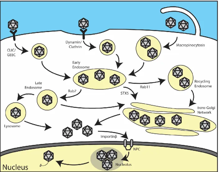
After rAAV enters the cell, it must traffic towards the nucleus in order to successfully deliver its genetic cargo. Immediately after uptake, AAV is presumably trafficked to Rab5+ early endosomal compartment, which is a feature conserved amongst many parvoviruses (67). From here, rAAV has been shown to be trafficked through a number of different compartments. Studies have demonstrated that rAAV2 traffics through both Rab7+ late endosomes and Rab11+ recycling endosomes (68). From here, AAV has been shown by numerous studies to traffic to the Golgi apparatus (32, 69, 70). One recent study showed that siRNA-mediated knockdown of syntaxin 5 (STX5), as well as disruption of STX5 by the small molecule Retro2.1, reduced rAAV transduction, suggesting that retrograde transport of rAAV to the trans-Golgi network (TGN) mediated by syntaxin 5 is important for transduction (71). Interestingly, this study was unable to confirm that Rab7+, Rab9+, or Rab11+ vesicles play a role in trafficking of rAAV to the Golgi apparatus. Recently, we demonstrated that inhibition of endoplasmic
reticulum-associated degradation (ERAD) by eeyarestatin I (EerI) in HeLa cells reroutes rAAV to enlarged Lamp1+ lysosomes, thereby increasing transduction, indicating that trafficking of rAAV through the lysosome may be an important step in infection (Discussed in Chapter 2) (72). It is important to note that, as of now, no studies have observed rAAV in the endoplasmic reticulum (ER). Therefore, it is unlikely that rAAV traffics to the ER prior to nuclear entry.
in the capsid (74), namely, the exposure of the N-terminal domains of VP1 and VP2, which are located inside the capsid prior to infection (75). Exposure of these domains for successful transduction is required, as it has been demonstrated that microinjection of both complete virions, as well as VP3-only virions, directly into the cytosol do not properly transduce the cell (76). It was suggested that the AAV capsid itself has protease activity that is pH-dependent, which could possibly be triggered by the acidification of the endosome (77). However, it has yet to be determined if the self-cleavage events mediated by this activity also play a role in the exposure of the VP1/VP2 N-termini, or in other events related to transduction.
After trafficking of rAAV through the endomembrane system, rAAV escapes the endosome into the cytosol. Endosomal escape is dependent on a phospholipase A2 (PLA2) domain located in the VP1 unique region of AAV (30, 78). Studies have shown that mutation or deletion of the PLA2 domain prevents endosomal escape and subsequent transduction (79).
As is the case with AAV cell entry, differential, and sometimes conflicting, data exists regarding the intracellular trafficking of rAAV. It is likely that the trafficking pathway differs in a cell line-dependent and dependent fashion. This possibility is evident in the serotype-dependent axonal trafficking of rAAV in neurons. Furthermore, rAAV9 was identified in Rab5+, Rab7+, and Rab11+ vesicles in neurons in cell culture, but was shown to only traffic effectively along axons in Rab7+ endosomes, further supporting the notion that intracellular trafficking of rAAV is likely dependent on cell type (80).
investigated for their potential function as nuclear localization signals (NLS). BR4 is located within VP3 and was shown to have no impact on nuclear import of rAAV virions, but mutation of BR4 did result in virion assembly defects (31). However, BR3, located within both VP1 and VP2, is essential for AAV transduction (76). In addition, BR1 and BR2, to a lesser extent, are also important for AAV transduction. However, confocal microscopy studies of BR-negative mutants demonstrated that BR2 and BR3 are important for nuclear translocation (32). A recent study demonstrated that rAAV2 enters the nucleus through the nuclear pore complex (NPC) by blocking nuclear entry of rAAV2 with wheat germ agglutinin, a lectin that binds the NPC and blocks and cargo from traversing through (81). This study also demonstrated that importin-β1 is the host protein responsible for import of rAAV2 particles through the NPC. Capsid interaction with members of the importin-α family of proteins was also shown by co-IP, but their
involvement in nuclear import of rAAV2 remains unclear. This route of nuclear translocation was further supported by live cell imaging technology that witnessed labeled rAAV2 particles traverse the nuclear envelope through labeled NPCs (82).
between cell types and serotypes will likely be helpful in dissecting the role of the nucleolus in rAAV transduction.
After AAV reaches the nucleus, the virus undergoes uncoating to release the genome. The mechanisms underlying uncoating of AAV remain unclear, though it is known that genome composition may play an important role in uncoating and genome release (86). After uncoating, the virus must undergo the crucial step which is conversion of the single-stranded genome into a double-stranded form, a process known as second-strand synthesis. This process has been shown to be a rate limiting step in infection in the absence of a helper virus (87). Second-strand
1.5 AAV Vectorology
Two of the factors driving the use of AAV vectors is the simplicity of the genome and the ease of manufacturing. As previously mentioned, the only cis-acting elements necessary for packaging of recombinant genomes into AAV capsids are the ITRs that flank either side of the genetic cargo (9). Therefore, replacement of the rep and cap genes with any desired genetic information can yield rAAV particles that carry any promoter and transgene of interest, provided the total vector DNA length does not exceed ~5 kb. In order to manufacture these particles, the rep and cap genes with the ITRs removed are simply provided in trans on a separate plasmid, named pXR (98). Further, it was soon discovered that fine-tuning the levels of rep expression could allow for greater production of the cap genes (99). Specifically, it was determined expression levels of Rep78 and Rep68 inversely correlated with the expression levels of cap (21). Therefore, mutation of the Rep78 and Rep68 start codon to a weak alternate start codon was performed on the pXR plasmid to reduced Rep78 and Rep68 expression levels, thereby increasing capsid protein production.
When rAAV was first manufactured, co-infection of a virus with helper activity, specifically Adenovirus or herpes simplex virus, was needed in order to promote rAAV
plasmid, called pXX6-80, is combined with a pXR plasmid and the ITR plasmid containing the gene of interest, it allows for the triple plasmid transfection method, a method that remains one of the most commonly used rAAV production method. It is also necessary to note that
concurrently with the development of the triple plasmid transfection method, another system was developed in a similar fashion, but with a different approach (100). This study generated a
plasmid called pDG, which contained both the Adenovirus genes necessary for rAAV production as well as the rep and cap genes lacking ITRs. To downregulate Rep78 and Rep68 expression, instead of mutating the start codon, this plasmid replaced the endogenous p5 promoter with a much weaker mouse mammary tumor virus long terminal repeat (MMTV-LTR) promoter. Use of this plasmid instead of pXX6-80 and a pXR plasmid allows for a two plasmid transfection method for production of rAAV. This method is also widely used to generate rAAV.
After the triple plasmid transfection system was standardized, several improvements began to be made to allow for production of broad range of serotypes as well as increased vector production. To achieve the first goal, the rep portion of the pXR2 plasmid was engineered such that the C-terminal portion of rep was replaced with the rep that matched the cap serotype being produced, but leaving the N-terminal portion of rep from AAV2 intact. This allowed packaging of genetic cargo flanked by AAV2 ITRs into the capsid of any serotype, a practice known as cross-packaging (101). This development sparked a revolution in rAAV research, allowing the production of potentially thousands of rAAV capsids, natural and synthetic, for study.
1.6 Synthetic rAAV Strains
generally used for intramuscular injections, as it transduces muscle more efficiently than AAV2 (102). AAV8 is generally used for liver-directed gene therapy, as it transduces mouse liver more efficiently than AAV2 (103). However, it must be noted that recent studies have demonstrated that AAV8 transduces non-human primate and human hepatocytes poorly compared to AAV3 (104). AAV9 is known to transduce many tissues types as well as having the ability to cross the blood-brain barrier (BBB) (54, 63). However, even with a broad range of capsids to choose from, targeted transduction of specific tissues while limiting or eliminating off target transduction remains one of the most vital requirements as individual therapies approach the clinic. To this end, many synthetic strains of AAV have been generated with a vast array of strategies that are able to overcome some of these obstacles.
One of the first methods used to produce synthetic rAAV capsids was rational design. One of the earliest successes with this approach was the generation of AAV2i8. This capsid was generated by reengineering the heparan sulfate binding footprint on AAV2 with a region from an interloop of AAV8, yielding a new synthetic vector that transduced the liver with much lower efficiency (105). Therefore, AAV2i8 became the first liver-detargeted vector. This finding let to the development of many more vectors that had similar liver-detargeted qualities. Shortly thereafter, another synthetic AAV capsid, AAV2.5, was produced. AAV2.5 was a rationally designed AAV2 capsid containing amino acid residues from AAV1 that contribute to the ability of AAV1 to transduce muscle with high efficiency (102). Additionally, this capsid demonstrated a lower level of cross reactivity with AAV2 neutralizing antibodies (Nabs), thereby making treatment available to more patients.
unique capsid DNA sequences. The individual directed evolution approaches are then designed to yield new capsids with one or several desirable properties. These libraries can be generated a number of different ways. However, there are two methods that are most often utilized. One method is capsid shuffling, which utilizes random assembly of fragmented capsid DNA based on reannealing at regions of complementarity (106, 107). The number of parental strains used to generate capsid shuffled libraries can range from only two to several. A number of different studies have utilized libraries generated using most or all serotypes AAV1-9 (108, 109). One such study utilized a unique directed evolution approach to generate a capsid that efficiently transduces oligodendrocytes in the CNS (110). The other commonly used method randomizes individual regions of interest on a parental strain to generate capsids that can have new properties or that can shed light on the biology of a particular serotype. For example, one particular library generated on an AAV9 background was able to generate several capsids of interest (111). One particular capsid, AAV9.45, was detargeted from the liver, where another capsid, AAV9.24, was later determined to be deficient in galactose binding, highlighting the eventually discovered galactose binding footprint on the AAV9 capsid (112).
circulation when administered intracranially. The use of this type of approach is sure to grow as more is discovered regarding the function of particular motifs on the capsid surface.
An additional strategy sometimes used to restrict transgene expression to the tissue of interest relies upon modification of the recombinant genome. For instance, while constitutive promoters such as the cytomegalovirus (CMV) promoter or the chicken beta actin (CBA)
These binding sites allow for tissue specific microRNA-based degradation of the transgene. One prime example is the addition of miR-122 binding sites to the 3’ UTR of a gene to inhibit
transgene expression in the liver (128). Taken together, combining different capsids, promoters, and 3’ UTR elements will allow for great levels of tissue specific gene expression as rAAV-mediated gene therapies approach the clinic.
1.7 Strategies to Augment rAAV Transduction
One of the strategies currently used to increase AAV transduction is modification of the viral genome. As previously mentioned, second-strand synthesis has been shown to be a major rate-limiting step in AAV transduction. Knowledge of this bottleneck led to the development of self-complementary AAV (scAAV) vectors (129). These were generated by mutating one of the ITRs to remove the terminal resolution site. This had the effect of generating vector genomes that were flanked by standard ITRs but retained the mutated ITR in the middle. This mutated ITR has the effect of causing the genome to base pair together down the molecule upon
uncoating, thereby bypassing the requirement for second-strand synthesis. These scAAV vectors demonstrate a striking increase in transduction that is greater than 10-fold. While this is a
Another set of strategies that are being investigated involve modification of the AAV capsid to increase transduction. One of the most successful examples of this approach are the tyrosine-to-phenylalanine mutants. It was postulated that tyrosine residues on the capsid surface act as sites for phosphorylation and subsequent ubiquitination, therefore leading to capsid degradation. The resulting Y-F mutations resulted in rAAV capsids that showed marked transduction increases (133). Further mutation of possible phosphorylation sites led to the so-called Y-T quadruple mutant, a modification of the rAAV2 capsid that dramatically increases transduction (134). Further studies leveraging the knowledge of rAAV trafficking to perform rational capsid mutagenesis are important to generate vectors that more efficiently transduce tissues.
mechanism, or combination of mechanisms, by which this may occur is unclear. For instance, hydroxurea is a DNA-damaging drug, putting in a similar class as cisplatin or the topoisomerase inhibitors. However, hydroxyurea has also been shown to disrupt the nucleolus, a process which itself has been shown to be productive for transduction (85). Furthermore, hydroxyurea has been shown to inhibit phosphorylation and subsequent activation of FKBP52, a protein that has been shown to be detrimental to AAV transduction (138).
Another class of small molecules that has been thoroughly studied for their potential to augment AAV transduction are the proteasome inhibitors. It has been hypothesized that the proteasome plays a strong inhibitory role in rAAV transduction by ubiquitin-dependent degradation of capsids before they can traffic to the nucleus. This hypothesis is supported by several studies, the first of which demonstrated a large increase in transduction of rAAV2 when cells were treated with the proteasome inhibitor MG132 (73). Since then, several more studies have shown an increase with other proteasome inhibitors, namely LLnL, bortezomib
Figure 2: Model of the intracellular trafficking pathway of AAV. AAV binds to a
CHAPTER 2: Chemical modulation of endocytic sorting augments adeno-associated viral transduction2
2.1 Overview
Intracellular trafficking of viruses can be influenced by a variety of inter-connected cellular sorting and degradation pathways involving endo-lysosomal vesicles, the ubiquitin-proteasome system, autophagy-based or ER-associated machinery. In case of recombinant adeno-associated viruses (AAV), proteasome inhibitors are known to prevent degradation of ubiquitinated AAV capsids, thereby leading to increased nuclear accumulation and transduction. However, the impact of other cellular degradation pathways on AAV trafficking is not well-understood. In the current report, we screened a panel of small molecules focused on modulating different cellular degradation pathways and identified Eeyarestatin I (EerI) as a novel reagent that enhances AAV transduction. EerI improved AAV transduction by an order of magnitude regardless of vector dose, genome architecture, cell type, or serotype. This effect was preceded by sequestration of AAV within enlarged vesicles that were dispersed throughout the cytoplasm. Specifically, EerI treatment redirected AAV particles towards large vesicles positive for late endosomal (Rab7) and lysosomal (LAMP1) markers. Notably, MG132 and EerI (proteasomal and ERAD inhibitors, respectively) appear to enhance AAV transduction by increasing the intracellular accumulation of viral particles in a mutually exclusive fashion. Taken together, our
2 This chapter includes the original publication: Berry, G.E., Asokan, A. Chemical modulation of endocytic sorting
results expand on potential strategies to redirect recombinant AAV vectors towards more productive trafficking pathways by deregulating cellular degradation mechanisms.
2.2 Introduction
Eukaryotic cells utilize tightly regulated pathways to sort and degrade internalized cargo by exploiting lysosomal proteases, the ubiquitin-proteasome system, ER-associated or
autophagy-based machinery. Endoplasmic reticulum (ER) associated degradation, or ERAD, is a critical eukaryotic process that involves extraction and ubiquitination of misfolded proteins followed by their degradation by the proteasomal machinery (148). Several viral pathogens exploit this process to infect and replicate within host cells (149, 150). For instance,
polyomaviruses appear to interact with ER lumen components to rearrange capsid proteins and subsequently retrotranslocate into the cytosol (151). Another constitutive degradation pathway essential for maintaining cellular homeostasis is autophagy (152). As with ERAD, several viruses have now been shown to subvert or mimic autophagy to facilitate replication and/or dissemination (153-155). The specific degradation pathway and the subsequent intracellular fate of viral proteins as well as their genomic cargo within the host cell are often dictated by a variety of preceding vesicular sorting events.
Recently, vesicular transport of non-enveloped parvoviruses such as the Minute Virus of Mice (MVM) through the ER and Golgi has been shown to accelerate progeny virus release (156). A particularly interesting member of the same parvovirus family is the helper-dependent Adeno-associated virus (AAV), which replicates upon co-infection with Adenoviruses or other viruses such as Herpes Simplex Virus or Papillomavirus. This small, non-pathogenic
glycans such as heparan sulfate, sialic acid or galactose as primary receptors for attachment (158). Subsequent internalization of AAV particles into endocytic vesicles is thought to be mediated by integrins and/or specific transmembrane receptors. In addition, several diverse and cell-specific mechanisms of endocytic uptake ranging from macropinocytosis to the CLIC/GEEC pathway have been described (58, 60). Despite these differences, perinuclear accumulation within the Golgi apparatus (32, 69-71, 159) and exploitation of the nuclear import machinery for nuclear entry appear to be broadly conserved, downstream trafficking events (81).
Although these studies provide a detailed map of AAV transport within the host cell, it remains unclear whether the modulation of cellular degradation pathways such as ERAD or autophagy outlined earlier can influence AAV trafficking. Most studies to date have focused on proteasome inhibitors such as MG132 (73), LLnL (160) and bortezomib or carfilzomib (85, 142), which have been shown to increase AAV transduction through increased nuclear/nucleolar accumulation of viral particles. In the current study, we tested the effect of several small molecules that modulate the ubiquitin-proteasome system, autophagy and/or ERAD on AAV transduction. The overall goal of the study was to understand the interplay (or lack thereof) between these different cellular degradation pathways in facilitating or restricting AAV
trafficking within host cells. In doing so, we identified an ERAD inhibitor (Eeyarestatin I / EerI) that deregulates endocytic sorting of AAV particles and redirects viral transport towards
2.3 Materials and Methods
Cell culture. HeLa, HepG2, and Huh7 cells were maintained in Dulbecco’s Modified Eagle’s
Medium with 10% FBS, 100 U/ml of penicillin, 100 µg/ml of streptomycin, and 2.5 µg/ml of amphotericin B (Sigma-Aldrich, St. Louis, MO). Human fibroblasts (AG05244) were obtained from Coriell Cell Repositories (Camden, NJ) and were maintained in Dulbecco’s Modified Eagle’s Medium with 15% FBS, 100 U/ml of penicillin, and 100 µg/ml of streptomycin. All cells were maintained at 37°C and 5% CO2.
Antibodies, chemicals, and cell labeling reagents. Mouse VCP (ab11433), rabbit
Recombinant AAV Production. Recombinant AAV packaging chicken beta actin (CBA)
promoter driven firefly luciferase (fLuc) as well as single-stranded and self-complementary vectors packaging a truncated CBA promoter driving green florescent protein (GFP) reporters were produced in HEK293 cells using the triple plasmid transfection protocol, purified and titers determined as described earlier (113, 163).
Transduction and cell viability assays. Cells were plated at a density of 5x10⁴ cells/well in
24-well plates and allowed to adhere overnight. Unless otherwise indicated, cells were treated with DMSO vehicle control or EerI for 4 hours before transduction with AAV2-CBA-fLuc at 1,000 (vector genomes) vg/cell. Cells were lysed 24 hours after using the luciferase assay system from Promega (Madison, WI) according to manufacturer instructions and read on a Wallac® 1420 Victor3 automated plate reader. Cell viability was assayed using the CellTiter Glo® Luminescent Cell Viability assay from Promega (Madison, WI) according to manufacturer instructions on the same instrument. Experiments were all performed in quadruplicate. For time course studies, EerI was provided at the designated time points by adding concentrated EerI stock solution in media to a final concentration of 10 μM.
3 times with PBS to remove all un-internalized virions. Viral DNA was then extracted using a DNeasy kit (Qiagen).
Confocal Fluorescence Microscopy. Cells were plated on slide covers in 24-well plates at a
density of 5x10⁴ cells/well and allowed to adhere overnight. Cells were plated at a density of 2.5x10⁴ cells/well and infected concurrently with the BacMam baculovirus encoding for GFP-tagged endosomal markers (20 copies/cell). Baculovirus was removed from cells 24 hours, and cells allowed to recover for an additional 24 hours prior to further studies. Cells were then treated with EerI 4 hours before incubation with AAV2 at 50,000 vg/cell. After 8 hours, cells were fixed with 2% paraformaldehyde for 15 min and then permeabilized with 0.2% Triton-X for 5 min. Cells were stained with primary antibody overnight at 4°C, and then stained with secondary antibody for 1 hour at 37°C. Cells were then mounted with Prolong Gold Antifade with DAPI (Life Technologies) and imaged using a Zeiss 710 scanning confocal microscope.
Image Analysis. Quantification of AAV particle co-localization with subcellular markers was
carried out using ImageJ software using the Colocalization Analysis tools from the Wright Cell Imaging Facility website (http://www.uhnresearch.ca/facilities/wcif). In case of BacMam 2.0 incubated cells, care was taken to include only cells expressing the GFP-tagged vesicle markers in the colocalization analysis. Data is represented as Pearson’s correlation coefficients. Briefly, a Pearson’s correlation coefficient of 0 represents random localization where a value of 1
Statistical analysis. All data is expressed as mean with error bars representing standard error of
the mean (SEM). A two-tailed unpaired student t-test was used for all statistical analysis. P values less than 0.05 were considered significant. Asterisks are used to indicate P values as follows: *P < 0.05; **P < 0.01; ***P < 0.005.
2.4 Results
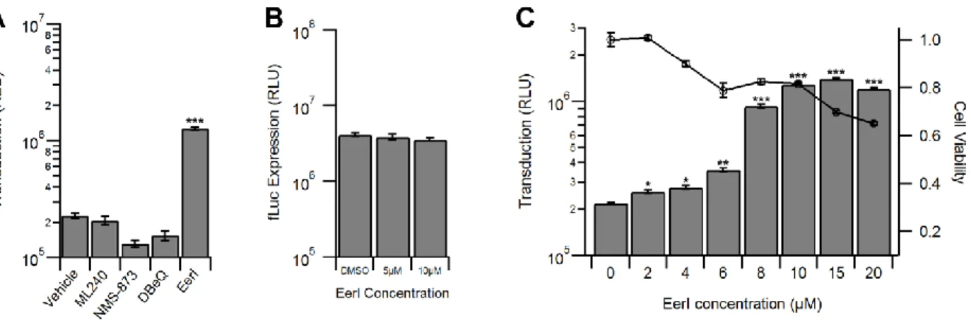
Eeyarestatin I increases AAV transduction. Small molecule inhibitors that augment cellular
degradation mechanisms have been used extensively to dissect virus-host interactions (151, 164, 165). We first treated HeLa cells with various small molecules that modulate the cellular
ubiquitin proteasome system (UPS) and then incubated the cells with AAV2-CBA-fLuc.
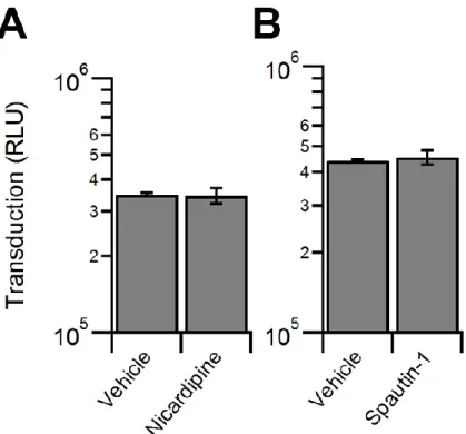
Inhibitors of the proteasome, MG132 and bortezomib, increased transduction by approximately a log order (Fig. 3), as reported previously (73, 141). Interestingly, PR-619, a pan-deubiquitinase (DUB) inhibitor, did not alter transduction, while PYR-41, an inhibitor of the ubiquitin activating enzyme UBA1, increased transduction to a degree similar as the proteasome inhibitors (Fig. 3). We then investigated a potential role for autophagy in AAV transduction, which has recently been shown to be critical in the infection pathway of numerous viruses (153-155). However, neither the autophagy inducer, nicardipine nor the autophagy inhibitor, spautin-1 altered AAV transduction (Fig. 4). We then tested endoplasmic reticulum associated degradation (ERAD) using two inhibitors, eeyarestatin I (EerI) and kifunensine (Kif). Interestingly, while Kif
treatment did not significantly alter transduction, EerI treatment led to a transduction increase of approximately a log order (Fig. 5).
results. However, as shown in Fig. 6A, none of the VCP inhibitors increased rAAV transduction with the exception of EerI. It should be noted that siRNA-mediated knockdown of VCP/p97 accompanied by significant cytotoxicity (>50%) precluding efforts to directly address the
potential (indirect) role of VCP/p97 in AAV transduction (data not shown). To confirm whether the increase in transduction efficiency was due to pleiotropic effects of EerI, we transfected HeLa cells with the pTR-CBA-fLuc packaging plasmid as control, allowed 48 hours for transgene expression, and then treated the cells with EerI for 24h. We observed no change in fLuc expression as a result of EerI treatment (Fig. 6B), confirming that the effect seen
exclusively affects AAV transduction and does not enhance transcription, transcript or protein stability in general.
Next, we investigated the effect of altering EerI concentration or virus dose on transduction. We observed a dose dependent increase of AAV transduction at EerI
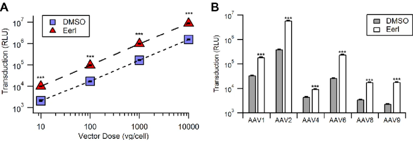
concentrations up to 15 µM, after which a decrease in transduction efficiency was observed due to cytotoxicity (Fig. 6C). Further, at an optimal EerI concentration of 10 µM, we observed a uniformly beneficial effect on transduction efficiency in HeLa cells over a broad range of viral doses ranging from 10 to 10,000 vg/cell (Fig. 7A).
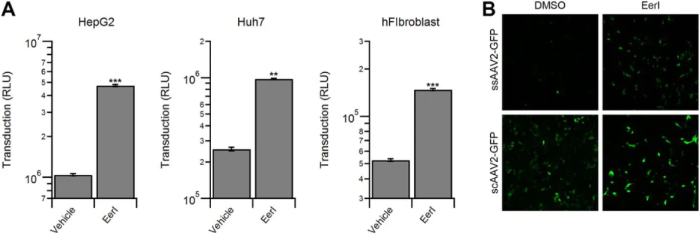
EerI enhances AAV transduction independent of capsid, vector genome or cell type. We
on the toxicity profile (Fig. 8A). Further, we determined that EerI enhances transduction by AAV vectors packaging single-stranded (ss) or self-complementary (sc) GFP reporter cassettes in a similar fashion (Fig. 8B). Taken together, these data demonstrate that EerI increases transduction regardless of capsid, second strand synthesis or cell type.
EerI redistributes AAV from a perinuclear pattern to large, dispersed vesicles in the
cytoplasm. Given the broad impact of EerI on AAV transduction, we assessed whether these
effects were preceded by notable changes in the intracellular trafficking of AAV particles through confocal fluorescence microscopy studies. Specifically, we pre-treated HeLa cells with EerI and incubated the cells with AAV2 particles, followed by immunofluorescent labeling of AAV capsids and VCP at 2 hours, 4 hours, and 8 hours post-incubation. We observed no signification correlation between the intracellular patterns of VCP (the apparent target of EerI discussed later), and AAV capsid immunostaining at 2, 4 or 8 hours post-incubation with or without EerI treatment (Figs. 9A-C). As seen in higher magnification images (bottom panels), a steady increase in the perinuclear accumulation of AAV particles was observed over time. At 8 hours post-incubation (Fig. 9C), we observed that AAV particles redistributed from a perinuclear location to a large and dispersed punctate pattern throughout the cytoplasm upon VCP inhibition. This observation strikingly contrasts with DMSO treated cells, in which AAV virions remain concentrated in the perinuclear region (Fig. 9C, bottom panel).
EerI redirects AAV particles to late endosomes and lysosomes. In an effort to identify the
versions of different endosomal markers, specifically Rab7 (late endosome) and LAMP1 (lysosome) using baculoviral expression vectors. In addition, we utilized an EEA1 antibody to stain for early endosomes. At 8 hours post-incubation, AAV particles did not appear to
colocalize with EEA1+ vesicles (data not shown), indicating that AAV is not associated with early endosomes at this time interval. However, AAV particles colocalized prominently with Rab7+ (Fig. 10A) and more extensively with LAMP1+ vesicles in EerI treated cells (Fig. 10B). This increased colocalization was further confirmed by quantitation of fluorescent signal using Image J software as outlined in methods. In particular, we determined the Pearson coefficients (Table 1) for AAV particle colocalization with different subcellular markers including EEA1 (early endosomes), Rab7a (late endosomes), LAMP1 (lysosomes), Golgin-97 (Golgi) and STX5 (syntaxin 5). A statistically significant increase in Pearson’s coefficients was noted for
redistribution of AAV particles to Rab7 and Lamp1+ vesicles.
EerI influences an early, post-entry trafficking event during AAV transduction. To further
with viral particles increased AAV transduction, albeit, to a lesser degree compared to drug pre-treatment. By 8 hours post AAV incubation, addition of EerI no longer affected transduction efficiency. When considered together with confocal fluorescence data, these results suggest that EerI treatment remodels endocytic sorting of AAV particles after cellular uptake, but prior to nuclear entry.
EerI and MG132 enhance AAV transduction in a mutually exclusive manner. We next
sought to characterize any similarities and/or differences of proteasome inhibitors and EerI on AAV transduction. Confocal microscopy experiments revealed a stark difference in capsid accumulation upon treatment with either EerI or MG132. As seen in Fig. 12A, where EerI treatment results in accumulation in enlarged vesicles, MG132 treatment leads to a more disseminated capsid accumulation in the perinuclear region. Further, proteasomal inhibition is thought to directly enhance AAV transduction by preventing capsid degradation in the cytosol. As a consequence, enhanced recovery of intact AAV capsids and genomes from the nuclear fraction has been reported by several groups (73, 85, 139, 142, 160). In the current study, upon EerI treatment, we observed a striking increase in the number of AAV genomes recovered after synchronized cellular uptake at later time intervals (Fig. 12B). These results were essentially identical to those obtained upon proteasomal inhibition with MG132. Specifically, no differences in recovered vg were observed at early time intervals up to 4 hours. However, as much as 80-90% of AAV genomes were recovered at 16-24 hours upon treatment with EerI in contrast to 25-35% of AAV genomes obtained from untreated cells. This data might indicate that EerI
The above described results prompted us to further consider the possibility that EerI might not only remodel endocytic sorting, but also influence AAV capsid degradation in a pleiotropic manner. To assess this possibility, cells were co-treated with EerI and MG132, alone or in combination. It is noteworthy to mention that these studies were optimized to minimize the combined cytotoxicity arising from dual drug treatment. As seen in Fig. 12C, EerI enhanced AAV transduction by 4-fold; while MG132 was twice as effective, resulting in a ~ 8-fold increase in transduction. Importantly, combined drug treatment was synergistic and enhanced AAV transduction by nearly 15-fold. These results demonstrate that cellular degradation
pathways can influence AAV infection in distinct ways, which, when modulated in conjunction, can have cumulatively enhance AAV transduction.
2.5 Discussion
studies that reported a dramatic increase in the size of endosomes upon EerI treatment (170). Further, these observations are consistent with earlier reports demonstrating that AAV transport through Rab7+ late endosomes/lysosomes is highly productive due to their high motility and faster retrograde velocities compared to early/recycling endosomes in neurons (80). Such a scenario is possible due to the low pH and high protease activity within such vesicles that could more effectively prime the AAV capsid (171) by exposing the VP1 phospholipase A2 (PLA2) domain (30) and nuclear localization signals (NLS) (31).
Another important observation is that EerI treatment completely remodeled AAV trafficking to the microtubule organizing center (MTOC) in the perinuclear region. Previous studies have indicated that AAV transduction efficiency is affected by knockdown of proteins involved in sorting retrograde cargo, such as syntaxin 5, calnexin, KDEL-R and other Golgi/ER-associated proteins (70, 71). In the current study, EerI treatment appears to decrease the
colocalization of AAV particles with markers such as STX5 and Golgin-97 (Table 1), although these results were not statistically significant. Nevertheless, our results suggest that efficient vesicular trafficking can enhance AAV transduction. This overall approach towards improved AAV trafficking (depicted in Fig. 13) is further supported by (i) the increased recovery of vector genome copy numbers from within cells at time intervals as late as 16-24 hours following EerI treatment similar to that observed with the proteasomal inhibitor, MG132 and (ii) the cumulative effects of EerI and MG132 on AAV transduction.
Figure 3: The ubiquitin proteasome system, but not deubiquitinases, impact AAV
Figure 4: Modulation of autophagy does not impact AAV transduction. HeLa cells
Figure 8: EerI increases vector transduction in a cell type and genome type independent manner. (A) HepG2, Huh7, and human fibroblasts (hFibro) were pretreated with EerI at
Figure 13: Schematic outlining a potential approach to enhance transduction by
Subcellular
marker DMSO EerI P-value
EEA1 0.198 ± 0.018 0.252 ± 0.040 n.s.
Rab7a 0.216 ± 0.019 0.342 ± 0.024 *P < 0.05
LAMP1 0.269 ± 0.036 0.439 ± 0.025 *P < 0.05
Golgin97 0.379 ± 0.025 0.312 ± 0.032 n.s.
STX5 0.453 ± 0.009 0.334 ± 0.042 n.s.
CHAPTER 3: Modulation of intracellular calcium influences recombinant AAV transduction3
3.1 Overview
Calcium is known to play a crucial role in numerous cellular processes, generally acting as a regulator of cell signaling. Intracellular calcium is also important in the life cycles of several viruses, such as Ebola virus and rotavirus. Several small molecules and clinical compounds exist that modify the cellular calcium environment, allowing further study of intracellular calcium in vivo. In the current study, we sought to determine if modulation of intracellular calcium levels affects AAV transduction. Increasing intracellular calcium levels using ionomycin decreased AAV transduction by an order of magnitude while decreasing intracellular calcium levels using BAPTA-AM increased transduction by 10 to 100-fold independent of vector serotype, vector dose, or vector genome. Ionomycin and BAPTA-AM altered binding of AAV particles to the cell surface by 2-fold. In addition, RNA transcription from the vector genome was also altered by ionomycin and BAPTA-AM. Furthermore, the effect of BAPTA-AM was dependent upon the presence of extracellular calcium. In addition, in vivo studies performed in mice illustrate that BAPTA-AM augments transduction by AAV when administered intracranially. In summary, our results demonstrate that modulation of intracellular calcium alters AAV transduction and
supports the preclinical evaluation of drugs and biologics that modulate intracellular calcium in the CNS as a strategy to increase AAV gene delivery.
3 This chapter includes the original publication in review: Berry, G.E., Nardone-White, D.T., Murlidharan, G.,
3.2 Introduction
Intracellular calcium is known to play an important role in cellular homeostasis, primarily acting as an important cell signal regulator (176). Dysregulation of intracellular calcium
homeostasis is a hallmark of several neurological diseases, including Alzheimer’s disease and spinocerebellar ataxia, generally leading to neuronal cell death (177-182). One of the most thoroughly characterized calcium-dependent cellular processes is the fusion of synaptic vesicles with the plasma membrane in neurons (183-185). However, it is also known that protein
transport within the cell can be controlled by calcium. Previously, calcium has been shown to be important in specific events during retrograde and anterograde transport of vesicles in the cell (186). While some viruses, such as influenza A, require calcium dependent cell signaling for infection (187), several viruses have demonstrated the need to modify the intracellular calcium environment to successfully infect cells. For instance, rotavirus encodes for a viroporin, a viral protein that permeabilizes the cell membrane, which specifically alters intracellular calcium to promote its own replication (188, 189). Ebolavirus (EBOV) requires calcium to successfully bud from the cell surface (190). Additionally, it was recently demonstrated that two host cell calcium pore channels are required for EBOV cellular entry, further suggesting that intracellular calcium plays a crucial role in EBOV infection (191).
Parvoviruses are single-stranded DNA viruses that contain a calcium-dependent
phospholipase A2 (PLA2) domain essential for infectivity (192). Specifically, parvoviruses with mutated PLA2 domains are endocytosed efficiently, but are defective in later infectivity steps, resulting in reduced genome delivery to the nucleus (193). In the case of human parvovirus B19, activity of the PLA2 domain has also been shown to activate the store-operated Ca2+ channel
calcium-dependent PLA2 is directly correlated to calcium concentration (195). Additionally, some parvoviruses, such as minute virus of mice and canine parvovirus, have been shown to have surface loops that bind calcium, and in some cases, this activity is essential for virus infectivity (196, 197). Adeno-associated virus (AAV) is a member of the Parvoviridae family that has demonstrated promise as a recombinant vector for gene delivery. This small (~25 nm in diameter), nonpathogenic virus contains an ssDNA genome of ~4.7 kb. AAV capsids are made of 3 viral proteins (VPs) that are the result of alternative splicing and start codons, named VP1, VP2, and VP3, in order of decreasing molecular weight (198). VP1 is the only AAV capsid subunit that contains a PLA2 domain (30). Additionally, AAV capsids are generally assembled with a VP1:VP2:VP3 ratio of approximately 1:1:10 (29). Therefore, each AAV capsid has ~5 calcium dependent PLA2 domains. There are numerous naturally occurring AAV isolates that utilize a range of glycan receptors for cell surface attachment and glycoprotein receptors for cell entry, respectively. Several strains have been shown to utilize integrins as glycoprotein receptors, and more recently, KIAA0319L, or AAVR, has also been shown to be an important glycoprotein receptor for many strains (49, 50, 52, 53). However, despite the diverse mechanisms employed for cell surface binding and entry, many downstream trafficking steps in the AAV infectious pathway appear to be broadly conserved (199).
the AAV infectious pathway that may be sensitive to intracellular calcium levels. Here, we establish two possible roles for intracellular calcium in the AAV infectious pathway. In addition, we demonstrate modulation of calcium as a strategy to increase AAV transduction in vivo, particularly for CNS gene transfer applications.
3.3 Materials and Methods
Cell culture. HeLa cells were maintained in Dulbecco’s Modified Eagle’s Medium with 10%
FBS, 100 U/ml of penicillin, 100 µg/ml of streptomycin. MB114 cells were maintained in Dulbecco’s Modified Eagle’s Medium with 5% FBS, 100 U/ml of penicillin, and 100 µg/ml of streptomycin. All cells were maintained at 37°C and 5% CO2.
Antibodies, chemicals, and cell labeling reagents. Rabbit anti-LaminB1 (12586) was obtained
from Cell Signaling (Danvers, MA). Goat anti-mouse-HRP antibody (32430) and Fluo-4-AM (F14201) was obtained from Thermo-Fisher (Waltham, MA). Rabbit anti-GFP (G10362) was obtained from Life Technologies (Carlsbad, CA). Anti-capsid protein antibody B1 was used to blot for capsid protein. Ionomycin (2092) and BAPTA-AM (2787) were obtained from Tocris Bioscience (Minneapolis, MN). Bortezomib (S1013) was obtained from Selleck Chemicals (Houston, TX).
Virus Production. Recombinant AAV packaging chicken beta actin (CBA) promoter driven
Cell viability assays. Cell viability was assayed using the CellTiter 96® AQueous One Cell
Proliferation assay from Promega (Madison, WI) according to manufacturer instructions and read on a Wallac® 1420 Victor3 automated plate reader.
Transduction assays. Cells were plated at a density of 5x10⁴ cells/well in 24-well plates and allowed to adhere overnight. Unless otherwise indicated, cells were incubated with DMSO vehicle control or drug for 4 hours before infection with AAV2-CBA-fLuc at a MOI of 1,000 vg/cell. Cells were lysed 24 hours after infection using the luciferase assay system from Promega (Madison, WI) according to manufacturer instructions and read on a Wallac® 1420 Victor3 automated plate reader. Experiments were all performed in quadruplicate. In the case of self-complimentary virus, cells were infected with scAAV2-CBh-GFP at the noted MOI and imaged were acquired using the Evos FL fluorescent microscopy system 24 hours after infection. For time course studies, either drug was provided at the designated time points by adding
concentrated drug stock solution in media to the final working concentration, or virus and drug containing media was removed at the designated time point and replaced with drug-containing media without virus.