JOURNAL OFVIROLOGY, May 2007, p. 5413–5417 Vol. 81, No. 10 0022-538X/07/$08.00⫹0 doi:10.1128/JVI.02554-06
Copyright © 2007, American Society for Microbiology. All Rights Reserved.
Independent Evolution of Human Immunodeficiency Virus Type 1
env
V1/V2 and V4/V5 Hypervariable Regions during Chronic Infection
䌤
Patrick R. Harrington,
1† Julie A. E. Nelson,
2Kathryn M. Kitrinos,
2,3‡ and Ronald Swanstrom
1,2,4*
Lineberger Comprehensive Cancer Center,1UNC Center for AIDS Research,2Curriculum in Genetics and
Molecular Biology,3and Department of Biochemistry and Biophysics,4University of North Carolina at
Chapel Hill School of Medicine, Chapel Hill, North Carolina 27599-7295
Received 20 November 2006/Accepted 20 February 2007
Using DNA heteroduplex tracking assays, we characterized human immunodeficiency virus type 1env
V4/V5 genetic populations in multiple blood plasma samples collected over an average of 7 months from 24 chronically infected human subjects. We observed complex and dynamic V4/V5 genetic populations in most subjects. Comparisons of V4/V5 and V1/V2 population changes over the course of the study showed that major shifts in genetic populations frequently occurred in one region but not the other, and these observations were independently confirmed in one subject by single-genome sequencing. These results suggest that the V1/V2 and V4/V5 regions ofenvoften evolve independently during chronic infection.
The V1/V2 and V4/V5 regions of the human immunodefi-ciency virus type 1 (HIV-1) env gene are highly variable in sequence and length and are the most genetically diverse re-gions of the HIV-1 genome (10, 14, 25, 27). These sequences code for highly accessible and often heavily glycosylated re-gions in the Env glycoprotein, and sequence evolution in these regions within infected individuals is thought to play an impor-tant role in virus evasion from neutralizing antibody (7, 8, 19, 23). Longitudinal analysis of env genetic populations in in-fected individuals can reveal key features of neutralizing anti-body or other selective pressures drivingenvsequence evolu-tion (1, 4, 9, 15, 21, 22), although few studies of chronically infected subjects have monitored viral genetic changes over short time intervals (⬍3 months).
We previously characterized blood plasma V1/V2 genetic populations at 2 to 4 week intervals over an approximately 6-to 9-month period in a cohort of 21 subjects in late chronic infection by using a DNA heteroduplex tracking assay (HTA) (12), which resolves mixtures of coexisting viral genotypes as a series of distinct bands on a polyacrylamide gel (5, 6, 16). Most subjects had complex V1/V2 genetic populations, with major population changes occurring in about two-thirds of the sub-jects over the course of study, suggesting continual evolution of selective pressures targeting the Env V1/V2 loops. The V1/V2 and V4/V5 regions are on opposite faces of the Env protein (3, 13), and it is unclear whether selective pressures on Env con-currently drive evolution of both V1/V2 and V4/V5 or whether host selection drives evolution of one region at a time. Fur-thermore, it is unknown whether natural selection driving se-quence evolution in one region affects the biological function
of the other, thus requiring subsequent coevolution of the latter. A better understanding of how the V1/V2 and V4/V5 regions evolve in vivo may provide further insight into their role in neutralizing antibody evasion, the persistence of host selective pressure, and Env protein function.
In the present study we characterized V4/V5 env genetic populations over time in 24 chronically infected subjects by using a V4/V5-specific HTA (11, 21). We then compared V4/V5 population changes to those previously observed in V1/V2 from the same subjects to assess the degree of indepen-dence or linkage of V1/V2 and V4/V5 sequence evolution. The subjects had failed antiretroviral therapy, had low CD4 cell counts, and were in the placebo arm of a clinical trial designed to investigate the addition of ritonavir to failing therapy regi-mens (2). Viral RNA was extracted from blood plasma sam-ples, reverse transcribed, and PCR amplified with V4/V5-tar-geted primers using conditions previously described (11, 21). The DNA amplicons were then characterized by HTA using a V4/V5 probe based on the HIV-1 NL4-3 clone, and the mi-gration and relative abundance of heteroduplex bands were analyzed and quantified by phosphorimaging (11, 21).
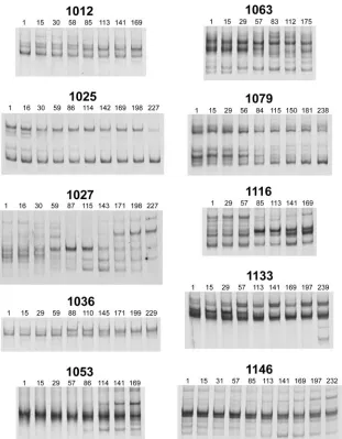
Characterization of V4/V5 genetic populations. We first characterized V4/V5envpopulations in the first and last time points of the study (separated by an average of 221 days) for each subject (Fig. 1). The vast majority of subjects had complex V4/V5 genetic populations, much like we previ-ously observed for V1/V2 (12), with up to 10 coexisting V4/V5 genetic variants detected at a single time point within an individual (e.g., subject 1067). Furthermore, the pattern of genetic variants changed for nearly all subjects over the course of study, with examples of changes in relative abun-dance for variants that remained present throughout the time course (e.g., subject 1012), the loss of major genetic variants (e.g., subject 1079), and the emergence of new major variants (e.g., subject 1027).
To examine the kinetics of viral population changes, we next characterized V4/V5 populations across all intervening time points for 10 subjects who were representative of the various HTA patterns observed in Fig. 1. We observed different kinet-* Corresponding author. Mailing address: University of North
Caro-lina at Chapel Hill, 22-062 Lineberger Cancer Center, CB#7295, Chapel Hill, NC 27599-7295. Phone: (919) 5710. Fax: (919) 966-8212. E-mail: [email protected].
† Present address: Carolina Vaccine Institute, University of North Carolina, Chapel Hill, NC 27599-7292.
‡ Present address: International Clinical Virology, GlaxoSmith-Kline, P.O. Box 13398, 5 Moore Dr., RTP, NC 27709-3398.
䌤Published ahead of print on 28 February 2007.
5413
on November 8, 2019 by guest
http://jvi.asm.org/
ics of V4/V5 changes among these subjects (Fig. 2). In some subjects we observed a gradual gain or loss of minor variants (e.g., subjects 1053 and 1025). We also observed striking pop-ulation shifts over short time spans (30 to 60 days) followed by periods of relative stability (e.g., subjects 1027 and 1116). Other subjects, such as subjects 1133 and 1146, had less dra-matic changes over the course of the study, limited primarily to changes in the relative abundance of minor variants. One in-dividual, subject 1036, had a relatively homogeneous V4/V5 genetic population at the start of the study, followed by the emergence of a new variant that first mixed with and then eventually replaced the entire preexisting population to yield a novel homogeneous population. Taken together, these results suggest that selective pressures targeting the V4/V5 region are often intense and continually evolving, much as we observed for V1/V2 in the same subjects (12).
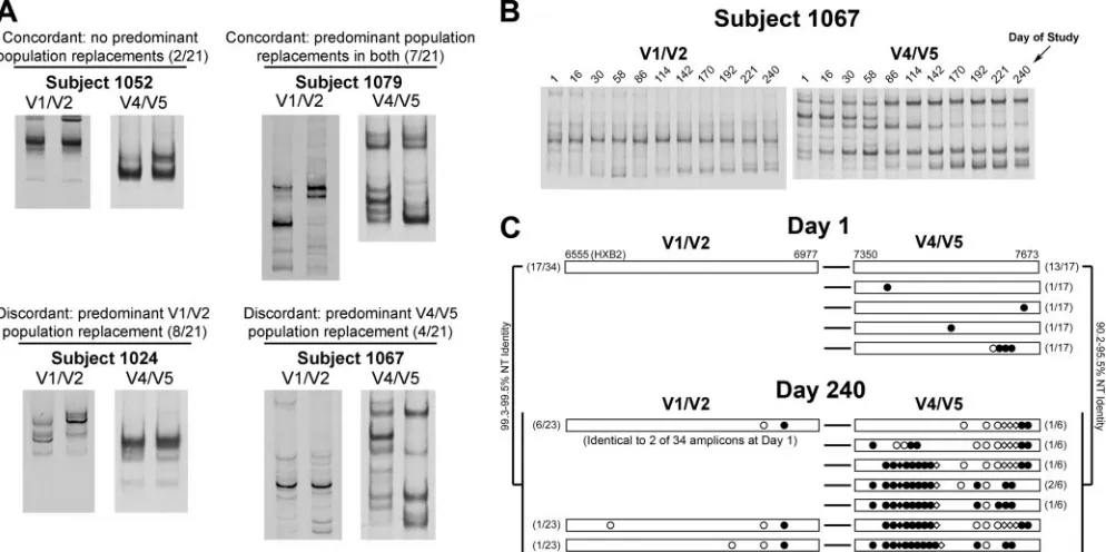
Independence of V1/V2 and V4/V5 population changes.We examined the timing of major V4/V5 population changes rel-ative to V1/V2. We first identified subjects who had one or more “predominant population changes” in either V1/V2 or V4/V5 or both over the course of the study, which were simply defined as replacements in the most abundant HTA variants as measured by phosphorimaging of HTA gels (11, 12). There-fore, changes in minor variants were not considered in this analysis. We observed variable patterns of predominant V1/V2 and V4/V5 genetic population changes, including (i) concor-dant patterns of predominant population stability (2 of 21 subjects), (ii) concordant replacement of both V1/V2 and V4/V5 predominant populations (7 of 21 subjects), and (iii) independent predominant population replacement in one re-gion with relative stability in the other (12 of 21 subjects). Representative examples are shown in Fig. 3A. In a few sub-jects, e.g., subject 1067 (Fig. 3B), we also observed stable, relatively homogeneous genetic populations in one region, with multiple dramatic population changes in the other.
To confirm the observation of independent evolution of V1/V2 and V4/V5 regions, we reverse transcribed blood plasma
RNA obtained from days 1 and 240 from subject 1067 and subjected the cDNA to a limiting-dilution, nested PCR proto-col targeting the full-lengthenvgene. To ensure a high prob-ability of amplification from single templates and thus limit artifactual recombination between templates, cDNAs were di-luted and amplified in 96-well plates such that nested PCR resulted in⬍50% of reactions positive for full-lengthenv. We then sequenced the V1/V2- and V4/V5-coding regions in the
envamplicons and discarded any sequences with evidence of frameshifts or mixtures at any nucleotide positions. This tech-nique, termed single-genome amplification and sequencing, was adapted from previously published protocols (17, 26) and will be described in greater detail elsewhere (J. Salazar and B. Hahn, unpublished data). In total, single genome sequences from 34 amplicons from day 1 plasma and 23 amplicons from day 240 plasma were obtained from subject 1067. Predominant V1/V2 sequence populations and their linked V4/V5 se-quences are shown in Fig. 3C. Half of theenvamplicons ob-tained from day 1 plasma had V1/V2 regions with 100% nu-cleotide identity, and nearly all of their linked V4/V5 sequences were identical, suggesting that a large proportion of theenvpopulation at this time point was comprised of a single variant. The remainder of V1/V2 and V4/V5 sequences de-tected at day 1 represented several minor populations (data not shown). At day 240, most of the V1/V2 sequences clustered into two groups, whereas the V4/V5 population was much more complex. Approximately one-third of the V1/V2 se-quences differed by only two to three nucleotides (one coding change) from the bulk of the V1/V2 population 239 days ear-lier, and 6 sequences were identical to 2 of 34 sequences obtained from day 1 plasma (Fig. 3C). In contrast, the linked V4/V5 sequences at day 240 differed dramatically from each other and from all V4/V5 sequences obtained from day 1 plasma, with several coding differences, insertions, and dele-tions (Fig. 3C). Taken together, these results confirm the HTA data for this subject and support the observation of indepen-dent V1/V2 and V4/V5 evolution.
FIG. 1. V4/V5envpopulations in infected subjects at the start and end of study. Blood plasma viral RNA populations were characterized by reverse transcription-PCR and HTA targeting the V4/V5 region ofenv. Bands representing the single-stranded probe and probe homoduplex are not shown. All DNA heteroduplex bands shown run between the single-stranded probe and probe homoduplexes, and represent coexisting HIV-1 V4/V5 genetic variants. The time points shown represent the day of blood sample collection relative to the start of the study.
on November 8, 2019 by guest
http://jvi.asm.org/
[image:2.585.111.473.67.252.2]We next quantified the overall concordance of V1/V2 and V4/V5 population changes over time by three different HTA measures: total change, entropy change, and HTA index. Total change is a measure of the difference in HTA band patterns for two different time points, taking into account both the pres-ence of unique variants in one time point relative to the other and the differences in relative abundance of variants shared between both time points (12, 21). Shannon entropy is a mea-sure of genetic population complexity at a single time point (4, 24). Therefore, entropy change measures the change in popu-lation complexity between two time points, with positive and negative values reflecting increasing and decreasing population complexity, respectively. The HTA index, recently described by Riddle et al. (21), is a novel algorithm for quantifying popu-lation changes that takes into account the timing of popupopu-lation changes and also emphasizes the emergence of new variants. We observed no correlation of V1/V2 and V4/V5 population changes using any of these three algorithms (Fig. 4).
Finally, we monitored viral populations at individual time points for three of the seven subjects who displayed predom-inant viral population changes in both V1/V2 and V4/V5. In subject 1027 we observed strongly concurrent population shifts in V1/V2 and V4/V5 at short, specific time intervals (data not shown). In the other two subjects—subjects 1036 and 1079— predominant population replacements in V1/V2 and V4/V5 occurred at different times and at generally different rates (data not shown). In summary, our results indicate that the V1/V2 and V4/V5 hypervariable regions of env frequently evolve independently in infected individuals during late chronic infection.
[image:3.585.137.448.66.465.2]One limitation of the present study is the fact that an HTA does not always resolve coexisting genetic variants with single or a few dispersed nucleotide differences. Therefore, it is pos-sible that for some subjects single nucleotide changes may occur in one or both regions ofenv where no predominant genetic changes are detected by HTA, although nucleotide FIG. 2. Kinetics of V4/V5envpopulation changes. V4/V5 RNA genetic variants present in blood specimens collected over 2- to 4-week intervals were characterized by reverse transcription-PCR and HTA. As described for Fig. 1, only heteroduplex bands are shown. The time points shown represent the day of blood sample collection relative to the start of the study.
VOL. 81, 2007 NOTES 5415
on November 8, 2019 by guest
http://jvi.asm.org/
changes near other clustered differences between the probe and target sequence are frequently detected by HTA (12, 20). Nevertheless, it seems unlikely that undetectable single nucle-otide changes in one region can account for all discordant population changes in V1/V2 and V4/V5, especially consider-ing that in several subjects we observed multiple strikconsider-ing pop-ulation shifts in one region over a period of approximately 6 to 9 months with little or no change detected in the other. Fur-thermore, single-genome sequencing data independently vali-dated the HTA observations for subject 1067 (Fig. 3C). In another recent cohort study (21), discordant V1/V2 and V4/V5
changes were observed at 6-month intervals for some subjects. Of note, in the present study the overall level of V1/V2 and V4/V5 change over time as measured by total change and HTA index was relatively low compared to the study by Riddle et al. (21). However, this is not surprising given that all of the sub-jects in the present study had low CD4 counts (⬍100). There-fore, generally reduced immune pressure on Env likely con-tributed to slower evolution of V1/V2 and V4/V5, which is consistent with the report by Delwart et al. (4).
[image:4.585.45.542.70.318.2]We speculate that at a given moment, at least a portion of a host neutralizing antibody response to HIV-1 can be prefer-FIG. 3. Independence of V1/V2 and V4/V5 predominant population changes. (A) Different patterns of concordance for V1/V2 and V4/V5 predominant population replacements over the full course of study are revealed by HTA. The data from first and last time points of the study are shown for representative subjects. The number of subjects representing each of the four patterns is shown, out of a total of 21 subjects characterized by both V1/V2 and V4/V5 HTA. (B) Time course HTAs for V1/V2 and V4/V5 for subject 1067. (C) Predominant V1/V2 populations and their linked V4/V5 sequences obtained from day 1 and day 240 plasma from subject 1067. Sequences were obtained by single-genome amplification. Numbers in parentheses on the left indicate the number of V1/V2 sequences represented (of the total number of amplicons) and on the right the number of V4/V5 sequences linked to a particular V1/V2 sequence. Sequence differences depicted are relative to predominant V1/V2 and V4/V5 sequence populations at day 1. Symbols:E, noncoding nucleotide difference;F, coding difference;〫, codon deletion;}, codon insertion.
FIG. 4. Lack of correlation by three different quantitative measures of V1/V2 and V4/V5 population change. Total change, entropy change, and HTA index algorithms were used to quantify V1/V2 and V4/V5 change between the first and last time points of the study for each subject (one interval per subject). Values were plotted for V1/V2 and V4/V5 to determine a correlation of changes between the two regions, and ther2andP values are indicated.
on November 8, 2019 by guest
http://jvi.asm.org/
[image:4.585.44.542.559.687.2]entially directed toward a subset of variable targets in Env, considering the following points: (i) the hypervariable regions of the Env protein are highly accessible (3, 13, 28) and are therefore major targets for host antibody responses; (ii) broadly reactive neutralizing antibodies are rarely detected in infected subjects and are difficult to induce by vaccination, as opposed to type-specific or autologous antibodies (reviewed in reference 18); (iii) preexisting antibody to heterologous Env antigen does not alter V1/V2 diversification in SIVsm-infected macaques (22); and (iv) as shown here, V1/V2 and V4/V5env
regions evolve independently in infected subjects. Unfortu-nately a type-specific neutralizing antibody response is difficult to assess in infected subjects, although careful characterization of env sequence evolution may provide a useful surrogate method, albeit indirect, for identifying regions of Env under neutralizing antibody selective pressure.
Nucleotide sequence accession numbers.All single-genome sequences have been deposited in GenBank (accession num-bers EF418433 to EF418546).
We thank Jesus Salazar and Beatrice Hahn for providing theenv single genome amplification protocol and Li-Hua Ping for assistance with this protocol.
This study was supported by NIH grant R37-AI44667 (R.S.), the UNC Center for AIDS Research (P30-AI50410), and an NIH post-doctoral training fellowship from the UNC Lineberger Cancer Center (T32-CA09156) to P.R.H.
REFERENCES
1.Blay, W. M., S. Gnanakaran, B. Foley, N. A. Doria-Rose, B. T. Korber, and N. L. Haigwood.2006. Consistent patterns of change during the divergence of human immunodeficiency virus type 1 envelope from that of the inocu-lated virus in simian/human immunodeficiency virus-infected macaques. J. Virol.80:999–1014.
2.Cameron, D. W., M. Heath-Chiozzi, S. Danner, C. Cohen, S. Kravcik, C. Maurath, E. Sun, D. Henry, R. Rode, A. Potthoff, and J. Leonard.1998. Randomised placebo-controlled trial of ritonavir in advanced HIV-1 disease. Lancet351:543–549.
3.Chen, B., E. M. Vogan, H. Gong, J. J. Skehel, D. C. Wiley, and S. C. Harrison.2005. Structure of an unliganded simian immunodeficiency virus gp120 core. Nature433:834–841.
4.Delwart, E. L., H. Pan, H. W. Sheppard, D. Wolpert, A. U. Neumann, B. Korber, and J. I. Mullins.1997. Slower evolution of human immunodefi-ciency virus type 1 quasispecies during progression to AIDS. J. Virol.71:
7498–7508.
5.Delwart, E. L., H. W. Sheppard, B. D. Walker, J. Goudsmit, and J. I. Mullins. 1994. Human immunodeficiency virus type 1 evolution in vivo tracked by DNA heteroduplex mobility assays. J. Virol.68:6672–6683. 6.Delwart, E. L., E. G. Shpaer, J. Louwagie, F. E. McCutchan, M. Grez, H.
Rubsamen-Waigmann, and J. I. Mullins.1993. Genetic relationships deter-mined by a DNA heteroduplex mobility assay: analysis of HIV-1 env genes. Science262:1257–1261.
7.Derdeyn, C. A., J. M. Decker, F. Bibollet-Ruche, J. L. Mokili, M. Muldoon, S. A. Denham, M. L. Heil, F. Kasolo, R. Musonda, B. H. Hahn, G. M. Shaw, B. T. Korber, S. Allen, and E. Hunter.2004. Envelope-constrained neutral-ization-sensitive HIV-1 after heterosexual transmission. Science303:2019– 2022.
8.Etemad-Moghadam, B., Y. Sun, E. K. Nicholson, G. B. Karlsson, D. Schenten, and J. Sodroski.1999. Determinants of neutralization resistance in the envelope glycoproteins of a simian-human immunodeficiency virus passaged in vivo. J. Virol.73:8873–8879.
9.Frost, S. D., T. Wrin, D. M. Smith, S. L. Kosakovsky Pond, Y. Liu, E. Paxinos, C. Chappey, J. Galovich, J. Beauchaine, C. J. Petropoulos, S. J.
Little, and D. D. Richman.2005. Neutralizing antibody responses drive the evolution of human immunodeficiency virus type 1 envelope during recent HIV infection. Proc. Natl. Acad. Sci. USA102:18514–18519.
10.Hahn, B. H., M. A. Gonda, G. M. Shaw, M. Popovic, J. A. Hoxie, R. C. Gallo, and F. Wong-Staal.1985. Genomic diversity of the acquired immune defi-ciency syndrome virus HTLV-III: different viruses exhibit greatest diver-gence in their envelope genes. Proc. Natl. Acad. Sci. USA82:4813–4817. 11.Harrington, P. R., D. W. Haas, K. Ritola, and R. Swanstrom.2005.
Com-partmentalized human immunodeficiency virus type 1 present in cerebrospi-nal fluid is produced by short-lived cells. J. Virol.79:7959–7966. 12.Kitrinos, K. M., N. G. Hoffman, J. A. E. Nelson, and R. Swanstrom.2003.
Turnover ofenvvariable region 1 and 2 genotypes in subjects with late-stage human immunodeficiency virus type 1 infection. J. Virol.77:6811–6822. 13.Kwong, P. D., R. Wyatt, J. Robinson, R. W. Sweet, J. Sodroski, and W. A.
Hendrickson.1998. Structure of an HIV gp120 envelope glycoprotein in complex with the CD4 receptor and a neutralizing human antibody. Nature
393:648–659.
14.Modrow, S., B. H. Hahn, G. M. Shaw, R. C. Gallo, F. Wong-Staal, and H. Wolf.1987. Computer-assisted analysis of envelope protein sequences of seven human immunodeficiency virus isolates: prediction of antigenic epitopes in conserved and variable regions. J. Virol.61:570–578. 15.Nelson, J. A. E., F. Baribaud, T. Edwards, and R. Swanstrom.2000. Patterns
of changes in human immunodeficiency virus type 1 V3 sequence popula-tions late in infection. J. Virol.74:8494–8501.
16.Nelson, J. A. E., S. A. Fiscus, and R. Swanstrom.1997. Evolutionary variants of the human immunodeficiency virus type 1 V3 region characterized by using a heteroduplex tracking assay. J. Virol.71:8750–8758.
17.Palmer, S., M. Kearney, F. Maldarelli, E. K. Halvas, C. J. Bixby, H. Bazmi, D. Rock, J. Falloon, R. T. Davey, Jr., R. L. Dewar, J. A. Metcalf, S. Hammer, J. W. Mellors, and J. M. Coffin.2005. Multiple, linked human immunode-ficiency virus type 1 drug resistance mutations in treatment-experienced patients are missed by standard genotype analysis. J. Clin. Microbiol.43:
406–413.
18.Pantophlet, R., and D. R. Burton.2006. GP120: target for neutralizing HIV-1 antibodies. Annu. Rev. Immunol.24:739–769.
19.Pinter, A., W. J. Honnen, Y. He, M. K. Gorny, S. Zolla-Pazner, and S. C. Kayman.2004. The V1/V2 domain of gp120 is a global regulator of the sensitivity of primary human immunodeficiency virus type 1 isolates to neu-tralization by antibodies commonly induced upon infection. J. Virol. 78:
5205–5215.
20.Resch, W., N. Parkin, E. L. Stuelke, T. Watkins, and R. Swanstrom.2001. A multiple-site-specific heteroduplex tracking assay as a tool for the study of viral population dynamics. Proc. Natl. Acad. Sci. USA98:176–181. 21.Riddle, T. M., N. J. Shire, M. S. Sherman, K. F. Franco, H. W. Sheppard,
and J. A. E. Nelson.2006. Sequential turnover of human immunodeficiency virus type 1envthroughout the course of infection. J. Virol.80:10591–10599. 22.Rybarczyk, B. J., D. Montefiori, P. R. Johnson, A. West, R. E. Johnston, and R. Swanstrom.2004. Correlation between env V1/V2 region diversification and neutralizing antibodies during primary infection by simian immunode-ficiency virus sm in rhesus macaques. J. Virol.78:3561–3571.
23.Sagar, M., X. Wu, S. Lee, and J. Overbaugh.2006. Human immunodefi-ciency virus type 1 V1–V2 envelope loop sequences expand and add glyco-sylation sites over the course of infection, and these modifications affect antibody neutralization sensitivity. J. Virol.80:9586–9598.
24.Shannon, C. E.1948. A mathematical theory of communication. Bell Syst. Technol J.27:379–423.
25.Starcich, B. R., B. H. Hahn, G. M. Shaw, P. D. McNeely, S. Modrow, H. Wolf, E. S. Parks, W. P. Parks, S. F. Josephs, R. C. Gallo, et al.1986. Identification and characterization of conserved and variable regions in the envelope gene of HTLV-III/LAV, the retrovirus of AIDS. Cell45:637–648.
26.Wei, X., J. M. Decker, S. Wang, H. Hui, J. C. Kappes, X. Wu, J. F. Salazar-Gonzalez, M. G. Salazar, J. M. Kilby, M. S. Saag, N. L. Komarova, M. A. Nowak, B. H. Hahn, P. D. Kwong, and G. M. Shaw.2003. Antibody neutral-ization and escape by HIV-1. Nature422:307–312.
27.Willey, R. L., R. A. Rutledge, S. Dias, T. Folks, T. Theodore, C. E. Buckler, and M. A. Martin.1986. Identification of conserved and divergent domains within the envelope gene of the acquired immunodeficiency syndrome retro-virus. Proc. Natl. Acad. Sci. USA83:5038–5042.
28.Zhu, P., J. Liu, J. Bess, Jr., E. Chertova, J. D. Lifson, H. Grise, G. A. Ofek, K. A. Taylor, and K. H. Roux.2006. Distribution and three-dimensional structure of AIDS virus envelope spikes. Nature441:847–852.
VOL. 81, 2007 NOTES 5417