Multimodality imaging to guide
cardiac interventional procedures
The studies described in this thesis were performed at the Department of Cardiology of the
Leiden University Medical Center, Leiden, the Netherlands, and the Division of Cardiology,
Department of Medicine of Johns Hopkins University, Baltimore, USA.
Cover: Optima Grafi sche Communicatie, Rotterdam, The Netherlands
Lay-out and print: Optima Grafi sche Communicatie, Rotterdam, The Netherlands
ISBN: 978-90-8559-949-4
Copyright © Laurens F. Tops, Leiden, the Netherlands. All rights reserved. No part of this book
may be reproduced or transmitted, in any form or by any means, without prior permission of
the author.
Financial support to the costs associated with the publication of this thesis from Biosense
Web-ster, Biotronik Nederland BV, Boston Scientifi c BV, Edwards Lifesciences BV, 3mensio Medical
Imaging BV, Bracco Imaging Europe BV, Daiichi Sankyo Nederland BV, HagaZiekenhuis, Siemens
Nederland NV, Sorin Group Nederland NV, St. Jude Medical Nederland BV, Stichting Imago,
Stichting J.E. Jurriaanse, Vital Images BV, Toshiba Medical Systems Nederland, AstraZeneca BV,
Boehringer Ingelheim BV, Eli Lilly Nederland BV, Merck Sharp & Dohme BV, Schering-Plough BV,
Brahms Nederland, Guerbet Nederland BV, Servier Nederland Farma BV, and Sanofi -Aventis BV
Multimodality imaging to guide
cardiac interventional procedures
Proefschrift
ter verkrijging van
de graad van Doctor aan de Universiteit Leiden,
op gezag van Rector Magnifi cus prof. mr. P.F. van der Heijden,
volgens besluit van het College voor Promoties
te verdedigen op donderdag 15 april 2010
klokke 16.15 uur
door
Laurens Franciscus Tops
PROMOTIECOMMISSIE
Promotores: Prof. dr. J.J. Bax
Prof. dr. M.J. Schalij
Overige leden: Prof. dr. D. Poldermans (Erasmus MC, Rotterdam)
Prof. dr. A. de Roos
Prof. dr. E.E. van der Wall
Dr. E.R. Holman
Dr. J.D. Schuijf
Dr. K. Zeppenfeld
Financial support by the Netherlands Heart Foundation for the publication of this thesis is
Great minds have purposes,
others have whishes
CONTENTS
Chapter 1 General introduction and outline of the thesis 9
PART I Catheter ablation for atrial fi brillation
Chapter 2 Multi-modality imaging to assess left atrial size, anatomy and function
Heart 2007;93:1461-70
29
Chapter 3 Imaging and atrial fi brillation: the role of multimodality imaging in
patient evaluation and management of atrial fi brillation Eur Heart J 2010, in press
51
Chapter 4 Fusion of multislice computed tomography imaging with
three-dimensional electroanatomic mapping to guide radiofrequency catheter ablation procedures
Heart Rhythm 2005;2:1076-81
73
Chapter 5 Real-time integration of intracardiac echocardiography and multislice
computed tomography to guide radiofrequency catheter ablation for atrial fi brillation
Heart Rhythm 2008;5:1403-10
85
Chapter 6 Impact of pulmonary vein anatomy and left atrial dimensions on the
outcome of circumferential radiofrequency catheter ablation for atrial fi brillation
Submitted
101
Chapter 7 Eff ect of radiofrequency catheter ablation for atrial fi brillation on left atrial cavity size
Am J Cardiol 2006;97:1220-2
113
Chapter 8 Comparison of left atrial volumes and function by real-time
three-dimensional echocardiography in patients having catheter ablation for atrial fi brillation with persistence of sinus rhythm versus recurrent atrial fi brillation three months later
Am J Cardiol 2008;102:847-53
121
Chapter 9 Left atrial strain predicts reverse remodeling after catheter ablation for
atrial fi brillation Submitted
135
Chapter 10 Long-term improvement in left ventricular strain after successful
catheter ablation for atrial fi brillation in patients with preserved left ventricular systolic function
Circ Arrhythmia Electrophysiol 2009;2:249-57
PART II Ventricular pacing and dyssynchrony
Chapter 11 The eff ects of right ventricular apical pacing on ventricular function and dyssynchrony: implications for therapy
J Am Coll Cardiol 2009;54:764-76
171
Chapter 12 Right ventricular pacing can induce ventricular dyssynchrony in patients
with atrial fi brillation after atrioventricular node ablation J Am Coll Cardiol 2006;48:1642-8
195
Chapter 13 Acute eff ects of right ventricular apical pacing on left ventricular synchrony and mechanics
Circ Arrhythmia Electrophysiol 2009;2:135-45
209
Chapter 14 Speckle-tracking radial strain reveals left ventricular dyssynchrony in
patients with permanent right ventricular pacing J Am Coll Cardiol 2007;50:1180-8
227
Chapter 15 The eff ect of right ventricular pacing on myocardial oxidative
metabo-lism and effi ciency: relation with left ventricular dyssynchrony Eur J Nucl Med Mol Imaging 2009;36:2042-8
245
Chapter 16 Prevalence and pathophysiologic attributes of ventricular dyssynchrony
in arrhythmogenic right ventricular dysplasia/cardiomyopathy J Am Coll Cardiol 2009;54:445-51
257
PART III Percutaneous valve procedures
Chapter 17 Percutaneous valve procedures: an update
Curr Probl Cardiol 2008;33:417-57
275
Chapter 18 Noninvasive evaluation of coronary sinus anatomy and its relation
to the mitral valve annulus: implications for percutaneous mitral annuloplasty
Circulation 2007;115:1426-32
307
Chapter 19 Assessment of mitral valve anatomy and geometry with multislice
computed tomography
J Am Coll Cardiol Img 2009;2:556-65
321
Chapter 20 Percutaneous aortic valve therapy: clinical experience and the role of
multimodality imaging Heart 2009;95:1538-46
339
Chapter 21 Noninvasive evaluation of the aortic root with multislice computed
tomography: implications for transcatheter aortic valve replacement J Am Coll Cardiol Img 2008;1;321-30
359
Chapter 22 Role of multislice computed tomography in transcatheter aortic valve
replacement
Am J Cardiol 2009;103:1295-301
Summary, conclusions and future perspectives 389
Samenvatting, conclusies en toekomstperspectieven 401
List of publications 415
Acknowledgements 423
1
Chapt
er 1
Intr
oduc
tion and outline of the thesis
11
In the past decades, tremendous advances have been made in both the imaging and
interventional fi eld of clinical cardiology. Dedicated imaging techniques such as multi-slice
computed tomography have been introduced and enable detailed non-invasive evaluation
of cardiac anatomy. Furthermore, conventional techniques such as echocardiography have
been improved and nowadays allow a more comprehensive assessment of cardiac
morphol-ogy and function. At the same time, percutaneous interventional procedures for arrhythmias
and valvular heart disease have been further explored. While conventional invasive treatment
of these conditions requires open-heart surgery, nowadays it has become feasible to perform
these procedures with minimal-invasive techniques.
These advances allow a more integrative approach to cardiac imaging and interventions.
The combination and integration of diff erent imaging modalities and subsequent use of these
techniques during interventional procedures will further enhance the evaluation and
treat-ment of cardiac arrhythmias and valvular heart disease. In this thesis, the role of multimodality
imaging to guide cardiac interventional procedures is investigated. In particular, catheter
abla-tion for atrial fi brillaabla-tion (AF), cardiac pacing and resynchronizaabla-tion therapy, and percutaneous
valve procedures are explored.
CATHETER ABLATION FOR ATRIAL FIBRILLATION
Atrial fi brillation is the most commonly encountered cardiac arrhythmia. It is characterized by
rapid, irregular activity of the atria. In the general population, the prevalence of AF is
approxi-mately 1% (1). Since the prevalence of AF increases with age, it may become ‘epidemic’ in the
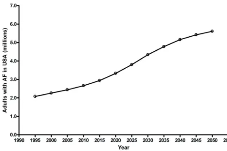
coming decades, with an estimated 3.3 million patients in the United States in 2020 (Figure 1).
Importantly, AF is associated with an increased risk of both cardiac morbidity and mortality (2).
The most important goals in the treatment of AF are: reduction of the risk of
thrombo-embolism and control of AF-related symptoms (3). To reduce the risk of thrombothrombo-embolism, a
tailored anti-thrombotic regimen (e.g. anticoagulation or aspirin) should be chosen depending
on clinical characteristics (4). To control symptoms of AF, both ‘rate control’ and ‘rhythm control’
strategies can be chosen. Again, an individualized approach is preferred, since the superiority
of one strategy has not been proven. Large randomized trials have not demonstrated diff
er-ences in mortality or quality of life between the two strategies (5,6).
If a ‘rhythm control’ strategy is chosen, anti-arrhythmic drugs and/or electrical cardioversion
are used to restore sinus rhythm. Unfortunately, anti-arrhythmic drugs may have side-eff ects
and often fail to maintain sinus rhythm. In the past decade, catheter ablation procedures have
been introduced as a new therapeutic option in the treatment of patients with AF.
Haissaguerre et al. demonstrated that the pulmonary veins (PVs) are the main source of
ectopic beats that initiate AF (7). Subsequently, it was shown that electrical isolation of these
12
maintenance of sinus rhythm. During the catheter ablation procedure, the PVs are isolated from
the left atrial (LA) wall by applying radiofrequency current around the PV ostia (Figure 2).
Sev-eral randomized controlled trials have compared anti-arrhythmic drugs and catheter ablation
procedures regarding the effi cacy to maintain sinus rhythm during long-term follow-up (8-11).
From these studies, it has become apparent that catheter ablation may be more eff ective than
anti-arrhythmic drugs (Table 1). It should be noted however, that serious complications may
occur in up to 5% of patients undergoing catheter ablation for AF (12). Therefore, at present
catheter ablation still is considered a second-line therapy, but an excellent treatment option
Figure 1. Estimated number of patients with atrial fi brillation (AF) in the United States of America (USA). The total number of patients may increase up to 2.5-fold in the coming 4 decades. Adapted with permission from Go AS et al., reference (1).
Chapt
er 1
Intr
oduc
tion and outline of the thesis
13
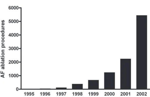
after at least one anti-arrhythmic drug has failed (3). Interestingly, an increasing number of AF
patients worldwide are treated with catheter ablation (Figure 3).
The cornerstone of AF ablation procedures is electrical isolation of the PVs. However,
ana-tomical studies have demonstrated that PV anatomy is highly variable (13). In particular, the
exact number and location of the PVs has large inter-individual variation. Therefore, careful
identifi cation of the PVs is important both before and during the catheter ablation procedure.
Several imaging modalities are available to evaluate the anatomy of the LA and the PVs.
Multi-slice computed tomography and magnetic resonance imaging provide excellent images of
the PVs. However, they do not provide real-time information since the images are acquired
before the actual ablation procedure. On the other hand, intracardiac echocardiography and
electroanatomic mapping enable online visualization of the PVs in relation with the ablation
catheters. However, these techniques are limited by the two-dimensional character and the
use of reconstructed anatomy, respectively. Ideally, the information of the diff erent imaging
Table 1. Randomized studies comparing catheter ablation and anti-arrhythmic drugs Study
(reference)
Number of patients
Type of AF Follow-up Primary endpoint: Freedom from AF (Ablation vs. AAR)
Secondary endpoints Complications ablation
Oral et al. (9) 146 146 persistent (100%)
12 months 74% vs. 58%, p=0.05
- Decrease in LA diameter in successful ablation patients - Increase in LVEF in successful ablation patients - Improvement in symptoms in all ablation patients None reported Pappone et al. (10) 198 198 paroxysmal (100%)
12 months 86% vs. 22%, p<0.001
N/A - TIA: 1
- Pericardial eff usion: 1 Stabile et al.
(11) 137 92 paroxysmal (67%) 45 persistent (33%)
12 months 66% vs. 9%, p<0.001
N/A - Stroke: 1 - Transient phrenic nerve paralysis: 1 - Pericardial eff usion: 1
Jais et al. (8) 112 112 paroxysmal (100%)
12 months 89% vs. 23%, p<0.001
- No diff erences in LA diameter and LVEF at follow-up
- Greater reduction in AF burden in ablation patients
- Improvement in quality of life and exercise capacity in ablation patients
- Cardiac tamponade: 2 - Hematoma: 2 - PV stenosis: 1
14
techniques is integrated, providing highly detailed on-line anatomical information during the
catheter ablation procedure.
Another important issue is the eff ect of catheter ablation procedures for AF on cardiac size
and function. It has been well recognized that there is a close relation between AF and LA size
(14). After catheter ablation, the ablation lesions may result in scarring of the LA wall. This may
negatively aff ect LA size and contractile function. On the other hand, a reduction in LA size
may result in lower susceptibility to AF (15). In addition, the restoration of sinus rhythm may
ultimately result in an improved LA function. Furthermore, normalization of heart rhythm may
result in more effi cient left ventricular (LV) function. It has been demonstrated that catheter
ablation results in signifi cant improvement in LV ejection fraction in patients with AF and
sys-tolic heart failure (16). However, the eff ect of catheter ablation on LV function in patients with
preserved LV ejection fraction is unclear.
Multimodality imaging and image integration may enhance AF ablation procedures by
improved visualization of cardiac structures, and may result in better understanding of the
eff ects of catheter ablation on cardiac function. Accordingly, the aims of the studies described
in this thesis are to test the feasibility of image integration to guide catheter ablation
proce-dures and to assess the eff ects of catheter ablation proceproce-dures on LA and LV function.
Chapt
er 1
Intr
oduc
tion and outline of the thesis
15
VENTRICULAR PACING AND DYSSYNCHRONY
In 1958, the fi rst pacemaker implantation was performed in a patient with high degree
atrio-ventricular block. Since then, cardiac pacing has been an eff ective treatment in the
manage-ment of patients with symptomatic brady- and tachy-arrhythmias. The annual number of new
pacemaker implantations in the Netherlands is about 6000, and is steadily increasing (17). High
degree atrioventricular block and sick sinus syndrome are the most important indications for
implantation of a conventional pacemaker (Figure 4).
Typically, the endocardial ventricular pacing lead is positioned at the right ventricular (RV)
apex. However, large randomized trials have revealed a possible association between RV apical
pacing and deterioration of cardiac function (18,19). In the Mode Selection Trial (MOST), it was
demonstrated that a high percentage of ventricular pacing is associated with an increased risk
of heart failure hospitalization (Figure 5). Furthermore, other studies have shown that RV apical
pacing results in changes in myocardial perfusion (20) and ventricular remodeling (21). At the
same time, minimizing RV apical pacing with dedicated algorithms can prevent harmful eff ects
of cardiac pacing (22).
The deleterious eff ects of conventional RV apical pacing may be associated with the
abnor-mal electrical and mechanical activation pattern of the cardiac chambers. During RV apical
AV block
SSS
AF
Other
16
pacing, the electrical wave front propagates through the myocardium, rather than through the
His-Purkinje conduction system. Due to the diff erences in conduction velocity, heterogeneity
in electrical activation of the cardiac chambers occurs (23). Simultaneously, changes in the
mechanical activation pattern are noted. In particular, the onset and magnitude of mechanical
contraction of various LV walls change (24). The temporal occurrence of peak strain of diff erent
ventricular segments exhibits an asynchronous pattern during RV apical pacing. This is referred
to as ‘ventricular mechanical dyssynchrony’ (Figure 6).
Mechanical dyssynchrony can be assessed with various non-invasive imaging modalities.
Already in 1977, Gomes et al. noticed the asynchronous contraction pattern of the LV during RV
apical pacing with the use of transthoracic echocardiography (25). A signifi cant delay between
the (early) posterior motion of the interventricular septum and the (delayed) contraction of the
posterior wall was observed immediately after onset of pacing. Nowadays, additional
echocar-diographic techniques are available for assessment of ventricular dyssynchrony (26). With the
use of tissue Doppler imaging, myocardial velocities of diff erent ventricular segments can be
assessed throughout the cardiac cycle (Figure 6). Off -line analysis of the regional time-to-peak
systolic velocity enables quantifi cation of ventricular mechanical dyssynchrony (27).
Speckle-tracking strain analysis is another echocardiographic technique that allows assessment of
regional timing of peak strain (28). The assessment of myocardial strain permits diff erentiation
Chapt
er 1
Intr
oduc
tion and outline of the thesis
17
between active contraction and passive motion of the myocardium. Again, by calculating
dif-ferences in time-to-peak systolic strain of various segments, ventricular dyssynchrony can be
assessed (29).
Importantly, it has been demonstrated that the presence of mechanical dyssynchrony has
prognostic value in heart failure patients (30). However, the association between mechanical
dyssynchrony and the deterioration of cardiac function and functional class in pacemaker
patients has not been fully elucidated yet. Furthermore, in the past decade cardiac
resynchro-nization therapy has become a well-established therapeutic option for patients with severe
drug-refractory heart failure and signs of electrical or mechanical dyssynchrony (31,32). In
cardiac resynchronization therapy, an LV pacing lead is added to a conventional pacing system,
allowing simultaneously pacing of the RV and LV to re-synchronize the cardiac chambers. It
may well be that cardiac resynchronization therapy is able to (partly) reverse the detrimental
eff ects of conventional RV apical pacing.
New imaging techniques may be valuable tools for the detection of mechanical
dyssyn-chrony, and may help in monitoring patients with conventional pacemakers and selecting
potential candidates for upgrade of RV pacing to biventricular pacing. Accordingly, the
cur-rent studies explore the possible association between deterioration of cardiac function and
ventricular mechanical dyssynchrony after onset of RV pacing, and reversal of the detrimental
eff ects and LV dyssynchrony with cardiac resynchronization therapy.
18
PERCUTANEOUS VALVE PROCEDURES
Valvular heart disease is an important entity in clinical cardiology. Aortic stenosis (AS) and mitral
regurgitation (MR) are the most common native single-valve disease. In the general population,
the prevalence of AS is estimated between 2 and 7% (33). Various pathophysiologic processes
can attribute to the development of AS or MR, but most frequently the etiology is degenerative
(Table 2). Importantly, the presence of severe AS (34) or MR (35) is associated with a
substan-tially increased risk of cardiac morbidity and mortality. The poor natural history of untreated
AS and MR emphasizes the importance of treatment of patients with these conditions (36,37).
Surgical aortic valve replacement is the treatment of choice in severe, symptomatic AS.
Operative mortality is about 3-5%, and good long-term survival has been reported (Figure 7)
(38). Importantly, the severity of symptoms, LV ejection fraction and age are important
predic-tors of good outcome after surgical aortic valve replacement (39). For MR, surgical treatment
is more complex, due to its variety in etiologies. Mitral valve repair using undersized mitral
annuloplasty is most frequently used for degenerative and ischemic MR (40). The outcome of
surgical mitral valve repair depends largely on the etiology of MR, but also severity of
symp-toms, LV ejection fraction and age are important predictors of outcome (33). In particular for
organic MR (e.g. mitral valve prolapse), good long-term results have been reported (41).
Despite the good outcome after surgical treatment of AS and MR, a large proportion of
patients does not undergo surgery. The Euro Heart Survey on Valvular Heart Disease explored
the characteristics, treatment and outcome of 5001 patients with valvular heart disease from 25
countries in Europe (42). From this Euro Heart Survey, it has become apparent that up to 30%
Table 2. Results from the Euro Heart Survey on Valvular Heart Disease on the etiology and surgical treatment of aortic stenosis and mitral regurgitation.
Aortic stenosis Mitral regurgitation
Etiology *
Degenerative, % 82 61 Rheumatic, % 11 14 Endocarditis, % 1 4 Infl ammatory, % 0 1 Congenital, % 5 5 Ischaemic, % 0 7
Other, % 1 8
Surgical intervention †
Mechanical prosthesis, % 49 43 Bioprosthesis, % 50 10 Valve repair, % 0 47
Other, % 1 0
Chapt
er 1
Intr
oduc
tion and outline of the thesis
19
of patients with severe symptomatic valvular disease do not undergo surgical intervention,
while a clear indication exists. Most frequently, this is because of co-morbidity and age (42).
Obviously, there is a need for a less invasive approach, in particular in elderly patients with
co-morbidity and severe valvular heart disease.
In recent years, various new percutaneous procedures for the treatment of AS and MR have
been introduced. The implantation of an aortic valve prosthesis through the femoral artery or
the LV apex has become feasible (Figure 8). The feasibility of a balloon-expandable (43) and
a self-expanding valve (44) for the percutaneous treatment of severe AS have been
demon-strated. Importantly, large multi-center studies (45) and mid-term follow-up studies (46) have
demonstrated the safety and effi cacy of these procedures.
Furthermore, new percutaneous devices have been introduced for the percutaneous
treatment of MR (Figure 8). The feasibility of a mitral valve clip mimicking edge-to-edge repair
has been demonstrated (47), and diff erent prostheses that target mitral annulus remodeling
through the coronary sinus have been introduced (48,49). The safety and mid-term effi cacy of
these procedures have also been demonstrated (50,51).
However, percutaneous valve procedures still have limitations and severe complications
can occur. An important issue is failure of the procedure as a result of unfavorable cardiac
anatomy. For example, the close relation between the native valve leafl ets, valve annulus
and the coronary arteries may preclude save percutaneous implantation of a device. In the
Mitral Annuloplasty Device European Union Study (AMADEUS), the coronary sinus device was
20
recaptured because of potential coronary compromise in up to 30% of the non-implanted
patients (51). Furthermore, acute coronary occlusion during percutaneous aortic valve
implan-tation has been reported (43).
Imaging may be of great value in the percutaneous treatment of valvular heart disease. It
may improve the selection of patients and may enhance real-time guidance of the procedures.
In the studies described in the present thesis, the potential role of multi-slice computed
tomog-raphy in the selection for candidates for new percutaneous valve procedures for AS and MR is
explored.
OUTLINE OF THE PRESENT THESIS
The aim of this thesis is to evaluate the role of multimodality imaging to guide cardiac
interven-tional procedures. In particular, catheter ablation procedures for AF, conveninterven-tional pacing and
cardiac resynchronization therapy, and percutaneous valve procedures are studied. Therefore,
the present thesis consists of three distinct parts.
PART I: Catheter ablation for atrial fi brillation
In the fi rst part, catheter ablation procedures for AF are studied. These procedures are considered
a good treatment option in patients with drug-refractory AF, after at least one anti-arrhythmic
drug has failed. Visualization of the PVs with diff erent imaging modalities, the integration of
various imaging techniques, and the eff ect of catheter ablation on LA and LV function are
important issues in AF ablation. Chapter 2 and Chapter 3 provide two extensive reviews on
the role of multimodality imaging in the assessment of PV and LA anatomy, and in catheter
ablation procedures for AF. In Chapter 4, the fi rst clinical experience with a new image integra-tion system that allows integraintegra-tion of MSCT images and electroanatomic mapping is described.
Subsequently, the integration of intracardiac echocardiography with electroanatomic mapping
and multi-slice computed tomography is studied in Chapter 5. The assessment of PV anatomy
with multi-slice computed tomography, and its impact on the outcome of catheter ablation
procedures is explored in Chapter 6.
In the next chapters, the eff ect of catheter ablation for AF on LA and LV function is studied.
In Chapter 7, conventional transthoracic two-dimensional echocardiography is used to assess the eff ect of catheter ablation on LA size. The fi ndings of this study are further extended in
the following chapters. In Chapter 8, real-time three-dimensional echocardiography is used to assess LA function after catheter ablation. Subsequently, the eff ect of catheter ablation on LA
systolic and diastolic strain is investigated in Chapter 9. Furthermore, the predictive value of LA strain for LA reverse remodeling is studied. Finally, the eff ect of sinus rhythm maintenance after
Chapt
er 1
Intr
oduc
tion and outline of the thesis
21
PART II: Ventricular pacing and dyssynchrony
In the second part, conventional RV apical pacing, cardiac resynchronization therapy and
ven-tricular mechanical dyssynchrony are studied. In particular, the association between
dyssyn-chrony and the deterioration of LV function after long-term RV apical pacing, and the reversal
of the negative eff ects with cardiac resynchronization therapy are investigated. In Chapter 11, an extensive review of the available evidence on the eff ects of RV apical pacing on LV function
is provided. Chapter 12 describes the initial observation that long-term RV apical pacing can induce LV mechanical dyssynchrony assessed with conventional echocardiography and tissue
Doppler imaging. Subsequently, the acute eff ects of RV apical pacing on ventricular
dyssyn-chrony are studied with speckle-tracking echocardiography in Chapter 13. Furthermore, the
eff ects of RV apical pacing on LV strain and LV twist are investigated in this study. Subsequently,
speckle-tracking echocardiography is used to assess ventricular dyssynchrony, and in particular
the site of latest activation in a cohort of patients with long-term RV apical pacing in Chapter 14. Importantly, the eff ect of upgrade to biventricular pacing is investigated in this study. In Chapter 15, the eff ect of RV apical pacing and ventricular dyssynchrony on myocardial oxida-tive metabolism and effi ciency is studied with the use of positron emission tomography
scan-ning. Finally, the prevalence of ventricular dyssynchrony in patients with arrhythmogenic right
ventricular dysplasia/cardiomyopathy is studied in Chapter 16.
PART III: Percutaneous valve procedures
In the third part, the role of cardiac imaging in percutaneous valve procedures is explored.
Recently, various percutaneous procedures for aortic valve and mitral valve disease have been
introduced. The background of these procedures and the diff erent prostheses are reviewed in
Chapter 17. The relation between the mitral annulus, the LA posterior wall and the coronary arteries determines the feasibility of percutaneous mitral annuloplasty. The assessment of this
critical relation with the use of multi-slice computed tomography is described in Chapter 18.
22
Subsequently, multi-slice computed tomography is used for the assessment of the mitral valve
itself, and exploration of the anatomical mechanism underlying functional MR in Chapter 19.
For percutaneous aortic valve procedures, other anatomical considerations are important. In
particular, the extent and location of aortic valve calcifi cations, and the relation between the
aortic valve annulus and the coronary arteries are important issues. The role of multimodality
imaging in the selection of patients and performing percutaneous aortic valve procedures is
discussed in Chapter 20. Furthermore, the clinical experience with percutaneous aortic valve procedures is extensively reviewed in this chapter. In Chapter 21, a systematic analysis with the use of multi-slice computed tomography of the aortic valve and the relation with the coronary
arteries is performed in a large cohort of patients. Finally, this methodology is used in patients
Chapt
er 1
Intr
oduc
tion and outline of the thesis
23
REFERENCES
1. Go AS, Hylek EM, Phillips KA et al. Prevalence of diagnosed atrial fi brillation in adults: national implica-tions for rhythm management and stroke prevention: the AnTicoagulation and Risk Factors in Atrial Fibrillation (ATRIA) Study. JAMA 2001;285:2370-5.
2. Benjamin EJ, Wolf PA, D’Agostino RB, Silbershatz H, Kannel WB, Levy D. Impact of atrial fi brillation on the risk of death: the Framingham Heart Study. Circulation 1998;98:946-52.
3. Fuster V, Ryden LE, Cannom DS et al. ACC/AHA/ESC 2006 guidelines for the management of patients with atrial fi brillation--executive summary. J Am Coll Cardiol 2006;48:854-906.
4. Gage BF, Waterman AD, Shannon W, Boechler M, Rich MW, Radford MJ. Validation of clinical clas-sifi cation schemes for predicting stroke: results from the National Registry of Atrial Fibrillation. JAMA 2001;285:2864-70.
5. Wyse DG, Waldo AL, DiMarco JP et al. A comparison of rate control and rhythm control in patients with atrial fi brillation. N Engl J Med 2002;347:1825-33.
6. Van Gelder IC, Hagens VE, Bosker HA et al. A comparison of rate control and rhythm control in patients with recurrent persistent atrial fi brillation. N Engl J Med 2002;347:1834-40.
7. Haissaguerre M, Jais P, Shah DC et al. Spontaneous initiation of atrial fi brillation by ectopic beats originating in the pulmonary veins. N Engl J Med 1998;339:659-66.
8. Jais P, Cauchemez B, Macle L et al. Catheter ablation versus antiarrhythmic drugs for atrial fi brillation: the A4 study. Circulation 2008;118:2498-505.
9. Oral H, Pappone C, Chugh A et al. Circumferential pulmonary-vein ablation for chronic atrial fi brilla-tion. N Engl J Med 2006;354:934-41.
10. Pappone C, Augello G, Sala S et al. A randomized trial of circumferential pulmonary vein ablation versus antiarrhythmic drug therapy in paroxysmal atrial fi brillation: the APAF Study. J Am Coll Cardiol 2006;48:2340-7.
11. Stabile G, Bertaglia E, Senatore G et al. Catheter ablation treatment in patients with drug-refractory atrial fi brillation: a prospective, multi-centre, randomized, controlled study (Catheter Ablation For The Cure Of Atrial Fibrillation Study). Eur Heart J 2006;27:216-21.
12. Cappato R, Calkins H, Chen SA et al. Up-dated Worldwide Survey on the Methods, Effi cacy and Safety of Catheter Ablation for Human Atrial Fibrillation. Circ Arrhythm Electrophysiol 2010, in press. 13. Ho SY, Cabrera JA, Tran VH, Farre J, Anderson RH, Sanchez-Quintana D. Architecture of the pulmonary
veins: relevance to radiofrequency ablation. Heart 2001;86:265-70.
14. Casaclang-Verzosa G, Gersh BJ, Tsang TS. Structural and functional remodeling of the left atrium: clinical and therapeutic implications for atrial fi brillation. J Am Coll Cardiol 2008;51:1-11.
15. Chen MC, Chang JP, Guo GB, Chang HW. Atrial size reduction as a predictor of the success of radio-frequency maze procedure for chronic atrial fi brillation in patients undergoing concomitant valvular surgery. J Cardiovasc Electrophysiol 2001;12:867-74.
16. Hsu LF, Jais P, Sanders P et al. Catheter ablation for atrial fi brillation in congestive heart failure. N Engl J Med 2004;351:2373-83.
17. Mond HG, Irwin M, Morillo C, Ector H. The world survey of cardiac pacing and cardioverter defi brilla-tors: calendar year 2001. Pacing Clin Electrophysiol 2004;27:955-64.
18. Sweeney MO, Hellkamp AS, Ellenbogen KA et al. Adverse eff ect of ventricular pacing on heart failure and atrial fi brillation among patients with normal baseline QRS duration in a clinical trial of pace-maker therapy for sinus node dysfunction. Circulation 2003;107:2932-7.
19. Wilkoff BL, Cook JR, Epstein AE et al. Dual-chamber pacing or ventricular backup pacing in patients with an implantable defi brillator: the Dual Chamber and VVI Implantable Defi brillator (DAVID) Trial. JAMA 2002;288:3115-23.
24
21. van Oosterhout MF, Prinzen FW, Arts T et al. Asynchronous electrical activation induces asymmetrical hypertrophy of the left ventricular wall. Circulation 1998;98:588-95.
22. Sweeney MO, Bank AJ, Nsah E et al. Minimizing ventricular pacing to reduce atrial fi brillation in sinus-node disease. N Engl J Med 2007;357:1000-8.
23. Vassallo JA, Cassidy DM, Miller JM, Buxton AE, Marchlinski FE, Josephson ME. Left ventricular endo-cardial activation during right ventricular pacing: eff ect of underlying heart disease. J Am Coll Cardiol 1986;7:1228-33.
24. Prinzen FW, Hunter WC, Wyman BT, McVeigh ER. Mapping of regional myocardial strain and work during ventricular pacing: experimental study using magnetic resonance imaging tagging. J Am Coll Cardiol 1999;33:1735-42.
25. Gomes JA, Damato AN, Akhtar M et al. Ventricular septal motion and left ventriclular dimensions during abnormal ventricular activation. Am J Cardiol 1977;39:641-50.
26. Marsan NA, Breithardt OA, Delgado V, Bertini M, Tops LF. Predicting response to CRT. The value of two- and three-dimensional echocardiography. Europace 2008;10 Suppl 3:iii73-iii79.
27. Bax JJ, Bleeker GB, Marwick TH et al. Left ventricular dyssynchrony predicts response and prognosis after cardiac resynchronization therapy. J Am Coll Cardiol 2004;44:1834-40.
28. Leitman M, Lysyansky P, Sidenko S et al. Two-dimensional strain-a novel software for real-time quanti-tative echocardiographic assessment of myocardial function. J Am Soc Echocardiogr 2004;17:1021-9. 29. Suff oletto MS, Dohi K, Cannesson M, Saba S, Gorcsan J, III. Novel speckle-tracking radial strain from
routine black-and-white echocardiographic images to quantify dyssynchrony and predict response to cardiac resynchronization therapy. Circulation 2006;113:960-8.
30. Bader H, Garrigue S, Lafi tte S et al. Intra-left ventricular electromechanical asynchrony. A new inde-pendent predictor of severe cardiac events in heart failure patients. J Am Coll Cardiol 2004;43:248-56. 31. Bristow MR, Saxon LA, Boehmer J et al. Cardiac-resynchronization therapy with or without an
implant-able defi brillator in advanced chronic heart failure. N Engl J Med 2004;350:2140-50.
32. Cleland JG, Daubert JC, Erdmann E et al. The eff ect of cardiac resynchronization on morbidity and mortality in heart failure. N Engl J Med 2005;352:1539-49.
33. Vahanian A, Baumgartner H, Bax J et al. Guidelines on the management of valvular heart disease: The Task Force on the Management of Valvular Heart Disease of the European Society of Cardiology. Eur Heart J 2007;28:230-68.
34. Otto CM, Lind BK, Kitzman DW, Gersh BJ, Siscovick DS. Association of aortic-valve sclerosis with cardiovascular mortality and morbidity in the elderly. N Engl J Med 1999;341:142-7.
35. Ling LH, Enriquez-Sarano M, Seward JB et al. Clinical outcome of mitral regurgitation due to fl ail leafl et. N Engl J Med 1996;335:1417-23.
36. Rosenhek R, Binder T, Porenta G et al. Predictors of outcome in severe, asymptomatic aortic stenosis. N Engl J Med 2000;343:611-7.
37. Grigioni F, Avierinos JF, Ling LH et al. Atrial fi brillation complicating the course of degenerative mitral regurgitation: determinants and long-term outcome. J Am Coll Cardiol 2002;40:84-92.
38. Kvidal P, Bergstrom R, Horte LG, Stahle E. Observed and relative survival after aortic valve replace-ment. J Am Coll Cardiol 2000;35:747-56.
39. Mihaljevic T, Nowicki ER, Rajeswaran J et al. Survival after valve replacement for aortic stenosis: implications for decision making. J Thorac Cardiovasc Surg 2008;135:1270-8.
40. Borger MA, Alam A, Murphy PM, Doenst T, David TE. Chronic ischemic mitral regurgitation: repair, replace or rethink? Ann Thorac Surg 2006;81:1153-61.
41. Mohty D, Orszulak TA, Schaff HV, Avierinos JF, Tajik JA, Enriquez-Sarano M. Very long-term survival and durability of mitral valve repair for mitral valve prolapse. Circulation 2001;104:I1-I7.
42. Iung B, Baron G, Butchart EG et al. A prospective survey of patients with valvular heart disease in Europe: The Euro Heart Survey on Valvular Heart Disease. Eur Heart J 2003;24:1231-43.
Chapt
er 1
Intr
oduc
tion and outline of the thesis
25
44. Grube E, Laborde JC, Gerckens U et al. Percutaneous implantation of the CoreValve self-expanding valve prosthesis in high-risk patients with aortic valve disease: the Siegburg fi rst-in-man study. Circu-lation 2006;114:1616-24.
45. Piazza N, Grube E, Gerckens U et al. Procedural and 30-day outcomes following transcatheter aortic valve implantation using the third generation (18 Fr) corevalve revalving system: results from the multicentre, expanded evaluation registry 1-year following CE mark approval. EuroIntervention 2008;4:242-9.
46. Grube E, Buellesfeld L, Mueller R et al. Progress and Current Status of Percutaneous Aortic Valve Replacement: Results of Three Device Generations of the CoreValve Revalving System. Circ Cardio-vasc Intervent 2008;1:167-75.
47. Feldman T, Wasserman HS, Herrmann HC et al. Percutaneous mitral valve repair using the edge-to-edge technique: six-month results of the EVEREST Phase I Clinical Trial. J Am Coll Cardiol 2005;46:2134-40. 48. Webb JG, Harnek J, Munt BI et al. Percutaneous transvenous mitral annuloplasty: initial human
experi-ence with device implantation in the coronary sinus. Circulation 2006;113:851-5.
49. Maniu CV, Patel JB, Reuter DG et al. Acute and chronic reduction of functional mitral regurgitation in experimental heart failure by percutaneous mitral annuloplasty. J Am Coll Cardiol 2004;44:1652-61. 50. Herrmann HC, Kar S, Siegel R et al. Eff ect of percutaneous mitral repair with the MitraClip device on
mitral valve area and gradient. EuroIntervention 2009;4:437-42.
2
Multi-modality imaging to
assess left atrial size, anatomy
and function
Laurens F. Tops
Ernst E. van der Wall
Martin J. Schalij
Jeroen J. Bax
Department of Cardiology, Leiden University Medical Center, Leiden, the Netherlands
Chapt
er 2
Imag
ing of the lef
t atrium
31
INTRODUCTION
The left atrium (LA) anterior-posterior diameter was one of the fi rst standardized
echocar-diographic parameters. However, the clinical importance of LA size assessment has been
neglected for a long time. Recent population-based studies have demonstrated the prognostic
value of LA size for long-term outcome. Furthermore, with new dedicated techniques such as
tissue Doppler imaging, it has become feasible to assess (regional) LA function. In addition, the
introduction of catheter ablation procedures has changed the treatment of patients with
drug-refractory atrial fi brillation (AF) dramatically. New image integration systems have become
available for these catheter ablation procedures. With the use of image integration systems,
a real anatomical ‘roadmap’ of the LA is provided for catheter ablation procedures. All these
factors may explain the renewed interest in LA anatomy.
In the present manuscript, the importance of assessment of LA size and LA anatomy is
discussed. Furthermore, the various imaging modalities that are available for the non-invasive
visualization of the LA will be reviewed. In addition, the role of these imaging techniques in
catheter ablation procedures for AF will be discussed.
CAUSES AND MECHANISMS OF LA DILATATION
In large population-based studies, it has been demonstrated that LA size is an important
predictor of cardiovascular outcome (1-3). Tsang et al (3) recently demonstrated that a larger
indexed LA volume predicted a higher risk of cardiovascular events after adjustment for age,
gender and other covariates. Patients with a severely increased left atrium (≥40 ml/m2) had the highest risk for the development of cardiovascular events (hazard ratio 6.6) (3).
Left atrium dilatation can occur in a broad spectrum of cardiovascular diseases including
hypertension, left ventricular dysfunction, mitral valve disease and AF.In general, two major
conditions are associated with LA dilatation: pressure overload and volume overload (4). LA
volume overload frequently occurs in the setting of mitral regurgitation. Pressure overload is
most frequently caused by an increased LA afterload, secondary to mitral valve disease or LV
dysfunction (4). Pritchett et al (5) demonstrated a close correlation between LA volume and the
severity of diastolic dysfunction after adjusting for the presence of covariates including age,
gender, cardiovascular disease, ejection fraction and left ventricular mass. Accordingly, it has
been suggested that whereas LA volumes represent long-term exposure to elevated pressures,
Doppler measures of fi lling pressures rather represent the actual LV fi lling pressures at one
point in time (6).
Atrial fi brillation is another important factor associated with LA dilatation. Atrial fi brillation
is the most commonly encountered cardiac arrhythmia, and the association of LA enlargement
32
versa still remains controversial. Several studies suggest that LA enlargement may cause AF
(1,7,8). In the Framingham Heart Study (7), M-mode derived LA size was an independent risk
factor for development of AF. More recently, Tsang et al (1) demonstrated that LA volume
(assessed with a modifi ed biplane method) was a strong predictor of AF, incremental to clinical
risk factors (1). However, other studies have revealed that LA enlargement may be the
conse-quence of AF (9,10). Dittrich et al (10) demonstrated that AF was an independent predictor of
LA size in a large cohort study with 3465 patients with AF.
THE IMPORTANCE OF LA SIZE AND ANATOMY ASSESSMENT
Assessment of LA size is important since it has been shown to provide strong prognostic
infor-mation. The incremental value of LA size over conventional risk factors has been demonstrated
in several studies (3,11-13). In the Framingham Heart study (13) it was demonstrated that LA
enlargement was a signifi cant predictor of death in both men and women. The relative risk of
death per 10 mm increment in LA size was 1.3 for men (95% CI 1.0-1.5) and 1.4 for women (95%
CI 1.1-1.7).
In particular, assessment of LA size is important in patients with AF. The guidelines on
management of patients with AF recommend a standard 2-dimensional and Doppler
echocar-diogram, with assessment of LA size and function, in the clinical evaluation of all patients with
AF (14). Osranek et al (12) demonstrated the predictive value of LA dilatation in patients with
lone AF. In this population-based study with a median follow-up of 27 years, it was noted that
in patients with lone AF, LA volume was a strong predictor of adverse events (cerebrovascular
event/ acute myocardial infarction/ heart failure hospitalization/ death), independent of age
and clinical risk factors (12).
The assessment of LA anatomy is important in the setting of catheter ablation procedures
for AF. Although there is still debate concerning the best ablation strategy and the exact lesion
set, knowledge on LA and pulmonary vein anatomy is mandatory, both before and during the
ablation procedure. Both anatomical (15) and in vivo studies with diff erent imaging
modali-ties (16-18) have shown that LA and pulmonary vein anatomy is highly variable. Diff erent
non-invasive imaging modalities are available for assessment of LA size and anatomy. The
various techniques and their clinical relevance/ applications will be discussed in the following
Chapt
er 2
Imag
ing of the lef
t atrium
33
MULTI-MODALITY IMAGING OF THE LEFT ATRIUM
Echocardiography
For assessment of LA size various echocardiographic techniques are available, including
trans-thoracic, transesophageal and intracardiac echocardiography. Transthoracic echocardiography
is most commonly used in daily clinical practice to assess LA size. Both transesophageal and
intracardiac echocardiography are mainly used during interventions for AF, such as
cardiover-sion (transesophageal echocardiography) and catheter ablation procedures (intracardiac
echocardiography).
Transthoracic echocardiography Feigenbaum was the fi rst to demonstrate the correlation between LA dimension assessed with one-dimensional M-mode echocardiography and
angio-graphic LA size (19). Afterwards, the development of two-dimensional echocardiography has
expanded the insight in LA size and morphology. Nowadays, various established parameters
for assessment of LA size are available (20). The LA anteroposterior diameter of the left atrium
as assessed with M-mode is most commonly used in daily clinical practice and in large
stud-ies. However, it is not suffi cient to determine true LA size, since M-mode represents only one
dimension of the LA (21). In particular in LA enlargement, which may result in an asymmetrical
geometry of the LA, M-mode echocardiography may underestimate LA size. Therefore, optimal
assessment of LA size should include LA volume measurements (20,21). Various methods for
the assessment of LA volume with two-dimensional echocardiography are available, including
the cubical method, area-length method, ellipsoid method, and Modifi ed Simpson’s rule (Table
1 and Figure 1). In a prospective study including 631 patients (22), it was demonstrated that the
biplane area-length method and the biplane Simpson’s method compared closely (mean LA
volume 39 ± 14 ml/m2 and 38 ± 13 ml/m2, correlation coeffi cient 0.98) , whereas the ellipsoid
method systematically underestimated LA volume (mean LA volume 32 ± 14 ml/m2). Recently,
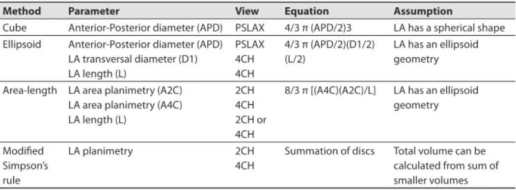
Table 1. Methods for left atrial volume quantifi cation with two-dimensional echocardiography
Method Parameter View Equation Assumption
Cube Anterior-Posterior diameter (APD) PSLAX 4/3 π (APD/2)3 LA has a spherical shape Ellipsoid Anterior-Posterior diameter (APD)
LA transversal diameter (D1) LA length (L)
PSLAX 4CH 4CH
4/3 π (APD/2)(D1/2) (L/2)
LA has an ellipsoid geometry Area-length LA area planimetry (A2C)
LA area planimetry (A4C) LA length (L)
2CH 4CH 2CH or 4CH
8/3 π [(A4C)(A2C)/L] LA has an ellipsoid geometry
Modifi ed Simpson’s rule
LA planimetry 2CH 4CH
Summation of discs Total volume can be calculated from sum of smaller volumes
34
three-dimensional echocardiography has been introduced (Figure 2). A number of studies have
demonstrated the feasibility of three-dimensional echocardiography for the assessment of LA
volumes (23,24), and it has been validated against magnetic resonance imaging (25). Jenkins
et al (23) have demonstrated that three-dimensional echocardiography allows accurate LA
volume assessment, with a low test-retest variation, and a lower intra-and inter-observer
vari-ability as compared to two-dimensional echocardiography. However, there still remain some
technical limitations such as the spatial and temporal resolution. In addition, since a relatively
constant RR interval is needed, three-dimensional echocardiography may not be feasible in
patients with AF and a high ventricular response rate.
Transesophageal echocardiography Transesophageal echocardiography (TEE) provides good views on the LA and the left atrial appendage (LAA). However, visualizing the complete left
atrium to determine LA size with TEE may be hampered by the close proximity of the probe
to the LA and the variable position of the esophagus to the posterior LA. As a result,
measure-ments of LA size with TEE have not been standardized. Only few studies have compared the
assessment of LA size with TEE and TTE. Block et al (26) assessed diff erent LA dimensions with
TEE and TTE in 109 patients. The authors noted that the 30- to 60-degree short-axis equivalent
at the level of the aortic valve was the only view in which the entire LA dimension could be
reliably obtained. Although TEE slightly underestimated LA size, it provided good correlation
with TTE (26).
TEE is considered the procedure of choice for assessment of thrombi in the LA cavity or LAA.
It can detect thrombi with a high degree of sensitivity and specifi city varying from 93 - 100%
(27). In addition, TEE is helpful in assessment of LAA emptying velocities, which are correlated
with thrombus formation (velocities <20 cm/s) and with maintenance of sinus rhythm after
cardioversion (velocities >40 cm/s) (28). Furthermore, TEE may be of great value in performing
transseptal punctures in AF ablation procedures.
Chapt
er 2
Imag
ing of the lef
t atrium
35
Intracardiac echocardiography Intracardiac echocardiography (ICE) is only used during inter-ventional procedures, such as percutaneous closure of atrial septal defects and catheter
abla-tion procedures. Therefore, no standardized measurements of LA size or volume are available.
During these interventional procedures, ICE can accurately visualize LA anatomy and related
structures (29). Furthermore, it allows visualization of intracardiac devices and catheters, and
it is helpful in monitoring potential complications during catheter ablation procedures (30).
Examples of intracardiac echocardiograms are shown in Figure 3.
In addition, the Doppler capacities of ICE allow for monitoring of pulmonary vein
narrow-ing and may predict the recurrence of AF after ablation (31). Furthermore, LA function can be
assessed with ICE. Rotter et al (32) demonstrated a good correlation between ICE and TEE for
measurement of mitral E wave velocity (correlation coeffi cient 0.759, mean diff erence 6.9 cm/s)
and LAA emptying velocity (correlation coeffi cient 0.991, mean diff erence 0.7 cm/s). Although
ICE is limited by the monoplane character and the lack of standardized measurements of LA
size, it is a valuable tool for interventional procedures.
Multi-slice computed tomography
The application of multi-slice computed tomography (MSCT) in cardiac imaging has rapidly
expanded in the past few years. Since MSCT has an excellent spatial and temporal resolution,
36
it can accurately quantify LA volumes, by using the modifi ed Simpson’s method (33). However,
because of the radiation exposure and the use of contrast agents, MSCT is not routinely used
for the assessment of LA size.
For AF ablation procedures, MSCT is a valuable tool to depict LA anatomy (34). With the use
of volume-rendered reconstructions, MSCT can provide detailed information on LA and
pulmo-nary vein anatomy (Figure 4). Since LA and pulmopulmo-nary vein anatomy is highly variable, MSCT
may off er a ‘road-map’ for ablation. The exact role of MSCT in ablation procedures is discussed
in one of the following paragraphs.
Magnetic resonance imaging
Magnetic resonance imaging (MRI) is considered the most accurate technique for the
non-invasive assessment of atrial volumes, because of the high spatial resolution and the excellent
myocardial border detection. Detailed information of LA size and volumes throughout the
cardiac cycle can be acquired with MRI (Figure 5). Anderson et al (35) recently reported fi ndings
on LA dimensions and LA area assessed with MRI in 20 healthy controls and in 20 patients with
cardiomyopathy. It was noted that a LA systolic area <24 cm2 was the upper 95th percentile of the normal range, and best discriminated normal from abnormal hearts (35). Similar to
MSCT, a modifi ed Simpson’s method can be used to determine LA volumes. However, due to
its relatively long acquisition times and the cumbersome data analysis, LA volume assessment
with MRI is not performed in daily clinical practice. MRI can provide detailed information on LA
and pulmonary vein anatomy before catheter ablation procedures, and is a useful tool in the
follow-up of patients after the ablation procedure. This will be discussed in more depth in one
of the following paragraphs.
Several studies have compared the value of the diff erent imaging modalities for the
assess-ment of LA size and volumes (23-26,36). Two-dimensional transthoracic echocardiography
Chapt
er 2
Imag
ing of the lef
t atrium
37
(using the biplane methods) may underestimate true LA size, as compared with computed
tomography (36) or magnetic resonance (25). However, these three-dimensional techniques
are not preferred for LA size assessment in daily clinical practice. In this respect, new
three-dimensional echocardiography is a promising technique that is widely available and provides
accurate information on LA size (24).
Figure 4. Volume-rendered three-dimensional reconstruction of a 64-slice MSCT scan. The dorsal view clearly demonstrates the anatomy of the LA and pulmonary veins. In this patient, normal pulmonary vein anatomy is present including four pulmonary veins, all with their own insertion into the LA. LIPV = left inferior pulmonary vein; LSPV = left superior pulmonary vein; RIPV = right inferior pulmonary vein; RSPV = right superior pulmonary vein.
38
ASSESSMENT OF REGIONAL LA FUNCTION
Regional LA function is not routinely assessed, and therefore no standardized parameters for
regional LA function are available. This can be partly explained by the fact that non-invasive
evaluation of regional LA function may be hampered by the relative thin LA walls. However,
assessment of regional LA function may provide more insight in atrial electromechanical
remodeling and may be helpful in the management of AF with surgical or catheter ablation.
New echocardiographic techniques, such as tissue Doppler imaging and strain (rate) imaging,
allow non-invasive measurement of regional function of the myocardium. Tissue Doppler
imag-ing quantifi es regional tissue velocities of the myocardium. Strain and strain rate represent local
tissue deformation and the rate (speed) of local deformation, respectively (37). Both techniques
have been well validated for the assessment of regional left ventricular function. Recently,
several studies (38-42) have applied these new techniques to the left atrium.
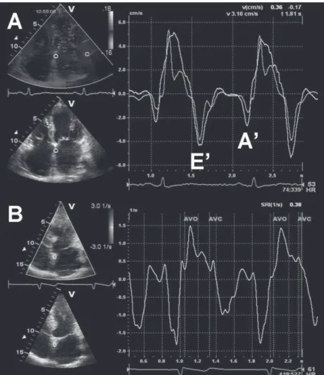
Tissue Doppler imaging allows quantifi cation of regional myocardial velocities, and
assess-ment of the timing of peak systolic and diastolic velocities of the myocardium (Figure 6, panel
A). Thomas et al (38) used tissue Doppler imaging in 92 healthy volunteers to evaluate regional
LA function. The authors noted that atrial contraction velocities were signifi cantly increased in
the annular segments, compared with the more superior segments.
Tissue Doppler imaging also provides information on the timing of regional velocities of
the myocardium. Therefore, it may quantify regional electromechanical LA function, such as the
total electromechanical activity of the atria (represented by the interval between the onset of
the P-wave on the ECG to the end of the A’ wave on the tissue Doppler images) (39). However,
the clinical relevance and the exact correlation of these new tissue Doppler derived parameters
of regional LA function with conventional parameters, such as mitral infl ow A wave velocity and
LA volumes, needs further investigation. Furthermore, a limitation of tissue Doppler imaging
for evaluation of regional LA function is the angle dependency of the technique. Therefore,
careful adjustment of the beam and gain settings should be made to avoid aliasing and to allow
reliable measurement of tissue velocities of the LA.
Strain imaging and strain rate imaging are new tools for the assessment of regional
myocar-dial deformation of the LA (40). An example of strain rate imaging of the LA is shown in Figure
6, panel B. In contrast to tissue Doppler imaging, strain imaging is not hampered by myocardial
tethering. Furthermore, strain imaging allows for diff erentiation between active contraction
and passive motion (37). However, the thin atrial walls may not generate clear strain curves
and therefore require careful interpretation. Several studies have demonstrated the value of
regional atrial strain in the analysis of patients with AF undergoing cardioversion (41,42). Di
Salvo et al (41) studied 65 patients with AF and performed tissue Doppler imaging of standard
apical images of the LA. It was noted that all tissue Doppler imaging derived parameters of the
LA, including tissue velocities, strain and strain rate, were signifi cantly reduced in patients with
Chapt
er 2
Imag
ing of the lef
t atrium
39
inferior wall peak systolic strain rate and atrial septal peak systolic strain were the best
predic-tors of maintenance of sinus rhythm after cardioversion (41). The assessment of regional LA
function by tissue Doppler imaging or strain imaging may be of value in the clinical follow-up
of patients with AF undergoing catheter ablation or cardioversion. It has been suggested that
diminished regional atrial strain values may warrant prolonged use of anti-arrhythmic drugs
and anti-coagulation (41,42). However, more studies are needed to appreciate the value of
regional left atrial strain and its role to guide use of medication in patients with AF.
40
IMAGING OF LA FUNCTION IN THERAPY FOR AF
As previously discussed, the association between LA remodeling and AF has been well
rec-ognized. Restoration of normal sinus rhythm by catheter ablation or cardioversion may result
in reverse remodeling of the LA, with subsequent improvement of LA function. However,
electrical or pharmacological cardioversion may cause transient atrial mechanical dysfunction
or ‘stunning’ (28,43). It has been demonstrated that conventional parameters of LA function,
such as A wave velocity or A wave velocity time integral, are decreased immediately after
cardioversion (43). The subsequent depressed LA appendage fl ow velocities increase the risk of
thromboembolic events after cardioversion.
With the use of new techniques such as strain imaging, LA dysfunction following
cardiover-sion can also be assessed (42). In 37 patients with chronic AF, it was noted that immediately after
cardioversion regional LA function was depressed compared with healthy controls. However,
6 months after successful cardioversion a signifi cant increase in LA strain was observed. The
maximal increase in regional LA strain occurred within 1 month after cardioversion (42). This
observation is in concordance with previous studies (43,44) and suggests that ‘atrial stunning’
following cardioversion is a function of the preceding AF, rather than the cardioversion itself.
Catheter ablation has been demonstrated to be successful in the restoration of sinus
rhythm, and is performed in an increasing number of patients with symptomatic
drug-refrac-tory AF. It has been demonstrated that maintenance of sinus rhythm after catheter ablation is
associated with a decrease in LA volumes (Figure 7) (45). Reant et al studied 48 patients with
lone AF treated with catheter ablation (46). Serial echocardiograms up to 12 months after the
procedure revealed a progressive decrease in LA dimensions. Interestingly, with the use of
new tissue Doppler derived parameters it was noted that in parallel to the improvement in LA
function, both LV systolic and diastolic function improved in the patients who maintained sinus
rhythm (46). Furthermore, with the use of MRI it has been demonstrated that in addition to
LA reverse remodeling, the area of the pulmonary venous ostia may decrease after successful
catheter ablation procedures (47).
IMAGING IN CATHETER ABLATION PROCEDURES FOR AF
Multimodality imaging
Catheter ablation procedures are being performed in an increasing number of patients
world-wide. The recent guidelines on management of patients with AF propose catheter ablation
as a reasonable option when fi rst-line anti-arrhythmic drugs have failed (14). Various ablation
strategies have been proposed, including segmental ostial ablation and anatomically based
circumferential ablation, and there is still debate concerning the exact lesion set. Regardless of
Chapt
er 2
Imag
ing of the lef
t atrium
41
essential during the ablation procedure. The veno-atrial junctions and anatomical landmarks
in the LA, such as the ridge between the left superior pulmonary vein and the LAA, are critical
structures to identify during catheter ablation procedures.
Anatomical studies have demonstrated that LA and pulmonary vein anatomy is highly
variable (15). Most frequently, two left-sided pulmonary veins and two right-sided pulmonary
veins drain separately into the LA (Figure 4). Anatomical variations include a single insertion
or ‘common ostium’ of the pulmonary veins, and an additional pulmonary vein (Figure 8). A
‘common ostium’ is most frequently found on the left-sided pulmonary veins, whereas an
addi-tional pulmonary vein is most frequently noted on the right side. In 201 patients undergoing
MSCT scanning, Marom et al (48) noted a left-sided ‘common ostium’ in 14% of the patients,
and an additional right-sided pulmonary vein in 28% of the patients. In addition, variations in
LAA morphology and LA roof anatomy may be present in patients with AF (49). Because of the
complex anatomy of the LA and the variability in pulmonary vein anatomy, a detailed ‘roadmap’
for the ablation procedure is mandatory. The various imaging modalities that are available for
assessment of LA and pulmonary vein anatomy in catheter ablation procedures include MSCT,
MRI, ICE and electroanatomical mapping systems.
MSCT and MRI (Figures 8 and 9) provide detailed information on the anatomy of the LA
and pulmonary veins. With the use of volume-rendered three-dimensional reconstructions and
cross-sectional images, the number of pulmonary veins and their branching pattern can be
accurately assessed (Figure 10). Furthermore, the diameters of the pulmonary vein ostia can
be measured on the diff erent orthogonal planes. In addition, MSCT may identify the presence
of thrombi in the LAA and provide detailed information on surrounding structures, such as the
42
esophagus and coronary arteries. However, the pre-procedural acquired MSCT and MRI images
only provide off -line information.
In contrast to MSCT and MRI, ICE allows real-time assessment of the pulmonary veins and
the veno-atrial junction during the ablation procedure. The Doppler capacities and the ability
to monitor the catheter position in relation to the pulmonary veins are great advantages of this
technique (29). In addition, ICE is helpful in assessment of the transmural extent of the ablation
lesions (50) and ICE has been used to titrate ablation energy, thereby increasing the safety of
the ablation procedure (30). Still, the major limitation of ICE is the mono-dimensional
char-acter of the technique. It has been demonstrated that three-dimensional imaging modalities
Figure 8. Variations in pulmonary vein (PV) anatomy shown on volume-rendered three-dimensional reconstructions of MSCT scans. Panel A demonstrates an additional PV on the right side. Panel B shows a ‘common’ ostium of the left-sided PVs.
Chapt
er 2
Imag
ing of the lef
t atrium
43
provide the most accurate information on LA and pulmonary vein anatomy (16,17). In a direct
comparison between MSCT and ICE (Figure 11), it was noted that MSCT has a higher sensitivity
for the detection of additional pulmonary veins and that ICE underestimated the size of the
pulmonary venous ostia (16).
During the ablation procedure, electroanatomical mapping systems such as CartoTM
(Biosense-Webster, Diamond Bar, California, USA) are available to assess LA and pulmonary
vein anatomy. These systems combine on-line electrophysiological data with anatomical
information, acquired with mapping catheters positioned in the LA (51). When performing the
actual ablation, the ablation points can be marked on the acquired electroanatomical map.
The major limitation of these systems is the use of reconstructed anatomy. Ideally, the detailed
anatomical information acquired with three-dimensional imaging techniques (such as MSCT
and MRI) could be combined with the on-line electrophysiological information. Recently, image
integration systems have been introduced that allow the on-line use of MSCT or MRI images
during the actual ablation procedures.