A ROLE FOR THE CRL4-WDTC1 E3 LIGASE IN THE SUPPRESSION OF ADIPOGENESIS
Beezly Sultana Groh
A dissertation submitted to the faculty of the University of North Carolina at Chapel Hill in partial fulfillment of the requirements for the degree of Doctor of Philosophy in the
Department of Biochemistry and Biophysics.
Chapel Hill 2013
Approved by: Yue Xiong
ABSTRACT
Beezly Sultana Groh: A Role For The CRL4-WDTC1 E3 Ligase In The Suppression Of Adipogenesis
(Under the direction of Yue Xiong)
Excess lipid accumulation in fat tissues underlies obesity and a myriad of associated pathologies that range from type 2 diabetes to cancer. Lipid synthesis in adipocytes, the primary cells of fat tissues, is regulated via extracellular and intracellular mechanisms.
WDTC1 encodes an evolutionarily conserved suppressor of lipid accumulation in
multicellular organisms. Decreased WDTC1 expression is associated with obesity in mice and humans. Yet, the molecular mechanism underlying its anti-obesity function remains elusive. WDTC1 is a candidate DWD protein (DDB1 binding WD40 repeat protein) that potentially functions as a substrate specificity factor for a cullin 4 E3 ligase (CRL4) complex. I hypothesized that WDTC1 mediates its anti-adipogenic effect through targeting substrates for ubiquitylation by the CRL4 complex. In this dissertation, I aim to understand the molecular function of WDTC1 in adipogenesis. I demonstrate that WDTC1 is indeed a CRL4 substrate receptor and its interaction with a CRL4 complex is central to its function. Using 3T3-L1 cell culture model of adipogenesis, I show that WDTC1 mutations disrupting its interaction with CRL4 impair the suppression of lipid accumulation and increase adipogenic gene expression. Rescue experiments demonstrate that the WDTC1 knockdown phenotypes can be rescued by ectopic expression of wild-type but not CRL4 binding mutants, underscoring the critical importance of the CRL4-WDTC1 interaction to the observed adipogenic suppression. In addition, I found that Cul4a knockout mice exhibit defects analogous to those reported in
Wdtc1+/- mice, such as adipocyte hypertrophy and poor metabolic parameters.
evidence that the CRL4WDTC1 complex promotes histone H2AK119 monoubiquitylation, an epigenetic modification that is associated with transcriptional silencing. Hence, I propose that CRL4WDTC1 E3 ligase may mediate its anti-adipogenic effect, at least in part, by repressing a subset of proadipogenic genes through histone H2AK119 monoubiquitylation. I also describe proteomic screens in 3T3-L1 cells to identify WDTC1 interacting proteins via mass
ACKNOWLEDGEMENTS
I would first like to thank my mentor, Dr.Yue Xiong, for his support and guidance throughout my dissertation research. I am thankful for the confidence he had in my scientific ability, which gave me this amazing opportunity to independently design and pursue my own research project. Most of all, I am thankful for all the ways he challenged me to meet my individual potential as a scientist. I also thank members of my committee Drs. Henrik Dohlman, Robert Duronio, Ben Major and Bill Marzluff, for their helpful suggestions,
encouragement and support over the years. I especially thank Dr. Bill Marzluff for supporting me from the beginning and meeting with me whenever research aims became intractable. Lastly, I thank past and present members of the Xiong laboratory. I had the rare opportunity to work with not only some very talented scientists, but also genuinely wonderful people.
TABLE OF CONTENTS
LIST OF TABLES... viii
LIST OF FIGURES... ix
LIST OF ABBREVIATIONS... x
CHAPTER 1: INTRODUCTION ... 1
Summary ... 1
Protein post translational modification with ubiquitin... 2
Enzymes of the ubiquitylation cascade ... 5
The cullin family of RING E3 ligases... 7
Regulation of CRL RING E3 ligases ... 10
CRL4 complexes ... 12
Chromatin related functions of CRL4 complexes ... 16
WDTC1 (Adipose), an anti-obesity factor and a putative CRL4 substrate receptor..19
Research summary... 21
Figures and tables ... 22
CHAPTER 2: BIOCHEMICAL ANALYSIS OF WDTC1 IN ADIPOGENESIS... 26
SUMMARY ... 26
BACKGROUND ... 27
RESULTS ... 28
WDTC1 is a substrate receptor of CRL4 E3 ubiquitin ligases... 28
The interaction of WDTC1 with CRL4 is critical for WDTC1-mediated adipogenic suppression ... 31
Cul4a-/-mice exhibit adipocyte hypertrophy and poor metabolic profiles ... 34
CRL4WDTC1 promotes histone H2AK119ub in a lineage specific manner ... 36
CRL4WDTC1-mediated H2AK119ub is linked to altered levels of H3K4me3 ... 39
DISCUSSION ... 40
EXPERIMENTAL PROCEDURES ... 43
FIGURES AND TABLES... 50
CHAPTER 3: PROTEOMIC SCREEN FOR CRL4WDTC1 SUBSTRATES... 66
SUMMARY ... 66
BACKGROUND ... 67
RESULTS ... 69
WDTC1 interacting proteins in preadipocyte and induced 3T3-L1 cells ... 69
Fatty acid synthase interacts with dominant negative WDTC1 but its protein level is not regulated by WDTC1... 72
Fatty acid synthase is not a CRL4WDTC1 substrate ... 73
DISCUSSION ... 75
EXPERIMENTAL PROCEDURES ... 79
FIGURES... 81
CHAPTER 4: CONCLUSIONS AND PERSPECTIVES... 89
Summary ... 89
Is WDTC1 the mammalian ortholog of yeast TUP1-SSN6 transcriptional repressor? ... 90
Regulation of adipogenesis through H2AK119ub ... 92
How many histone H2AK119ub E3 ligases are there? ... 95
Additional cellular substrates and functions of CRL4WDTC1 E3 ligase... 96
LIST OF TABLES
Table
LIST OF FIGURES
Figure
1.1 The ubiquitin conjugation system and the types of ubiquitin linkages ... 22
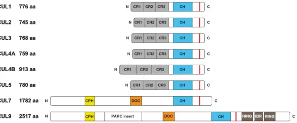
1.2 Schematic representation of cullin protein domain organization. ... 23
1.3 Composition of multisubunit CRL E3 ligase complexes. ... 24
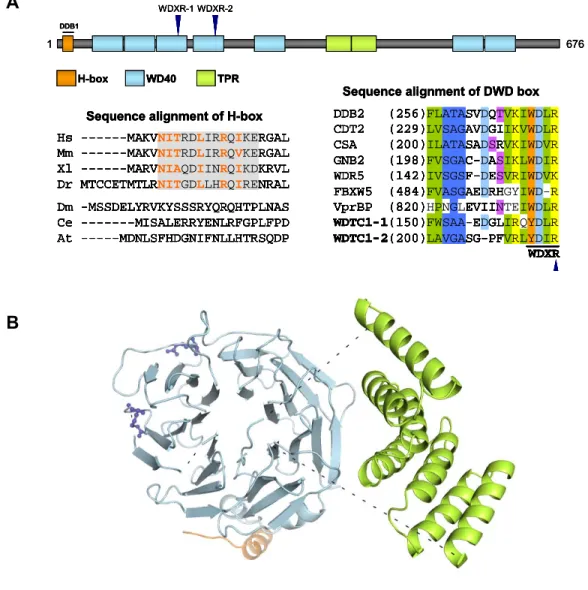
2.1 WDTC1 encodes a DWD protein with a novel structure. ... 50
2.2 WDTC1 is a substrate receptor of CRL4 complexes ... 51
2.3 WDTC1 suppresses adipogenesis in a CRL4-dependent manner... 53
2.4 CUL4A and CUL4B RNAi phenotype cannot be clearly assessed in 3T3-L1 differentiation ... 55
2.5 Wild-type WDTC1, but not CRL4 binding mutants, rescues the WDTC1 Knockdown phenotype...56
2.6 Cul4a-/-mice exhibit adipocyte hypertrophy and poor metabolic profiles... 57
2.7 WDTC1 binds histones and promotes histone H2AK119 monoubiquitylation... 58
2.8 CRL4WDTC1 catalyzes histone H2AK119 monoubiquitylation in vitro ... 60
2.9 CRL4WDTC1-dependent increase in H2AK119 monoubiquitylation is associated with reduced H3K4 trimethylation ... 61
2.10 A model of CRL4WDTC1-dependent transcriptional repression ... 62
2.11 Schematic summary of experimental procedures performed with 3T3-L1 cells...63
3.1 Proteomic analysis of WDTC1 interacting proteins in uninduced 3T3-L1 cells. ... 81
3.2 Proteomic analysis of WDTC1 interacting proteins in induced 3T3-L1 cells ... 82
3.3 Dominant negative WDTC1 binds FAS and WDTC1 expression alters FAS protein expression in adipogenically induced 3T3-L1 cells ... 83
3.4 FAS protein expression is transcriptionally regulated in 3T3-L1 cells ... 84
3.5 FAS is not a CRL4WDTC1 substrate...85
3.6 FAS polyubiquitylation is rapidly altered following 3T3-L1 induction... 87
LIST OF ABBREVIATIONS
CAND1 Cullin-associated Nedd8-dissociated 1
CDT Chromatin licensing and DNA replication factor
CRL Cullin RING E3 ligase
CSA Cockayne syndrome gene A
CSB Cockayne syndrome gene B
CSN COP9 signalosome
CUL Cullin
DCAF DDB1-CUL4 associated factors
DDB Damaged DNA Binding
DUB Deubiquitinating enzyme
DWD DDB1-binding WD40 protein
E1 Ubiquitin activating enzyme E2 Ubiquitin conjugating enzyme
E3 Ubiquitin ligase
EV Empty vector
FAS Fatty acid synthase
HA Hemagglutinin
HDAC Histone deacetylase
HECT Homologous to the E6AP Carboxyl Terminus IB Immunoblot
LC-MS Liquid chromatography–mass spectrometry MEF Mouse embryonic fibroblast
NEDD8 Neural precursor cell expressed, developmentally downregulated 8 NER Nucleotide excision repair
PCNA Proliferating Cell Nuclear Antigen
PRC Polycomb repressive complex
RING Really interesting new gene
RNAi Ribonucleic Acid (RNA) interference
ROC RING of cullins
SDS-PAGE Sodium dodecyl sulfate-polyacrylamide gel electrophoresis
shRNA Small hairpin RNA
siRNA Small interfering RNA
TPR Tetratricopeptide repeat
Ub Ubiquitin UBE1 Ubiquitin-activating enzyme 1 UBE2D3 Ubiquitin-conjugating enzyme E2 D3
WCE Whole cell extract
WD40 Tryptophan-aspartic acid domain-40 amino acid residues WDTC1 WD40 and tetratricopeptide repeats 1
CHAPTER 1:INTRODUCTION
Summary
Protein posttranslational modification constitutes a key regulatory mechanism to control the
function and composition of the proteome and thereby, underlies all biological processes.
Proteins can be modified by the addition of small chemical groups such as phosphate,
methyl and acetyl groups or by the addition of small proteins such as ubiquitin, SUMO1 and
NEDD8. Of particular interest is protein modification with ubiquitin, a highly conserved 76
amino acid (8.5 KDa) protein that is ubiquitously present in all eukaryotes but absent in
bacteria and archaea. Ubiquitin can be covalently attached to substrate protein or to another
ubiquitin through sequential enzymatic activities, a process termed ubiquitylation. Since its
discovery nearly 40 years ago in the covalent modification of histone H2A, ubiquitylation has
emerged as a widely utilized modification to alter properties of a protein, including stability,
activity, localization and interactions. As such, protein ubiquitylation has a critical role in
Protein post translational modification with ubiquitin
In mammals, ubiquitin is encoded by four different genes (UBB, UBC, UBA52 and
RPS27A). While UBB and UBC encode several copies of ubiquitin in a tandem configuration,
UBA52 and RPS27A encode a single copy of ubiquitin fused to ribosomal proteins L40 and
S27a, respectively (Varshavsky, 2006). Accordingly, ubiquitin is initially synthesized as an
inactive precursor. Specific isopeptidases called deubiquitinases (DUBs) cleave precursor
ubiquitin fusion proteins at their C-termini into conjugation competent monomeric units
(Jonnalagadda et al., 1989). Ubiquitin possesses two key functional features, a conserved
C-terminal diglycine motif (G75 and G76) and seven lysine residues (K6, K11, K27, K29, K33,
K48 and K63). The diglycine motif is required for ubiquitin conjugation since the carboxyl
group of G76 is the site of covalent conjugation to substrate lysine or lysine residue of
another ubiquitin (Pickart and Eddins, 2004). All seven internal lysine residues, as well as the
N-terminal methionine (M1), of the ubiquitin monomer are cellular substrates for conjugation
to the G76 carboxyl group of a donor ubiquitin, which yield ubiquitin polymers or polyubiquitin
chains (Kulathu and Komander, 2012).
Although many mechanistic details of ubiquitylation are still impending, much of the
identification and characterization of the enzymes involved in the catalysis of the chemical
steps were carried out by the Hershko laboratory in the early 1980s. Ubiquitylation proceeds
through a concerted enzymatic cascade comprising an E1 ubiquitin activating enzyme, an E2
ubiquitin conjugating enzyme, and an E3 ubiquitin ligating enzyme (Hershko, 1983; Pickart,
2004) (Figure 1.1A). E1 and E2 enzymes catalyze the ATP-dependent activation and
conjugation of ubiquitin, while E3s confer reaction specificity through substrate recruitment
and facilitate ubiquitin transfer by bridging the interaction between substrates and E2s.
Ubiquitylation is initiated by the activation of conjugation competent ubiquitin via a
two-step reaction catalyzed by the E1 activating enzyme (Kerscher et al., 2006; Pickart,
2001a). By coupling ATP hydrolysis, E1 first adenylates the C-terminal carboxyl group of
intermediate is subsequently the substrate for attack by the sulfhydryl group of the E1
catalytic cysteine, and thus resulting in the formation of a thioester linkage between E1 and
ubiquitin (E1-Ub). Formation of the E1-Ub complex induces structural changes that expose
cryptic E2 binding sites in E1, promoting its binding to an E2 conjugating enzyme (Ye and
Rape, 2009). In the second step of the ubiquitylation reaction, ubiquitin is transferred from
the E1-Ub to E2 through a transthiolation reaction involving the C-terminus of ubiquitin and
the E2 catalytic cysteine, resulting in the formation of the E2 and ubiquitin thioester complex
(E2-Ub). In the final enzymatic step of the ubiquitin conjugation reaction, charged E2-Ub can
directly transfer ubiquitin to a substrate protein, but more commonly, E2-Ub combines with
an E3 ligase which recruits a specific protein for ubiquitylation. Lastly, ubiquitin is conjugated
to a lysine residue in target protein or acceptor ubiquitin through the formation of an
isopeptide bond between the C-terminal glycine carboxyl group of ubiquitin and the target
lysine ε-amino group of substrate. Although noncanonical, the N-terminal α-NH2 group of the substrate can also be modified by ubiquitin, as well as serine and threonine hydroxyl and
cysteine thiol groups of target proteins (McDowell and Philpott, 2013).
Substrates can be monoubiquitylated through the conjugation of a single ubiquitin
monomer, mulitiply monoubiquitylated, or polyubiquitylated through repeated reactions that
generate polymeric ubiquitin chains of distinct or mixed linkages (Komander and Rape, 2012;
Ye and Rape, 2009) (Figure 1.1B). Polyubiquitylation generally targets proteins for
proteolytic degradation via the 26S proteasome, while monoubiquitylation is linked to
nonproteolytic functions ranging from protein trafficking to chromatin regulation (Hicke, 2001;
Pickart, 2001b). Modification of substrate lysine without further modification to ubiquitin itself,
referred to as the specificity of the monoubiquitylation reaction, is thought to be determined
by a number of factors including structural constraints imposed by a particular substrate-E3
complex or specificity might be encoded by the distinct E2 or E3 pairings (Komander and
transcription factor, for example. Polyubiquitylation can either proceed through either
homotypic or heterotypic linkages (Kulathu and Komander, 2012). Polyubiquitin chains are
homotypic when the donor ubiquitin is successively conjugated to the same residue in each
acceptor ubiquitin (M1, K6, K11, K27, K29, K33, K48 or K63) during elongation. By contrast,
polyubiquitin chains are referred to as heterotypic (branched and nonbranched) when
assembled by mixed linkages or an array of atypical linkages. Although K48 and K63
linkages remain best characterized, all possible linkage types have been detected in
mammalian cells and presently, their regulated assembly and biological significance is a
subject of intensive research.
Ubiquitylated substrates are recognized by a large number of proteins that contain
ubiquitin-binding domains (UBDs), which bind discreet interaction surfaces on ubiquitin
(Husnjak and Dikic, 2012; Komander and Rape, 2012). UBDs may be broadly classified into
α-helix based, zinc-finger based, plekstrin homology-like and ubiquitin conjugating (UBC)-like domains. Many UBDs preferentially bind a specific type of ubiquitin conjugate and can thus
“decode” information encoded by differentially ubiquitylated substrates into the appropriate
cellular response—the ubiquitin signal encoded by monoubiquitin versus polyubiquitin chains
of specific and mixed linkages, for example. Similar to the reversal of phosphorylation by
protein phosphatases, a family of DUBs (~95 in humans) cleaves conjugated ubiquitin from
substrates, thus rendering ubiquitylation a highly dynamic and reversible posttranslational
modification (Kulathu and Komander, 2012). Collectively, the type of ubiquitin modification,
the ubiquitin binding proteins that decode the signal carried by a distinct conjugate and the
DUBs that dynamically regulate the ubiquitylated proteome constitute the “ubiquitin code”
and determine the functional outcome of substrate ubiquitylation (Husnjak and Dikic, 2012;
Enzymes of the ubiquitylation cascade
The ubiquitin conjugation system is hierarchically organized with the mammalian
genome encoding 2 E1s, 28 active E2s and a large array of E3 ligases that is estimated to
exceed 600 (Deshaies and Joazeiro, 2009; Groettrup et al., 2008; Pickart, 2001a). This
organizational hierarchy is predicted to increase specificity through additional regulation and
generate functional diversity in the ubiquitin system. Examples illustrating this include the
exquisite linkage specificity achieved by several E2 enzymes through differential positioning
of ubiquitin in their active sites or when the specific E2-E3 interactions dictate substrate
monoubiquitylation versus polyubiquitylation (Komander and Rape, 2012).
The canonical cascade for protein ubiquitylation involves ubiquitin activation by the
E1 UBE1. However, the recent identification of a second E1 enzyme UBE1l2 (Chiu et al.,
2007; Jin et al., 2007; Pelzer et al., 2007), a dual system of ubiquitin activation, adds an
unexpected level of regulation in the ubiquitin pathway. The two E1s exhibit different
specificities towards E2s, partly owing to subtle differences in their ubiquitin fold domains
which recruit E2s. While all E2s except USE1 can be charged by UBE1, UBE1l2 charges a
small cohort of E2s but specifically charges USE1 with ubiquitin. Consistent with the notion
that UBE1l2 promotes a discreet subset of ubiquitylation reactions, UBE1l2 mice are
embryonic lethal likely due to altered neuronal development (Chiu et al., 2007; Lee et al.,
2013).
The 28 E2 ubiquitin conjugating enzymes (UBE2s) can be grouped into
monoubiquitylating, chain initiating or chain elongating E2s (Kulathu and Komander, 2012). A
common feature shared by all active E2s is a core ubiquitin-conjugating domain (UBC)
comprising ~150 residues, which binds E1s and includes the catalytic cysteine as well as a
conserved asparagine residue at the active site. Although E2 enzymes are critical to
ubiquitylation reactions, their substrate affinity in the absence of an E3 is too low for efficient
physiological E2-E3 pairs have been characterized (such as APC/C and UBE2S) (Ye and
Rape, 2009). While some E2s dictate structure of the ubiquitin modification (mono- vs
polyubiquitylation) and confer linkage specificity to polyubiquitin chains, substrate specificity
is largely dictated by the E3 ligase. Nevertheless, E2s add functional versatility to the
ubiquitin system by modulating the type of ubiquitin modification, and therefore the biological
outcome of substrate ubiquitylation.
The large number of E3 ligases is thought to reflect the specific targeting of an even
larger number of cellular proteins by the ubiquitin system (Pickart, 2001a). In addition to
conferring substrate specificity, E3s generally serve a scaffolding role to bridge the
interaction between a substrate lysine and the E2-Ub intermediate to facilitate efficient
ubiquitylation. As such, a shared property of all E3 ligases is distinct substrate and E2
binding domains, which may be included in a single protein or comprise distinct subunits.
There are two mechanistically distinct classes of E3 ligases: HECT (homology to E6AP
C-terminus) domain and RING (really interesting new gene) domain families (Figure 1.1A). To
note, there are at least two other subfamilies of E3s: the U-box (UFD2 homology) which is
structurally similar to RING E3s and the recently classified RBR (RING-in-between-RING)
E3s which are RING/HECT hybrids (Kulathu and Komander, 2012).
Named after the founding member E6AP protein, the mammalian genome encodes
~30 HECT domain E3 ligases (Metzger et al., 2012). Substrate ubiquitylation by HECT E3s
follows a covalent mechanism. Their C-termini contain the conserved ~40 kDa (~350 amino
acids) HECT domain, while variable N-termini mediate substrate recognition. The HECT
domain has a bilobal architecture consisting of an E2 interacting N-lobe and a C-lobe that
harbors the active site cysteine. Ubiquitylation proceeds through an obligate transfer of
ubiquitin from E2-Ub to the catalytic cysteine of HECT E3 by a transthiolation reaction.
Following the formation of the HECT E3-Ub thioester intermediate, ubiquitin is directly
and among their diverse functions, HECT E3s regulate the trafficking of many receptors,
transporters and channels (Rotin and Kumar, 2009).
The RING family comprises the vast majority of E3 ligases with the mammalian
genome potentially encoding over 600 RING proteins (Deshaies and Joazeiro, 2009). Unlike
HECT E3s, RING E3s do not transfer ubiquitin directly to substrate. Instead, RING E3s
minimally function in “catalysis by proximity” by simultaneously binding the targeted substrate
and the E2-Ub conjugate (Pickart and Eddins, 2004). The catalytic step involves a
nucleophilic attack by the substrate lysine ε-amino group on the E2-Ub reactive thioester bond, resulting in isopeptide bond formation between substrate and ubiquitin. In addition to
its scaffolding role, RING E3s may induce subtle conformational changes in E2s to stabilize
the transition state intermediate (an oxyanion) of the ubiquitin conjugation reaction (Deshaies
and Joazeiro, 2009; Ye and Rape, 2009). Structurally, RING E3s share a domain (~70 amino
acids) of distinctively spaced histidine and cysteine residues which coordinate two zinc
cations, forming a ‘cross-brace’ structure termed the RING domain. RING E3s either
possess an intrinsic RING domain or contain a separate RING domain subunit. The RING
domain binds the E2-Ub conjugate while a separate domain or subunit mediates specific
recruitment of diverse substrates. As such, RING E3s can function as monomers or in the
context of multisubunit complexes. The cullin family of RING E3 ligases typify multisubunit
assemblies and is the subject of remaining discussion on E3 ligases.
The cullin family of RING E3 ligases
The evolutionarily conserved cullin RING ligases (CRLs) represent the largest known
family of E3 ligases (Deshaies and Joazeiro, 2009; Petroski and Deshaies, 2005; Sarikas et
al., 2011). The CRL enzymatic core contains a cullin protein which functions as a molecular
scaffold to assemble a substrate targeting module and a ubiquitin conjugation module into a
Mammals encode six canonical (CUL1, CUL2, CUL3, CUL4A, CUL4B and CUL5) and three
distantly related (CUL7, CUL9 and APC2) cullin proteins. With the exception of CUL7 and
CUL9—which evolved more recently in the common chordate ancestor, cullin orthologs are
present in Drosophila melanogaster (5), Caenorhabditis elegans (6), Arabidopsis thaliana (5)
and yeast (3). Structurally, cullins are characterized by three key features: a highly
conserved C-terminal cullin-homology domain, an N-terminal domain containing a series of
cullin repeats along with divergent sequences and a conserved lysine residue proximal to the
cullin-homology domain (Figure 1.2). The following discussion will focus on the canonical
cullins.
Although cullins do not possess an intrinsic RING domain, the CRL catalytic core
includes ROC1 (for RING of cullins; also known as RBX1/HRT1), a zinc-binding RING-H2
domain subunit (Kamura et al., 1999b; Ohta et al., 1999; Seol et al., 1999; Tan et al., 1999).
A notable exception is the CUL5-based complexes which preferentially include the related
RING subunit ROC2 (discussion will refer to ROC1 for clarity). A highly conserved C-terminal
globular domain in cullins harbors the cullin homology domain (~180 amino acids). At this
domain, cullins bind ROC1 through an interlocking mechanism that tightly integrates the
RING domain (Sarikas et al., 2011). ROC1 in turn recruits E2-Ub to form the active CRL
conjugation apparatus. This modular assembly of a catalytic core is one of two defining
structural features of CRL complexes, the substrate targeting module being the other.
On the basis of the crystal structures of CRL1 and CRL4 complexes (Angers et al.,
2006; Zheng et al., 2002b), cullins have a arched but rigid stalk-like N-terminal domain that
consists of three helical cullin repeats (CR1-CR3). This rigid architecture presumably
positions the ROC1 bound E2-Ub at a proper distance from the substrate to promote efficient
ubiquitin transfer. Indeed, inserting a linker in CUL1 to render flexibility to its N-terminal
domain abolished CRL1 activity in vitro (Zheng et al., 2002b). Despite the anticipated
structural conservation among cullin proteins, the very N-terminal sequences in each cullin is
cullins proteins—they each assemble distinct substrate targeting modules at their N-termini
in a combinatorial manner to recruit structurally diverse substrates to a common catalytic
core. The substrate targeting module comprises an interchangeable substrate receptor that
binds substrate and thus dictates the substrate specificity of its cognate CRL complex and in
most cases, a distinct adaptor protein that links the substrate receptor to the core complex.
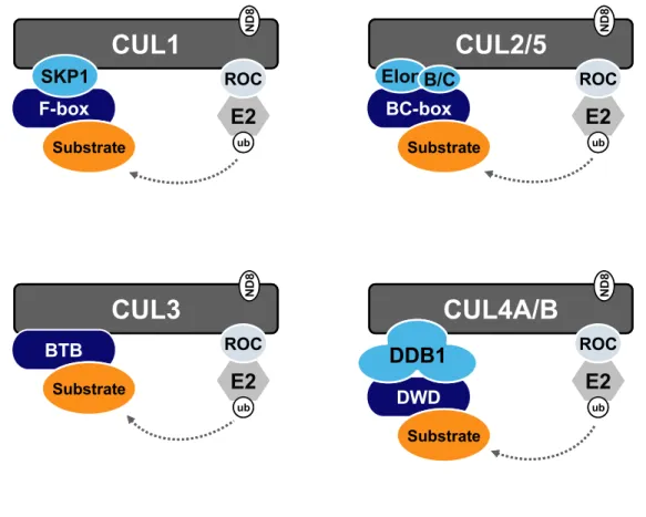
The multisubunit organization of the CRL complexes is shown schematically in Figure 1.3.
CUL1 is the founding member of the CRL E3 ligase family. Independent genetic
studies on cell cycle control in C. elegans and S. cerevisiae led to the identification of CUL1
and the cullin gene family (Kipreos et al., 1996; Mathias et al., 1996), and set the stage for
the subsequent discovery of the CUL1-based CRL1 complexes (also known as SCF
complexes) (Feldman et al., 1997; Skowyra et al., 1997). CRL1 complexes are composed of
four subunits: CUL1 scaffold, ROC1 RING subunit, SKP1 adaptor and F-box containing
proteins as variable substrate receptors. The F-box domain (~40 amino acid motif named
after cyclin F where it was first identified) in the substrate receptor mediates binding to SKP1,
which in turn links it to the CRL1 catalytic core (Bai et al., 1996). The human genome
encodes 69 F-box proteins while the number of F-box proteins in other organisms ranges
from 20 in S. cerevisiae to an astonishing 700+ in A. thaliana (Gagne et al., 2002; Skaar et
al., 2009). The different combinations of F-box proteins generate a multitude of distinct CRL1
complexes and thereby greatly expand the diversity in substrate targeting by the CRL1 E3
ligase. This general principle applies to other CRLs (discussed below).
CUL2- and CUL5-based CRLs both utilize Elongin C as the adaptor, which binds the
two cullins as a heterodimeric complex with Elongin B (Kamura et al., 2004). Although they
share the same adaptor, CUL2 and CUL5 each assemble distinct complexes by interacting
with two separate classes of substrate receptors. Elongin C binds proteins with two similar
motifs: von Hippel-Lindau (VHL)-box and suppressor of cytokine-signaling (SOCS)-box. Yet,
et al., 2011). Examples of differential substrate recruitment by VHL-box and SOCS-box
proteins includes proteolytic targeting of hydroxylated HIF-1α transcription factor by CRL2VHL complex (Ivan et al., 2001) and of antiviral protein APOBEC3G by the virally subverted
CRL5VIF complex (Yu et al., 2003). CUL3-based CRL complexes uniquely omit an adaptor
protein, instead binding directly to a class of substrate receptors through their Broad complex,
Tramtrack, Bric-a-brac (BTB) domains (Furukawa et al., 2003; Geyer et al., 2003; Pintard et
al., 2003; Xu et al., 2003). Although this direct binding may implicate that nearly all BTB
proteins potentially assemble into CRL3 complexes, as is the apparent case for all three S.
cerevisiae BTB proteins, this has yet to be experimentally verified for the large number of
BTB proteins present in higher organisms (humans encode over 400) (Pintard et al., 2004).
CUL4-based CRLs employ a similar multisubunit assembly with the specific adaptor DDB1
and a distinct class of DDB1-binding substrate receptors (CRL4 complexes are discussed in
detail in separate sections). Given their relatively recent discovery and the extraordinary
number of diverse complexes that cullins potentially assemble in the cell (estimated 300-500
distinct CRLs), future research should be able to better define the functional repertoire of
CRL E3 ligases.
Regulation of CRL RING E3 ligases
Despite the diversity in subunit composition and cellular function, the assembly and
ubiquitin ligase activity of CRL complexes are similarly regulated by a regulatory cycle
involving several mechanisms. All cullins are covalently modified by the ubiquitin-like protein
NEDD8 (~60% identity with ubiquitin) at a conserved lysine residue in their C-termini,
proximal to the cullin homology domain (Hori et al., 1999). The NEDD8 conjugation (termed
neddylation) drives the formation of active CRL E3 complexes and is essential for their
function in mammalian cells (Bosu and Kipreos, 2008). Underlying the activating function,
neddylation induces a conformation change in cullins such that it optimally reorients the
facilitating ubiquitin transfer across a ~50 Å gap (Duda et al., 2008; Saha and Deshaies,
2008). Indeed, cullins appear to be the primary substrates of the NEDD8 conjugation
pathway. Similar to ubiquitylation, neddylation comprises an enzymatic cascade which
includes the activating E1 enzyme NAE (NAE1/UBA3 heterodimer) and the
NEDD8-conjugating E2 enzymes UBE2M/UBE2F. Although not essential for in vivo neddylation
reactions, DCN1 interacts with cullin bound ROC1 and possibly functions as a NEDD8 E3
ligase (Kamura et al., 1999a; Kurz et al., 2008; Yang et al., 2007). Demonstrating the
importance of neddylation to CRL4 activity, a small molecule inhibitor of NAE, MLN4924,
was recently developed to disrupt CRL function in human tumor cells and is currently in
clinical trials (Soucy et al., 2009).
Analogous to deubiquitylation, cullin neddylation is reversed by the multisubunit
COP9 signalosome (CSN) complex (termed deneddylation), thus converting active CRLs into
inactive complexes (Lyapina et al., 2001). The CSN5 subunit encodes a critical
metalloprotease active site for the isopeptidase activity towards neddylated cullins (Cope et
al., 2002). Another key player in the regulation of CRL complexes is CAND1 (or TIP120A),
which interacts with unneddylated cullins and in a mutually exclusive manner with cullin
substrate receptors (Hwang et al., 2003; Liu et al., 2002; Min et al., 2003; Oshikawa et al.,
2003; Zheng et al., 2002a). Mechanistically, CAND1 was proposed to inactivate cullins
through a sequestration based mechanism. Indeed, the crystal structure of
CAND1-CUL1-ROC1 complex shows that CAND1 forms an inhibitory complex with CUL1 that
simultaneously masks the neddylation site and the adaptor binding site (Goldenberg et al.,
2004). In this regulatory cycle, cullin neddylation displaces bound CAND1 and prevents
re-association, thus enabling CRL activation and substrate recruitment by associated substrate
receptors.
While biochemical and structural studies demonstrated that CSN and CAND1
phenocopies the loss of cullin function in multiple organisms and despite the increase in
neddylated cullins, CSN inactivation impairs the activity of CUL1, CUL3 and CUL4-based
complexes (Bosu and Kipreos, 2008; Petroski and Deshaies, 2005). Similarly, CAND1
inactivation in Arabidopsis and human cells suggested it is a positive regulator of cullin
function. To reconcile these disparate findings, a number of models have been proposed
(Bosu and Kipreos, 2008). First, a cycle of neddylation/deneddylation prevents proteolytic
degradation of substrate receptors, which are frequently autoubiquitylated by their cognate
CRL after ubiquitylation of target substrate is completed. In this view, sustained neddylation
of cullins may promote ‘instability’ of select substrate receptors and thereby limit their
availability. Consistent with this model, depletion of CSN5 resulted in both a failure to
deneddylate CUL1 and the accumulation of CRL1 substrates, presumably from decreased
stability of substrate receptors (Cope and Deshaies, 2006). Second, assembly of functionally
distinct CRL complexes must require regulated substrate receptor exchange. A recent study
suggests CAND1 facilitates assembly of new and rare CRL1 complexes by promoting the
dissociation of pre-existing or high affinity F-box substrate receptors in exchange for new or
less abundant F-box proteins (Pierce et al., 2013). It should be noted that an alternative
model favors that substrate receptor abundance drives neddylation and the formation of
active CRL complexes, and suggests neddylation-CSN-CAND1 cycle accounts for a minor
fraction of CRL regulation (Bennett et al., 2010). Clearly, multiple layers of regulation are
present to control the assembly of distinct CRL complexes to dynamically respond to cellular
conditions.
CRL4 complexes
The core CRL4 complex comprises either CUL4A or CUL4B as the scaffold
(collectively referred to as CUL4), ROC1 as the RING subunit in trans and DDB1 (DNA
damage binding 1) as the CUL4 specific adaptor. DDB1 is evolutionarily conserved from
with DDB1 orthologs, such as A. thaliana and S. pombe. DDB1 was first characterized as a
DNA damage sensor that functions in a heterodimeric complex with DDB2 to recognize
UV-induced DNA lesions in the nucleotide excision repair (NER) pathway (Chu and Chang,
1988). A decade later, in an attempt to characterize DDB1-DDB2 interacting proteins, DDB1
was found to interact with CUL4A (Shiyanov et al., 1999). The pleiotropic effects uncovered
by genetic studies of DDB1 mutants in model organisms implicated that DDB1 is a
multifunctional protein with roles beyond DNA damage repair (Jackson and Xiong, 2009).
Finally, the presence of DDB1 in several distinct CUL4 complexes including those containing
DDB2 and CSA established a role for DDB1 as the adaptor for CUL4 E3 ligases (Groisman
et al., 2003; Hu et al., 2004; Sugasawa et al., 2005; Wertz et al., 2004), and thus accounted
for its pleiotropic effects.
Structurally distinct, DDB1 is a large protein (127 kDa) containing three β-propeller domains (BPA-BPC; spanning 100 Å) compared with the small size and structural complexity
of all other cullin adaptors (<20 kDa) (Li et al., 2006). Additionally, cullin adaptors (SKP1,
Elongin C or BTB substrate receptors) share sequence homology with each other and share
a common structural element termed the SKP1/BTB/POZ fold to interact with their cognate
cullin N-terminus (Sarikas et al., 2011). DDB1 does not contain this recognition fold. The
flexibly linked BPB domain of DDB1 binds the N-terminus of CUL4, while the BPA-BPC
domains form a rigid double propeller fold (resembling a clam shell) and was initially believed
to function in substrate presentation (Li et al., 2006). The structural complexity and variation
in cullin-adaptor interaction seen with CUL4-DDB1 possibly reflect the requirement for
multiple interaction surfaces to accommodate structurally diverse receptors, or to bind
cofactors for efficient CRL4 activity, or both.
Despite DDB1 firmly established as the adaptor for CRL4 complexes, substrate
recruitment mechanism for CUL4-based complexes remained elusive. It was reasonably
DDB2 and CSA, assembled into identical CRL4 complexes strongly hinted at this possibly
(Groisman et al., 2003). Further supporting this proposition were the findings that XPC and
CSB were substrates of CRL4DDB2 and CRL4CSB complexes, respectively (Groisman et al.,
2006; Sugasawa et al., 2005). A pattern emerged that suggested the involvement of a
shared structural motif when a third WD40 protein, CDT2, was reported to target replication
licensing factor CDT1 for CRL4-mediated proteolysis (Higa et al., 2006a; Ralph et al., 2006;
Sansam et al., 2006). Ultimately, combined proteomic, structural and biochemical
approaches to systematically search for specificity factors led to the identification of a diverse
family of WD40-repeat proteins as putative substrate receptors of CRL4 complexes (Angers
et al., 2006; He et al., 2006; Higa et al., 2006b; Jin et al., 2006). These four independent
studies collectively identified 52 DDB1 binding WD40 proteins (referred to as DWDs
hereafter; also referred elsewhere as DCAFs for DDB1-CUL4 associated factors or CWDs
for CUL4-DDB1 associated WDRs).
WD40 repeats are structural motifs of ~40 amino acids that are characterized by the
frequent occurrence of a glycine-histidine (GH) dipeptide and a tryptophan-aspartic acid
(WD) dipeptide at the end of the repeat. WD40 domain proteins typically contain several
tandem or intervening WD40 repeats that often fold into a circular β-propeller structure. Search for a signature motif that defines DWDs led to the identification of a submotif within
WD40 repeats, termed “WDXR”; comprises the WD dipeptide followed an X (any residue)-
arginine/lysine dipeptide. More extensive sequence analyses yielded the “DWD-box”, a 16
residue stretch terminating in the WDXR motif (He et al., 2006). Most DWD proteins contain
at least one, frequently two tandem and rarely, three DWD boxes. A disease derived
mutation in the WDXR of DDB2 (R273H) impairs its DDB1 binding (Rapic-Otrin et al., 2003;
Shiyanov et al., 1999), underscoring the importance of this arginine in WDXR. Surprisingly,
the crystal structure of the D. rerio DDB1-DDB2 complex revealed that the corresponding
residue (R309) did not directly contact DDB1 but it may contribute to DNA binding and
the interaction between many DWD proteins and DDB1, not all DWDs contain a WDXR motif
(Jin et al., 2006). Moreover, mutational analysis revealed that while many DWDs interact with
DDB1 though its BPC domain, different BPC surfaces are utilized (Jin et al., 2006). These
observations highlight the possibly that multiple sequence and structural elements determine
the association between DWDs and DDB1. Indeed, the H-box, a recently discovered
structural motif found in seven DWD proteins, is likely to be important for their association
with DDB1 but this has yet to be experimentally tested for all H-box containing DWDs (Li et
al., 2010).
Results from the four studies clearly demonstrated that only a subset of the ~300
WD40 proteins encoded by the human genome interacts with DDB1. Database searches of
the DWD box motif estimated ~90 unique DWD proteins are present in human cells (He et al.,
2006). Besides WD40 domain, the array of presumptive functional domains in DWDs,
ranging from bromodomain to tetratricopeptide, predicts the versatility of the CRL4 core
complex. The interaction between DDB1 and the 52 DWDs has been confirmed, but primarily
through transient overexpression followed by immunoprecipitation experiments. Importantly,
the functional interaction between CRL4 and DWDs remains unexplored for the vast majority
of them. As such, a key question regards whether all or most DWDs function as substrate
receptors for CRL4 complexes to specify ubiquitylation of distinct substrates. Systematic
validation of the putative substrate receptors will certainly require linking specific CRL4
complexes to biological processes and identifying the physiological substrates through
combined genetic, proteomic and biochemical approaches. Nevertheless, given the
extraordinary number of distinct CRL4 complexes that may be assembled in the cell, it is
reasonable to anticipate that these DWDs potentially extend the functional range of the
CRL4 catalytic core. Elucidating the molecular function of one DWD protein, WDTC1 (also
Chromatin related functions of CRL4 complexes
Besides the well established role for DDB1 in DNA repair, early genetic analysis of
CUL4 deletion mutants in several organisms overwhelmingly implicated a role for CUL4 in
chromatin regulation (Jackson and Xiong, 2009). Deleting cul4 (or pcu4) in S. pombe
resulted in a viable but slow growth phenotype and two obvious phenotypes: abnormally
elongated cells and decondensed chromatin (Osaka et al., 2000). The later discoveries that
S. pombe cul4 interacts with Clr4 histone H3K9 methyltransferase to maintain
heterochromatin and is required for S-phase destruction of Cdt1 at least partially accounts
for the decondensed chromatin and growth retarded phenotypes of Δcul4 cells, respectively
(Horn et al., 2005; Jia et al., 2005; Ralph et al., 2006). RNAi-mediated inactivation of cul4 in
C. elegans causes growth arrest at the L2 larval stage and severe polyploidy arising from
massive re-replication of the genome (Zhong et al., 2003). A failure to effect S
phase-coupled degradation of CDT1 (replication licensing factor) explained the re-replication in cul4
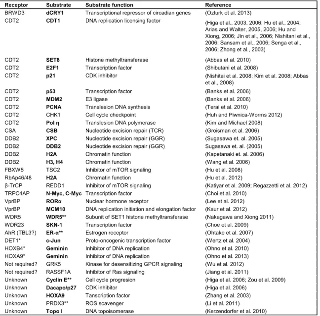
RNAi cells. Known CRL4 substrates are listed and those related to chromatin based
processes are indicated in bold (Table 1.1). The following discussion with focus on reports
that directly link CUL4 to chromatin related functions: DNA damage repair, replication and
transcriptional regulation.
CRL4 in the maintenance of genome integrity
At least two substrate receptors of CRL4 complexes are mutated in human
hereditary diseases: DDB2 in xeroderma pigmentosum E group (XPE) and CSA in Cockayne
syndrome, both diseases are associated with a defect in NER. In response to UV-induced
helix-distorting DNA damage, DDB2 and CSA each function in separate NER pathways,
global genome repair (GGR) and transcription coupled repair (TCR), respectively. In GGR,
CRL4DDB2 is rapidly recruited to site of damage following UV. In turn, DDB2 recruits repair
factor XPC to chromatin where both are polyubiquitylated by CRL4, but undergo different
(and presumably CRL4 as well), displaces it from chromatin and targets it for degradation
(El-Mahdy et al., 2006). In contrast, XPC ubiquitylation increases its DNA binding affinity and
is protected from degradation to carry out repair. Additionally, CRL4DDB2 also have reported
roles in histone H2A, H3 and H4 monoubiquitylation following UV irradiation to facilitate GGR,
possibly through recruitment of repair factors (Kapetanaki et al., 2006; Wang et al., 2006). In
TCR, CRL4CSA complex is recruited to chromatin through its interaction with CSB, but in a
CSN bound inactive state (Groisman et al., 2006). The precise mechanism and order of
events are still unclear but CSB is thought to link the DNA damage signal, marked by stalled
RNAPII, to the recruitment of NER repair factors. Although it is unknown how CRL4CSA is
activated following repair, CSB degradation is critical for restoring transcription post-TCR.
To maintain genome integrity, DNA replication must be restricted to only once per
cell cycle. Of the checkpoint mechanisms evolutionarily conserved from S. pombe to humans,
includes CRL4CDT2-mediated degradation of CDT1 after S phase initiation to prevent
re-replication and in response to DNA damage to prevent re-replication before damage repair. An
interesting requirement of the CRL4CDT2-mediated degradation of CDT1 is that only PCNA
bound CDT1 is ubiquitylated (Arias and Walter, 2006; Jin et al., 2006). Through mutational
analysis, sequences in CDT1 that specify its interaction with PCNA was identified and
termed the “PIP-box” (for PCNA-interacting motif). Similar to CDT2 depletion, mutating the
PIP-box results in a loss of CRL4CDT2 chromatin recruitment, CDT1 stabilization and
re-replication, indicating that the PCNA-CDT1 interaction is critical for CRL4CDT2 recruitment to
chromatin (Arias and Walter, 2006). Following this discovery, the PIP-box was demonstrated
to be critically important for targeted degradation of other CRL4CDT2 substrates including p21
CDK inhibitor, E2F1 transcription factor and SET8 histone methyltransferase (Abbas et al.,
2010; Nishitani et al., 2008; Shibutani et al., 2008). As no other CRL4 complexes have been
reported to require PCNA, this feature may be unique to CRL4CDT2 substrates. It remains to
CRL4 complexes in transcriptional regulation
Apart from directly targeting transcriptional regulators, recent reports indicate a role
for CRL4 complexes in the direct regulation of chromatin dynamics—histone lysine
methylation, in particular. Early studies in S. pombe showed that Cul4 recruits histone H3K9
methyltransferase Clr4 to maintain heterochromatin silencing (Horn et al., 2005; Jia et al.,
2005). Although Cul4 ubiquitin ligase activity is evidently important for H3K9 methylation as
neddylation defective Cul4 mutant cannot restore heterochromatin silencing, the activity
linking Cul4 and Clr4 remains unknown. In mammalian cells, at least three DWD proteins
have established roles in histone methylation: WDR5 and RBBP5 are core components of
the MLL histone H3K4 methyltransferase complex (activates transcription) and EED is a
component of EZH2 containing PRC2 H3K27 methyltransferase complex (silences
transcription). One study reported that WDR5/RBBP5 and EED are functionally linked to
CRL4 as depletion of DDB1 or CUL4A leads to a dramatic loss of H3K4 and H3K27
methylation (Higa et al., 2006b). However, the idea that CRL4-WDR5functionally interact to
promote H3K4 methylation is challenged by the finding that WDR5 itself is the ubiquitylation
target of CRL4B and that rather than decreasing, the loss of CRL4B increases H3K4
trimethylation (H3K4me3) with the concomitant activation of gene transcription (Nakagawa
and Xiong, 2011).
A recent study finally shed some light on a probable link between CRL4 activity and
transcriptional regulation through direct alteration to chromatin structure. Histone H2AK119
and H2BK120 are the nucleosomal substrates of ubiquitylation, however, while H2AK119ub
is associated with gene silencing, H2BK120ub activates transcription and is a prerequisite
modification for H3K4me3 (Hammond-Martel et al., 2012). Polycomb complex PRC1 was
once believed to be the sole E3 ligase for H2AK119; however, recent findings challenge this
premise. Indeed, CRL4BRBPB4/7 directly catalyzes H2AK119ub to silence transcription of a set
of genes, including the tumor suppressor genes PTEN and CDKN2A (p16) (Hu et al., 2012).
recruitment and H2K27me3 enrichment, revealing a crosstalk mechanism to tightly enforce
gene silencing. In this example, the RBPB4/7 DWD protein is speculated to recruit CRL4B to
chromatin and functions in the downstream recruitment of PRC2, thus connecting two
distinct enzymatic activities on chromatin.
WDTC1 (Adipose), an anti-obesity factor and a putative CRL4 substrate receptor
More than half a century ago, Dr. Winifred W. Doane hypothesized that
environmental conditions with marked nutrient shortage may select for mutations that
enhance fat storage capacity. The rationale being that although deleterious under normal
conditions, such alleles could provide a survival advantage in nutrient deprived conditions.
During course of her doctoral dissertation work at Yale University, she isolated such a
mutation in a wild D. melanogaster population from Sub-Saharan Kaduna, Nigeria. The most
obvious phenotype she observed was massive hypertrophy of the fat body (fly fat organ)
accompanied by “enormous oil globules”, and thus named the mutant “adipose” (adp)
(Doane, 1960a). Not surprisingly, adp mutants exhibit a selective advantage in starvation
survival tests (Doane, 1960b).
About forty years after characterizing the adp mutant phenotype, Dr. Doane
collaborated with research groups in Germany to identify the adp gene (Hader et al., 2003).
Comparative sequence analysis of open reading frames (ORFs) was performed between
mutant and wild-type sequences within a 70 kb candidate region. Compared to the wild-type,
a frameshift-causing 23 bp deletion was identified in one single ORF, the candidate adp
gene. This deletion is expected to cause premature termination of the Adp protein; induces a
stop codon one amino acid following the frameshift. Transgenic expression of the candidate
wild-type adp ORF in mutant files completely rescued the mutant phonotype and firmly
established the identify of the adp gene. The adp gene is evolutionarily conserved from
Chapter 2). The fly Adp protein shares 37% sequence identity to human Adp but functional
conservation was unknown.
To elucidate whether the anti-obesity function of adp is conserved in vertebrates, the
Graff laboratory generated transgenic mice to study the loss and gain of function phenotypes
of the mammalian adp homolog (referred to as Wdtc1 hereafter) (Suh et al., 2007). Because
Wdtc1-/- mice are partially embryonic lethal and enough mice could not be generated, the
authors evaluated Wdtc1 heterozygous mice. The loss of a single Wdtc1 allele yielded a
clear obese phenotype, indicating evolutionary conservation of WDTC1 function and dose
sensitivity. The Wdtc1-/+ mice were obese based on appearance, total body weight and fat
content, histological assessment of adipocyte size and plasma analysis of metabolites. Quite
strikingly, transgenic overexpression of Wdtc1 in mature adipocytes produced mice that were
leaner and showed improved metabolic profiles compared to their wild-type littermates. The
3T3-L1 cell culture adipogenic model was then utilized to explore potential molecular function
of WDTC1. WDTC1 overexpression also inhibited 3T3-L1 adipogenesis. Additionally, nuclear
exclusion of WDTC1, by attaching a nuclear export signal, mimicked the WDTC1 RNAi
phenotype, suggesting WDTC1 may have a nuclear function. Following this lead, WDTC1
was shown to bind histones and HDAC3 histone acetyltransferase in coimmunoprecipitation
experiments. Suh and colleagues speculated that WDTC1 may function in transcriptional
regulation through regulating chromatin structure. The molecular mechanism underlying the
anti-obesity function of WDTC1 remains unknown.
Of the four studies reporting the discovery of DWD proteins, the human WDTC1
protein (676 amino acids) was first reported to interact with CRL4 components in two
separate studies (Angers et al., 2006; Jin et al., 2006) and predicted in another through
sequence analyses with the DWD box (He et al., 2006). In the Angers et al. study, tandem
affinity purification followed by mass spectrometry identified WDTC1 in DDB1 and CUL4A
complexes. The reciprocal analysis of WDTC1 complexes confirmed the association,
vast majority of DWD proteins, the functional interaction between CRL4 and WDTC1 remain
uncharacterized.
Research summary
The main goal of my dissertation research was to study the molecular mechanism underlying
the anti-adipogenic function of WDTC1. In Chapter 2 (modified from a first author manuscript
currently in revision), I characterized the functional interaction between CRL4 and WDTC1
and I elucidated a potential mechanism, histone H2AK119 monoubiquitylation, by which
CRL4WDTC1 complex suppresses adipogenesis. In Chapter 3, I describe the data obtained
from screening for WDTC1 interacting proteins via mass spectrometry and include
characterization of a putative CRL4WDTC1 substrate, fatty acid synthase. Finally, in Chapter 4,
I provide perspective on the outstanding questions in WDTC1 research and discuss future
A
B
FIGURES AND TABLES
Figure 1.1. The ubiquitin conjugation system and the types of ubiquitin linkages.
Figure and modified figure legend from Husnjak and Dikic, 2012.
(A) The E1-E2-E3 enzymatic cascade is required for ubiquitin conjugation to target protein (ubiquitylation). The catalytic activities of HECT and RING E3 ligases, and deubiquitinases (DUBs; enzymes that reverse ubiquitylation) are shown schematically. Substrates can be modified by a single ubiquitin monomer (monoubiquitylation) or multiple ubiquitin monomers (multi-monoubiquitylation) or by the sequential addition of multiple ubiquitin monomers to one of eight residues (M1, K6, K11, K27, K29, K33, K48, or K63) of a previously conjugated ubiquitin (polyubiquitylation).
CUL1
CUL2
CUL3
CUL4A
CUL4B
CUL5
CUL7
CUL9 776 aa
745 aa
768 aa
759 aa
913 aa
780 aa
1782 aa
2517 aa CUL1
CUL2
CUL3
CUL4A
CUL4B
CUL5
CUL7
CUL9 CUL1
CUL2
CUL3
CUL4A
CUL4B
CUL5
CUL7
CUL9 776 aa
745 aa
768 aa
759 aa
913 aa
780 aa
1782 aa
2517 aa 776 aa
745 aa
768 aa
759 aa
913 aa
780 aa
1782 aa
2517 aa
Figure 1.2. Schematic representation of cullin protein domain organization.
Figure and modified figure legend from Sarikas et al., 2011.
The N-terminal Cullin repeat 1 (CR1) binds a specific adaptor and the C-terminal cullin homology domain (CH) binds the RING subunit ROC1/2. The neddylation site is indicated by the red vertical line; CUL7 and CUL9 neddylation site has not been experimentally
CUL1
E2
ub ROC ND 8 F-box Substrate SKP1CUL2/5
E2
ub ROC ND 8 BC-box Substrate ElonB/CCUL4A/B
E2
ub ROC ND 8 DWD SubstrateDDB1
CUL3
E2
ub ROC ND 8 BTB SubstrateCUL1
E2
ub ROC ND 8 F-box Substrate Substrate SKP1CUL2/5
E2
ub ROC ND 8 BC-box Substrate Substrate ElonB/CCUL4A/B
E2
ub ROC ND 8 DWD Substrate SubstrateDDB1
DDB1
CUL3
E2
ub ROC ND 8 BTB Substrate SubstrateFigure 1.3. Composition of multisubunit CRL E3 ligase complexes.
Receptor Substrate Substrate function Reference BRWD3 dCRY1 Transcriptional repressor of circadian genes (Ozturk et al. 2013)
CDT2 CDT1 DNA replication licensing factor
CDT2 SET8 Histone methyltransferase (Abbas et al. 2010)
CDT2 E2F1 Transcription factor (Shibutani et al. 2008)
CDT2 p21 CDK inhibitor
CDT2 p53 Transcription factor (Banks et al. 2006)
CDT2 MDM2 E3 ligase (Banks et al. 2006)
CDT2 PCNA Translesion DNA synthesis (Terai et al. 2010)
CDT2 CHK1 Cell cycle checkpoint (Huh and Piwnica-Worms 2012)
CDT2 Pol η Translesion DNA polymerase (Kim and Michael 2008)
CSA CSB Nucleotide excision repair (TCR) (Groisman et al. 2006)
DDB2 XPC Nucleotide excision repair (GGR) (Sugasawa et. al. 2005)
DDB2 DDB2 Nucleotide excision repair (GGR) Sugasawa et. al. (2005)
DDB2 H2A Chromatin function (Kapetanaki et. al. 2006)
DDB2 H3, H4 Chromatin function (Wang et al. 2006)
FBXW5 TSC2 Inhibitor of mTOR signaling (Hu et al. 2008)
RbAp46/48 H2A Chromatin function (Hu et al. 2012)
β-TrCP REDD1 Inhibitor of mTOR signaling (Katiyar et al. 2009; Regazzetti et al. 2012)
TRPC4AP N-Myc, C-Myc Transcription factor (Choi et al. 2010)
VprBP RORα Nuclear hormone receptor (Lee et al. 2012)
VprBP MCM10 DNA replication initiation and elongation factor (Kaur et al. 2012)
WDR5 WDR5** Subunit of SET1 histone methyltransferase (Nakagawa and Xiong 2011)
WDR23 SKN-1 Transcription factor (Choe et al. 2009)
AhR (TBL3?) ER-α** Estrogen receptor (Ohtake et al. 2007)
DET1* c-Jun Proto-oncogenic transcription factor (Wertz et al. 2004)
HOXB4* Geminin Inhibitor of DNA replication (Ohno et al. 2010)
HOXA9* Geminin Inhibitor of DNA replication (Ohno et al. 2013)
Not required? GRK5 Kinase for desensitizing GPCR signaling (Wu et al. 2012)
Not required? RASSF1A Inhibitor of Ras signaling (Jiang et al. 2011)
Unknown Cyclin E** Cell cycle progresion (Higa et al. 2006; Zou et al. 2009)
Unknown Dacapo/p27 CDK inhibitor (Higa et al. 2006)
Unknown HOXA9 Tanscription factor (Zhang et al. 2003)
Unknown PRDX3** ROS scavenger (Li et al. 2011)
Unknown Topo I DNA topoisomerase (Kerzendorfer et al. 2010)
Proteins in bold indicate chromatin related substrates *Not a WD40 domain protein
**CRL4B specific
(Nishitai et al. 2008; Kim et al. 2008; Abbas et al., 2008)
(Higa et al., 2003, 2006; Hu et al., 2004; Arias and Walter, 2005, 2006; Hu and Xiong, 2006; Jin et al., 2006; Nishitani et al., 2006; Sansam et al., 2006; Senga et al., 2006; Zhong et al., 2003)
Table 1.1. Substrates of CRL4 E3 ligase complexes.
CHAPTER 2: BIOCHEMICAL ANALYSIS OF WDTC1 IN ADIPOGENESIS
SUMMARY
WDTC1, an anti-adipogenic gene product and a putative substrate receptor of a cullin 4
RING E3 ligase (CRL4), suppresses adipogenesis by an unknown mechanism. I
hypothesized that the anti-adipogenic function of WDTC1 is mediated through CRL4 activity.
In this study, I characterized the interaction between WDTC1 and CRL4, and delineated the
molecular function of WDTC1 using 3T3-L1 cell culture model of adipogenesis. I
demonstrated that WDTC1 mutations that disrupt DDB1 binding mimic the loss of function
defects observed in WDTC1 knockdown cells, impaired suppression of triglyceride
accumulation and increased adipogenic gene expression. Rescue experiments showed that
WDTC1 RNAi defects can be restored by wild-type WDTC1 but not CRL4 binding mutants,
confirming that the CRL4 interaction is critical for WDTC1 function. Furthermore, I found that
Cul4a knockout mice exhibit adipocyte hypertrophy and metabolic defects, these phenotypes
are analogous to Wdtc1 heterozygous mice. Mechanistically, CRL4WDTC1 complex promotes
H2AK119 monoubiquitylation, an epigenetic modification linked to transcriptional repression.
Collectively, the results in this chapter identify CRL4WDTC1 E3 ligase as a suppressor of
adipogenesis and implicate a role for this complex in transcriptional repression during
BACKGROUND
Over 50 years ago, Dr. Winifred Doane isolated and extensively characterized a
naturally-derived D. melanogaster mutant, which she termed adipose (adp) (Doane, 1960a;
Doane, 1960b). The most obvious mutant phenotype observed was hypertrophy of the fly fat
organ due to excessive lipid storage (Doane, 1960a). Recently, Doane and colleagues
identified the fly adp gene, which is evolutionarily conserved from files to humans as a single
copy gene (Hader et al., 2003). The mammalian adp ortholog is WD40 and tetratricopeptide
repeats 1 (WDTC1), encoding a protein that contains WD40 repeat and TPR repeat domains.
Specifically, the fat suppressive function of Wdtc1 is evolutionarily conserved in mammals
(Suh et al., 2007). Loss of a single Wdtc1 allele results in obese mice with poor metabolic
parameters, and conversely, transgenic Wdtc1 expression in fat cells yields skinnier mice
(Suh et al., 2007). Further, population studies have recently linked intronic WDTC1 single
nucleotide polymorphic gene variants (Lai et al., 2009) and reduced WDTC1 gene
expression (Galgani et al., 2013) to human obesity. Despite the strong genetic evidence
linking WDTC1 to anti-adipogenic function, its molecular function remains elusive.
In eukaryotic cells, covalent attachment of the 76 amino acid protein ubiquitin to
substrate proteins, known as ubiquitylation, has a critical role in virtually all cellular
processes. Perturbations in this system have been linked to diseases ranging from cancers
to neurodegeneration (Petroski, 2008). Proteins can be polyubiquitylated, which typically
targets it for proteolytic degradation, or monoubiquitylated, which regulates the property and
thus the function of the protein (Hicke, 2001; Pickart, 2001b). Ubiquitylation proceeds via an
enzymatic cascade where E1 and E2 enzymes catalyze the activation and conjugation of
ubiquitin, while E3s confer reaction specificity through substrate recruitment (Hershko, 1983;
Pickart, 2004). Belonging to the largest family of E3 ligases—the cullins, cullin 4-RING
ubiquitin ligases (CRL4s) are a large subclass of multisubunit E3 enzymes. The core
2009). Substrate targeting to the catalytic core typically requires the interaction between
DDB1 and substrate receptors. As recently elucidated, a subset of DDB1 binding WD40
repeat (DWD) proteins likely function as substrate receptors for CRL4 complexes (Angers et
al., 2006; He et al., 2006; Higa et al., 2006b; Jin et al., 2006). The human genome encodes
~90 DWD proteins (He et al., 2006), but their function remain vastly unexplored. CRL4 E3
ligases are strongly linked to chromatin-related processes through ubiquitylation of histones
and factors controlling DNA repair, replication and transcription protein (Jackson and Xiong,
2009; O'Connell and Harper, 2007).
Indeed, WDTC1 (DCAF9) is a DWD protein and as such, a putative substrate
receptor of CRL4 E3 ligases (Angers et al., 2006; He et al., 2006; Jin et al., 2006). However,
the in vivo interaction between WDTC1 and CRL4 has not yet been explored. I hypothesized
that WDTC1 functions as a substrate receptor of the CRL4 E3 ligase to mediate its
anti-adipogenic activity. The aim of this chapter is to examine the functional significance of the
WDTC1 and CRL4 interaction, and elucidate the molecular mechanism underlying the
anti-adipogenic function of WDTC1. I demonstrated that WDTC1 functions as part of a CRL4
complex to suppress adipogenesis. I also uncovered a function of the CRL4WDTC1 E3
complexin promoting histone H2AK119 monoubiquitylation, a modification associated with
transcriptional repression and thus a potential mechanism by which WDTC1 suppresses
adipogenesis.
RESULTS
WDTC1 is a substrate receptor of CRL4 E3 ubiquitin ligases
To begin to characterize the interaction between WDTC1 and CRL4, I compared a
few key structural and sequence elements. WDTC1 encodes a protein with a novel structure,
and its linear domain organization is predicted to be conserved from mammals to
Arabidopsis (Figure 2.1A, top). The modeled human WDTC1 structure depicts the unique
repeats which are spatially separated by unstructured regions (Figure 2.1B). Of note, the
structural basis of the interaction between WDTC1 and DDB1 is predicted to include a short
N-terminal α-helical motif termed the H-box (Li et al., 2010). Although not strictly conserved at the primary sequence level, the H-box is a shared structural feature among a few DWD
proteins, and surprisingly, several viral proteins that hijack the CRL4 complex. The alignment
of H-box sequences revealed substantial sequence homology among vertebrates, consistent
with their shared mode of DDB1 interaction (Figure 2.1A, bottom left). Additionally, the
signature motif present in nearly all DWD proteins is the DWD box, a highly conserved 16
residue sequence positioned within WD40 repeats (He et al., 2006). Similar to most DWD
proteins, WDTC1 contains two tandem DWD boxes with conserved WDXR submotifs (Figure
2.1A, bottom right). Interestingly, although the H-box of the DWD protein DDB2 makes a
large contribution to DDB1 binding (Jin et al., 2006; Li et al., 2010), the importance of the
arginine in the WDXR of DDB2 is indicated by its mutation (R273H) in some human XPE
patients and its loss of DDB1 binding (Rapic-Otrin et al., 2003; Shiyanov et al., 1999). In fact,
this arginine in WDXR submotifs is critically important for the interaction between many DWD
proteins and DDB1 (Higa and Zhang, 2007). These observations therefore underscore the
presence of multiple binding determinants for the functional interaction between DWD
proteins and CRL4 complexes.
To experimentally confirm the prediction that WDTC1 and CRL4 interact in vivo,
endogenous CUL4A or CUL4B complexes were immunoprecipitated from HEK293T cells,
and the presence of WDTC1 in the immunoprecipitates was determined by immunoblot
analysis. I found that WDTC1 forms both CUL4A and CUL4B endogenous complexes
(Figure 2.2A). While in vitro binding assays suggested that the H-box of WDTC1 is the key
determinant of binding to DDB1 (Li et al., 2010), this has not been experimentally verified in
cells. Additionally, in line with the aim to characterize WDTC1 molecular function, I sought to
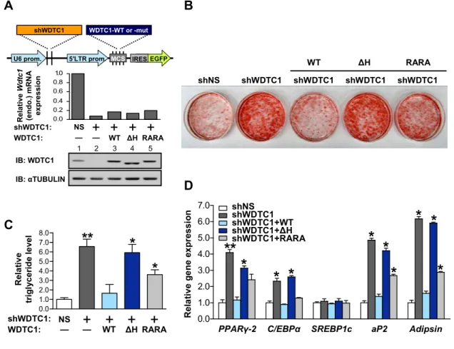
residues spanning the H-box (referred to as ΔH) and the other substituted the arginine residues in the tandem WDXR motifs to alanines (referred to as RARA). I first confirmed their
expression and determined that their relative stability is similar by cycloheximide chase
(Figure 2.2B). I then examined their effect on binding CRL4 components by
coimmunoprecipitation assays.
As expected, wild-type (WT) Flag-tagged WDTC1 (Flag-WDTC1-WT)
coimmunoprecipitated all CRL4 proteins tested (Figure 2.2C, lane 6). Consistent with the
reported importance of the H-box, its deletion in WDTC1-ΔH completely ablated complex formation (lane 7). Interestingly, WDTC1-RARA mutant showed a marginal decrease in
DDB1 binding but its interaction with CUL4 and ROC1 was markedly reduced (lane 8),
indicating that the WDXR motifs in WDTC1 are required for forming stable CRL4 complexes.
A superimposed structure of WDTC1 is shown in complex with CRL4 (Figure 2.2D). WDTC1
is anchored through its H-box into the DDB1 BPC domain. As in the case of DDB2-DDB1
binding (Fischer et al., 2011), the tandem WDXR residues of WDTC1 are not predicted to
make direct contact with DDB1 but are solvent-exposed on the bottom surface of the
propeller fold.
Besides DDB2 (Nag et al., 2001; Sugasawa et al., 2005), the Xiong laboratory
recently reported the second example of a DWD protein, WDR5, that is ubiquitylated by its
cognate CRL4B complex and targeted for proteolysis (Nakagawa and Xiong, 2011). I
therefore tested whether WDTC1 is a CRL4 substrate by an in vivo ubiquitylation assay. I
found that although WDTC1 was extensively ubiquitylated, its ubiquitylation status was
unchanged by either DDB1 knockdown or ΔH mutation that disrupts DDB1 binding (Figure 2.2E). Finally, the steady state levels of transiently expressed Flag-WDTC1 protein are
largely unaltered by knockdown of DDB1 or either CUL4A or B (Figure 2.2F), indicating that
CRL4 does not regulate WDTC1 protein stability. Although DDB1 depletion resulted in a
slight decrease in WDTC1 protein (lane 2), this is possibly due to instability arising from the
component of CRL4 complexes and likely functions as a substrate receptor of CRL4 E3
ligases.
The interaction of WDTC1 with CRL4 is critical for WDTC1-mediated adipogenic suppression
To determine the biological significance of the CRL4-WDTC1 interaction, I
hypothesized that the fat suppressive role of WDTC1 is mediated through CRL4. I first
confirmed expression of CRL4 proteins in adipose tissues out of a panel of adult mouse
tissues (Figure 2.3A). In addition to adipose tissues, WDTC1 protein appears to be broadly
expressed, in agreement with a previous report on Wdtc1 mRNA expression pattern (Suh et
al., 2007). To test my hypothesis, I assayed the adipogenic differentiation of 3T3-L1
preadipocytes, the best characterized and most extensively used cell culture model to study
adipogenesis. Originally derived from mouse embryonic fibroblasts (Green and Meuth, 1974),
3T3-L1 adipogenesis is thought to closely recapitulate adipogenesis in mice. Treating 3T3-L1
cells to an adipogenic media triggers transcriptional activation of the terminal differentiation
program and morphological changes following lipogenic accumulation of triglycerides
(MacDougald and Lane, 1995).
I first confirmed the endogenous interaction between WDTC1 and CRL4 subunits
during the course of 3T3-L1 differentiation, which I monitored by phenotypic changes (data
not shown) and Fatty Acid Synthase (FAS) protein induction. WDTC1 was
immunoprecipitated from cells collected at specific time points and the presence of CRL4
proteins in the immunoprecipitates was determined by immunoblot analysis (Figure 2.3B).
Although anti-WDTC1 antibody shows low immunoprecipitation efficiency, the results show
that WDTC1 forms both CUL4A and CUL4B endogenous complexes. The levels of CRL4
proteins remained relatively constant during differentiation (Figure 2.3B, input lanes),
consistent with the notion that CRL4 has diverse roles in the process of 3T3-L1 proliferation,