The Varicocele: Clinical Presentation, Evaluation,
and Surgical Management
Jason R. Lomboy, MD
1Robert M. Coward, MD
1,21Department of Urology, University of North Carolina School of
Medicine, Chapel Hill, North Carolina
2UNC Fertility, Raleigh, North Carolina
Semin Intervent Radiol 2016;33:163–169
Address for correspondence Jason R. Lomboy, MD, Department of
Urology, University of North Carolina School of Medicine, 2113 Physician’s Office Building, CB 7235, 170 Manning Drive, Chapel Hill, NC 27599-7235 (e-mail: [email protected]).
Objectives: Upon completion of this article, the reader will be able to identify the diagnosis and interventions for varico-celes, including what is known of its clinical significance, the diagnosis and necessary workup, as well as the different surgical options for treatment.
Accreditation: This activity has been planned and im-plemented in accordance with the Essential Areas and Policies of the Accreditation Council for Continuing Medical Education (ACCME) through the joint providership of Tufts University School of Medicine (TUSM) and Thieme Medical Publishers, New York. TUSM is accredited by the ACCME to provide continuing medical education for physicians.
Credit: Tufts University School of Medicine designates this journal-based CME activity for a maximum of 1 AMA PRA Category 1 Credit™. Physicians should claim only the credit commensurate with the extent of their participation in the activity.
A varicocele is an abnormal dilatation and tortuosity of the veins of the spermatic cord. It is a common condition among men of all ages, affecting approximately 15% of the male population.1In addition, as the most common cor-rectable cause of male infertility, it affects 19 to 41% of men with primary infertility and 45 to 81% of men with second-ary infertility.2The etiology and pathophysiology of vari-coceles remain incompletely understood with only a few understudied theories. In addition to affecting semen parameters contributing to male factor infertility, histori-cally varicoceles have also been treated for testicular pain and testicular atrophy. Although evidence has shown an association between varicocele repair and improvement in semen parameters, there is some controversy on patient selection and method of repair. In addition, there continues to be controversy on the indications and timing for treat-ment of a varicocele in an adolescent. When considering treatment for a patient with a varicocele, consultation by a
Keywords
►
varicocele
►
varicocelectomy
►
infertility
►
interventional
radiology
Abstract
A varicocele is an abnormal dilatation and tortuosity of the veins of the spermatic cord.
Although varicoceles are common in the general population and are frequently found on
routine physical examinations, they represent the most common correctable cause of
male factor infertility. Varicoceles are also often incidental
fi
ndings on imaging studies,
particularly scrotal ultrasound. Importantly, not all varicoceles should be treated equally
(or at all), and basic guidelines on the evaluation and indications for treatment of adult
varicoceles should be reviewed before counseling and treatment. A semen analysis
should be obtained for any male patient of reproductive age considering intervention.
The adolescent varicocele is managed much differently than the adult varicocele and
remains a source of controversy. This review describes the clinical presentation and the
evaluation of adult and pediatric varicoceles, and provides guidance on their diagnosis
and workup. It also describes options for surgical repair and the success and
complica-tion rates associated with each surgical approach, ultimately supporting microsurgical
subinguinal varicocele repair as the current surgical standard.
Issue Theme Men’s Health;
Guest Editor, Charles Burke, MD
Copyright © 2016 by Thieme Medical Publishers, Inc., 333 Seventh Avenue, New York, NY 10001, USA.
Tel: +1(212) 584-4662.
DOI http://dx.doi.org/ 10.1055/s-0036-1586143. ISSN 0739-9529.
urologist, particularly a fellowship-trained urologist in male infertility when fertility is of concern, is indicated. It is imperative that providers diagnosing and treating varicoceles understand their presentation and diagnostic criteria, the necessary evaluation, the indications and op-tions for repair, and the appropriate follow-up and/or observation of patients both with treated and untreated varicoceles. Treating providers should continue to look for future studies, as more valuable, prospective data are needed to better understand the controversies surrounding varicocele management.
Presentation
Although some men will present with scrotal discomfort, varicoceles are typically asymptomatic. Adult men with varicoceles typically are diagnosed during evaluation of male factor infertility, while adolescent varicoceles are usually discovered incidentally on physical examination. As part of the initial evaluation, a complete reproductive and sexual history should be obtained. Physical examina-tion is the standard diagnostic test for varicoceles. To encourage relaxation of the cremasteric and dartos muscle
fibers to facilitate inspection and palpation, the examina-tion should be performed in a warm room. The examinaexamina-tion is performed by inspection and palpation of the patient’s scrotum in the standing position while in the relaxed position and while Valsalva is induced. Varicocele grading is based on the ability to visualize and/or palpate the varicocele in both the relaxed state and while inducing Valsalva. Grade I varicoceles are palpable only with Val-salva, grade II varicoceles are palpable without ValVal-salva, and grade III varicoceles are readily visible through the scrotal skin (►Table 1). ►Fig. 1provides an example of a grade III varicocele. Subclinical varicoceles are not visible or palpable and are typically diagnosed incidentally with ultrasonography. The majority of varicoceles are left sided due to the drainage of the left spermatic vein into the higher resistance left renal vein compared with the vena cava for drainage of the right spermatic vein. Right-sided varico-celes are usually discovered when bilateral varicovarico-celes are present. However, when isolated or irreducible in the supine position, right-sided varicoceles warrant further investigation into underlying retroperitoneal pathology.3
Use of Adjunctive Diagnostic Testing
Scrotal ultrasound, although very sensitive (97%) and specific (94%),4should not be routinely ordered simply for confi rma-tion of clinically palpable varicoceles or to look for subclinical varicoceles. Ultrasound can, however, be helpful when the physical examination is difficult or indeterminate, such as in situations where a patient is obese, has had prior scrotal surgery, has a small scrotum, or has thick scrotal skin. Criteria for determination of the presence of a varicocele by ultraso-nography include dilation of spermatic veins with demon-stration of reversal of flow with color Doppler. Some commonly used cutoffs between normal and abnormal veins are 2 to 3 mm in diameter, although these may vary. Dilation of veins without demonstrated reversal of flow on color Doppler does not represent a varicocele, as in the example of a patient with a surgically repaired varicocele who may have permanently dilated veins. Although there is no con-sensus on this use for ultrasound, several studies have sought to correlate internal spermatic vein diameter measurements with the presence of a clinical varicocele. Pilatz et al deter-mined cutoff measurements of vein diameter to detect pal-pable varicoceles in 217 men to be a diameter greater than 2.45 mm at rest (sensitivity 84% and specificity 81%) and 2.95 mm (sensitivity 84% and specificity 84%) during Val-salva.5However, known ultrasound operator bias and lack of well-studied standardized criteria for assessment with ultra-sound limit the ability to reliably correlate diameter measure-ments with clinically relevant varicoceles that would benefit from treatment.6
In addition to ultrasonography, other diagnostic measures—
such as thermography, radionuclide scanning, and spermatic
Table 1 Classification of varicoceles
Grade Examination
Subclinical Not visible, not palpable
Grade I Palpable varicocele detected upon Valsalva maneuver, not visible
Grade II Palpable varicocele detected while standing up, not visible
Grade III Large visible varicocele while standing up
Source: Adapted from Dubin and Amelar.47
Fig. 1 Grade III varicocele. Note the dilated and tortuous veins seen on the scrotum (arrow).
venography—should also not be routinely used for the detection of subclinical varicocele in a patient without a palpable varico-cele on physical examination.7If used at all, these studies could be helpful in patients with a recurrent varicocele.
In addition to the impact of varicocele on fertility, evidence also suggests varicoceles impair testicular Leydig cell function with a downstream effect on testosterone production. Several of these studies have demonstrated significant testosterone level improvements in patients with hypogonadism after repair of a clinical varicocele.8–10 As part of the evaluation of a varicocele, hormone laboratory testing should be offered to the patient to help characterize any degree of androgen deficiency as well as screen for other potential endocrine causes for infertility. These laboratory tests include total and free testosterone levels, luteinizing and follicle-stimulating hormones, prolactin level, and estrogen (E2) level. Although a patient with clinical varicocele may exhibit laboratory results consistent with hypergonadotropic hypogonadism, it is im-portant to consider other causes for infertility based on these results.
Indications for Treatment
The American Society for Reproductive Medicine (ASRM) Practice Committee guideline indicates treatment of varico-celes should be considered when most or all of the following conditions are met: (1) the varicocele is palpable on physical examination; (2) the couple is attempting to conceive and has known infertility; (3) the female partner has normal fertility or a potentially treatable cause of infertility, and time to conception is not a concern; and (4) the male partner has abnormal semen parameters.7
Although several studies have sought to show subclinical (nonpalpable) varicoceles could offer improved fertility, it is widely accepted—based on multiple randomized controlled trials—that only palpable varicoceles have been associated with infertility and that treatment of subclinical varicoceles do not offer the benefit of increased paternity rate. One trial noted an improvement in sperm density; however, no signif-icant differences in sperm motility, morphology, or pregnan-cy rate.11 Another randomized controlled trial compared treatment of subclinical varicocele with clomiphene versus surgery demonstrated no statistically significant difference in terms of seminal improvement and pregnancy rate.12
The World Health Organization (WHO) defines infertility as the failure of a couple to achieve pregnancy after 12 months or more of regular, unprotected sexual intercourse.13While men with varicocele in couples actively trying to achieve pregnancy are a clear indication for repair, men who are not sexually active (and thus, do not meet the definition of infertility) should still be offered repair if they present with a clinical varicocele and abnormal semen analysis.7When considering treatment, it is important to assess the fertility of the female before varicocele repair because the potential required use of other advanced reproductive techniques may preclude the benefit of varicocele repair. Varicocele repair is not routinely indicated when in vitro fertilization (IVF) with or without intracytoplasmic sperm injection (ICSI)
are otherwise required; however, there are certain circum-stances that have shown benefit. For example, the potential cost effectiveness of varicocele repair compared with IVF with or without ICSI may warrant varicocele treatment.14In addi-tion, treatment of varicoceles in men with nonobstructive azoospermia (NOA), although highly controversial, has been shown to restore sperm to the ejaculate to allow IVF without testicular sperm extraction. A meta-analysis conducted in 2010 with 233 patients with known NOA reported motile sperm in 39% and a natural pregnancy rate of 6% following varicocele treatment.15
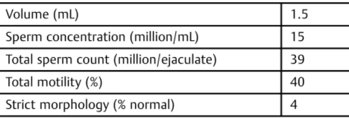
At least two semen analyses should be performed during the evaluation of any male patient with infertility. Men with a clinically palpable varicocele may demonstrate reduced total sperm count, decreased motility, and/or abnormal morphology as defined by the WHO (►Table 2).16However, the validity of the semen parameters set by the WHO with regard to varicoceles has recently been questioned, as a recent study showed that 58.8% of men with a clinical varicocele whose semen analyses were abnormal based on the WHO 1999 reference but normal on the 2010 reference still demonstrated meaningful improvements in sperm count or motility after varicocele repair.17Thus, interpreta-tion of what defines an abnormal semen analysis is difficult, and providers should not withhold treatment based on hard cutoffs such as the latest reference values alone; rather, consideration for repair may be made based on borderline semen parameter values in a scenario where repair would otherwise be indicated.
Scrotal pain associated with varicocele with or without infertility is an alternative indication for repair. Pain from varicoceles is typically a dull ache that worsens with activity and improves with rest. It is imperative to rule out other organic causes of testicular pain such as epididy-mitis, testicular mass, or inguinal hernia. Similar to the varicocele grading indication for repair for infertility, sub-clinical varicoceles should not be offered repair for pain. Higher grade varicoceles are increasingly likely to be a source of pain; however, any clinically palpable varicocele with a convincing story for pain may be considered for repair. Patients with a longer duration of scrotal pain preoperatively are more likely to have resolution of pain, with complete or marked resolution of pain reported in 83 to 92% following treatment for varicocele.18–20
Table 2 Lower limits of normal semen analysis
Volume (mL) 1.5
Sperm concentration (million/mL) 15
Total sperm count (million/ejaculate) 39
Total motility (%) 40
Strict morphology (% normal) 4
Note: WHO 2010 reference lower limit is derived from thefifth percentile of normal fertile men.
Source: Adapted from Cooper et al.16
Adolescent Varicocele
Varicoceles in patients of adolescent age are treated differ-ently than in those of adults, in that the goal of treatment is to prevent future testicular failure and/or infertility. Nonethe-less, treatment of adolescent varicoceles is controversial due to the unknown predictability of the future effects an un-treated adolescent varicocele could have on a patient later in life. There is no consensus on indications for treatment for adolescent varicocele. A recent review by Chiba et al provided additional possible indications in this population,21which is shown in►Table 3. Physical examination is still the mainstay of diagnosis; however, in cases of adolescent varicocele, objective measurements of testicular volume should be made either by an orchidometer or ultrasound. Scrotal ultra-sonography has been shown to offer greater accuracy than an orchidometer for volume measurement using the Lambert formula (LWH0.71),22 and thus can be a useful adjunct in determining testicular size differences that could indicate treatment. Typically, the contralateral testis volume acts as a control for the affected testis to determine volume discrepancy. In cases of bilateral varicoceles, it is recom-mended to use the standardized testis volume measurements at different Tanner stages,23shown in►Table 4. A testicular volume discrepancy of>10% has been associated with both testicular atrophy24and abnormal semen analysis (decreased sperm concentration and total motile sperm count)25 that would likely benefit from treatment. Testicular volume dis-crepancies>20% have been associated with a more dramatic effect on semen analysis.25,26
Spontaneous catch-up growth without intervention has been noted in the adolescent varicocele, indicating the possibility for additional indications to direct treatment.27Though markedly underused, the ASRM Practice Committee recommends obtain-ing a semen analysis in adolescents presentobtain-ing with a varicocele in the absence of significant testicular atrophy.7Although the WHO criteria for normal semen parameters is based on adults, Tanner stage 5 adolescents have comparable semen analysis parameters and should be prompted for a semen analysis with sufficient education and support.28 In addition to abnormal semen analysis, elements of scrotal ultrasonography other than testicular volume have demonstrated an association with clinical varicocele, including the presence of venous reflux29and elevated peak retrogradeflow.30
No single criteria can predict which varicoceles discovered in adolescence will affect future testicular function and fertility, which can complicate the ability of the clinician to direct treatment versus observation. At this time, decisions for this population should be driven by some combination of symptoms, physical examination, testicular volume discrep-ancy, abnormal semen parameters, and ultrasoundfindings. Consultation to a fellowship-trained urologist specialized in reproductive medicine may be considered for adolescent varicoceles as the topic continues to be frequently debated and studied further.
Surgical Options
The goal of treatment for varicocele is preventing retrograde
flow within the internal spermatic veins. This is performed either by percutaneous selective embolization, sclerotherapy, or surgical correction, commonly known as varicocelectomy (although this is a misnomer, as varicocele veins are not surgically removed as the term implies). The possible surgical approaches are high retroperitoneal (Palomo),31 laparosco-pic, inguinal (Ivanissevich),32and subinguinal (►Fig. 2). Each approach carries different degrees of complexity, success, and complication and recurrence rates.
The retroperitoneal approach is typically performed as a conventional open procedure. A horizontal incision is made medial and inferior to the ipsilateral anterior superior iliac spine and extended medially. To access the internal spermatic veins, which at this level are proximal to the internal inguinal ring, the external oblique fascia is opened and the internal oblique muscle is retracted cranially. In this approach, the testicular artery is often not dissected; however, if identified, all attempts at preservation are usually made. A recent prospective controlled study demonstrated the use of the surgical microscope in this approach with good results; however, additional larger studies are required to demon-strate this benefit.33 The open retroperitoneal approach, although thefirst technique for varicocele repair described in 1949,31is used less frequently in today’s practice. It initially became popular by its lack of requisite microsurgical or laparoscopic training, thus allowing surgeons across multiple backgrounds to perform the procedure.
However, as technological advances gave way to laparos-copy, the open retroperitoneal approach was less commonly
Table 3 Possible indications for treatment of adolescent
varicocele
Any palpable varicocele
Testicular volume discrepancy>20%
Abnormal semen parameters (Tanner stage 5)
Testicular discomfort due to varicocele
Ultrasoundfindings when physical examination is
inconclusive, including spontaneous venous reflux and peak retrogradeflow>30 cm/s
Source: Adapted from Chiba et al.21
Table 4 Mean testicular volumes by Tanner stage
Tanner stage Left testis volume (mL)
Right testis volume (mL)
1 4.762.76 5.203.86
2 6.403.16 7.083.89
3 14.586.54 14.776.1
4 19.806.17 20.456.79
5 28.318.52 30.259.64
Source: Data from Kass et al.48
used to decrease postoperative pain and hospitalization duration. One meta-analysis of 1,015 patients undergoing varicocelectomy found a significant difference in the time to return to work between open repair and either laparoscopic or microsurgical repair.34The laparoscopic approach typically involves ligating the spermatic veins near the entry point into the left renal vein. At this level, fewer veins are present needing ligation and the testicular artery has typically not yet branched to be at risk for injury. Laparoscopic repair of varicoceles remains commonly used, particularly by pediatric urologists. A recent survey demonstrated it is the most common approach for pediatric urologists, likely because of the overall increased use of laparoscopy in pediatrics as well as the reduced operative time and costs compared with open techniques. Regardless of the use of laparoscopy, a retroperi-toneal approach carries the highest relative risk of recurrence and hydrocele formation.35In addition, although the risk is low, injury to visceral organs in this approach is a possible complication. Thus, inguinal and subinguinal approaches are often preferred due to their ability to ligate the most distal venous contributions to a clinical varicocele.
The inguinal and subinguinal approaches are typically performed with the assistance of intraoperative magnifi ca-tion. For the inguinal approach, an inguinal incision is made over the inguinal canal to open the external oblique fascia above the inguinal ring to deliver the spermatic cord into the
field. In the subinguinal approach, the position of the external inguinal ring is identified, a small 2.5-cm transverse incision is made directly below the external ring, and the spermatic cord is elevated gently into thefield. The external spermatic fascia is opened, and the testicular artery, lymphatics, and vas deferens are identified and preserved with the assistance of magnification, ideally with a surgical microscope. As the differing surgical approaches approximate the level of the scrotum and testes, injuries to the artery or lymphatics at a more distal, end-organ level could be more likely to cause atrophy or postoperative hydrocele. Thus, their identification and preservation in the inguinal and subinguinal approaches
are important and typically performed. The biggest differ-ences between the two anatomical approaches are the degree of postoperative pain and recovery as well as the length of operative time possibly related to complexity. Studies com-paring inguinal to subinguinal approach have shown the opening of the external oblique aponeurosis in inguinal repair may lead to more pain and longer recovery times.36,37One of those studies showed significantly longer average operating time with the subinguinal approach, likely a result of more veins and more complex anatomy as it is the most distal of all surgical approaches.36
Results and Complications
It has been well studied with randomized controlled trials that men with clinical varicoceles and abnormal semen analyses have higher pregnancy rates after varicocele repair compared with control, that is, no treatment.38To date, there is insufficient evidence to suggest a“gold standard”approach to treatment. However, large studies and meta-analyses have demonstrated differences in success rate—measured by either improvement in semen analysis or pregnancy rate—and complication rate. The most common complications are hydrocele and recurrent or persistent varicocele.
Data on studies comparing semen analyses and pregnancy rates before and after treatment are limited by many factors, and there are few meta-analyses comparing these factors across different surgical approaches. In one meta-analysis with four randomized controlled trials and 1,015 patients, microsurgical varicocelectomy had higher pregnancy rates than open and laparoscopic approaches (40.2 vs. 29.3% and 39.0 vs. 31.8%, respectively).34Another meta-analysis of 36 studies with 4,473 men demonstrated a postprocedure preg-nancy rate of 42.0% in the microsurgical technique compared with 37.7% in the retroperitoneal technique, 30.1% in the laparoscopic technique, and 36.0% in the macroscopic ingui-nal technique.39 In both studies, these differences were all statistically significant. However, more large multicenter trials are still needed to further support or refute thefindings that a microscopic approach provides the highest success rate. Postoperative hydrocele and recurrent or persistent vari-coceles are common complications, which again are under-studied when stratified across different surgical approaches. In the largest of meta-analyses mentioned previously, hydro-cele formation rates were 0.4% in the microsurgical approach, 8.2% in the retroperitoneal approach, 2.8% in the laparoscopic approach, and 7.3% in the macroscopic inguinal approach.39 Similarly, recurrence rates were 1.1, 15.0, 4.3, and 2.6%, respectively.39 Persistence and recurrence of varicocele treated by the retroperitoneal approach is likely due to the inability to ligate the contributions of the external spermatic vein, which has been found to be dilated in 16 to 74% of cases.40In addition to having the ability to ligate the most distal contributions to a varicocele, approaches closer to the affected testis also have the ability to identify and preserve individual lymphatics to reduce the risk of hydrocele forma-tion. The recognition of these improved complication rates has made a major impact on the evolution of adult urologic
Fig. 2 Anatomical locations of the incision in each approach for varicocelectomy.
practice toward the treatment of varicoceles primarily with microsurgical inguinal and subinguinal approaches.
With respect to recurrence and complication rates in the inguinal and subinguinal approaches, there also may be a correlation with the degree of magnification used. One study with 100 patients undergoing repair by both inguinal and subinguinal approaches demonstrated a 0% recurrence with the microscope, 2.9% with loupe magnification, and 8.8% without magnification.41 This same study showed corre-sponding postoperative hydrocele rates of 0, 2.9, and 5.9%, respectively.41Two recent studies also compared microscopic varicocelectomy to open and laparoscopic techniques to support these conclusions, reporting 0% postoperative hydro-cele rates and a 2 to 3% recurrence rate.42,43
The most reported disadvantage for the use of microsur-gical repair is the level of training and expertise required as well as the longer operative times associated with its use. However, the two may be inversely related and suggest that this may not be an issue at high volume centers performing microscopic repair on a regular basis. In one Japanese study of 144 varicocele repairs performed by open, laparoscopic, and microsurgical approaches, the microsurgical approach was actually associated with significantly shorter operative times compared with the open and laparoscopic approaches.44 Practically speaking, the use of the microscope adds very little time to the case itself regarding the setup and docking times; the longer operative times reported are due to the identification of more veins and arteries, and thus, a more lengthy procedure. For urologists with microsurgical training, subinguinal microsurgical varicocele repair appears to be the technique that is most effective, safe, and with the quickest convalescence compared with other approaches.
Observation and Follow-up
As previously discussed, the controversy surrounding varico-cele management is highly centered on who to treat and when to treat. Although catch-up growth and improvements in semen analysis have been shown in the adolescent population both with and without surgery, physicians are faced with the difficult decision to pursue treatment and potentially subject the patient to the inherent risks and costs of surgery, or to observe with the risks of potentially causing testicular injury or contributing to future infertility. If observed, one recom-mended standardized approach described at the Children’s Hospital of Philadelphia includes yearly examinations with an orchidometer (or every other year if normal total testicular volume) until the patient reaches Tanner 5 maturity, at which point, a semen analysis and androgen hormone level testing are performed.45If at that point, the patient is symptomatic, or testicular volume, semen parameters, or serum hormone results are low, surgical correction is discussed; otherwise, if observation is continued, follow-up with an adult urologist should be encouraged until paternity is achieved.45
In patients who have undergone surgery for varicocele, follow-up typically involves one or more routine postopera-tive visit to perform examination of the wound as well as the scrotum to evaluate for persistence or recurrence. If
persis-tence or recurrence is noted, internal spermatic venography may help identify the site of persistent reflux to direct any further repair,46whether surgically or with embolization or sclerotherapy. For adolescent patients who have undergone treatment, routine examinations should include orchidom-eter measurements to follow catch-up growth.
In patients who have previously provided a semen analysis before treatment, repeat analyses should be offered at approx-imately 3- to 6-month intervals during thefirst year after treatment or until pregnancy is achieved.7It is important to inform patients that recognizable improvements in semen analysis (thus, an improvement in pregnancy rate) after repair may take several spermatogenic cycles, each lasting approxi-mately 3 months on average. In addition, men with clinical varicocele but with normal sperm parameters remain at risk for progressive testicular dysfunction and should be offered monitoring by semen analysis every 1 to 2 years.7
Summary
Varicoceles are a common entity both in adolescents and in the infertile male that can be diagnosed by any physician. A thorough history and physical examination and semen anal-ysis are warranted. Adjunctive imaging studies may be used in difficult cases; however, treatment of subclinical or inci-dental varicoceles is not necessary, and searching for them should be discouraged. Referral to a urologist, particularly one with fellowship training in reproductive medicine, is an appropriate consideration at any stage of diagnosis and management. When observed, regular follow-up for varico-celes is indicated in adolescents and in those seeking current and/or future fertility. When treatment is indicated, there are several options, either surgical or with interventional radiol-ogy, that have been shown to be successful. Each varies in its degree of risks and potential benefits; however, the micro-surgical subinguinal approach may be considered the current surgical standard.
References
1 Nagler HM, Martinis FG. Varicocele. In: Lipshultz LI, Howards SS, eds. Infertility in the Male. 3rd ed. St. Louis: Mosby; 1997:336–359 2 Agarwal A, Deepinder F, Cocuzza M, et al. Efficacy of varicocelec-tomy in improving semen parameters: new meta-analytical ap-proach. Urology 2007;70(3):532–538
3 Masson P, Brannigan RE. The varicocele. Urol Clin North Am 2014; 41(1):129–144
4 Trum JW, Gubler FM, Laan R, van der Veen F. The value of palpation, varicoscreen contact thermography and colour Doppler ultrasound in the diagnosis of varicocele. Hum Reprod 1996;11(6):1232–1235 5 Pilatz A, Altinkilic B, Köhler E, Marconi M, Weidner W. Color Doppler ultrasound imaging in varicoceles: is the venous diameter sufficient for predicting clinical and subclinical varicocele? World J Urol 2011;29(5):645–650
6 Stahl P, Schlegel PN. Standardization and documentation of vari-cocele evaluation. Curr Opin Urol 2011;21(6):500–505
7 Practice Committee of the American Society for Reproductive Medicine; Society for Male Reproduction and Urology. Report on varicocele and infertility: a committee opinion. Fertil Steril 2014;102(6):1556–1560
8 Abdel-Meguid TA, Farsi HM, Al-Sayyad A, Tayib A, Mosli HA, Halawani AH. Effects of varicocele on serum testosterone and changes of testosterone after varicocelectomy: a prospective controlled study. Urology 2014;84(5):1081–1087
9 Hsiao W, Rosoff JS, Pale JR, Powell JL, Goldstein M. Varicocelectomy is associated with increases in serum testosterone independent of clinical grade. Urology 2013;81(6):1213–1217
10 Tanrikut C, Goldstein M, Rosoff JS, Lee RK, Nelson CJ, Mulhall JP. Varicocele as a risk factor for androgen deficiency and effect of repair. BJU Int 2011;108(9):1480–1484
11 Yamamoto M, Hibi H, Hirata Y, Miyake K, Ishigaki T. Effect of varicocelectomy on sperm parameters and pregnancy rate in patients with subclinical varicocele: a randomized prospective controlled study. J Urol 1996;155(5):1636–1638
12 Unal D, Yeni E, Verit A, Karatas OF. Clomiphene citrate versus varicocelectomy in treatment of subclinical varicocele: a prospec-tive randomized study. Int J Urol 2001;8(5):227–230
13 Zegers-Hochschild F, Adamson GD, de Mouzon J, et al; Interna-tional Committee for Monitoring Assisted Reproductive Technol-ogy; World Health Organization. International Committee for Monitoring Assisted Reproductive Technology (ICMART) and the World Health Organization (WHO) revised glossary of ART termi-nology, 2009. Fertil Steril 2009;92(5):1520–1524
14 Schlegel PN. Is assisted reproduction the optimal treatment for varicocele-associated male infertility? A cost-effectiveness analy-sis. Urology 1997;49(1):83–90
15 Weedin JW, Khera M, Lipshultz LI. Varicocele repair in patients with nonobstructive azoospermia: a meta-analysis. J Urol 2010; 183(6):2309–2315
16 Cooper TG, Noonan E, von Eckardstein S, et al. World Health Organization reference values for human semen characteristics. Hum Reprod Update 2010;16(3):231–245
17 Lee YJ, Cho SY, Paick JS, Kim SW. Usefulness of 2010 world health organization reference values for determining indications for varicocelectomy. Urology 2015;85(4):831–835
18 Karademir K, Senkul T, Baykal K, AteşF, Işeri C, Erden D. Evaluation of the role of varicocelectomy including external spermatic vein ligation in patients with scrotal pain. Int J Urol 2005;12(5): 484–488
19 Al-Buheissi SZ, Patel HR, Wazait HD, Miller RA, Nathan S. Pre-dictors of success in surgical ligation of painful varicocele. Urol Int 2007;79(1):33–36
20 Abd Ellatif ME, Asker W, Abbas A, et al. Varicocelectomy to treat pain, and predictors of success: a prospective study. Curr Urol 2012;6(1):33–36
21 Chiba K, Ramasamy R, Lamb DJ, et al. The varicocele: diagnostic dilemmas, therapeutic challenges and future perspectives. Asian J Androl 2016;18(2):276–281
22 Sakamoto H, Saito K, Oohta M, Inoue K, Ogawa Y, Yoshida H. Testicular volume measurement: comparison of ultrasonography, orchidometry, and water displacement. Urology 2007;69(1): 152–157
23 Kass EJ. The adolescent varicocele: treatment and outcome. Curr Urol Rep 2002;3(2):100–106
24 Li F, Chiba K, Yamaguchi K, et al. Effect of varicocelectomy on testicular volume in children and adolescents: a meta-analysis. Urology 2012;79(6):1340–1345
25 Diamond DA, Zurakowski D, Bauer SB, et al. Relationship of varicocele grade and testicular hypotrophy to semen parameters in adolescents. J Urol 2007;178(4, Pt 2):1584–1588
26 Kurtz MP, Zurakowski D, Rosoklija I, et al. Semen parameters in adolescents with varicocele: association with testis volume differ-ential and total testis volume. J Urol 2015;193(5, Suppl):1843–1847 27 Poon SA, Gjertson CK, Mercado MA, Raimondi PM, Kozakowski KA, Glassberg KI. Testicular asymmetry and adolescent varicoceles managed expectantly. J Urol 2010;183(2):731–734
28 Fine RG, Gitlin J, Reda EF, et al. Barriers to use of semen analysis in the adolescent with a varicocele: survey of patient, parental, and practitioner attitudes. J Pediatr Urol 2015;12(1):e1–e6
29 Hussein AF. The role of color Doppler ultrasound in prediction of the outcome of microsurgical subinguinal varicocelectomy. J Urol 2006;176(5):2141–2145
30 Kozakowski KA, Gjertson CK, Decastro GJ, Poon S, Gasalberti A, Glassberg KI. Peak retrogradeflow: a novel predictor of persistent, progressive and new onset asymmetry in adolescent varicocele. J Urol 2009;181(6):2717–2722, discussion 2723
31 Palomo A. Radical cure of varicocele by a new technique; prelimi-nary report. J Urol 1949;61(3):604–607
32 Ivanissevich O. Left varicocele due to reflux; experience with 4,470 operative cases in forty-two years. J Int Coll Surg 1960;34:742–755 33 Zhang H, Li H, Hou Y, et al. Microscopic retroperitoneal varico-celectomy with artery and lymphatic sparing: an alternative treatment for varicocele in infertile men. Urology 2015;86(3): 511–515
34 Ding H, Tian J, Du W, Zhang L, Wang H, Wang Z. Open non-microsurgical, laparoscopic or open microsurgical varicocelec-tomy for male infertility: a meta-analysis of randomized con-trolled trials. BJU Int 2012;110(10):1536–1542
35 Wang J, Xia SJ, Liu ZH, et al. Inguinal and subinguinal micro-varicocelectomy, the optimal surgical management of varicocele: a meta-analysis. Asian J Androl 2015;17(1):74–80
36 Gontero P, Pretti G, Fontana F, Zitella A, Marchioro G, Frea B. Inguinal versus subinguinal varicocele vein ligation using magni-fying loupe under local anesthesia: which technique is preferable in clinical practice? Urology 2005;66(5):1075–1079
37 Pan F, Pan L, Zhang A, Liu Y, Zhang F, Dai Y. Comparison of two approaches in microsurgical varicocelectomy in Chinese infertile males. Urol Int 2013;90(4):443–448
38 Marmar JL, Agarwal A, Prabakaran S, et al. Reassessing the value of varicocelectomy as a treatment for male subfertility with a new meta-analysis. Fertil Steril 2007;88(3):639–648
39 Cayan S, Shavakhabov S, Kadioğlu A. Treatment of palpable varicocele in infertile men: a meta-analysis to define the best technique. J Androl 2009;30(1):33–40
40 Beck EM, Schlegel PN, Goldstein M. Intraoperative varicocele anatomy: a macroscopic and microscopic study. J Urol 1992; 148(4):1190–1194
41 Cayan S, Acar D, Ulger S, et al. Adolescent varicocele repair: long-term results and comparison of surgical techniques according to optical magnification use in 100 cases at a single university hospital. J Urol 2005;174(5):2003–2006; discussion 2006–2007 42 Al-Kandari AM, Shabaan H, Ibrahim HM, Elshebiny YH, Shokeir AA.
Comparison of outcomes of different varicocelectomy techniques: open inguinal, laparoscopic, and subinguinal microscopic varicoce-lectomy: a randomized clinical trial. Urology 2007;69(3):417–420 43 Al-Said S, Al-Naimi A, Al-Ansari A, et al. Varicocelectomy for male
infertility: a comparative study of open, laparoscopic and micro-surgical approaches. J Urol 2008;180(1):266–270
44 Watanabe M, Nagai A, Kusumi N, Tsuboi H, Nasu Y, Kumon H. Minimal invasiveness and effectivity of subinguinal microscopic varicocelectomy: a comparative study with retroperitoneal high and laparoscopic approaches. Int J Urol 2005;12(10):892–898 45 Kolon TF. Evaluation and management of the adolescent
varico-cele. J Urol 2015;194(5):1194–1201
46 Punekar SV, Prem AR, Ridhorkar VR, Deshmukh HL, Kelkar AR. Post-surgical recurrent varicocele: efficacy of internal spermatic venog-raphy and steel-coil embolization. Br J Urol 1996;77(1):124–128 47 Dubin L, Amelar RD. Varicocele size and results of varicocelectomy
in selected subfertile men with varicocele. Fertil Steril 1970;21(8): 606–609
48 Kass EJ, Stork BR, Steinert BW. Varicocele in adolescence induces left and right testicular volume loss. BJU Int 2001;87(6):499–501