Structural and Biological Identification of Residues on the
Surface of NS3 Helicase Required for Optimal Replication of
the Hepatitis C Virus
*
□SReceived for publication, November 10, 2005Published, JBC Papers in Press, November 22, 2005, DOI 10.1074/jbc.M512100200
Samuel G. Mackintosh
‡1, Jeff Zhiqiang Lu
§1,2, John B. Jordan
§, Melody K. Harrison
‡, Bartek Sikora
‡,
Suresh D. Sharma
¶, Craig E. Cameron
¶, Kevin D. Raney
‡3, and Joshua Sakon
§4From the
‡Department of Biochemistry and Molecular Biology, University of Arkansas for Medical Sciences, Little Rock, Arkansas
72205, the
§Department of Chemistry and Biochemistry, University of Arkansas, Fayetteville, Arkansas 72701, and the
¶Department
of Biochemistry and Molecular Biology, Pennsylvania State University, University Park, Pennsylvania 16802
The hepatitis C virus (HCV) nonstructural protein 3 (NS3) is a multifunctional enzyme with serine protease and DEXH/D-box helicase domains. A crystal structure of the NS3 helicase domain (NS3h) was generated in the presence of a single-stranded oligonu-cleotide long enough to accommodate binding of two molecules of enzyme. Several amino acid residues at the interface of the two NS3h molecules were identified that appear to mediate a protein-protein interaction between domains 2 and 3 of adjacent molecules. Mutations were introduced into domain 3 to disrupt the putative interface and subsequently examined using an HCV subgenomic replicon, resulting in significant reduction in replication capacity. The mutations in domain 3 were then examined using recombinant NS3h in biochemical assays. The mutant enzyme showed RNA binding and RNA-stimulated ATPase activity that mirrored wild type NS3h. In DNA unwinding assays under single turnover condi-tions, the mutant NS3h exhibited a similar unwinding rate and only ⬃2-fold lower processivity than wild type NS3h. Overall biochemi-cal activities of the mutant NS3h were similar to the wild type enzyme, which was not reflective of the large reduction in HCV replicative capacity observed in the biological experiment. Hence, the biological results suggest that the known biochemical proper-ties associated with the helicase activity of NS3h do not reveal all of the likely biological roles of NS3 during HCV replication. Domain 3 of NS3 is implicated in protein-protein interactions that are neces-sary for HCV replication.
Hepatitis C virus (HCV),5a member of the familyFlaviviridae, is a leading cause of liver cirrhosis and hepatocellular carcinoma and has infected more than 170 million people worldwide (1–3). Standard
treat-ment with interferon-␣and ribavirin is frequently ineffective or toxic, and no vaccine for HCV is currently available (4). Identification of a novel viral target for therapeutic intervention could lead to the devel-opment of a more effective treatment.
The HCV genome is a 9.6-kb positive, single-stranded RNA that sup-ports translation of a 3000-amino acid polyprotein, which is subse-quently cleaved to produce both structural (C, E1, and E2) and non-structural (NS2, NS3, NS4A, NS4B, NS5A, and NS5B) viral proteins (5). Two viral proteases are encoded by the NS2 and NS3 genes (6, 7). In addition to its protease domain, the NS3 protein also contains a DEXH/ D-box helicase domain (8). NS4A encodes a peptide co-factor for NS3 protease (9), and NS5B encodes an RNA-dependent RNA polymerase (10). The functions of NS4B and NS5A have not been as well character-ized, but NS5A is involved in viral interferon sensitivity (11) and in adaptation of subgenomic replicons for growth in cultured hepatoma cells (12).
One obstacle to studying viral protein interactions is that HCV can-not be maintained efficiently in cell culture, although recent progress may overcome this difficulty (13). In order to circumvent this problem, selectable replicons carrying the neomycin phosphotransferase gene in addition to essential HCV genes and untranslated flanking regions have been constructed (14). Viral genes are translated independently of the neomycin phosphotransferase gene from an encephalomyocarditis virus internal ribosome entry site. Replication in cell culture has been further enhanced by the introduction of adaptive mutations primarily within theNS5Agene (12), although the specific mechanism by which these mutations influence replication is not known. These adaptive rep-licon mutants provide a means of studying HCV replication in cultured cells.
Many aspects of the HCV replication mechanism remain unknown, but it appears that at least five of the viral nonstructural proteins are required for replication (5). Several nonstructural protein-protein inter-actions have been identified (15, 16), and co-localization of nonstruc-tural proteins, including NS3 and NS5B, on membrane structures within cells has been observed (17, 18), indicating formation of a mul-tiprotein replication complex containing both polymerase and helicase enzymes. Understanding the mechanism of nucleic acid unwinding by NS3 is therefore a key step in characterizing the viral replication mechanism.
Helicases are molecular motor proteins that utilize energy from ATP hydrolysis to manipulate nucleic acids as well as protein-nucleic acid complexes (19 –21). NS3 helicase (NS3h) has been expressed and puri-fied independently of NS3 protease in a recombinant bacterial system and retains itsin vitrounwinding activity (8). The NS3h structure con-sists of three distinct domains, with domains 1 and 2 containing all of the *This work was supported by National Institutes of Health (NIH) COBRE Research Grant
P20 RR15569 (to J. S. and K. D. R.), NIH R01 AI060563 (to K. D. R., C. E. C., and J. S.), NIH BRIN Award P20 RR016460, the Arkansas Biosciences Institute, and the United States Department of Agriculture Cooperative State, Research, Education, and Extension Service. The costs of publication of this article were defrayed in part by the payment of page charges. This article must therefore be hereby marked “advertisement” in accordance with 18 U.S.C. Section 1734 solely to indicate this fact.
□SThe on-line version of this article (available at http://www.jbc.org) contains supple-mental Figs. 1 and 2.
The atomic coordinates and structure factors (code 2F55) have been deposited in the Protein Data Bank, Research Collaboratory for Structural Bioinformatics, Rutgers University, New Brunswick, NJ (http://www.rcsb.org/).
1These authors contributed equally to this work.
2Present address: Dept. of Molecular Microbiology and Immunology, Johns Hopkins
Bloomberg School of Public Health, Baltimore, Maryland 21205.
3To whom correspondence may be addressed. E-mail: [email protected]. 4To whom correspondence may be addressed. E-mail: [email protected].
5The abbreviations used are: HCV, hepatitis C virus; NS3h, NS3 helicase domain; MR,
conserved motifs common to superfamily II helicase enzymes (22, 23). ATP binds at the interface between domains 1 and 2, and nucleic acid binds within a cleft formed at the interface between domain 3 and domains 1 and 2 (23). Interactions between the protein and nucleic acid are not sequence-specific and primarily involve the phosphate back-bone of the nucleic acid.
Despite extensive structural and biochemical characterization, the oligomeric state of the functional species of NS3 has not been deter-mined conclusively. Biochemical studies have resulted in reports of monomeric (24), dimeric (25, 26), and oligomeric (27, 28) forms of the protein. Crystal structures of NS3h (22), NS3h bound to an 8-mer poly(dU) substrate (23), and a full-length NS3/NS4A fusion protein (29) demonstrate that a single protein monomer can form crystals. However, none of these structures includes a substrate molecule of sufficient length to accommodate binding of multiple NS3h monomers. Cross-linking experiments indicate that NS3h can form oligomeric structures in solution (28). DNA unwinding activity of full-length NS3 is optimal at concentrations high enough to allow binding of multiple NS3h mono-mers to each nucleic acid molecule (30). However, no cooperative effect has been observed in binding studies (31), leaving open the possibility that NS3 is a nonprocessive monomer. A recently reported biochemical model suggests that monomeric NS3h is functional but that multiple NS3h molecules are required for optimal processivity (32).
We have crystallized NS3h bound to a 16-mer poly-dU nucleic acid. The structure shows two NS3h molecules bound to a single DNA mol-ecule and reveals an apparent interface between the two protein mole-cules. A mutational analysis of the protein interface region was per-formed in order to determine its importance for helicase activity and viral replication.
EXPERIMENTAL PROCEDURES
Purification of NS3h—BL21(DE3) cells were co-transformed with pET26b expression plasmid containing a wild type or mutant NS3h gene with an NH2-terminal ubiquitin tag and pCG1 expressing the ubiquitin protease Ubp1. 3.5 liters of transformed cells were cultured at 37 °C to anA600of 1.5 in NZCYM broth containing 30g/ml kanamycin and 30g/ml chloramphenicol. Overexpression was induced by the addition of 3.5 liters of chilled NZCYM, 0.2% dextrose, and 0.5 mM
isopropyl 1-thio--D-galactopyranoside. Antibiotic concentrations were kept constant. Cells were cultured at 15 °C for 16 –18 h following induction. All subsequent steps were performed at 4 °C. NS3h purifica-tion was followed by measuring poly(U)-dependent ATPase activity and by SDS-PAGE. Cells were centrifuged at 6000 rpm for 10 min in a Beckman JLA-10.500 rotor and resuspended in lysis buffer (25 mMTris/
Cl, pH 7.5, 1 mMEDTA, 500 mMNaCl, 5 mM-mercaptoethanol, 10%
glycerol) at 5 ml/g. Cells were lysed by the addition of 4 mM
phenyl-methylsulfonyl fluoride, 4g/ml pepstatin A, and 2.25 mg of lysozyme/g of cells, followed by a freeze/thaw in liquid nitrogen, pressurization at 1500 p.s.i. for 3 min in a nitrogen bomb, and sonication. The lysate was centrifuged at 20,000 rpm for 20 min in a Beckman JA-25.50 rotor. The supernatant was further centrifuged at 45,000 rpm for 2 h in a Beckman 50.2 Ti rotor. The supernatant from the final centrifugation was further purified by anion exchange chromatography. NS3h was eluted in the wash from the anion exchange column with lysis buffer and precipitated by the addition of 55% (NH4)2SO4followed by centrifugation at 20,000 rpm for 60 min in a Beckman JA-25.50 rotor. Pellets were resuspended in lysis buffer plus 500 mM(NH4)2SO4. NS3h was further purified using
methyl-hydrophobic (30 ml), heparin-Sepharose (30 ml), and poly(U) affinity (20 ml) chromatography columns. NS3h was eluted from each column with a low salt/high salt buffer gradient (10 mM/1MNaCl in 25
mMTris/Cl, pH 7.5, 1 mMEDTA, 5 mM-mercaptoethanol, 10%
glyc-erol). The gradient was reversed for the hydrophobic column. NS3h eluted from the hydrophobic and heparin-Sepharose columns between 250 and 450 mMNaCl and from the poly(U) column between 450 and
650 mMNaCl. Fractions containing NS3h were combined and dialyzed
into low salt buffer following elution from each column. Fractions from the final column were dialyzed into storage buffer (25 mMTris/Cl, pH 7.5, 1 mMEDTA, 100 mMNaCl, 5 mM-mercaptoethanol, 20% glycerol) and concentrated to 7–10M.
Crystallization and Structural Determination of NS3h—Purified NS3h was concentrated to 32.2 mg/ml for crystallization. Examination of sample purity and determination of molecular weight were per-formed using SDS-PAGE on a Phast Gel system (AP Biotech). The apparent molecular mass of the helicase was⬃50 kDa. The (dU)16 oli-gonucleotide was synthesized using an Expedite nucleic acid synthesis system and purified by polyacrylamide gel electrophoresis to a final concentration of 4.6 mg/ml. Immediately before crystallization, NS3h was mixed with the (dU)16oligonucleotide at a 2:1 molar ratio. Crystals of diffraction quality were obtained by using the hanging drop method at room temperature with 4l of the mixture and 1l of reservoir solution (2.4M(NH4)2SO4, 0.1MTris hydrochloride, pH 8.5).
Diffrac-tion data were collected at 1.5418 Å by using a single crystal of 0.2⫻ 0.4⫻0.1 mm mounted in a thin-walled glass capillary at room temper-ature. The x-ray radiation was produced by an in-house Rigaku RU-H3RHB generator and focused by Osmic’s Gutman multilayer mir-rors (Woodland, TX). The diffraction pattern was recorded on the R-AXIS IV phosphor image plate detector. The diffraction data were reduced, integrated, and scaled with Denzo/Scalepack. Molecular replacement analysis was performed using AMORE software (33), and structure refinements and model building/adjustment were done using CNS (34) and XtalView (35), respectively.
The scaled data of resolution 3.3 Å indicated that the NS3h/(dU)16 crystal belongs to the orthorhombic space group (Table 1). Space group assignment between P21212 and P212121could not be made due to the absence of some (00l) reflections. Matthews’ coefficient suggested that the number of protein molecules in one asymmetric unit could be 3 or 4, with solvent content being 67 or 50%, respectively.
We completed the structure determination by molecular replace-ment (MR), using the published 1A1V structure (23) as an initial model. The 1A1V structure consists of an NS3h monomer bound to a (dU)8 oligonucleotide. Prior to MR, the oligonucleotide was removed from the model. The MR results from space group P21212 did not display any reasonable correlation among the individual solutions; however, the MR solutions in space group P212121showed strong correlation as well as realistic crystal packing (not shown).
A composite omit map was calculated using CNS (34) to identify any missing components. The resulting electron density map showed clearly the presence of the oligonucleotide at the nucleic acid binding sites of the protein molecules. The (dU)xfragments were inserted man-ually into the structure using XtalView (35). The maximum likelihood from structure factors refinement on the model was done in CNS and is presented in Table 1.
restriction sites followed by ligation. Mutant NS3 sequences were trans-ferred to pET26b-ubiquitin expression plasmid by PCR followed by digestion at SacII and EcoRI restriction sites and ligation. Quality of all final plasmid products was confirmed by sequencing.
Replicon RNA Synthesis—DNA template was prepared by digestion of replicon plasmid (10g) with ScaI restriction endonuclease at 37 °C for 4 h. Complete linearization of plasmid was confirmed by agarose gel electrophoresis. RNA was synthesizedin vitroby incubating 0.5g of linear DNA template with 0.5g of T7 RNA polymerase in 350 mM
HEPES, pH 7.5, 32 mMmagnesium acetate, 40 mMdithiothreitol, 2 mM
spermidine, and 28 mMNTPs at 37 °C for 3 h. Template DNA was removed by incubation with 2 units of DNase I at 37 °C for 30 min. RNA was precipitated overnight in LiCl at⫺20 °C. RNA purity and quality were verified by agarose gel electrophoresis.
Colony Formation Assays—Huh-7.5 cells were trypsinized 1 day before transfection. Cells (2⫻106) were washed once with DMEM lacking serum and suspended in 0.9 ml of the same medium. RNA-liposome complexes were prepared using the TransMessenger reagent from Qiagen. Cells and and RNA-liposome complexes were mixed and incubated at 37 °C for 1 h. After incubation, cells were pelleted by cen-trifugation (525⫻g) for 4 min and suspended in DMEM containing 10% fetal bovine serum. Cells were plated in 100-mm Petri dishes in the absence of feeder cells at the following densities: 1⫻106, 2⫻105, and 2⫻104. After 2 days of recovery, cells were treated with 0.5 mg/ml G418, and selection was continued for 3 weeks with medium changes every 4 days. Cells were fixed and stained using crystal violet. At least three independent experiments were performed for each transfection.
Western Analysis—Huh-7.5 cells were transiently transfected with HCV RNA using TransMessenger Lipofectin Reagent (Qiagen) accord-ing to the manufacturer’s instructions. Cells were lysed at 4 h post-transfection in SDS-PAGE denaturing sample buffer. Lysate from 5⫻ 105cells from each transfection was run on 10% SDS gel. Proteins were transferred from gel to polyvinylidene difluoride membrane using a Bio-Rad electrophoretic transfer cell at 100 V for 1 h at 4 °C in transfer buffer (25 mMTris, 192 mMglycine). Membranes were blocked in 5%
dry milk/TBST (20 mMTris/Cl, pH 7.5, 137 mMNaCl, 0.1% Tween 20)
for 1 h. Blocked membranes were exposed to rabbit polyclonal anti-NS3 in 5% bovine serum albumin/TBST for 90 min and then exposed to horseradish peroxidase-conjugated goat anti-rabbit IgG (PerkinElmer Life Sciences) in 5% dry milk/TBST for 1 h. Chemiluminescent detec-tion was done by an ECL Western blotting analysis system (Amersham Biosciences).
Nucleic Acid Binding Assays—NS3h was incubated with 500 pM
5⬘-fluorescein labeled rU20(Integrated DNA Technologies) in 50 mM MOPS (pH 7.0), 10 mMNaCl, 50MEDTA, 0.1 mg/ml bovine serum albumin for 5 min at 37 °C. Binding was measured as a function of fluorescence polarization using a Beacon fluorescence polarization instrument. Data were fit to a hyperbola using Kaleidagraph software.
ATPase Assays—NS3h was incubated with 5 mMATP in 50 mM
HEPES (pH 7.5), 0.5 mMEDTA, 10 mMMgCl2, 10 mMNaCl, 0.1 mg/ml bovine serum albumin, 4 mMphosphoenolpyruvate, 10 units/ml pyru-vate kinase/lactate dehydrogenase, and 0.7 mg/ml NADH. Absorbance of NADH at 380 nm was measured at 1-s intervals for a period of 30 s in the presence of the indicated concentrations of poly(U). Hydrolysis rates were calculated using an extinction coefficient of 1,210M⫺1cm⫺1 for NADH. Data were fit to a hyperbola using Kaleidagraph software.
DNA Unwinding Assays—Unwinding assays were carried out using a Quench-Flow apparatus (RQF-3, KinTek Instruments, Austin, TX) and a two-step mixing protocol (32). The temperature was held constant at 37 °C with a circulating water bath. Reactions were carried out in 25 mM
MOPS (pH 7.0), 10 mMNaCl, 0.1 mMEDTA, 2 mM-mercaptoethanol, and 0.1 mg/ml bovine serum albumin; all concentrations are postmix-ing. NS3h (500 nM) was mixed with 2 nMDNA substrate (30-base pair duplex with 3⬘T15 overhang; the longer strand was radiolabeled with 32P) for 10 s before adding 5 m
MATP, 10 mMMgCl2, 60 nMannealing
trap (complementary to the displaced strand), and 100Mpoly(dT)
protein trap. The reaction was quenched after 0.1–15 s by ejection into a tube containing 200 mM EDTA, 0.7% SDS. Double- and single-stranded DNA were resolved on a native 20% polyacrylamide gel, detected using a PhosphorImager (Amersham Biosciences), and quan-tified using ImageQuant software. Bench top experiments were used to determine the amplitude of product formation under single turnover conditions. NS3h was added to substrate (2 nM), followed by a 10-s
incubation. ATP (5 mM), MgCl2(10 mM), and a protein trap (poly(dT),
100M) were added to initiate the unwinding reaction. The reaction was stopped after 10 s by adding 200 mMEDTA. Data were fit to Equa-tion 1, using Kaleidagraph (Synergy Software, Reading, PA). This equa-tion describes a five-step mechanism for DNA unwinding that is neces-sary to fit the substantial lag phase associated with unwinding of the substrate (32, 36, 37).
Y
⫽
A
(1
⫺共
1
⫹
k
obst
⫹
1/ 2
共
k
obst
兲
2
⫹
1/6
共
k
obst
兲
3
⫹
1/ 24
共
k
obst
兲
4兲
e
⫺kobst)
(Eq. 1)
RESULTS
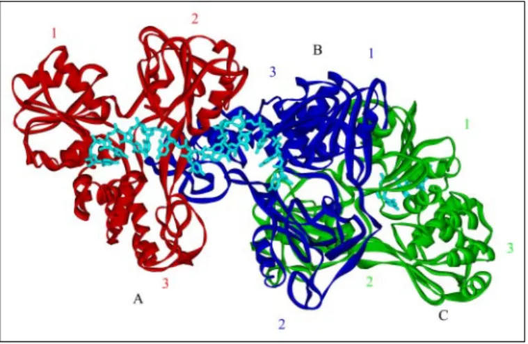
Crystallization and Structural Analysis of NS3h Bound to (dU)16 —Crys-tals of NS3h in the presence of (dU)16oligonucleotide belonged to space group P212121with unit cell dimensionsa⫽108.3 Å,b⫽109.8 Å, andc⫽ 183.4 Å (Table 1). We determined the structure by the MR method, using 1A1V.pdb (23) as an initial model, in which its (dU)8fragment was manu-ally removed prior to the rotational function search. We identified three helicase molecules (chains A, B, and C in Fig. 1) per asymmetric unit in our structure, with two helicase molecules bound to a single (dU)16molecule. The final atomic model shows no major unfavorable steric interactions between the helicase molecules, and the crystal packing shows no conflicts between the protein molecules in adjacent asymmetric units. We found no apparent noncrystallographic symmetric operations among the three monomers in this helicase model, nor did we observe any dramatic differ-ences among the monomers. All three helicase molecules retain the basic Y TABLE 1
Data collection and refinement statistics
Parameter Value
Data collection
Unit cell ␣⫽⫽␥⫽90°
A (Å) 108.3
B (Å) 109.8
C (Å) 183.4
Space group P212121
Wavelength (Å) 1.5418
Resolution (Å) 3.3
Unique reflections 33,594
Completeness (%) 91.1
Rsym
a(%) 0.34
具I/(I)典 4.4
Refinement
Resolution (Å) 30 to 3.3
No. of reflectionsb
27,936 (1,673) Rb,c
(%) 24.8 (27.4)
AverageB-factors (Å) 34.25 r.m.s. deviation bond lengths (Å) 0.010 r.m.s. deviation bond angles (degrees) 1.495
aR
sym⫽冱兩I⫺ 具I典兩/冱I, whereIrepresents intensity. bValue forR
freeset containing 5% of randomly chosen reflections. cr⫽冱兩F
shape characteristic of previously reported structures, with minor confor-mational differences at the surface loop regions.
Chains A and B of the complex are bound to a 13-nucleotide span of one (dU)16molecule, with chain B rotated 90° relative to chain A. The binding mode of both chains is consistent with that of the 1A1V struc-ture, with the binding cleft at the interface of domains 1 and 2 with domain 3 in each protein molecule. Chain C appears to be independent of the dimer-oligonucleotide complex. The nucleic acid binding cleft of chain C faces away from the dimer structure and is occupied by a second oligonucleotide molecule. Chain C does not have any evident structural or mechanistic relationship with the other two protein molecules, and there appear to be no suitable contacts between chains B and C to indicate a functional interaction between these two molecules.
To determine the degree of similarity between chains A and B, we superimposed the two using domain 1 (the NTPase domain) as an anchor (the backbone r.m.s. deviation for residues 190 –324 was 0.7 Å). We observed only minor conformational differences between the two chains in each of the three domains (Fig. 2). However, it appears that the relative orientation of domain 2 with respect to the anchored domain 1 is slightly different between chains A and B with a small but detectable tilting angle, resulting in a slight widening of the nucleic acid binding groove in chain B. This flexibility of domain 2 relative to domain 1 is consistent with the ratchet mechanism for nucleic acid translocation proposed by Kimet al.(23). Domain 3 displays a small degree of rotation between chains A and B but is otherwise similar.
To assess the relationship of the apparent dimer structure to that of the monomeric NS3-oligonucleotide complex reported by Kimet al.
(23), we independently aligned each of the two monomer structures (chains A and B) with the 1A1V structure (data not shown). Both chains of the dimer structure align well with 1A1V, with chain A being a slightly better fit than chain B (overall r.m.s. deviation⬃0.9 and⬃1.3 Å, respec-tively). Aside from the slight widening of the groove between domains 2 and 3 in chain B of our structure, no significant structural differences exist between the structure of 1A1V and the structures of our A and B chains.
In the dimer structure, the oligonucleotide is bound to each NS3h molecule within the groove formed at the interface of domains 1 and 2 with domain 3 (Fig. 1). The DNA interactions with chain A are virtually identical to those observed in the 1A1V structure. In the region between chains A and B, the DNA appears to be bent by nearly 90 degrees. It is
possible that this bend is stabilized by the apparent base stacking between nucleotides dU8and dU10. However, the electron density in this region is weak, and although structure validation by WHAT_ CHECK (38) suggested that the bent conformation of the DNA is allow-able according to Parkinson (39), we accept the possibility that the bend may indeed exist in another conformation. Comparisons of the 3⬘ bind-ing regions in both chains A and B to 1A1V exhibited high similarity in nucleotide binding, suggesting that the binding mode of the oligonu-cleotide between chains A and B is likely to exist as presented. The overall DNA binding mode with respect to chain B is similar to that with respect to chain A (Fig. 3,AandB). However, due to the displacement of domain 2 in chain B, domains 2 and 3 are farther apart than in chain A. In turn, the binding groove of chain B is slightly wider and causes the oligonucleotide to tilt toward domain 2, allowing domain 1 and the oligonucleotide backbone to retain the same interactions observed in chain A. The domain displacement, however, does not dramatically alter the interactions of oligonucleotide with domains 1 and 3. For example, the Trp501side chain retains its ring-to-ring stacking position, and Thr269 remains in position to allow hydrogen bonding with the phosphate backbone of the DNA at dU11(Fig. 3,AandB). The DNA fragment in chain B is pushed slightly out of the binding groove and shifted away from the␣-helical domain. As a result, the electron density for this fragment is less clear than that in chain A.
There are numerous close interactions between chains A and B involving multiple sets of amino acid residues, including His545–Ala553 and Cys584–Thr591of chain B, and Thr435–Gln453, Thr477–Ser488, and Val524–Gln536of chain A (Fig. 4A). In addition, Thr450of chain A and Gln549of chain B appear to be in position to allow hydrogen bond formation between the two monomers at the dimeric interface (Fig. 4B). To assess the strength of interaction between chains A and B, we calcu-lated the extent of buried surface area (SAB) at the interaction site using WHATIF molecular modeling software (40). This value is defined as
SAB⫽AA⫹AB⫺AAB, whereAis the total surface area of the folded polypeptide molecule. The surface areas of chains A and B are 5883 and 5839 Å2, respectively. The total surface area calculated for the dimer is ⬃11,279 Å2, leaving a difference of 443 Å2buried at the interface. This area is not sufficient to support independent dimer formation in the absence of nucleic acid, which is consistent with the observed mono-meric behavior of NS3h in size exclusion chromatography.6
6K. D. Raney and Y. Chen, unpublished observations.
FIGURE 1.X-ray crystal structure of NS3h bound to a 16-mer poly(dU) substrate.
Three molecules of NS3h are found in the asymmetric unit, and two of the helicase monomers, chain A (red) and chain B (blue), are bound to a single DNA substrate mole-cule (aqua). The third molecule of the asymmetric unit (chain C, shown ingreen) is bound to a separate strand of nucleic acid. The DNA passes across the face of chain A, emerging between domains 2 and 3, where it enters the binding site of chain B.
Biological Analysis of Surface Residues—To assess the importance of the protein-protein interface region observed in the crystal structure, we performed a mutational analysis involving two clusters of residues (Asp543/His545/Gln549 and Arg587/Leu588/Lys589/Thr591) in domain 3 (Fig. 4). These residues are situated at the interface of the two NS3h molecules and appear to be of particular importance in the protein-protein interaction. We did not introduce mutations at the domain 2 interface site because of its proximity to the conserved helicase motifs. Amino acid residues 543–545 were deleted (⌬543–545), and two sets of substitution mutations (D543K/H545D/Q549A and R587D/L588D/ K589D/T591D) were introduced independently into the HCV-neo-I377/NS3–3⬘-untranslated region replicon (14) containing an S1179I adaptive mutation (12).
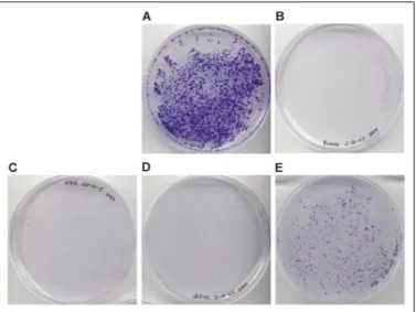
We transfected Huh-7.5 human hepatoma cells with mutant HCV RNA and monitored the cells for replication-dependent growth. Cells transfected with S1179I HCV RNA formed large, densely spaced colo-nies after 2–3 weeks of growth (Fig. 5A). Cells transfected with the mutant HCV RNA showed significantly reduced colony formation, indicating that the targeted NS3 surface region is important for efficient viral replication. The ⌬543–545 and D543K/H545D/Q549A (NS3h KDA) mutants were of particular interest, since they supported little or no cell growth (Fig. 5,CandD). The R587D/L588D/K589D/T591D mutation (NS3h DDDD), at a different site within the protein interface than the⌬543–545 and NS3h KDA mutations, had a visible but less dramatic effect on growth (Fig. 5E).
We performed a Western analysis with NS3 antibody on transfected cell lysates in order to determine whether NS3 protein expression was affected by any of the mutations (Fig. 6). The⌬543–545 mutant NS3 protein was present at significantly lower concentration than observed in S1179I-transfected cells. However, no impairment of translation was observed for either of the two substitution mutants. Quantitative com-parison with purified NS3h standards indicated that these similar expression levels were not an artifact of saturating assay conditions (supplemental Fig. 1).
Biochemical Analysis of NS3h Mutants—In order to determine the effects of the surface mutations on the biochemical activities of NS3h, we overexpressed and purified NS3h KDA and NS3h DDDD in a pro-karyotic expression system and compared the binding and enzymatic activities of the mutant NS3h proteins with those of wild type. We measured the RNA binding affinity of the mutant and wild type enzymes
FIGURE 3.A, the terminal region of the oligonucleotide (aqua) interacts with NS3h chain A (red) via a hydrogen bond between Thr269
(T269) and the phosphate backbone and ring stacking between nucleotide dU1and Trp
501
(W501;green).B, at the point where the oligonucleotide (aqua) enters the binding groove of chain B (blue), interactions identical to those with chain A are observed (Trp501
-dU ring stacking and Thr269
-phosphate hydrogen bond formation).
FIGURE 4.Depiction of the dimer structure involving chains A (red) and B (blue) illustrating the residues involved in the protein-protein interaction.A, five sets of residues are identified at the dimer interface, including His545
–Ala553
(purple) and Cys584
–Thr591
(orange) of chain B and Thr435
–Gln453
(yellow), Thr477
–Ser488
(green), and Val524
–Gln536
(cyan) of chain A.B, a close-up view of the dimer interface. A pair of hydro-gen bonding partners, Thr450
of chain A and Gln549
using a fluorescein-labeled oligonucleotide substrate (Fig. 7). The NS3h KDA mutation did not impair binding to RNA, but the NS3h DDDD mutation severely reduced binding affinity as measured by fluorescence polarization. Poly(U)-stimulated ATP hydrolysis of the mutant and wild type enzymes was measured, and no significant differences were observed between the activities of the wild type and NS3h KDA mutant (Fig. 8). The activity of the NS3h DDDD mutant was severely impaired, probably as a consequence of its reduced nucleic acid binding affinity.
Unwinding of nucleic acid was measured by using a standard helicase assay (Fig. 9A) with a substrate (45/30-mer) containing 30 base pairs with a 15-nucleotide 3⬘single-stranded overhang. Very little unwinding of an RNA substrate by NS3h was observed (Fig. 9B), consistent with a recent report describing the lack of RNA unwinding activity of NS3h (27), so unwinding rates were determined using a DNA substrate (30). Under single turnover conditions, NS3h exhibited a lag phase similar to that observed previously for a substrate of similar length (Fig. 10) (32). The lag phase represents multiple steps that are believed to occur prior to complete unwinding of the duplex (36, 37, 41). However, NS3h KDA unwound the substrate with a similar lag phase as the wild type enzyme but somewhat lower amplitude (Fig. 10). Very little unwinding was observed with NS3h DDDD, consistent with the poor binding to nucleic acid. The amplitude for unwinding reflects the degree of processivity of the enzyme (30, 32, 42); therefore, NS3h KDA has slightly lower
proces-sivity than NS3h. Increasing the enzyme concentration resulted in higher amplitudes for each enzyme. We previously reported that bind-ing of multiple full-length NS3 molecules to this substrate increases the amplitude for unwinding (30). The results here with NS3h are consist-ent with the observation for full-length NS3.
DISCUSSION
We describe here the first x-ray crystal structure of NS3 helicase bound to a DNA molecule of sufficient length to accommodate binding of two molecules of enzyme. Our structure shows a nucleic acid binding site for each monomer that is consistent with a published crystal struc-ture of a single monomer bound to a shorter DNA molecule (23). In each case, the DNA binds to a cleft between domains 1 and 2 on one side and domain 3 on the other. DNA binding is not sequence-specific, with the majority of protein-DNA contacts involving the phosphate back-bone. The structure also reveals a protein-protein interface between two DNA-bound helicase monomers. The protein-protein contacts are mostly hydrophobic and involve domain 2 of one subunit (chain A) and domain 3 of the second subunit (chain B). The two nucleic acid binding sites of the dimer are therefore aligned in such a way as to force a significant bend in the bound DNA that appears to induce a base stack-ing motif in the oligonucleotide.
The structure and function of HCV helicase have been investigated extensively. Important amino acid sequence motifs that play critical roles in biochemical function have been identified in both domains 1
FIGURE 5.Growth of Huh-7 cells after transfection with wild type and mutant forms of the HCV replicon.Colony formation of Huh-7 cells was monitored over a period of 3 weeks following transfection by HCV replicon RNA. Colonies were stained with 0.1% crystal violet. Representative plate images for each transfection are as follows.A, S1179I RNA;B, blank transfection (no RNA);C, D543K/H545D/Q549A mutant RNA;D,⌬543–545 mutant RNA;E, R587D/L588D/K589D/T591D mutant RNA. Colony-forming units per microgram of RNA for each experiment were as follows: 15,560⫾940, 2,720⫾440, and 7,760⫾1,072 for the S1179I RNA, D543K/H545D/Q549A mutant RNA, and R587D/ L588D/K589D/T591D mutant RNA, respectively. No colonies were observed for the blank treatment or for the⌬543–545 mutant RNA.
FIGURE 6.Western analysis of HCV-transfected Huh-7.5 cell lysates.5⫻105
cells from each lysate were loaded on a 10% polyacrylamide gel. The gel was blotted onto a polyvinylidene difluoride membrane, and the blot was incubated with rabbit anti-NS3. Primary antibody binding was detected by chemiluminescence with horseradish perox-idase-conjugated anti-rabbit IgG. Thelane marked NS3shows purified NS3. Thelane marked Cshows the control transfection with no HCV RNA.WT, wild type.
FIGURE 7.Binding of mutant NS3h to fluorescein-labeled U20RNA.Nucleic acid bind-ing was determined by measurbind-ing fluorescence polarization followbind-ing incubation of protein and nucleic acid at 37 °C. Data were fit to a hyperbola using Kaleidagraph soft-ware.A, NS3h wild type (F) bound to RNA with aK
Dof 47⫾5 nM, and NS3h KDA (E)
bound to RNA with aKDof 38⫾4 nM. NS3h DDDD (f) did not bind with high enough
affinity to determine a binding constant under these conditions.
FIGURE 8.ATPase activity of mutant NS3h (50 nM) was measured as a function of nucleic acid concentration in a coupled spectrophotometric assay.Data represent the average of three independent experiments and were fit to a hyperbola using Kalei-dagraph software. Specific activity of NS3h wild type (F) and NS3h KDA (E) were com-parable at 126 and 108 s⫺1, respectively, in the presence of saturating poly(U). Specific
activity of NS3h DDDD (f) was lower at 70 s⫺1. The concentration of nucleic acid (K act)
required to reach one-halfVmaxwas 0.89, 0.68, and 5.84Mfor NS3h wild type, NS3h KDA,
and 2. The function of domain 3 has been less well characterized. Domain 3 of superfamily II helicases is less conserved compared with domains 1 and 2, and there is no apparent homology in domain 3 among the different helicase superfamilies (43). However, the sequence of domain 3 is highly conserved among HCV isolates (supplemental Fig. 2). In the structure reported here, surface regions on domain 3 appear to interact with domain 2 of the second molecule. Biological studies dem-onstrate that mutations in these surface regions interfere with viral rep-lication in cell culture. Although it remains difficult to speculate as to its precise function, domain 3 may play a role in mediating the formation of protein-protein complexes between NS3 monomers or between NS3 and other proteins in addition to its critical role in substrate binding.
Several models for helicase function of NS3 have been proposed. Each mechanism is driven by binding and hydrolysis of ATP, which is thought to drive the enzyme through multiple conformational states. One model is the proposed inchworm-type mechanism in which the helicase repeatedly releases and rebinds a single strand of the nucleic acid substrate. Such a mechanism has been described for PcrA helicase (44) as well as for NS3h (45). The structure reported here shows two interacting monomers bound in series to the same DNA strand. Heli-cases that dimerize have been studied extensively by the Lohman labo-ratory, and a dimeric inchworm model has been suggested forE. coli
Rep helicase (46). The alignment of the NS3h molecules along nucleic acid would favor some form of inchworm mechanism rather than a hand-over-hand mechanism, because the protein interface does not appear to be sufficient to maintain protein-protien interactions in the absence of nucleic acid binding. It is interesting that the dimeric struc-ture reported here does not appear to impede flexible motions between domains 1 and 2 of the enzyme that are proposed to play a role in the inchworm mechanism (23). A model for NS3h activity involving Brownian motion was recently proposed based on the observation of two states of DNA binding (strong affinity and weak affinity) in the presence or absence of ATP (47). This “Brownian motor” model does not require protein-protein interactions for helicase activity. However, the model suggests that when multiple NS3h molecules bind to the same substrate molecule, the adjacent enzymes may enhance the net
activity by reducing the possibility for backwards sliding of the helicase during the unwinding cycle. Our own work is consistent with this gen-eral concept, in which multiple molecules of helicase bound to the same substrate enhance unwinding activity (30).
Recently, a model for unwinding by full-length NS3 was proposed that suggests a dimeric form of the enzyme based in part on an 18-bp step size determined by high resolution kinetic measurements (25). The structure reported here is consistent with the proposed dimeric form of the enzyme. However, unwinding data for the full-length form of NS3 indicate that a larger oligomer or substrate saturation may be required for optimal activity (30). NS3 is reportedly less processive in the absence of its co-factor, NS4A (42), so final conclusions regarding the oligomeric form required for optimal unwinding await detailed kinetic and bio-physical studies of the NS3-4A complex. NS3h exhibits relatively weak protein-protein interactions, whereas full-length NS3 appears to inter-act with itself more strongly,7which may account for some of the dif-ferences in activities observed between the two forms of the enzyme. For example, NS3h unwound only 17% of the 45/30-mer (Fig. 10), whereas full-length NS3 unwound greater than 80% of the same substrate under the same conditions (30). Therefore, NS3h exhibits lower processivity in DNA unwinding than the full-length protein, which may reflect the relative strength of protein-protein interactions.
The mutations at the interface of the putative NS3h dimer were
7K. D. Raney and Y. Chen, manuscript in preparation.
FIGURE 10.A two-step mixing technique was used to measure single-turnover unwinding in the presence of excess NS3h.A, NS3h (1M) or one of the mutant enzymes was rapidly mixed with substrate followed by a 10-s incubation. A second, rapid mixing step followed, in which ATP, Mg2⫹, and protein trap (poly(dT)) were added. The
quench solution was placed in the receiving vial. Single-stranded DNA (ssDNA) was sep-arated from double-stranded DNA by native polyacrylamide electrophoresis, and the fractions were quantitified by using ImageQuant software. Data were fit according to Equation 1, resulting in unwinding rates of 3.1⫾0.1 s⫺1, 3.7⫾0.1 s⫺1, and 4.5⫾0.9 s⫺1
for NS3h (F), NS3h KDA (⽧), and NS3h DDDD (f), respectively.B, unwinding amplitude
versusconcentration of NS3h and NS3h mutant enzymes. Bench top experiments were used to determine the amplitude of product formation under single turnover conditions. The amplitudes for NS3h (F), NS3h KDA (⽧), and NS3h DDDD (f) were plotted as a function of enzyme concentration. The error bars represent S.D. of triplicate measurements.
FIGURE 9.A, the assay for measuring helicase unwinding activity is depicted. A partial duplex substrate containing 30 bp and 15 nucleotides of single-stranded overhang (45/ 30-mer) was incubated with NS3h in the presence of ATP and Mg2⫹, leading to
unwind-ing of the duplex.B, comparison of unwinding of DNA and RNA substrates by NS3h (representative gels). Otherwise identical 250 nMduplexed DNA and RNA substrates were incubated with 100 nMNS3h. Reactions were initiated by the addition of 5 mMATP and 10 mMMgCl2and quenched by the addition of 200 mMEDTA, 0.7% SDS.ssDNAand
designed to disrupt protein-protein interactions. These mutations would not be expected to disrupt the biochemical activities of mono-meric NS3h. Indeed, NS3h KDA exhibits activities that mirror the wild-type enzyme in terms of nucleic acid binding and ATPase activity. The only biochemical activity of NS3h KDA that is reduced is processivity, which may reflect reduced protein-protein interactions. However, the reduction in amplitude for unwinding under single turnover conditions is only⬃2-fold, which would seem unlikely to be responsible for the dramatic reduction in HCV replicative capacity exhibited with the NS3h KDA mutations. Hence, the biochemical results are not sufficient to explain the biological results. The results with NS3h DDDD further emphasize this point. NS3h DDDD has much lower affinity for nucleic acid than wild-type NS3h, resulting in little product formation under single turnover unwinding conditions. However, the replicative capac-ity of the HCV replicon containing the DDDD mutation is greater than the HCV replicon containing the KDA mutation. Thus, the biochemical activities that are typically measured of NS3h do not appear to reflect all of the biological activities of this enzyme.
It remains a strong possibility that the protein surface implicated in for-mation of a dimeric helicasein vitromediates additional interactionsin vivo
that are required for formation of a multiprotein viral replication complex. For example, this protein surface might bind to the NS5B RNA polymerase to form part of the HCV replication machinery. NS5B has been shown to interact with NS3 (48). Several reports support the idea that HCV replica-tion takes place at specific membrane-associated sites (17, 49, 50). NS3, NS5B, and other nonstructural viral proteins co-localize in replicon-trans-fected cells within the endoplasmic reticulum (17). A recent report suggests that lipid rafts may support viral replication (18). Insertion of active viral transcription complexes into cellular membranes would presumably require some sort of coordinated membrane-targeting and assembly proc-ess. It is reasonable to hypothesize that this assembly process requires spe-cific protein-protein and protein-DNA interactions that are not necessary forin vitroenzyme activity. The crystal structure described here may rep-resent a protein complex required for some function of NS3 not directly related to unwinding. For example, the helicase dimer (or larger oligomer) may sequester single-stranded nucleic acids that have already been acted on by the HCV replication machinery or may play a role in formation of viral RNA secondary structures required for initiation of transcription or trans-lation. A recent report suggests that HCV RNA is an integral part of a viral nucleoprotein replication complex (18). The protein-protein interaction suggested by the crystal structure could be a subunit of such a complex.
By solving the x-ray crystal structure of two molecules of NS3 helicase bound to the same oligonucleotide, we have identified a region on the surface of the protein that is required for efficient viral replication. The amino acid residues in this region are highly conserved among different HCV isolates. The surface region appears to be capable of mediating protein-protein interactions but does not appear to be essential in the nucleic acid binding or known enzymatic activities of NS3h. These data illustrate that NS3 has additional biochemical activities and/or mediates protein-protein interactionsin vivo through domain 3 that are not reflected in standard helicase assays.
Acknowledgments—We thank Brandy Tharp and Jamie Arnold for providing technical assistance and Robert Eoff for synthesis and purification of oligonucleotides.
REFERENCES
1. Tan, S. L., Pause, A., Shi, Y., and Sonenberg, N. (2002)Nat. Rev. Drug Discov.1, 867– 881
2. Giannini, C., and Brechot, C. (2003)Cell Death. Differ.10,Suppl. 1, S27–S38 3. Penin, F., Dubuisson, J., Rey, F. A., Moradpour, D., and Pawlotsky, J. M. (2004)
Hepa-tology39,5–19
4. De Francesco, R., and Rice, C. M. (2003)Clin. Liver Dis.7,211–242
5. Blight, K. J., Kolykhalov, A. A., Reed, K. E., Agapov, E. V., and Rice, C. M. (1998)
Antivir. Ther.3,71– 81
6. Reed, K. E., Grakoui, A., and Rice, C. M. (1995)J. Virol.69,4127– 4136
7. Bartenschlager, R., Ahlborn-Laake, L., Mous, J., and Jacobsen, H. (1993)J. Virol.67, 3835–3844
8. Kim, D. W., Gwack, Y., Han, J. H., and Choe, J. (1995)Biochem. Biophys. Res. Com-mun.215,160 –166
9. Failla, C., Tomei, L., and De Francesco, R. (1994)J. Virol.68,3753–3760 10. Behrens, S. E., Tomei, L., and De Francesco, R. (1996)EMBO J.15,12–22 11. Enomoto, N., Sakuma, I., Asahina, Y., Kurosaki, M., Murakami, T., Yamamoto, C.,
Izumi, N., Marumo, F., and Sato, C. (1995)J. Clin. Invest.96,224 –230 12. Blight, K. J., Kolykhalov, A. A., and Rice, C. M. (2000)Science290,1972–1974 13. Heller, T., Saito, S., Auerbach, J., Williams, T., Moreen, T. R., Jazwinski, A., Cruz, B.,
Jeurkar, N., Sapp, R., Luo, G., and Liang, T. J. (2005)Proc. Natl. Acad. Sci. U. S. A.
14. Lohmann, V., Korner, F., Koch, J., Herian, U., Theilmann, L., and Bartenschlager, R. (1999)Science285,110 –113
15. Lin, C., Thomson, J. A., and Rice, C. M. (1995)J. Virol.69,4373– 4380
16. Qin, W., Luo, H., Nomura, T., Hayashi, N., Yamashita, T., and Murakami, S. (2002)
J. Biol. Chem.277,2132–2137
17. Mottola, G., Cardinali, G., Ceccacci, A., Trozzi, C., Bartholomew, L., Torrisi, M. R., Pedrazzini, E., Bonatti, S., and Migliaccio, G. (2002)Virology293,31– 43 18. Shi, S. T., Lee, K. J., Aizaki, H., Hwang, S. B., and Lai, M. M. (2003)J. Virol.77,
4160 – 4168
19. Delagoutte, E., and von Hippel, P. H. (2002)Q. Rev. Biophys.35,431– 478 20. Delagoutte, E., and von Hippel, P. H. (2003)Q. Rev. Biophys.36,1– 69 21. Hall, M. C., and Matson, S. W. (1999)Mol. Microbiol.35,867– 877
22. Cho, H. S., Ha, N. C., Kang, L. W., Chung, K. M., Back, S. H., Jang, S. K., and Oh, B. H. (1998)J. Biol. Chem.273,15045–15052
23. Kim, J. L., Morgenstern, K. A., Griffith, J. P., Dwyer, M. D., Thomson, J. A., Murcko, M. A., Lin, C., and Caron, P. R. (1998)Structure6,89 –100
24. Gallinari, P., Brennan, D., Nardi, C., Brunetti, M., Tomei, L., Steinkuhler, C., and De Francesco, R. (1998)J. Virol.72,6758 – 6769
25. Serebrov, V., and Pyle, A. M. (2004)Nature430,476 – 480
26. Locatelli, G. A., Spadari, S., and Maga, G. (2002)Biochemistry41,10332–10342 27. Frick, D. N., Rypma, R. S., Lam, A. M., and Gu, B. (2004)J. Biol. Chem.279,
1269 –1280
28. Levin, M. K., and Patel, S. S. (1999)J. Biol. Chem.274,31839 –31846
29. Yao, N., Reichert, P., Taremi, S. S., Prosise, W. W., and Weber, P. C. (1999)Struct. Fold Des7,1353–1363
30. Tackett, A. J., Chen, Y., Cameron, C. E., and Raney, K. D. (2005)J. Biol. Chem.280, 10797–10806
31. Levin, M. K., and Patel, S. S. (2002)J. Biol. Chem.277,29377–29385
32. Levin, M. K., Wang, Y. H., and Patel, S. S. (2004)J. Biol. Chem.279,26005–26012 33. Navaza, J. (2001)Acta Crystallogr. D. Biol. Crystallogr.57,1367–1372
34. Brunger, A. T., Adams, P. D., Clore, G. M., DeLano, W. L., Gros, P., Grosse-Kunstleve, R. W., Jiang, J. S., Kuszewski, J., Nilges, M., Pannu, N. S., Read, R. J., Rice, L. M., Simonson, T., and Warren, G. L. (1998)Acta Crystallogr. D Biol. Crystallogr.54, 905–921
35. McRee, D. E. (1999)J. Struct. Biol.125,156 –165 36. Ali, J. A., and Lohman, T. M. (1997)Science275,377–380
37. Lucius, A. L., Maluf, N. K., Fischer, C. J., and Lohman, T. M. (2003)Biophys. J.85, 2224 –2239
38. Hooft, R. W., Vriend, G., Sander, C., and Abola, E. E. (1996)Nature381,272 39. Parkinson, G. (1996)Acta Crystallogr. Sect. D52,57– 64
40. Vriend, G. (1990)J. Mol. Graph.8,52–56
41. Galletto, R., Jezewska, M. J., and Bujalowski, W. (2004)J. Mol. Biol.343,83–99 42. Pang, P. S., Jankowsky, E., Planet, P. J., and Pyle, A. M. (2002)EMBO J.21,1168 –1176 43. Singleton, M. R., and Wigley, D. B. (2002)J. Bacteriol.184,1819 –1826
44. Velankar, S. S., Soultanas, P., Dillingham, M. S., Subramanya, H. S., and Wigley, D. B. (1999)Cell97,75– 84
45. Kim, J. L., Morgenstern, K. A., Lin, C., Fox, T., Dwyer, M. D., Landro, J. A., Chambers, S. P., Markland, W., Lepre, C. A., O’Malley, E. T., Harbeson, S. L., Rice, C. M., Murcko, M. A., Caron, P. R., and Thomson, J. A. (1996)Cell87,343–355
46. Cheng, W., Brendza, K. M., Gauss, G. H., Korolev, S., Waksman, G., and Lohman, T. M. (2002)Proc. Natl. Acad. Sci. U. S. A.99,16006 –16011
47. Levin, M. K., Gurjar, M. M., and Patel, S. S. (2003)J. Biol. Chem.278,23311–23316 48. Piccininni, S., Varaklioti, A., Nardelli, M., Dave, B., Raney, K. D., and McCarthy, J. E.
(2002)J. Biol. Chem.277,45670 – 45679
49. Wolk, B., Sansonno, D., Krausslich, H. G., Dammacco, F., Rice, C. M., Blum, H. E., and Moradpour, D. (2000)J. Virol.74,2293–2304