THE EFFECT OF ESTROGEN ON INFLAMMATORY MARKERS FOLLOWING PROLONGED AEROBIC EXERCISE IN EUMENORRHEIC WOMEN
Elizabeth A. Walz
A thesis submitted to the faculty of the University of North Carolina at Chapel Hill in partial fulfillment of the requirement for the degree of Masters of Arts in the Department
of Exercise and Sports Science (Exercise Physiology).
Chapel Hill Spring 2014
Approved by:
Anthony C. Hackney
Abbie Smith-Ryan
ABSTRACT
Elizabeth A. Walz: The Effect of Estrogen on Inflammatory Markers Following Prolonged Aerobic Exercise in Eumenorrheic Women
(Under the direction of Anthony C. Hackney)
The study purpose was to determine if estrogen (E2) concentrations attenuate
inflammation after exercise-induced muscle damage. Blood responses of
pro-inflammatory cytokine biomarker TNF-, and pro- and anti-inflammatory cytokine biomarker IL-6 were measured. Ten, eumenorrheic, endurance-trained women
(Mean±SD; 21±1 years, 24.1±2.8 body fat%) were studied. They completed a 60 minute
running protocol at ~60-65% of their oxygen uptake (VO2peak 53.5±4.7ml/kg/min) during
two hormonal conditions (low E2 and high E2). Inflammation was assessed at rest,
immediately post exercise, 30 minutes post exercise, and 24 hours post exercise. There
was not a significant interaction effect for TNF- (p=0.60). There was a significant interaction effect for IL-6 (p=0.001). The response at 30 minutes post exercise was
significantly elevated from rest and significantly reduced in high E2. Results suggest high
E2 conditions attenuate the IL-6 response. Due to the pro- and anti-inflammatory
ACKNOWLEDGEMENTS
I am very appreciative of the efforts of Dr. Anthony C. Hackney for helping me
accomplish this project. I am grateful for his encouragement, guidance, and knowledge.
Thank you to my committee members for their efforts, reviewing my work and giving me
valuable feedback. I am in debt to my subjects; I am so grateful they volunteered to take
part in my thesis and I thank them for their time. Thank you to Michelle, Timmons,
TABLE OF CONTENTS
PAGE
LIST OF TABLES ... ix
LIST OF FIGURES ... x
CHAPTER I: INTRODUCTION ... 1
Basis For Study: ... 1
Purpose ... 5
Research Hypothesis ... 5
Definition of terms ... 5
Delayed Onset Muscle Soreness (DOMS) ... 6
Estrogen (E2) ... 6
Eumenorrhea ... 6
Moderate intensity prolonged exercise bout ... 6
Inflammatory Response ... 7
Delimitations ... 7
Limitations ... 8
Significance of study ... 8
CHAPTER II: REVIEW OF LITERATURE ... 9
Antioxidant, membrane stabilizing and gene regulating properties of E2: ... 9
Antioxidant properties of E2 ... 9
Gene regulating properties of E2 ... 11
Estrogenic effect on exercise-induced skeletal muscle damage, inflammation and repair... 12
Skeletal muscle damage ... 12
Acute inflammatory response ... 14
Skeletal muscle repair and regeneration ... 16
Interaction of E2 and progesterone ... 17
Role of E2 and cytokines in exercise induced muscle damage, inflammation and repair... 18
CHAPTER III: METHODOLOGY ... 24
Participants ... 24
Instrumentation ... 25
Protocol ... 26
Pre-Screening ... 26
Orientation Session I ... 26
Follow Up Blood Draws Session III and V ... 30
Blood Procedures ... 30
Hematocrit ... 30
Hemoglobin ... 31
Plasma Volume Shift ... 31
Cytokines (TNF-α and IL-6), E2 ... 31
Data Analysis ... 32
CHAPTER IV: RESULTS ... 33
VO2 peak testing ... 34
Hormonal condition determination ... 34
Prolonged treadmill running bout ... 35
Blood responses to prolonged exercise ... 37
Tumor necrosis factor-α (TNF-α) ... 37
Interleukin-6 (IL-6) ... 37
CHAPTER V: DISCUSSION ... 40
Hormonal condition determination ... 40
Prolonged aerobic exercise session ... 41
Blood responses ... 42
Tumor Necrosis Factor (TNF-α) ... 42
Interleukin-6 (IL-6) ... 43
APPENDIX A: SAMPLE SIZE CALCUATIONS ... 48
APPENDIX B: INFORMED CONSENT ... 49
APPENDIX C: MEDICAL SREENING FORMS ... 56
Physical Examination Screening ... 56
Medical History Questionnaire ... 57
APPENDIX D: ACSM METABOLIC EQUATION FOR DETERMINING EXERCISE INTENSITY .. 61
APPENDIX E: MENSTRUAL CYCLE QUESTIONNAIRE ... 62
APPENDIX F: FOOD LOG ... 63
APPENDIX G: DATA COLLECTIONS FORM ... 64
Experimental Session ... 65
Hematocrit and Hemoglobin ... 66
Subject Compliance ... 67
APPENDIX H: ASSAY SHEETS ... 68
Estrogen Assay Information ... 68
TNF- Assay Information ... 69
IL-6 Assay Information ... 72
LIST OF TABLES
Table
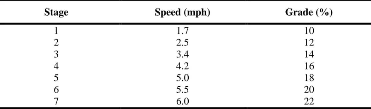
1. Speed (miles per hour; mph) and grade for each three-minute stage
of the Bruce protocol incremental exercise test……….………....28
2. Descriptive data for subjects………..34
3. Descriptive data (mean ± SD) for VO2, HR and RPE for each
prolonged running bouts in each hormonal condition (LE and HE…………...…36
4. Mean (± SD) plasma volume shifts from rest to immediately post exercise (R-IP), rest to 30 minutes post exercise (R-30P) and
rest to 24 hours post exercise (R-24P) ………..36
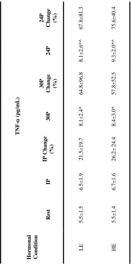
5. Mean (± SD) for TNF- at rest, immediately post exercise (IP), 30 minutes post exercise (30P), and 24 hours post exercise (24P). An * indicates significant increase from rest, while an ^ indicates
significant increase from IP………...………38
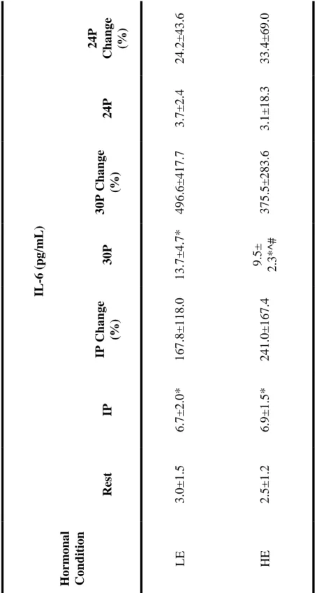
6. Mean (± SD) for IL-6 at rest, immediately post exercise (IP), 30 minutes post exercise (30P), and 24 hours post exercise (24P). An * indicates significance from rest, an ^ indicates significantly lower IL-6 compared to LE, while a # indicates an interaction effect
LIST OF FIGURES Figure
1. Theoretical model illustrating the potential protective role of estrogen in
skeletal muscle damage, inflammation, and repair………...….22
CHAPTER I
INTRODUCTION
Basis For Study:
There has been debate surrounding the influence of 17-estradiol (E2) on skeletal
muscle, and whether E2 has a protective influence on skeletal muscle damage,
inflammation, and repair after prolonged aerobic exercise. This area of research is limited.
There are multiple studies indicating that high levels of E2 attenuate circulating cytokines,
and as a result attenuate inflammation during non-exercise induced inflammatory
responses (Pfeilschifter et al., 2002; Pottratz et al., 1994; Puder et al., 2001; Schwarz et
al., 2000). This indicates that there may be a similar phenomenon occurring during
exercise induced inflammatory responses. A clear relationship between E2 and
inflammation after exercise induced muscle damage, in women, is not well defined in
existing literature. Some studies suggest there is an estrogenic effect on circulating
cytokines, while other do not (Dieli-Conwright et al., 2009; Ives et al., 2011; Timmons et
al., 2005; Timmons et al., 2006). These conflicting results may be due to study
limitations, such as small sample sizes, lack of a treatment condition, or inadequate
exercise stimuli (Ives et al., 2011). The intent of the current study was to overcome these
limitations, with sufficient power, a treatment condition, and adequate exercise stimulus.
Unfortunately, due to the difficulty of recruiting subjects, the study was underpowered
important consideration for women designing training programs to improve performance
and minimize risk.
E2 is a steroid hormone-molecule that plays an important role in maintaining and
regulating sexual and reproductive function in females. Additionally, E2 exerts an
influence on other physiological systems in females, such as the cardiovascular,
musculoskeletal, immune and central nervous systems. The form of E2 that has the
largest effect on these systems is 17β-estradiol (Enns & Tiidus, 2010). There are three
mechanisms by which E2 might exert a protective effect on skeletal muscle damage,
inflammation, and repair. E2 may act as an antioxidant, membrane stabilizer, and gene
regulator (Perskey et al., 1999). Similar to other antioxidants with similar structures, it
has been suggested that the phenolic hydroxyl group on E2 donates a hydrogen atom
which disrupts free radical damaging cascades, minimizes lipid peroxidation and thus
limits cell membrane damage (Perksy et al., 1999; Sugioka et al., 1987). E2 may also
stabilize membranes by intercalating with membrane phospholipids, similar to cholesterol
(Perksy et al., 1999). Finally E2 may also exert protective effects through gene regulation,
affecting cytokine and cell-adhesion activity, as well as activation of satellite cells (Smith
et al., 2000; Enns et al., 2008; Enns & Tiidus, 2008). With these protective properties, E2
may attenuate exercise-induced muscle damage, and inflammation while also facilitating
repair in skeletal muscle.
While exercise-induced oxidative stress and membrane damage are important
signals for the skeletal muscle tissue to adapt, there are potential negative side-effects,
2001; Komulainen et al., 1999). E2 may play a role in decreasing the severity of these
side-effects during high intensity exercise (Bar et al., 1988; Roth et al., 2000,
Komulainen et al., 1999). After exercise induced muscle damage, inflammatory
processes are activated to clear and repair the damaged cell. These processes include an
infiltration of fluid, plasma proteins and circulating leukocytes, which is mediated by a
variety of cytokines (St. Pierre Schneider et al. 1999; Tiidus et al. 2001). The cytokines
of interest in this study are tumor necrosis factor-alpha (TNF-α) and interleukin 6 (IL-6).
TNF-α is considered to be a pro-inflammatory cytokine, produced to recruit more
leukocytes to the inflamed tissues. IL-6 has been shown to be both a pro- and
anti-inflammatory cytokine. Until recently, researchers considered IL-6 to be a
pro-inflammatory cytokine. For example, MacIntyre et al. (2001) reported that neutrophil
and IL-6 increased up to 6 h post-exercise, and there was a significant relationship
between IL-6 and DOMS, suggesting that IL-6 is a pro-inflammatory cytokine that
initiates inflammation. It is thought that anti-inflammatory IL-6 may be released locally
in the muscle after strenuous exercise and the amount released is dependent on the type
of physical activity, as well as the duration of exercise (Hamer & Karageorghis 2007).
Currently, the literature is unclear as to the exact role this cytokine plays in the
inflammatory response (Pedersen et al., 2004; Smith et al., 2000).
Some researchers have suggested that E2 may positively influence inflammation
by attenuating pro-inflammatory cytokine TNF-α, reducing leukocyte activity and
collateral damage to healthy cells, as well as initiating satellite cell activity (Tiidus et al.,
2001). Satellite cells activation initiates cell growth and repair. An estrogenic effect on
displayed a greater increase in satellite cell number when compared to men. Additionally,
Enns et al. (2008) demonstrated that satellite cell activity increased in E2 supplemented,
exercised rats compared to other controls. The Enns study also suggests that E2 may
increase satellite cell activation through receptor-mediated mechanisms (Enns et al.,
2008). As evidenced here, there are strong reasons to believe that E2 exerts a protective
effect on exercise induced muscle damage, inflammation, and repair.
The present study focused on inflammatory mechanisms after exercise induced
skeletal muscle damage; specifically the effect of E2 on inflammatory markers, TNF-α
and IL-6. Since IL-6 has been shown to have both anti- and pro-inflammatory properties,
it is not primary variable of interest. It was included to add to investigations previously
produced from this research group. This particular study focused on the effect of E2 on
TNF-α during the inflammatory response after an exercise induced muscle damage bout.
Ostrowski et al. (1999) reported that strenuous exercise increased concentrations of TNF-α. Chao et al. (1995) found that TNF-α fluctuated with changes in estrogen
concentrations. Additionally Schwarz et al. (2000) found that the release of TNF-α was
diminished in pre-menopausal women during the luteal phase of the menstrual cycle,
when compared to the follicular phase, which suggests an anti-inflammatory response.
This author also found that TNF-α and IL-6 were inhibited in females, compared to male
controls. These studies provide evidence that there may be a relationship between E2
and inflammatory markers. Thus with this in mind, a key goal of the present study was to
clarify the role of E2 on TNF-α after exercise induced muscle damage. In this study, the
was IL-6; each was assessed at four time points (at rest, immediately post-exercise, 30
minutes post-exercise, and 24 hours post exercise).
Purpose
The purpose of this study was to determine if E2 levels influence
pro-inflammatory marker TNF-α following a moderate intensity exercise protocol in
eumenorrheic women.
A secondary purpose of this study was to determine if E2 levels influence pro- or
anti-inflammatory cytokine IL-6 following a moderate intensity exercise protocol in
eumenorrheic women.
Research Hypothesis
If E2 is related to pro-inflammatory cytokine TNF-α, then higher E2 levels in
eumenorrheic women will attenuate TNF-α concentrations after a moderate intensity
exercise protocol.
A secondary research hypothesis is if E2 is related to pro- or anti-inflammatory
cytokine IL-6, then higher E2 levels in eumenorrheic women will attenuate IL-6
concentrations after a moderate intensity exercise protocol.
Definition of terms
Cytokines
Cytokines are released in response to stress, infection, illness and inflammation,
as part of the innate immune response of the human body. Cytokines mediate immune
responses activated in order to repair and clear damaged cells. Cytokines can be pro- or
Delayed Onset Muscle Soreness (DOMS)
Following high intensity exercise, muscle injury can cause myofiber damage,
including sarcolemma disturbances, swelling or disturbances of the contractile proteins,
cytoskeletal and extracellular matrix damage and disturbance (Kendall & Eston, 2002).
Estrogen (E2)
18-carbon steroid molecule-hormone, secreted by the ovaries in women and (to a
much lesser extent) in the testes in men. E2 is important in the maintenance of normal
sexual and reproductive function in females, but also plays a role in many cardiovascular,
musculoskeletal, immune and central nervous system functions. E2 exists in several
forms, the most prominent is estradiol -17, and is known to fluctuate across the menstrual cycle (Ruggerio & Likis 2002; Enns & Tiidus 2010).
Eumenorrhea
Normal menstrual cycle, typically seen in women ages 18-30 years, in which
cycles establish an early follicular phase increase in follicle-stimulating hormone, a
pre-ovulatory luteinizing hormone peak, a luteal phase of at least 11 days, and a progesterone
peak greater than 10ng/mL (Sherman & Korenman, 1975). A typical menstrual cycle
lasts 28 days.
Moderate intensity prolonged exercise bout
Exercise performed on a treadmill at 0% grade with a speed equal to ~60-65% of
VO2max for 60 minutes. Previous investigations from the same research group indicate
Inflammatory Response
The inflammation process and satellite cell activation and proliferation is initiated
by local and systemic signals, such as cytokines, growth factors and leukocytes, released
by the injured muscle tissues (Hamer & Karageorghis, 2007).
Leukocytes
During the inflammatory process after muscle damage leukocytes, such as
neutrophils and macrophages, accomplish three tasks: breakdown damaged muscle tissue,
remove the damaged muscle tissues, and restore function of muscle tissues (Kendall &
Eston, 2002).
Delimitations
1. Participants were healthy females between the ages of 18-30 years.
2. Participants were eumenorrheic and not currently taking oral contraceptives or other
hormone therapy six months prior to participation in this study.
3. Had not sustained an injury within the last six months that limited the ability to
exercise or have a doctor’s clearance.
4. Had not been taking anti-inflammatory medicines, such as ibuprofen, naproxen, or
aspirin six months prior to participation in this study.
5. Were not pregnant or become pregnant during the study.
6. Become ill with an immune responding condition, such as a cold or respiratory
infection during the study.
7. Had a current minimum training volume of 3-5 days a week, 45-120 minutes per
session of aerobic activity, and a maximal oxygen consumption (VO2 max) of at least
8. Abstained from strenuous physical activity and maintained a diet similar in calories
and carbohydrate content 24 hours prior to experimental protocol.
Limitations
1. Results may not be applicable to men and some women (amenorrheic,
oligomenorrheic, or post-menopausal).
2. Subjects may not comply with specific pre-test instructions.
3. Cytokine concentrations will be measured in blood, there will not be another method
to verify local cytokine changes (such as muscle messenger RNA [mRNA]).
Significance of study
Understanding the influence of E2 on the inflammatory response following muscle
damage is an important consideration for both women and researchers. Women who
experience amenorrhea or are post-menopause may be missing the potential protective
influence of E2 related to muscle damage, inflammation, and repair. Additionally women
who are eumenorrheic would benefit from knowing the potential protective properties of
estrogen. This information could be used to design training programs that optimize
performance and minimize risk, such as periodization of training based on hormonal
condition. While there are many important training considerations for understanding
estrogenic influence, it is especially crucial to account for estrogenic influence when it
comes to designing research studies involving women. Failing to account for hormonal
fluctuations throughout the menstrual cycle may affect the results of these studies. Lastly,
research in this area is limited and results are contradictory. More research needs to be
done in this area to clarify the role of E2 on the inflammatory response after
CHAPTER II
REVIEW OF LITERATURE
In this review, the influence of E2 on skeletal muscle damage, inflammation, and
repair will be examined. There are three potential mechanisms by which E2 exerts a
protective effect on skeletal muscle. E2 may act as an antioxidant, membrane stabilizer,
and gene regulator (Kendall & Eston, 2002; Enns and Tiidus, 2010). It is a
well-documented phenomenon that muscle damage occurs after strenuous, unaccustomed
exercise (Clarkson et al., 2001). Following exercise-induced skeletal muscle damage,
pro- and anti-inflammatory cytokines and other chemo-attractants facilitate inflammation
and repair. This response involves the recruitment of leukocytes, such as neutrophils and
macrophages, and the activation and proliferation of satellite cells (Belcastro et al., 1998).
This review will discuss how E2 exerts protective mechanisms on skeletal muscle, the
effect of E2 on pro-inflammatory cytokine TNF-, and the pro- or anti-inflammatory
cytokine IL-6 after exercise-induced muscle damage.
Antioxidant, membrane stabilizing and gene regulating properties of E2:
Antioxidant properties of E2
It is well known that during strenuous exercise oxygen consumption increases to
meet metabolic demand. With this increase in oxygen consumption, there is a similar
reduced by oxidative metabolism, there is one free radical produced (Kanter 1998). Thus
oxygen free radicals may rapidly accumulate during strenuous exercise and result in
oxidative damage, production of reactive oxygen species (ROS), and lipid peroxidation,
all of which alter membrane fluidity and cell membrane stability (Sen 1995). In addition
to free radicals produced during oxidative metabolism, free radicals can be produced as a
result of enzyme activity with the recruitment of neutrophils in the inflammatory
response (Fantone et al., 1982). There is a link between excess free radicals, due to
either over-production or a decrease in the effectiveness of antioxidants, and developing
diseases, such as cancer, atherosclerosis, and Alzheimer’s (Persky et al., 1999).
Endogenous production of antioxidants, such as E2, serves to protect skeletal cell
membranes from free radical damage. More specifically, the phenolic hydroxyl group on
estrogen donates a hydrogen atom to disrupt free radical damaging cascades, minimize
lipid peroxidation and thus limit cell membrane damage (Perksy et al., 1999; Sugioka et
al., 1987). Both E2 status in females and intensity of exercise affect lipid peroxidation.
Ayers et al (1998) evaluated the difference between eumenorrheic (measured during high
E2) and amenorrheic athletes’ responses to exercise-induced oxidative stress. These
authors found that there was a greater potential for lipid peroxidation after 15 minutes of
maximal treadmill exercise for amenorrheic athletes when compared to eumenorrheic
athletes. Additionally, Feng et al. (2004) found that physiological levels of E2 could
increase membrane stability, reduce consumption of glutathione (GSH) and Vitamin E
(both of which are antioxidants), and maintain overall antioxidant capability of the
strained muscles in female rats. Additionally the results showed that there was decreased
These studies highlight how skeletal muscle cells might be susceptible to free radical
damage and how E2 may exert protective effects through direct antioxidant actions.
Membrane stabilizing properties of E2
E2 may also be able to exert protective effects through membrane stabilizing
mechanisms. The ability of fat-soluble E2 to interact with phospholipids also contributes
to the membrane stabilizing properties of E2, similar to cholesterol (Whiting et al., 2000).
Whiting et al. (2000) studied the effect of testosterone, progesterone, and E2 on various
liposomes, plasma membranes, and sarcoplasmic reticulum membranes. This group
suggests that E2 has the ability to intercalate with phospholipids, alter the fluidity of
phospholipids, and increase protein mobility in membrane bilayers, all of which may
affect protein function. Although the authors in this study found E2 to increase
membrane fluidity, they demonstrated a mechanism by which steroid hormones influence
these actions. Multiple researchers have suggested the opposite, that E2 decreases
membrane fluidity and stabilizes membrane phospholipids due to its structure and
antioxidant ability (Kendall & Eston 2002; Persky et al., 1999). Nonetheless, E2 may
play an important role in the stabilization of skeletal cell membranes and the conflicting
findings show the need for further research in this area.
Gene regulating properties of E2
Lastly, E2 may potentially exert protective effects on skeletal muscle through gene
regulation. Gene regulation by E2 may affect cytokine and cell adhesion activity, as well
as the activation of satellite cells. Research involving another known antioxidant,
suggested that tocopherol prevents signal transduction of leukocyte-endothelial cell
adhesion. Cytokine production and leukocyte-endothelial cell adhesion are important
factors regulating leukocyte infiltration. In another study Enns et al. (2008) demonstrated
that E2 may increase muscle satellite cell numbers through E2 receptor mediated
mechanisms, indicating upstream gene regulation of satellite cell activation. Furthermore
these authors suggested that the attenuation of exercise-induced muscle damage and
leukocyte infiltration via estrogenic effects was not mediated by E2 receptor mechanisms
(Enns et al., 2008; Enns & Tiidus, 2008). This supports the findings by Yoshikawa and
Yoshida, who suggested that the attenuation of exercise-induced muscle damage and
leukocyte infiltration may be attenuated via estrogenic regulation of endothelial cell
adhesion and leukocyte infiltration. The ability of E2 to decrease leukocyte infiltration
and increase satellite cell activation may be due to the gene regulating properties of E2.
Estrogenic effect on exercise-induced skeletal muscle damage, inflammation and
repair
Skeletal muscle damage
Skeletal muscle damage caused by strenuous, unaccustomed exercise can be
measured directly or indirectly. It can be directly measured via muscle biopsies, or
indirectly measured through muscle strength loss, muscle soreness, and increased muscle
proteins, such as creatine kinase (Clarkson et al., 2001). Additionally, the protein
β-glucoronidase activity can reflect the histopathological state of the cell, which indicates if
As previously mentioned, E2 may have the ability to attenuate skeletal muscle
damage by acting as an antioxidant and membrane stabilizer. As a result of exercise
induced oxidative stress and membrane damage, more creatine kinase is able to permeate
the membrane and elicit a larger inflammatory response (Bar et al., 198; Sewright, 2008,
Roth et al., 2000). Bar et al. (1988) suggested that E2 may have a protective effect on
skeletal muscle damage by showing reduced creatine kinase values after exercise induced
stress. In a more recent study, Sewright (2008) hypothesized that there would be similar
responses between men and women regarding indirect skeletal muscle damage markers,
but there would be sex differences in the variability and distribution of indirect skeletal
muscle damage markers. These authors found that women experienced greater immediate
strength loss, while men showed greater creatine kinase activity. These results indicate
sex differences in fatigue and muscle damage after intense exercise. Sex differences in
fatigue may be due to metabolism, blood flow or intracellular calcium, while sex
differences in muscle damage may be due to estrogenic protective mechanisms.
Additionally, Roth et al. (2000) suggests that E2 levels in women influences the degree of
muscle damage after heavy-resistance strength training three times a week for nine weeks.
Muscle damage increased significantly in older women, from 5 to 17% of muscle fibers
damaged, compared to younger women, from 2 to 5% of muscle fibers damaged.
Taking a different approach to evaluating skeletal muscle damage differences
between males and females, Komulainen et al. (1999) evaluated β-glucoronidase activity,
histological assessment of muscle samples for inflammation, and immunohistochemistry
of structural proteins of muscle fibers, such as actin, desmin, and dystrophin, and
downhill running exercise. β-glucoronidase activity was smaller and histological changes
were slower and less prominent in female rats compared to male rats. Additionally,
immunohistochemical changes in structural and extracellular matrix proteins were
unchanged in female rats when compared to male rats.
Both rodent and human studies suggest that the skeletal muscle cell membranes in
females compared to males, are stronger and better able to resist exercise-induced
skeletal cell membrane damage. This may be due to a protective estrogenic effect on
skeletal muscle cells following damaging exercise.
Acute inflammatory response
To clear and repair skeletal muscle tissue after an acute bout of high intensity
exercise there is an acute inflammatory response in which there is an increase in fluid,
plasma proteins and circulating leukocytes. Vascular endothelial cells, tissue-resident
leukocytes, and circulating leukocytes produce a variety of cytokines that mediate the
inflammatory response. For example, the up-regulation of pro-inflammatory TNF-α is
associated with resident macrophages in the damaged muscle tissue (Smith et al., 2000).
Interestingly IL-6 can be either pro- or anti-inflammatory, depending on how much of it
is released or what is being released along with it. Thus pro-inflammatory IL-6 may be
associated with resident macrophages, while anti-inflammatory IL-6 may be associated
with the exercising muscle tissue, specifically substrate mobilization (Pederson et al.,
2004). There are multiple families of cytokines that play a role in regulating the acute
inflammatory response, including; interleukins (IL), tumor necrosis factors (TNF),
interferons, growth factors, colony stimulating factors (CSFs), and cell adhesion
interest in this study, it is important to understand there are many cytokines that play a
crucial role in inflammation.
The infiltration of leukocytes, primarily neutrophils and macrophages, remove
damaged muscle tissues and stimulate repair processes. St. Pierre Schneider et al. (1999)
induced skeletal muscle injury using an in vivo lengthening contraction model in 50
sexually mature mice. Leukocyte infiltration was assessed after 1, 3, 5, and 7 days of
recovery. While leukocytes invaded muscle fibers in both sexes after 1 day, there were
differences in the subsets of leukocytes between the sexes. The authors concluded that
leukocyte activity associated with the inflammation may be prevented or delayed in
female mice after exercise induced injury. Both neutrophils and macrophages play a
crucial role in removing and repairing damaged muscle tissues. While this activity is
necessary, excessive infiltration of leukocytes can cause an increase in muscle membrane
and oxidative damage. It is still unknown whether E2 enhances or hinders the
inflammation process. E2 may positively influence inflammation by reducing leukocyte
activity, and thus reducing oxidative damage and collateral damage to healthy cells.
However, estrogenic influence that reduces inflammation may result in a diminished
ability to repair damaged muscle tissue (Tiidus et al., 1999, Tiidus et al., 2001).
With exercise induced muscle damage there are disruptions to the sarcomere Z
line and sarcoplasmic reticulum, resulting in changes in calcium concentrations and the
rate of protein degradation. Calpain is a non-lysosomal cysteine protease that degrades
the damaged cytoskeletal and myofibrillar proteins. Protein degradation, induced by
Calpain activity, produces peptide fragments that act as chemo-attractants to neutrophils
and membrane stabilizer, by reducing sarcomere disruption, and calcium disturbances
(Tiidus et al., 2001). Neutrophil activity contributes to further oxidative damage in
muscle tissues via the production of ROS from NADPH oxidase and the production of
hypochlorous acid from hydrogen peroxidase from the myeloperioxidase (MPO) reaction
(Suzuki et al., 1999; Tiidus et al., 1999). MPO activity is known as an indicator of
neutrophil activity in damaged muscle tissue. Tiidus et al., 1999 found there were
significant elevations in muscle MPO activity 24 hours post exercise in male rats
compared to female rats and estrogen supplemented male rats. The authors suggest that
while MPO represents neutrophil activity, infiltration of macrophages at 24 hours post
exercise may have begun, thus MPO activity at 24 hours post exercise is indicative of
overall leukocyte activity and inflammation. Increasing MPO activity plays an important
role in clearing damaged muscle tissue, but can be damaging to healthy muscle tissue.
Further clarification in the literature will be needed in order to elucidate whether an E2
mediated reduction in inflammation is advantageous or not.
Skeletal muscle repair and regeneration
After skeletal muscle cell damage, satellite cells are activated to proliferate and
provide the necessary materials to initiate muscle growth and repair. There may be an
estrogenic effect on the activation and proliferation of satellite cells. For example,
resistance trained women displayed a greater increase in satellite cells when compared to
men (Roth et al., 2001). In another study, histochemical analysis was used to show that
the greatest number of muscle fibers containing total, activated and proliferating satellite
cells were in the exercised, E2 supplemented group of female rats (Enns et al., 2007),
finally exercised and no E2 rats. E2 might increase satellite cell activation through
receptor-mediated mechanisms. For example, when E2 receptors are blocked or there is
an E2 receptor antagonist, exercise and E2-mediated increases in satellite cells are
inhibited (Enns et al., 2008).
Another important promoter of satellite cell propagation might be the infiltration
of leukocytes. Even though it is hypothesized that E2 decreases the leukocyte response
following muscle damage, new research proposes that E2 increases IL-6 levels and nitric
oxide, which are known activators of satellite cells. This means that E2 can both increase
satellite cell production and proliferation and still attenuate the leukocyte response (Enns
& Tiidus, 2010, Tiidus 2003). The effect of E2 on the inflammatory response requires
further research, in order to clarify E2’s role in activation of satellite cells and leukocyte
recruitment. As IL-6 is an important player in these mechanisms, the effect of E2 on IL-6
levels is an important step in identifying these mechanisms.
Interaction of E2 and progesterone
There has been limited research investigating the effects of progesterone and E2
on exercise induced damage, inflammation and repair. Studies involving female
ovariectomized rats treated with E2 showed less neutrophil and macrophage infiltration in
skeletal muscle following eccentric exercise, compared to rats not treated with E2 (Enns
et al., 2008; Iqbal et al., 2008) The reduced inflammatory response in rats treated with E2
may be related to reduced muscle damage as shown by reduced skeletal muscle damage markers, such as β-glucuronidase and creatine kinase (Enns et al., 2008). Reduced
skeletal muscle damage may be a result of the estrogenic muscle membrane stabilizing
progesterone receptors present and there is the possibility that there are interactive effects
in response to exercise induced muscle damage (Iqbal et al., 2008). To investigate these
interactive effects between E2 and progesterone, Iqbal et al., (2008), compared the
concentrations of each hormone at 24 hours post exercise. The female rats were divided
into 4 exercise and 4 control groups (sham, E2, progesterone, and a combination of E2
plus progesterone) following 8 days of hormone replacement. They confirmed that E2
attenuated leukocyte infiltration following exercise induced muscle damage. They also
found that progesterone also attenuated leukocyte infiltration, but to a smaller extent than
E2. In rats supplemented with both progesterone and E2, leukocyte infiltration was not
significantly different from the E2 only group, suggesting that progesterone does not
affect estrogenic influence.
Role of E2 and cytokines in exercise induced muscle damage, inflammation and
repair
The acute inflammatory response and satellite cell activation and proliferation are
initiated by local and systemic signals, such as cytokines, growth factors and leukocytes,
released by the injured muscle tissues. In response to moderate to high intensity exercise,
pro-inflammatory cytokines TNF-α and anti-inflammatory cytokine IL-6 are produced.
Pro-inflammatory cytokines up-regulate leukocytes, calpains, and nitric oxide in order to
initiate the inflammatory response at the site of tissue damage. Anti-inflammatory
cytokines are thought to limit the inflammatory response to exercise and inhibit
pro-inflammatory cytokines (Pedersen et al., 2003). In the pro-inflammatory response,
is the produced in large amounts. As muscle damage occurs, resident leukocytes
produce pro-inflammatory cytokine TNF-α to initiate inflammation and repair
mechanisms. While the inflammatory response is critical to muscle damage repair and
regeneration, the large response affects both damaged and healthy tissues, resulting in
more inflammation than might be needed to repair the tissue. It is hypothesized that if E2
plays a role in limiting the inflammatory response, less muscle damage will occur
(Kendall & Eston, 2002). TNF-α and IL-6 are major inflammatory markers during
exercise induced muscle damage, inflammation and repair. If these inflammatory
markers are attenuated as a result of E2 in the human body, then it could be inferred that
E2 has a protective role in limiting muscle damage and inflammation.
The cytokine TNF-α plays a major role in regulating the influx of leukocytes in
clearing damaged muscle tissue and stimulating repair. Ostrowski et al., (1999) reported
that strenuous exercise resulted in increases in pro-inflammatory cytokine TNF-α. In
addition this group suggested that anti-inflammatory cytokines, such as IL-6, may restrict
the potency and duration of the inflammatory response after exercise. E2 has been shown
to alter the concentration of TNF-α. For example Chao et al., (1995) found that TNF-α
fluctuates with changes in the E2 and progesterone. While this is an endotoxin model, not
an exercise model, these results suggest a relationship between E2 and TNF-α. In
another endotoxin model, E2 was shown to attenuate TNF-α and IL-6 in post-menopausal
women receiving E2 replacement. Bacterial endotoxin studies can serve as a model to
study TNF-α and other cytokines in the acute phase response because endotoxin
stimulates the production of cytokines and leukocytes, similar to the response seen during
IL-6 is an interesting cytokine because it has been shown to have both anti- and
pro-inflammatory properties. It is thought that anti-inflammatory IL-6 is released locally
in the muscle after strenuous exercise and the amount released is dependent on the type
of physical activity, as well as the duration of exercise (Hamer & Karageorghis, 2007).
After strenuous exercise, IL-6 is markedly increased more so than any other cytokine.
Pederson et al., (2004) suggests that IL-6 is produced first and plays an important role in
the inflammatory response. Previous studies suggest that muscle damage was related to
IL-6 production, while later studies showed that high intensity training elevated creatine
kinase levels, but failed to increase IL-6 levels (Pederson et al., 2004). This suggests that
IL-6 response may be independent of muscle damage; Although, this view is not held by
all immunology researchers. Furthermore a different study, comparing the levels of IL-6
production in the hind legs of rodents, found that there was no difference in IL-6
production in concentric and eccentric contractions (Jonsdottier et al., 2000). While
muscle damage may not be directly dependent on IL-6 levels, it has been shown that IL-6
produced locally from the exercising muscles exerts an anti-inflammatory effect on the
immune response. Until recently, researchers considered IL-6 to be a pro-inflammatory
cytokine. For example, MacIntyre et al., (2001) reported that neutrophil and IL-6
increased up to 6 h post-exercise, and there was a significant relationship between IL-6
and DOMS, suggesting that IL-6 as a pro-inflammatory cytokine initiates inflammation.
Further research needs to be completed in order to clarify the roles of pro- and
anti-inflammatory IL-6. Due to the uncertainty of the role of IL-6, it is not the primary
In the present study, the effects of E2 and TNF-α on muscle damage,
inflammation and repair are being evaluated. IL-6 is included as a secondary variable in
this study to investigate how IL-6 affects TNF-α and if there is a relationship between E2,
IL-6 and inflammation. Schwarz et al (2000) found that the release of TNF-α was
diminished in pre-menopausal females during the luteal phase of the menstrual cycle
when compared to the follicular phase of the menstrual cycle, as well as the inhibited
release of both TNF-α and IL-6 in females during the luteal phase when compared to
male controls. In a study by Pottratz et al (1994), E2 was shown to inhibit the expression
of the IL-6 gene through an E2 receptor mediated effect on the transcription of the gene’s
promoter region. Due to the unclear pro- or anti-inflammatory cytokine properties, IL-6
is hard cytokine to measure and accurately describe its effect on physiological systems
related to muscle damage and inflammation. Thus TNF-α is the primary inflammatory
marker measured in this study, because it is a known pro-inflammatory cytokine marker
and its effect on physiological systems related to exercise induced skeletal muscle
Figure 1: Theoretical model illustrating the potential protective role of estrogen in skeletal muscle damage, inflammation, and repair.
Exercise causing skeletal muscle cell
damage, inflammation, and
repair
E2 may have
protective effect by acting as an antioxidant, membrane stabilizer,
and gene regulator
Attenuate muscle damage Decrease free radicals, creatine kinase, and muscle membrane damage Attenuate inflammation Decrease TNF-α, IL-6, free radicals, muscle membrane damage, leukocyte cell adhesion, ROS, and MPO activity
Facilitate repair Due to potential estrogenic attenuation of muscle damage and inflammation there is less to
repair
Increase satellite cell
In summary, clarifying the role of E2 during exercise induced muscle damage,
inflammation, and repair is beneficial towards expanding this area of the research
literature, as well as providing women with more information that may be potentially
useful when designing training programs. As demonstrated in this review, E2 plays a role
in many physiological systems in addition to sexual and reproductive function.
Understanding E2’s role in these systems, especially these systems involving exercise
induced muscle damage, inflammation, and repair is especially important. It has been
suggested in this review that E2 may attenuate the inflammatory response, so that after
exercise induced muscle damage enough cytokines are produced and leukocytes recruited
to clear the damaged muscle tissues and limit excessive inflammation that may damage
healthy muscle tissues. The results of this study will contribute to this body of
knowledge and provide insight as to the role of estrogen on inflammatory markers TNF-α
CHAPTER III
METHODOLOGY
The recruited participants in this study made five visits to the Applied Physiology
Laboratory at the University of North Carolina, Chapel Hill. The first visit was an
orientation visit, where informed consent was obtained, subjects were determined eligible
for the study, descriptive characteristics were acquired, menstrual histories were recorded,
and maximal oxygen consumption (VO2max) tests were completed. The menstrual cycle
was used to create two hormonal conditions, low E2 (LE) and high E2 (HE). Participants
reported to the investigator the first day of menses, which was denoted as day 1. The LE
phase occurs early in the menstrual cycle, roughly between days 3-7, when E2 is lower,
while the HE phase occurs later, approximately between days 20-24, when E2 is much
higher. Session two and four was an experimental protocol where subjects performed 60
minutes of treadmill running at 65% of their predetermined VO2max in each hormonal
condition, LE and HE. The variables TNF-α, and IL-6 were measured at baseline,
immediately post-exercise, 30 minutes post-exercise. E2 was measured at baseline.
During session three and five, the days after the exercise protocol, the 24-hour
post-exercise blood draw was taken in each hormonal condition.
Participants
Healthy, highly trained, pre-menopausal women between the ages of 18-30 years
were recruited for this study. Samples size was estimated from previous research in the
participants were eumenorrheic for the past six months, had not taken oral contraceptives
or other hormone therapy six months prior to participation, and were not currently taking
anti-inflammatory medications for chronically diagnosed conditions, such as ibuprofen,
naproxen, or aspirin. Additionally participants had a current minimum training volume
of 3-5 days a week, 45-120 minutes per session of aerobic activity, a VO2max of 45
ml/kg/min, and no major injuries that limited the ability to engage in exercise.
Participants that had sustained an injury in the past 6 months were fully recovered with a
physician’s clearance for exercise before participating in the study. Participants that
became ill with a immune responding condition were dropped from the study. Once
cleared, participants signed informed consent after being thoroughly informed of the
experimental protocol and any risks or rewards related to the study. Additionally,
participants agreed to abstain from strenuous physical activity and maintain a diet similar
in calories and carbohydrate content for the 24 hours prior to the two experimental trials.
Compliance was assessed through the use of a 24-hour food log and compliance
questionnaire. Participants were asked to replicate their diet prior to each prolonged
treadmill bout.
Instrumentation
Height was determined using a portable stadiometer (Perspectives Enterprises,
Portage, MI USA). Body mass was measured using a mechanical scale (Detecto, Webb
City, MO USA). Skinfolds were measured with a Lange skin caliper (Beta Technology,
Inc., Santa Cruz, CA USA). Maximal oxygen consumption was determined during a
continuous, incremental treadmill test on a Quinton Q65 treadmill (Bothell, WA USA).
experimental sessions using the Parvo Medics TrueMax 2400 Metabolic System (Parvo
Medics, Salt Lake City, UT USA). Heart rate was monitored continuously using the
Polar telemetry system (Polar Electro, Inc., Lake Success, NY USA). Ratings of
perceived exertion were determined using Borg’s 20 point scale. Urine specific gravity
was assessed using CLINITEK Atlas Automated Urine Chemistry Analyzer (Bayer,
Erlangen, Germany). Hematocrit was determined using an Adams MHCT II
microhematocrit centrifuge (Becton Dickinson, Franklin Lakes, NJ USA) and an
International Microcapillary Reader (International Equipment Company, Needham
Heights, MA USA). Hemoglobin was determined from a Stanbio Lab Hemopoint H2 analyzer (Boerne, TX USA). Whole blood samples were placed in an IEC Centra-8R
refrigerated centrifuge (International Equipment Company, Needham Heights, MA USA)
and the resultant separated plasma was stored and frozen at -80˚C. Plasma TNF-α and
IL-6 were measured using ABNOVA ELISA kits (Taipei, Taiwan). Plasma E2 was
measured using the radioactive (125I) immunoassay technique, (Siemens Healthcare Technologies, Los Angeles, CA USA).
Protocol
Pre-Screening
Women interested in participating in this study emailed the investigator. During
this initial communication the investigator determined if they met the inclusion criteria.
Women accepted into the study were scheduled for an orientation session.
Orientation Session I
Participants arrived at the Applied Physiology Laboratory at The University of
of the possible risks and rewards associated with the protocol, and signed an informed
consent form (Appendix B). The Office of Human Research Ethics, Institutional Review
Board, of The University of North Carolina, Chapel Hill approved the informed consent
form. After giving informed consent, participants underwent a physical screening and a
12 lead electrocardiogram. Then participants filled out the Exercise and Sports Science
medical history questionnaire and passed the medical screening (Appendix C).
Descriptive characteristics, such as height, weight, age and percent body fat were
obtained. Percent body fat was measured via measurements at the triceps, thigh, and
suprailliac skinfolds, using the 3-site Jackson, Pollock, and Ward equation (Jackson et al.,
1980).
Next, participants completed a VO2max test using a continuous, incremental
treadmill test. They were allowed a five minute warm up consisting of running on the
treadmill at their preferred pace, followed by light stretching. Following the warm up,
resting oxygen consumption VO2 was recorded for at least three minutes. Participants
then began the graded exercise test, as determined by the Bruce treadmill protocol
(ACSM Guidelines, 2010). Table 1 lists the Bruce protocol treadmill test. Throughout
the test, heart rate (HR), 20 point scale ratings of perceived exertion (RPE), and
respiratory gases were collected. At the conclusion of the test, participants recovered
(actively or passively) and were permitted to leave the laboratory when HR dropped
below 100 beats per minute (bpm). In order to confirm that the VO2 test was a maximal
test (rather than a peak test) participants showed three of the four following criteria: a
plateau or decrease in VO2 with increases in workload, respiratory exchange ratio (RER)
(220-age, [ACSM Guidelines, 2010]). ACSM guidelines were used to estimate exercise
intensity corresponding to 65% of VO2max, as shown in Appendix D (ACSM Guidelines,
2010).
Table 1: Speed (miles per hour; mph) and grade for each three minute stage of the Bruce
protocol incremental exercise test.
Stage Speed (mph) Grade (%)
1 1.7 10
2 2.5 12
3 3.4 14
4 4.2 16
5 5.0 18
6 5.5 20
7 6.0 22
Experimental Sessions II and IV
Participants arrived at the Applied Physiology Laboratory once during the
mid-follicular (LE) phase of the menstrual cycle and once during the mid-luteal (HE) phase.
The forward counting method (Chavanne & Gallup 1998) was used to determine and
schedule sessions during the respective phases of the menstrual cycle. Participants
reported to the investigator the first day of menses, this was denoted as day 1. The LE
phase occurs early in the menstrual cycle, roughly between days 3-7, when E2 is lower,
while the HE phase occurs later, approximately between days 20-24, when E2 is much
higher. The LE and HE phases of the menstrual cycle were used for the two experimental
sessions to find the largest differences between naturally fluctuating E2 levels and
varied as a result of the actual length of a subjects’ menstrual cycle and when they were
scheduled for a testing session. A menstrual cycle questionnaire was used to determine
each participant’s respective phases of the menstrual cycle (Appendix E). The two
experimental sessions were counterbalanced to prevent order effects.
Subjects were asked to refrain from intense physical activity and replicated their
diet 24 hours prior to each experimental session. Subjects were asked if they followed all
guidelines prior to the testing sessions, and a food diary was used to ensure nutrient
intake was replicated between trials, as well as to make sure they consumed adequate
calories and carbohydrate (Appendix F). To ensure adequate hydration, a urine sample
was requested to assess urine specific gravity. If subjects were compliant, the
experimental trial was continued. Subjects rested supine in a relaxed, quiet environment
for 10 minutes. After the resting period, blood was obtained (3mL) via individual blood
draws. The blood sample was placed in a sterile K2 –EDTA (purple top) Vacutainer™
tube and immediately put on ice. Subjects then completed a five-minute warm-up
consisting of cycling and stretching, followed by 60 minutes of running at their
previously determined workload of 65% of VO2 max. This intensity and time frame was
chosen to make sure E2, IL-6 and TNF-α were elevated as a result of exercise (Bonen et
al., 1979; Mendham et al., 2011; Pederson 2000). During both treadmill bouts heart rate
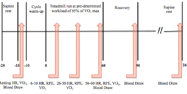
and VO2 was assessed at rest, while heart rate, ratings of perceived exertion, and VO2 was
assessed from 6-10 minutes, 26-30 minutes, and 56-60 minutes. This is shown in figure 2.
During the first treadmill bout, at 10 and 30 minutes, VO2 was checked to ensure
participants reached this intensity of 65%. If participants were below 65%, the running
trials were exactly replicated. Immediately post exercise and 30 minutes into recovery,
blood samples were taken (3mL) and promptly put on ice. Plasma was separated from
blood samples and stored until later analysis for E2, TNF-α, and IL-6. Blood samples are
stored for three years.
Follow Up Blood Draws Session III and V
Participants returned to the APL 24 hours post exercise for additional blood draws.
Upon entering the APL, participants laid supine, in a quiet environment, for 10 minutes.
Blood samples were obtained to measure E2, TNF-α and IL-6 using the same procedures
specified above. All data collection forms used are included in Appendix G.
Figure 2: A diagram showing the protocol of the experimental sessions.
Blood Procedures
Hematocrit
Immediately post each exercise test, resting and post-exercise Hematocrit (Hct)
into 75mm Allied Corporation microcapillary tubes (Fisher Scientific, Pittsburgh, PA)
and sealed using Critoseal (Krackeler Scientific, Albany, NY). Capillary tubes were spun
in a microhematocrit centrifuge for three minutes at 10,000 RPM and then placed on a
hematocrit wheel to determine the hematocrit values of each sample. A mean was
calculated from three samples and used in data analysis.
Hemoglobin
Resting, immediately post-exercise, and recovery hemoglobin (Hb) values from
each experimental session were measured in duplicate from the whole blood samples
using the Stanbiolab Hemopoint H2 analyzer (Boerne, TX). These values were determined immediately after completion of the exercise tests. A mean was calculated
from three samples and used in data analysis.
Plasma Volume Shift
Hb and Ht values were used to calculate exercise induced plasma volume shifts
according to the Dill and Costill equation (Dill & Costill 1974).
Cytokines (TNF-α and IL-6), E2
To separate plasma from whole blood, the blood samples were centrifuged at
3,000 x g for 10 minutes. The separated plasma was transferred to storage tubes and
stored until analyses are conducted. Radioactive (125I) immunoassay technique
(Siemens Healthcare Technologies, Los Angeles, CA USA) with solid-phase antibody
procedures was used to measure plasma E2 concentrations. The assay manufacturer
reports a minimum detectable concentration of 2.0 pg/mL. High-sensitivity
enzyme-linked immunosorbent assay kits (Abnova, Taipei, Taiwan) were used to measure both
5.0 pg/ml for TNF-α and 0.92 pg/mL for IL-6. All blood assays were performed in
duplicate while standards were done in triplicate. See Appendix H for assay sheets.
Data Analysis
Statistica statistical software was used to analyze the data in this study (version 6.3 Tulsa, OK USA). Significance for all data was set at α < 0.05. Descriptive statistics
were shown as means ± standard deviations (SD). Sample size of fifteen participants was
estimated from previous research in the literature to ensure adequate power (=0.80). Effect size was calculated for all significant measures to determine if statistically
significance effects had practical meaning.
Separate 2 x 4 (estradiol level x time) totally within, repeated measures ANOVAs
and where appropriate, Bonferoni post hoc test, was used to assess the effects of estradiol
on blood TNF-α and IL-6 concentrations.
At dependent t-test analysis was used to evaluate statistical significance between
CHAPTER IV
RESULTS
Due to the stringency of the inclusion criteria and study protocol, only 10 of the
15 initially recruited subjects completed all aspects the study. Three subjects completed a
VO2 max test, but did not meet the 45 ml/kg/min criteria. One subject dropped out for
personal health reasons. One subject was not able to complete the treadmill bouts due to
scheduling constraints. The remaining 10 subjects met and maintained all of the
inclusion criteria. However, due to medical reasons, one subject was unable to complete
the mid-luteal prolonged running bout. In order to not lose this subject’s data within the
statistical analysis, mean substitution was used to approximate their mid-luteal values. Additionally, several subject’s HR or RPE data were accidentally missed during data
collection and this data was also approximated using mean substitution.
Subject characteristics
As noted, ten eumenorrheic, aerobically trained females completed this study.
These subjects met all the inclusion criteria: healthy females between the ages of 18-30
years, eumenorrheic, not currently taking or have taken oral contraceptives or other
hormone therapy 6 months prior to participation, have not had an injury in the previous
six months, not currently taking anti-inflammatory medication, and have a current
minimum training volume of 3-5 days per week, 45-120 minutes per session of aerobic
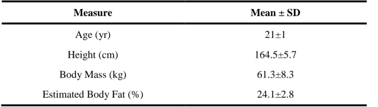
Table 2: Descriptive data for subjects.
Measure Mean ± SD
Age (yr) 21±1
Height (cm) 164.5±5.7
Body Mass (kg) 61.3±8.3
Estimated Body Fat (%) 24.1±2.8
VO2 peak testing
All the criteria for a maximal oxygen consumption test was not achieved by all
subjects, thus all maximal oxygen consumption tests are referred to as VO2 peak tests.
Average relative VO2 peak was 53.5 ± 4.7 ml/kg/min, while the average peak RPE
obtained was 18 ± 1 Borg units, and the average peak HR was 191 ± 7 bpm. The average
calculated 65% of VO2 peak to use during the submaximal 60 minute prolonged runs was
34.8 ± 3.0 ml/kg/min.
Hormonal condition determination
Average menstrual cycle length of subjects was 28 ± 1 days. Menstrual cycle
condition was determined using the protocol detailed in the Methodology chapter. With
the onset of menses denoted day 1, subjects were tested on 7 ± 2 days during the
mid-follicular (low E2; LE) phase, while subjects were tested on 23 ± 3 days for the mid-luteal
(high E2; HE) phase. Analysis of resting blood samples for E2 indicated appropriate
hormonal condition was achieved. The LE concentration was 39.3 ± 18.3 pg/mL and
the HE concentration was 148.1 ± 35.2 pg/mL. These concentrations were significantly
different from one another (p=0.003). The significant difference between hormonal
Prolonged treadmill running bout
Before each prolonged treadmill running bout, subjects submitted a 24 hour food
log, and answered a questionnaire regarding compliance of pre-testing guidelines.
Between the two testing sessions, all subjects complied with all guidelines (no strenuous
exercise or consumption of anti-inflammatory medication, replication of dietary intake,
and adequate consumption of fluids within 24 hours prior to the testing session). Three
subjects participated in light exercise within the 24 hour period prior to the first testing
session, and this exercise was replicated exactly prior to the second prolonged running
session. The resting urine specific gravity was well below 1.030 cc3 for all subjects for both LE and HE prolonged running sessions, indicating adequate hydration prior to
exercise. Mean body mass prior to exercise for LE was 61.4 ± 8.6 kg and for HE was
61.1 ± 8.3 kg; these values did not differ significantly (p>0.05).
Each prolonged running bout was performed for 60 minutes at the calculated running speed to elicit 65% of the individual’s VO2 peak. All subjects were able to
complete each of the 60 minute running bouts. Actual treadmill running speed was
replicated for each prolonged running bout, which was equal to 14.7 ± 1.3 km/hr with a
corresponding VO2 of 61.7 ± 5.0% during LE and 59.7 ± 2.8% during HE. The
prolonged running sessions were counterbalanced in order to prevent order balances, six
subjects completed LE then HE, while four subjects completed HE before LE. The mean
VO2, HR and RPE were nearly identical for each of the 60 minute running sessions.
Table 3: Descriptive data (mean ± SD) for VO2, HR and RPE for each prolonged running
bouts in each hormonal condition (LE and HE).
Hormonal
Condition Measure
Time (min)
Rest 10 30 60
LE
VO2
(mL/kg/min) 4.9±0.6 30.4±4.2 33.2±3.2 35.2±2.9
HR
(bpm) 63±7 150±15 157±13 165±13
RPE
(Borg units) - 11±1 12±1 14±1
HE
VO2
(mL/kg/min)
4.9±0.4 30.0±3.2 32.7±3.3 33.1±4.5
HR
(bpm) 61±4 149±7 154±6 164±6
RPE
(Borg units) - 11±1 12±1 14±2
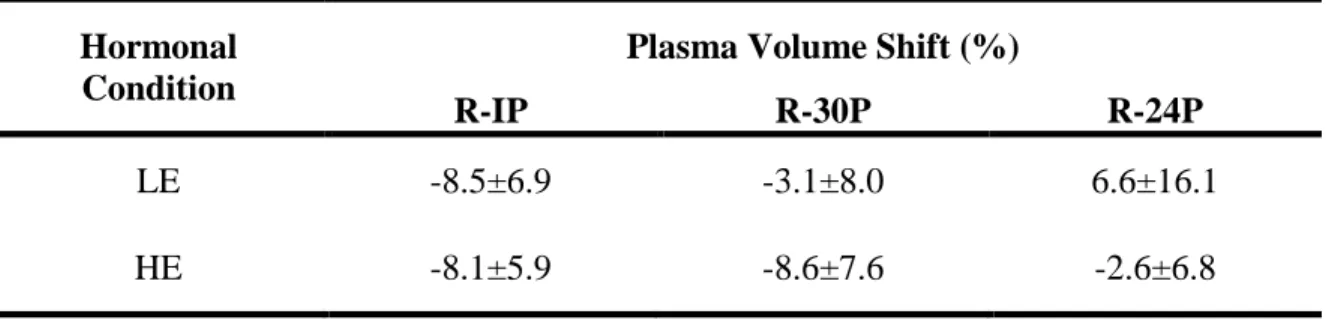
Using the Dill and Costill method of determining plasma volume shifts, plasma volume
decreased over the prolonged running bouts were calculated. These plasma volume shifts
are reported in Table 4.
Table 4: Mean (± SD) plasma volume shifts from rest to immediately post exercise (R-IP), rest to 30 minutes post exercise (R-30P) and rest to 24 hours post exercise (R-24P).
Hormonal Condition
Plasma Volume Shift (%)
R-IP R-30P R-24P
LE -8.5±6.9 -3.1±8.0 6.6±16.1
Blood responses to prolonged exercise
Tumor necrosis factor-α (TNF-α)
TNF- responses are reported in Table 5. The main effect for hormonal condition was not significant (p=0.48). The main effect for time was significant (p=0.001), with
post-hoc tests indicating there was a significant increase from rest to 30 minutes post
exercise (p=0.001), from rest to 24 hours post exercise (p=0.001), and from immediately
post exercise to 24 hours post exercise (p=0.03). There was not a significant interaction
effect for hormonal condition and time (p=0.60).
Interleukin-6 (IL-6)
Interleukin-6 responses are reported in Table 6. The main effect for hormonal
condition was significant (p = 0.022), with post hoc revealing IL-6 response was greater
in LE than HE. The main effect for time was also significant (p=0.001), with IL-6
elevated from rest to immediately post exercise, and from rest to 30 minutes post exercise.
There was a significant interaction effect between hormonal condition and time for IL-6
(p=0.001). Post hoc revealed that IL-6 was significantly increased from rest to
immediately post exercise, and from rest to 30 minutes post exercise in both hormonal
conditions. The response at immediately post exercise did not differ between LE and HE,
however, the response at 30 minutes post exercise was significantly elevated in LE when
Table 5: Mean (± SD) for TNF- at rest, immediately post exercise (IP), 30 minutes post exercise (30P), and 24 hours post exercise (24P). An * indicates significant increase from rest, while an ^ indicates significant increase from IP.
Table 6: Mean (± SD) for IL-6 at rest, immediately post exercise (IP), 30 minutes post exercise (30P), and 24 hours post exercise (24P). An * indicates significance from rest, an ^ indicates significantly lower IL-6 compared to LE, while a # indicates an interaction effect between hormonal condition and time.
IL -6 (pg/ m L ) 24P Chan ge (%) 24.2± 43.6 33.4± 69.0 24P 3.7± 2.4 3.1± 18.3 30P Chan ge (%) 496.6± 417.7 375.5± 283.6 30P 13.7 ± 4.7 *
9.5± 2.3*