Defining the function of Pyrin, the Familial Mediterranean Fever-associated
protein, in inflammation
Pamela Rose Hesker
A dissertation submitted to the faculty of the University of North Carolina at
Chapel Hill in partial fulfillment of the requirements for the degree of Doctor of
Philosophy in the Curriculum of Genetics and Molecular Biology.
Chapel Hill
2011
Approved by:
Beverly H. Koller, Ph.D.
Blossom A. Damania, Ph.D.
Mark T. Heise, Ph.D.
P. Kay Lund, Ph.D.
ii
©2011
iii
ABSTRACT
PAMELA ROSE HESKER: Defining the function of Pyrin, the Familial
Mediterranean Fever-associated protein, in inflammation.
(Under the direction of Dr. Beverly H. Koller, Ph.D.)
iv
v
ACKNOWLEDGEMENTS
Many wonderful people have helped me along this journey of earning a Ph.D. I’d especially like to acknowledgement the guidance, support, and friendship I’ve received from the following people.
My advisor, Dr. Bev Koller. Most importantly, she taught me to approach questions in a more scientific manner. I truly appreciate having the opportunity to be trained by a brilliant, creative, and ambitious scientist.
My dissertation committee: Blossom A. Damania, Ph.D., Mark T. Heise, Ph.D., P. Kay Lund, Ph.D., and Jenny P.Y. Ting, Ph.D. I appreciate their guidance as science experts and career mentors.
Members of the Koller lab, past and present. Especially: Martina Kovarova, Ph.D., for guidance and support on both an intellectual and technical level; Anne Latour, for her technical assistance and friendship; Leigh Jania, for experimental and statistical guidance and encouragement; MyTrang Nguyen, for technical expertise; Jay Snouwaert for advice with cloning schemes; my graduate student comrades: Coy Allen, Alysia Lovgren, Julie Ledford, Artiom Gruzdev, Jaime Cyphert, and Rachel Cote’ for moral support; Amy Pace, for getting me oriented to the lab and for technical assistance.
vi
Vilen, and Anthony LaMantia, who allowed me to rotate in their labs; Dr. Bob Duronio, director of the Curriculum of Genetics and Molecular Biology; Drs. Adrienne Cox and Frank Conlon, CMB directors; Dr. Vytas Bankaitis; Drs. Tom Maynard, Mark McDermott, and Elizabeth Guthrie, who guided me during my rotations and furthered my knowledge of basic cellular and molecular biology lab techniques; Drs. Pat Phelps and Patrick Brandt, TIBBS directors; Drs. Jama Darling and Kim Isaacs; Sausyty Hamreck, Curriculum of Genetics and Molecular Biology secretary; Kathy Allen, IBMS secretary.
Three faculty members at Bradley University, Erich Stabenau, Al MacKrell, and Keith Johnson, played instrumental roles in my decision to go to graduate school. I thank them for helping me to realize that I enjoy being a research scientist.
vii
viii
TABLE OF CONTENTS
LIST OF TABLES ... xiv
LIST OF FIGURES ... xv
LIST OF ABBREVIATIONS ... xvii
CHAPTER 1: Introduction... 1
Familial Mediterranean Fever (FMF) is a disorder of the innate immune system... 1
Regulation of the innate immune system ... 3
Interleukin-1β production ... 4
The inflammasome complexes ... 5
Inflammasome-mediated autoinflammatory disorders ... 13
The FMF-associated gene, Mefv, encodes the protein Pyrin... 15
Expression of Mefv ... 19
Molecular interactions ... 20
Protein interactions ... 22
Cytoskeletal interactions ... 22
Intramolecular interactions ... 24
Previous research implicates Pyrin in IL-1β production ... 24
Pyrin is a POP that negatively regulates IL-1β production ... 25
Pyrin forms an inflammasome which positively regulates IL-1β production ... 26
Pyrin forms a pyroptosome ... 28
ix
Pyrin’s interaction with the cytoskeleton may mediate IL-1β
production ... 30
Evidence using mouse models support conflicting hypotheses ... 30
Evidence from human patients supports conflicting hypotheses ... 33
The role of neutrophils in FMF ... 34
Neutrophil physiology ... 34
Neutrophil Production ... 34
Neutrophil recruitment ... 35
Neutrophil effector functions ... 36
Neutrophil lifespan ... 36
Evidence for altered neutrophil physiology in FMF ... 37
Neutrophil activation is altered in FMF patients ... 37
Neutrophil survival is altered in FMF patients... 37
Treatment of FMF with colchicine ... 38
The genetics of FMF ... 39
Mice and Man: Using the mouse as a model organism ... 42
Summary and Significance of this work ... 43
Research presented in this dissertation ... 45
References: ... 46
CHAPTER 2: Expression of Mefv ... 55
Introduction ... 56
Materials and Methods ... 58
Sources of RNA ... 58
Real-time PCR ... 58
Northern blot expression analysis ... 59
Statistical Analyses ... 59
Animal Care and Use ... 59
x
Cells of the myeloid lineage express high levels of Mefv ... 60
Mefv expression is increased upon maturation of neutrophils and macrophages ... 61
Mefv expression is increased upon activation of macrophages ... 62
Discussion ... 64
References ... 67
CHAPTER 3: Creation of Mefv null mice and characterization of naïve mice ... 69
Introduction ... 70
Materials and Methods ... 72
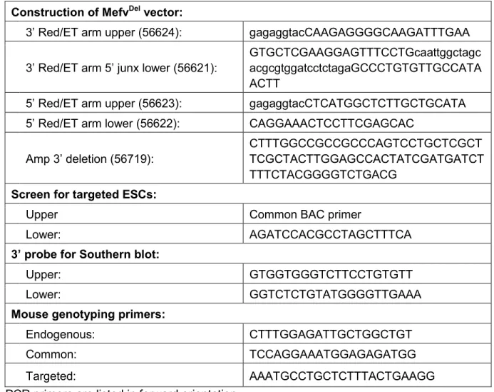
Creation of MefvDel targeting plasmid ... 72
Detection of ESC transformants and mice carrying a targeted allele ... 72
Northern blotting ... 73
Real-time PCR to detect Mefv expression ... 73
Mouse genotyping ... 74
Flow cytometry ... 74
Peritoneal lavage ... 74
Blood analysis ... 74
Use of animals ... 75
Results ... 76
Design of the Mefv targeting vector ... 76
Targeted mouse ESCs were identified ... 76
Mice carrying a null Mefv allele were generated ... 78
Naïve Mefv-deficient mice showed no developmental abnormalities ... 79
Discussion ... 83
References ... 87
CHAPTER 4: Genetic loss of murine Pyrin, the Familial Mediterranean Fever protein, increases Interleukin-1β levels ... 89
xi
Materials and Methods ... 93
In vitro macrophage activation studies ... 93
Real-time PCR Analysis ... 93
Protein analyses ... 93
LPS/ATP treatment in vivo ... 94
Animal Care and Use ... 94
Results ... 95
Pyrin-deficient mice displayed normal responses to LPS ... 95
NLRP3 Inflammasome-dependent IL-1β production is increased in Mefv-deficient macrophages ... 97
Negative regulation of IL-1β production is not limited to the NLRP3 inflammasome ... 99
IL-1β levels after LPS/ATP treatment in vivo were not affected by the loss of Pyrin ... 101
Discussion ... 103
Acknowledgments ... 108
References ... 109
CHAPTER 5: The role of Pyrin in neutrophils ... 113
Introduction ... 114
Methods ... 116
Isolation of bone marrow neutrophils ... 116
Peritoneal neutrophil recruitment ... 116
In vitro neutrophil survival studies ... 116
Trypan Blue... 117
LDH Assays ... 117
Flow cytometry ... 117
Animal Care and Use ... 118
Statistical analyses... 118
xii
Development of neutrophils is not impaired by the loss of Pyrin ... 119
Neutrophil recruitment in Mefv null mice is normal ... 120
Neutrophil survival... 122
Discussion ... 124
References ... 127
CHAPTER 6: In vivo models of peritonitis ... 129
Introduction ... 130
Materials and Methods ... 132
Thioglycolate-induced peritonitis ... 132
In vivo LPS treatment ... 132
Pseudomonas aeruginosa infection ... 132
Animal Care and Use ... 133
Statistical analyses... 133
Results ... 134
A loss of Pyrin does not affect cell recruitment or survival in vivo ... 134
A loss of Pyrin does not affect the response to LPS in vivo ... 138
A loss of Pyrin does not affect the outcome of Pseudomonas aeruginosa infection ... 140
Discussion ... 143
References ... 147
CHAPTER 7: Conclusions and Perspectives ... 150
Summary ... 150
Previous mouse models ... 152
Where in the pathway? ... 154
Endogenous regulation of inflammasome-dependent IL-1β production ... 158
Sequestration of inflammasome complex proteins ... 159
Feedback loops ... 160
xiii
Regulation of Pyrin’s function... 162
Splice variants ... 162
Cleavage of Pyrin ... 164
The genetics of FMF ... 166
xiv
LIST OF TABLES
Table
1.1 Inflammasome complexes ... 7 1.2. Symptoms and genetic alterations of the
xv
LIST OF FIGURES
Figure
1.1. LPS and ATP stimulated IL-1β production. ... 10
1.2. Mefv encodes Pyrin. ... 18
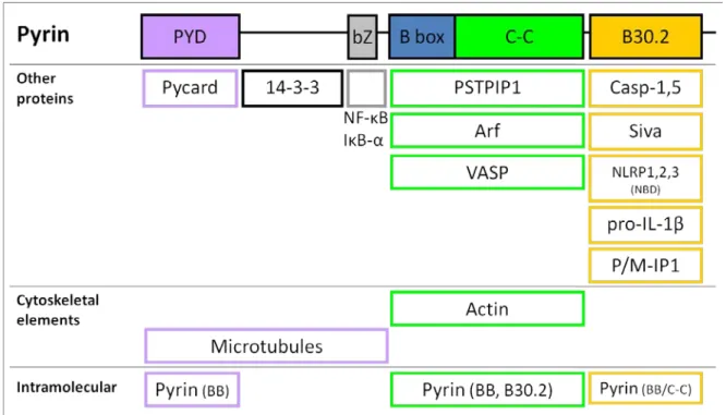
1.3. Pyrin’s molecular interactions. ... 21
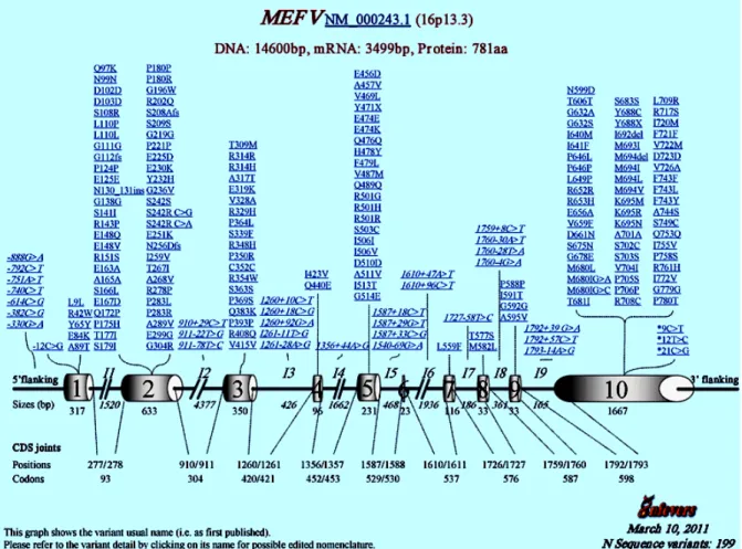
1.4. Mefv sequence variants. ... 40
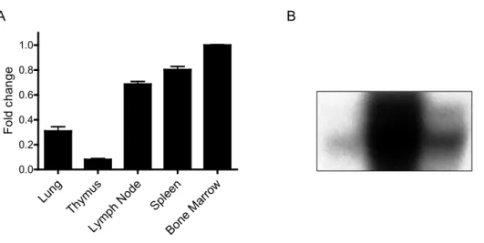
2.1. Mefv is expressed in immune tissues. ... 60
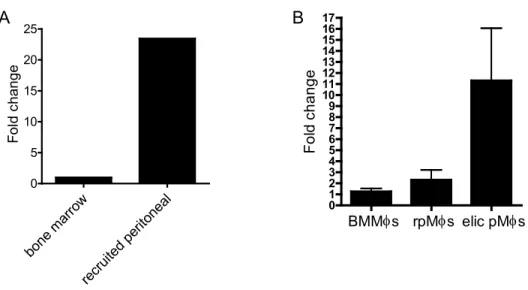
2.2. Mefv expression increases upon matuaration of neutrophils and macrophages collected from mice. ... 62
2.3. Mefv expression in macrophages is increased after pathogen and cytokine treatment. ... 63
3.1. Targeting of the Mefv locus and screening of ESC clones. ... 77
3.2. Verification of Mefv-/- mice. ... 79
3.3. Mefv-/- mice had normal body and organ weights. ... 80
3.4. Naïve Mefv-/- mice have a normal immune system profile. ... 82
4.1. Loss of Mefv does not affect the response to LPS. ... 96
4.2. A loss of Pyrin causes increased IL-1β protein levels in response to NLRP3 inflammasome stimuli. ... 98
4.3. A loss of Pyrin causes increased IL-1β protein levels in response to NLRC4 and NLRP1b inflammasome stimuli. ... 100
4.4. Loss of Pyrin does not affect the response to LPS/ATP treatment in vivo. ... 102
5.1. Mefv-deficiency does not affect neutrophil differentiation. ... 120
5.2. Mefv-deficiency does not affect neutrophil recruitment. ... 121
5.3. The loss of Pyrin does not affect neutrophil survival. ... 123
6.1. Loss of Mefv does not affect peritoneal cell recruitment or survival. ... 136
xvi
6.3. Loss of Mefv does not affect the response to LPS. ... 139 6.4. Loss of Pyrin does not affect the outcome of
xvii
LIST OF ABBREVIATIONS
AIM2 Absent in melanoma 2
ATP adenosine 5'-triphosphate
Arf ADP-ribosylation factor
Alum aluminum hydroxide
Ampr ampicillin resistance
ANOVA analysis of variance
An V Annexin V
ASC apoptosis-associated speck-like protein containing a CARD
LT Bacillus anthracis lethal toxin
BAC bacteria artificial chromosome
BB B-box domain
BMMΦ bone marrow-derived macrophages
BMMC bone marrow-derived mast cells
bZ bZip domain
CPPD calcium pyrophosphate dihydrate
COP CARD-only protein
CARD Caspase activation and recruitment domain
xviii cPOP2 Cellular pyrin-only protein 2
CD cluster of differentiation
C-C coiled-coiled domain
CFU colony forming units
cDNA complementary DNA
CRP C-reactive protein
CAPS Cryopyrin-associated periodic syndromes
Ct cycle number
Cre Cyclization recombinase
DAMP danger-associated molecular pattern
del deletion
DNA deoxyribonucleic acid
NATCH domain present in NAIP, CIITA, HET E, and TP1
dsDNA double-stranded deoxyribonucleic acid
elic pMΦs elicited peritoneal macrophages
ESC embryonic stem cell
ESR erythrocyte sedimentation rate
xix ext extension
FMF Familial Mediterranean Fever
FAF1 Fas-associated factor 1
FACS fluorescently activated cell sorting
FIIND Function to find domain
GM-CSF Granulocyte/moncyte-colony stimulating factor
G-CSF Granulocyte-colony stimulating factor
GFP Green fluorescent protein
TH1 helper T cell subset 1
TH2 helper T cell subset 2
HMGB1 High mobility group box protein 1
HIN HIN-200/IF120x domain
IκB Inhibitor of kappa B
INCA Inhibitory CARD
IFN Interferon
IL Interleukin
IL-1R Interleukin-1 receptor
xx
JAK-STAT Janus kinase-signal transducers and activators of transcription
Kanr kanomycin resistance
kb kilobase
KO knock-out
LDH Lactate dehydrogenase
LRR leucine rich repeat
LTB4 Leukotriene B 4
LPS lipopolysaccharide
LYM lymphocytes
MΦ macrophage
M-CSF Macrophage colony-stimulating factor
MIP-2 Macrophage inflammatory protein-2
MEFV Mediterranean fever gene
Mefv Mediterranean fever gene
MCP-1 Monocyte chemoattractant protein-1
MSU monosodium urate
Mefvtrunc/trunc mouse line carrying a targetted disruption of Mefv
xxi MDP muramyl dipeptide
MPO Myeloperoxidase
Neor neomycin resistance
NET neutrophil extracellular trap
NLRC NLR containing a CARD domain
NLRP NLR containing a pyrin domain
NAIP NLR family, apoptosis inhibitory protein
NF-κB Nuclear factor kappa-light-chain-enhancer of activated B cells
NBD nucleotide-binding domain
NLR nucleotide-binding domain, leucine-rich repeat
OCT octacalcium phosphate
OD optical density
PAMP pathogen-associated molecular pattern
PGN peptidoglycan
PFAPA periodic fever, aphthous stomatitis, pharyngitis, and adenitis
pMΦ peritoneal macrophages
PMA Phorbol myristate acetate
xxii
PSTPIP1 Proline-serine-threonine phosphatase-interacting protein 1
PI propidium iodide
PKC Protein kinase C
P2rX7 Purinergic receptor P2X, ligand-gated ion channel, 7
PAPA Pyogenic sterile arthritis, pyoderma gangrenosum, and acne
PYD pyrin domain
P/M-IP1 Pyrin/marenostrin interacting protein 1
POP Pyrin-only protein
POP1 Pyrin-only protein 1
rpMΦ resident peritoneal macrophages
RNA ribonucleic acid
PAI-2 Serpin plasminogen activator inhibitor 2
PI-9 Serpin proteinase inhibitor 9
SAA Serum amyloid A
SEM standard error of the mean
TiO2 titanium dioxide
TRAPS TNF-Receptor Associated Periodic Syndrome
xxiii TGF transforming growth factor
TRIM20 tripartite motif-20
trunc truncated
TNF Tumor necrosis factor
UTR untranslated region
VASP Vasodilator-stimulated phosphoprotein
CHAPTER 1
Introduction
Familial Mediterranean Fever (FMF) is a disorder of the innate immune system
Familial Mediterranean Fever (FMF) is an inherited autoinflammatory disease characterized by sudden episodes of fever and inflammation that typically last 1 – 3 days. Fever and acute abdominal pain are the most common symptoms, but symptoms can also include pain in the joints or lungs or lesions in the skin. The location and the severity of pain and inflammation differ between patients and between attack episodes within each patient. Acute attack periods are interspersed with remission periods of either low-grade or undetectable inflammation. FMF has long-term consequences that include tissue scarring, which is secondary to inflammation, and amyloid deposition within organs, especially the kidneys. Severe cases result in kidney failure and subsequent death. Disease onset and diagnosis usually occurs in children before age eight (1, 2).The innate, but not the adaptive, arm of the immune system mediates FMF pathologies. Local tissue inflammation is due, in part, to considerable deposition of neutrophils. Neither T lymphocyte infiltration, nor auto-reactive antibodies have been detected, indicating that T and B lymphocytes are unlikely to be involved in mediating inflammation. Patients have an increase in the acute phase reactants: erythrocyte sedimentation rate (ESR), C-reactive protein (CRP),
2
the Interleukin (IL) cytokines 6 and 10 (3), and the cytokine SA100A12 (4) in comparison to healthy controls. IL-1β cytokine levels are also elevated in some patients (3). IL-1β is of
particular interest because it is elevated in patients with several different overactive immune system disorders, and blocking the signaling of IL-1β can decrease inflammation. IL-1β is
produced predominantly by macrophages.
FMF was associated with mutations in Mefv (Mediterranean Fever) based on linkage analysis and was confirmed by positional cloning (5, 6). Mefv encodes the protein Pyrin. Consistent with IL-1β-mediated inflammatory responses and neutrophil accumulation in FMF, Pyrin expression is highest in macrophage and neutrophil cells (7, 8). Thus, the symptoms of FMF, clinical tests, and the properties of the Pyrin protein indicate that the innate immune system mediates disease. Specifically, neutrophils and macrophages, are likely to play an important role in FMF-associated inflammation that is caused by mutations in the Mefv gene. Accordingly, previous laboratory research demonstrates a role of Pyrin in macrophage-mediated
IL-1β production and in survival, chemotaxis, and phagocytosis processes of neutrophils. In this
chapter, I will summarize previous research on the regulation of IL-1β production and neutrophil
functions, as they pertain to Mefv and FMF, and then discuss the features of the MEFV gene and Pyrin protein. These data fit together nicely to provide support for a role of Pyrin in IL-1β
3
Regulation of the innate immune system
4
Appropriate pathogen recognition is critical for the prevention of autoinflammatory disorders. The innate immune system must make quick decisions about whether or not a pathogen or foreign particle threatens the survival of the organism. To confer this ability, cells often have co-stimulatory requirements or two independent pathways that converge and activate an immune response. In 1994, Polly Matzinger proposed “the danger hypothesis” to account for some of the gaps in the current model describing the process of self versus non-self decisions. “The danger hypothesis” proposes that the immune system does not directly detect self versus non-self stimuli to guide appropriate immune responses, but instead detects and protects against danger. Indications of danger are provided by a network of extrinsic communication signals from damaged tissues and intrinsic cell stress signals. Thus, the presentation of a pathogen in the context of a danger signal triggers the innate immune response. These independent signals come together to tailor the breadth and magnitude of the response (9-11).
Interleukin-1β production
Cytokine and chemokine signaling molecules circulate throughout the body and coordinate the strength and timing of pathogen responses. The cytokine IL-1β is released very early during the innate immune response and is an important mediator of the overall magnitude of the immune response. Its importance in the immune response is underscored by its contribution to sepsis, the autoimmune disorders rheumatoid arthritis, Crohn’s disease, and multiple sclerosis, and the autoinflammatory disorders CAPS, PAPA syndrome, and FMF. Controlled experiments implicate Pyrin, the protein mutated in FMF, in the regulation of IL-1β production. IL-1β is
5
that can be quantified by neutrophil recruitment (12). IL-1β binds to the IL-1 receptor (IL-1R) on the external surface of cells and promotes further downstream pro-inflammatory cytokine signaling, cell recruitment, fever, and overall metabolic changes (13).
IL-1β production involves two signaling pathways: 1) Pro-IL-1β transcripts are produced in
a pathogen-stimulated and NF-kB-dependent manner, and 2) Caspase-1 processes pro-IL-1β
protein to mature, or active, IL-1β cytokine (14). Another IL-1 family cytokine, IL-18, is cleaved by Caspase-1 in an analogous manner. Caspase-1, a cysteine-aspartic acid protease, is activated by autocatalytic cleavage, which is promoted by inflammasome complexes in response to cellular damage (15, 16). The vast majority of IL-1β is produced in a Caspase-1-dependent manner, although other proteases, produced by neutrophils and mast cells, can generate an active form of IL-1β with relatively low efficiency. Pyrin is implicated in the
regulation of IL-1β production during both the transcriptional and the inflammasome-dependent processing stages.
The inflammasome complexes
6
the indicated protein to IL-1β production. It is unclear whether heterocomplexes with multiple
7
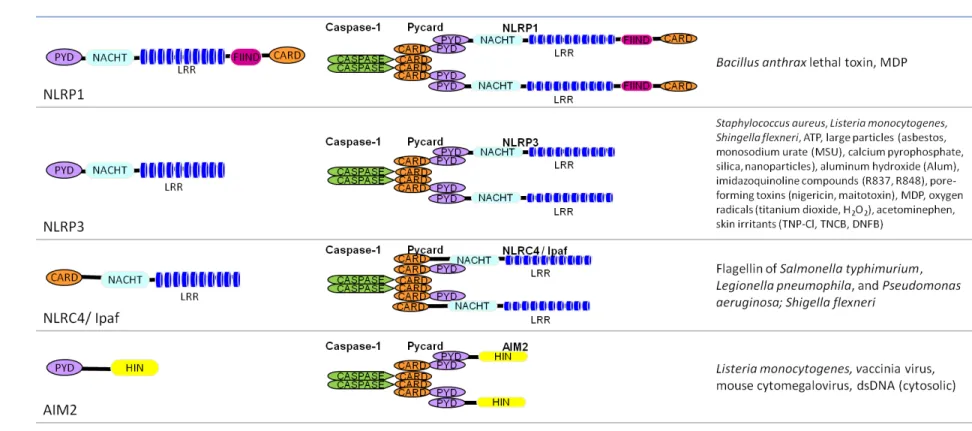
Table 1.1 Inflammasome complexes.
PYD, pyrin domain; LRR, leucine-rich repeat; FIIND, function to find; HIN, HIN-200/IF120x.
The inflammasome structures are depicted as dimers, but also form higher-order molecular complexes.
The indicated stimuli have been tested primarily in vitro, using a readout of Caspase-1 cleavage, Caspase-1 activity, or IL-1β
8
The protein domain structure of the NLRs is critical to their function. NLRP1 – 3 and 6 and NLRC4 are members of the NLR protein family, based upon the presence of two domains for which they are named: the nucleotide-binding domain (NBD or NATCH) and the leucine-rich repeat (LRR). These proteins also contain a pyrin domain (PYD) and/or a caspase recruitment domain (CARD). NLRP1 contains an additional domain, a function to find (FIIND) domain. AIM2 contains a PYD and HIN-200/IF120x domain (HIN) and thus shows little structural similarity to the NLRs, but functional analysis indicates that it nucleates inflammasome complexes. Unfolding of the NLR/AIM2 exposes the binding surfaces of the PYD and CARD domains and is regarded as the “ignition switch” for inflammasome complex formation. The PYD and CARD form homodimers with cognate domains present in ASC (apoptosis-associated speck-like protein containing a CARD) or Caspase-1 to bring the inflammasome complexes together. The inflammasome complexes are often depicted as dimers, but electron micrographs of in vitro reconstituted NLRP1 protein complexes show that they actually form higher order complexes, similar to apoptosomes, with 7 NLRP1 proteins forming a circular perimeter around the ASC and Caspase-1 proteins at the core (17).
The NLRP1 inflammasome was the first inflammasome to be described (18), and it remains the only inflammasome complex verified by reconstitution using purified human recombinant proteins (17). The Nlrp1 gene was identified in mice based upon differential susceptibility of inbred mouse strains to Bacillus anthrax lethal toxin (LT) induced death. The susceptibility region was mapped to a locus containing three orthologs analogous to the human NLRP1 gene: Nlrp1a, Nlrp1b, and Nlrp1c (19). Further studies using genetically-deficient mice have verified that Nlrp1b is indeed necessary for LT-induced Caspase-1 activation and IL-1β production.
9
is required for IL-1β production in response to pathogens and toxins (20). NLRP3 mediates IL
-1β production following exposure to a diverse set of stimuli, including some bacteria, ATP, uric
acid crystals common in gout, the vaccine adjuvant alum, antiviral drug compounds, reactive oxygen species, and cell membrane permeabilizing toxins. It remains unclear how such a diverse set of stimuli can activate NLRP3, and it will be interesting for future studies to determine where the danger response pathways converge to trigger NLRP3 inflammasome complex formation. Under physiological conditions, NLRP3 folds upon itself to prevent activation. NLRP3 unfolds in response to low intracellular potassium levels induced by hypotonic cell medium or treatment with a toxin that permeabilizes the cell membrane. These treatments favor potassium efflux (21-23). It is likely that NLRP3 unfolding requires interaction between a phosphorylated nucleotide and the NACHT domain to transfer energy that enables protein unfolding. Once unfolded, NLRP3 forms an inflammasome complex that brings together NLRP3 and Caspase-1 through the adaptor protein ASC (gene: Pycard). Inflammasomes aggregate to form larger protein complexes called ASC specks. Proximity of multiple Caspase-1 molecules causes autocatalytic cleavage and activation of Caspase-Caspase-1. Active Caspase-Caspase-1 cleaves pro-IL-1β (and pro-IL-18) to their mature forms, and these molecules are released from the cell (24). The NLRP3-mediated response to a combined exposure of LPS and ATP is well described. LPS activates NF-κB-dependent transcription of IL-1β, and ATP triggers NLRP3
10
Figure 1.1. LPS and ATP stimulated IL-1β production. IL-1β production is mediated by 1)
NF-κB dependent transcription, and 2) Caspase-1 mediated cleavage of pro-IL-1β to form a
mature protein capable of signaling. The pathogen-associated molecular pattern (PAMP) endotoxin (LPS) is recognized by Toll-like receptor 4 (TLR4). Signaling through TLR4 results in
NF-κB activation and induced transcription of IL-1β. IL-1β production is also regulated at the
protein level by the NLRP3 inflammasome. The NLRP3 inflammasome is activated by the danger-associated molecular pattern (DAMP) of high extracellular ATP (concentrations >500 times the physiological level) (25). ATP binds to P2rX
7 (purinergic receptor P2X, ligand-gated ion channel, 7), which is an ATP-dependent potassium channel. P2rX7 allows rapid efflux of K+ from the cell. Subsequently, NLRP3 unfolds, and its exposed pyrin domain forms homodomain interactions with the pyrin domain of ASC. ASC is an adaptor protein that also binds pro-Caspase-1 through CARD homodomain interactions. Pro-pro-Caspase-1 is autocatalytically cleaved and activated Caspase-1 then cleaves pro-IL-1β inside of intracellular vesicles. IL-1β and
11
NLRC4, or Ipaf, responds to intracellular bacterial virulence factors and nucleates an inflammasome that contains either NLRC4 and Caspase-1 with ASC, or NLRC4 and Caspase-1 alone and requires NAIP5 for activation. Salmonella typhimurium, Pseudomonas aeruginosa, and Shingella flexneri activate a ASC-dependent NLRC4 inflammasome, whereas Caspase-1 activation in response to Legionella pneumophila requires NAIP5, but not ASC (26). Purified recombinant flagellin has also been shown to increase IL-1β production in an NLRC4 -dependent manner, indicating that flagellin alone can activate the NLRC4 inflammasome. Correspondingly, S. typhimurium and L. pneumophila that are deficinent in flagellin have a reduced ability to activate Caspase-1. However, S. flexneri does not express flagellin; its recognition factor is unknown. The intracellular delivery of virulence factors through a Type III or Type IV secretion system is critical for NLRC4 activation. This finding helps to separate the NLRC4 inflammasome pathway as a virulence factor-sensing pathway that is independent of TLR5, which senses flagellin that is presented extracellularly (21, 27).
12
The NLRP6 inflammasome was recently described. NLRP6 complexes with ASC and Caspase-1 to form an inflammasome which mediates pro-IL-18 cleavage (31, 32). In vivo, Nlrp6-deficient mice have exacerbated intestinal inflammation and an increased propensity for tumors in dextran sodium sulfate models of colitis (31-33). Phenotypes are similar to those of Pycard-deficient mice (31).
The inflammasomes differ slightly between humans (depicted in Table 1.1) and mice. The PYD of mouse NLRP1b is non-functional, and instead the C-terminal CARD binds directly to Caspase-1, whereas the CARD of human NLRP1 binds to caspase-5, another pro-inflammatory caspase. Human NLRP3 inflammasome complex may also contain CARD8/CARDINAL. The molecular contribution of CARD8 towards Caspase-1 activation and IL-1β production is not well -described, and CARD8 is absent from the mouse genome (34). However mutations in CARD8 are associated with increased risk for Crohn’s disease and ulcerative colitis and increased IL-1β
levels in some populations (35, 36). However, despite these differences, the function of the inflammasomes is well-conserved between humans and mice.
The inflammasome complex provides a unique signaling pathway that is amendable to manipulation to specifically affect IL-1β and IL-18 production without affecting production of other cytokines. Previous research indicates that both endogenous and pathogen-derived molecules can interfere with this pathway. This is important on a clinical level, because it suggests that continued research will help to direct production of pharmaceutical agents that can interfere with IL-1β production in a more specific manner. In theory, the inhibition of
aberrant, disease-associated IL-1β production will be possible without limiting crucial pathogen-stimulated IL-1β and IL-18 signaling. Pyrin regulates inflammasome-mediated IL-1β production.
13
therapies to influence Pyrin’s molecular functions are a promising avenue for improved therapies for immune disorders.
Inflammasome-mediated autoinflammatory disorders
Autoinflammatory disorders, by definition, are innate immune responses in the absence of an identifiable stimulus. Familial Mediterranean Fever (FMF) is part of a group of hereditary autoinflammatory disorders. It was originally identified as a unique disorder in 1948, and named benign paroxysmal peritonitis (37) and later periodic disease (38), periodic fever, and Siegal-Cattan-Mamou syndrome. Today, periodic disease describes a family of genetic autoinflammatory disorders that include: Cryopyrin-Associated Periodic Syndromes (CAPS), PAPA syndrome (Pyogenic sterile arthritis, pyoderma gangrenosum, and acne), Blau Syndrome, early-onset Sarcoidosis, Familial Hibernian Fever (also known as TNF-Receptor Associated Periodic Syndrome, or TRAPS), Hyperimmunoglobulinemia D with recurrent fever syndrome (also known as mevalonate kinase deficiency), PFAPA syndrome (Periodic fever, aphthous stomatitis, pharyngitis, and adenitis), and Deficiency of the Interleukin-1–receptor antagonist syndrome.
FMF, CAPS, and PAPA diseases are all characterized by spontaneous inflammation or inflammation in response to innocuous stimuli, such as cold temperatures (39), and have similar symptoms, including elevated levels of IL-1β and neutrophil deposition within inflamed tissues
14
function, and why there are discrepancies in the results of various studies, is unclear. The proposed mechanisms by which Pyrin alters IL-1β production will be discussed further in the following sections. PSTPIP1 (also known as CD2BP1) binds to Pyrin, and this interaction influences the binding of Pyrin to ASC. Mutations in PSTPIP1 increase PSTPIP1’s binding affinity for Pyrin (40, 41). In one model, an interaction between Pyrin and PSTPIP1 leads to decreased binding between Pyrin and ASC. In this model, Pyrin and ASC interaction functions to decrease IL-1β production (41). However, an alternative model depicts that PSTPIP1-Pyrin interaction unfolds Pyrin and reveals the domains necessary for Pyrin to bind ASC. In this model, Pyrin and ASC interaction forms a pyroptosome that promotes IL-1β production and cell
death (40). Despite the differences in these two models, they both posit that PAPA syndrome-associated mutations in PSTPIP1 increase IL-1β production through a mechanism mediated by
15
Table 1.2. Symptoms and genetic alterations of the inflammasome-mediated autoinflammatory disorders. .
Disorder Symptoms Genetic Alteration
Familial Mediterranean Fever (FMF)
Fever, Peritonitis, pleuritis, Arthritis, Skin rashes, polymorphonuclear infiltration at sites of inflammation, Amyloidosis, Elevated IL-1β
Missense mutations in Mefv;
Autosomal recessive Cryopyrin-associated
Periodic Syndromes (CAPS) • Familial cold autoinflammatory
syndrome • Muckle-Wells syndrome
• Neonatal-onset multisystem inflammatory disorder
Skin rashes (uticaria) in response to cold, polymorphonuclear infiltration at uticarial sites, Fever, Hearing loss, Arthritis, Amyloidosis, Meningitis, Inflammation of the eyes, Elevated
IL-1β
Missense mutations in NLRP3 (formerly Cryopyrin);
Autosomal dominant
PAPA syndrome (Pyogenic sterile arthritis, pyoderma gangrenosum, and acne)
Arthritis, polymorphonuclear infiltration at sites of inflammation, Skin rashes with ulcerative lesions, cystic acne, Elevated IL-1β
Point mutations in PSTPIP1;
Autosomal dominant
The similarities among these diseases, and especially the common pathologies of elevated
IL-1β and neutrophilia support a model that NLRP3, Pyrin, and PSTPIP1 function within the
same signaling pathway to affect innate immune responses. Further evidence to support a function for Pyrin in IL-1β production and neutrophil dynamics will be discussed later in this
chapter. First, I will provide a conceptual framework for these function of Pyrin by describing the expression pattern of Mefv and the molecular interactions previously reported for Pyrin.
The FMF-associated gene, Mefv, encodes the protein Pyrin
16
The promoter region of human Mefv contains regulatory elements associated with inflammation-dependent transcription. Specifically, C/EBPα and NF-κB response elements are necessary for mediating the increase in MEFV expression induced by tumor necrosis factor (TNF)-α (42). In mice, cytokine-induced Pyrin expression is prevented by the genetic loss of Stat6 and NF-κB, indicating that these elements are critical to the regulation of Mefv expression (43), although the regulation of Mefv may be a downstream effect of another cytokine that is regulated by Stat6 and NF-κB elements, such as IL-4 or IFNγ. Thus, it appears that Mefv expression is regulated somewhat differently in humans and mice.
Alternative splicing of both the human and mouse transcripts has been detected. In humans, there are isoforms lacking one or more exons: exon 2 (del2), exons 3 and 4 (del34), exons 2,3,4 (del234), exons 2,3,4,5 (del2345), exon 7 (del7), and exon 7 and 8 (del78). These are found in both FMF-patients and healthy controls, suggesting that mutations in MEFV may not influence splicing, and that alternative splicing may not have a pathological effect (44). In peripheral blood leukocytes and sonovial fibroblasts, splice variants with alternative exons 2a and 4a in place of 2 and 4, respectively, have been identified. The 4a exon substitution creates a frame-shift and a predicted truncated protein ending in exon 5. A transcript that contains an extended version of exon 8 (8ext) and lacks exons 9 and 10 is induced by LPS and accounts for 27% of transcripts in sonovial fibroblasts. Transcripts with del2 or 2a combined with 4a or 8ext were also detected (45). In mice, alternative splicing replaces exon 9 with 9a. It remains unclear if these splice variants provide wild-type function in a similar, increased, or decreased capacity, or if they have novel function or no function at all.
17
family proteins (46). Within this family are NLRs, several of which function in innate immunity, and specifically, IL-1β production. As previously mentioned, the PYD mediates homodomain
18
Figure 1.2. Mefv encodes Pyrin. The orthologous Mefv genes of the human and mouse genomes both contain ten exons and encode proteins with a high degree of homology. Gene exons are shown as unfilled boxes and align with protein domains (filled boxes) as indicated. Exons 1-8, except exon 2, are especially similar between humans and mice. The B30.2 domain of the human protein is missing from the mouse. The mouse protein contains the bZip domain (bZ) sequence, but it is unclear if its function is conserved. PYD, pyrin domain; BB, B-box; C-C, coiled-coiled domain.
19
within the B30.2 region of the human protein, creating a conundrum as to how function is conserved between mice and humans. It is, however, clear that there are disease associated mutations which lie outside of the B30.2 region (50-54).
Expression of Mefv
Mefv expression is highest in immune tissues in both humans and mice. In humans, significant expression can be detected in the spleen, lung, and muscle tissues and very low levels of expression are detected in several other human organs by RT-PCR (8). At the cellular level, expression is highest in neutrophils and macrophages, and there is low expression in B220+ B cells, CD3+ T cells, eosinophils, dendritic cells, and epithelial cells of the lung, peritoneum, and synovium (8, 45, 55). Rodents may have a more restricted expression pattern of Mefv in immune tissues. By northern blot analysis, expression was detected in the spleen of mice and rats and also in the lung and kidney of rats. Expression was not detected during mouse embryonic development. In the murine spleen, in situ hybridization showed that expression was concentrated in the primary follicle and marginal zone regions, which contain mostly granulocytes and B cells. At the cellular level, Pyrin is expressed in granulocytes and macrophages, but not lymphocytes (7).
20
overexpressing human Pyrin, localization was unaffected by M694V or V726A mutations (8). Less is known about the cellular localization of murine Pyrin. Since there are fewer splice variants described for the mouse transcript and protein cleavage is not described, protein localization may be less complex in mouse cells.
Pyrin expression in humans and rodents is regulated by sterile immune stimuli and cytokines. Lipopolysacchride (LPS), interferon (IFN)-γ, and IFN-α increase expression of MEFV in human macrophages (55), while IL-4, IL-10, and transforming growth factor (TGF)-β can
abrogate the effect of increased expression following a combined treatment of LPS and IFN-γ
(55). There are some differences between humans and mice in the induction of Pyrin expression. IFN-γ dose-dependently increases expression at the transcript and protein level in human macrophages (55, 57). In mice, IFN-γ increases Mefv expression at the transcript level, although it has also been reported that a difference in Pyrin expression was not seen at the protein level in murine macrophages (43). LPS, TNF-α, IL-1β, IL-2, IL-4, IL-6, IL-10, and IL-12 increase expression of Pyrin in murine macrophages. The TH2-inducing cytokines 4 and IL-10 are the strongest inducers of mouse Pyrin expression (43), but perhaps not human Pyrin. Instead, IL-4 treatment inhibits expression at the transcript level, and IL-10 induces cleavage of the human Pyrin protein (57). In vivo intratracheal administration of LPS or silica increased levels of Mefv in rats (60). Thus, Pyrin expression is regulated by endogenous and exogenous immune stimulants, both in vitro and in vivo. Mutations in Mefv that are associated with FMF may increase, decrease, or have no effect on levels of Pyrin expression.
Molecular interactions
21
hypotheses for the function of Pyrin. In this section, I will describe the previously-reported molecular interactions of Pyrin. The functional relevance of these interactions will be discussed in subsequent sections, as they pertain to IL-1β production and neutrophil physiology.
22
Protein interactions
An interaction between Pyrin and ASC was demonstrated through a yeast two-hybrid screen of the human genome and through immunofluorescence microscopy and coimmunoprecipitation of endogenous human and mouse Pyrin with ASC proteins (47). Several studies indicate that Pyrin’s interaction with ASC affects production of IL-1β (40, 43, 61, 66, 67) and inflammation -associated cell death (40, 43, 47).
The SPRY domain of Pyrin interacts with the NACHT domain of NLRPs 1-3 the pro domain of pro-IL-1β, and Caspase-1 based upon co-immunoprecipitation experiments (61, 62). These interactions support multiple models of ASC-independent contributions of Pyrin to IL-1β
regulation.
An interaction between Pyrin and 14-3-3 has also been shown. 14-3-3 is implicated in multiple signal transduction pathways and in protein shuttling across the nuclear membrane (64). It is reasonable to hypothesize that 14-3-3 mediates cytoplasmic-nuclear shuttling of Pyrin.
A co-immunoprecipitation experiment identified an interaction between Pyrin and Siva1. Siva1 is proapoptotic protein, and its interaction with Pyrin reduced the ability of Siva1 to induce apoptosis (63). However, in a screen of gene networks involved in FMF pathogenesis, Siva1-mediated apoptosis was not linked to FMF. Gene network analysis in THP-1 human monocytes supports an interaction of Mefv with Pycard, Caspase-1, Nlrp3, Pstpip1, NF-κB, and Interferon-α, but the gene networks for Mefv and Siva1 do not overlap (68).
Cytoskeletal interactions
23
P/M-IP1 and Pyrin co-localized to perinuclear structures in Cos-7 cells (69). Based upon these findings, Pyrin was believed to have a role in Golgi-mediated transport. A direct interaction in human cells has not been demonstrated by coimmunoprecipitation experiments. Further experiments have failed to demonstrate co-localization of Pyrin with Golgi structures. In fact, paclitaxel or colchicine treatment of Cos-7 cells, at a dose capable of diffusing Golgi structures, did not disrupt the cellular distribution of Pyrin (58).
Immunofluorescent co-localization and an in vitro binding assay were used to show that Pyrin associates with microtubules. However, the common FMF-associated mutations M680I, M694V and V726A mutations did not affect binding, which limits support for the hypothesis that Pyrin’s association with microtubules has functional consequences that are important in FMF pathophysiology. In the same study, Pyrin also co-localized with actin depolymerization ends located at cell membrane ruffles on the lagging end of HeLa cells (58). Subsequently, another group found that Pyrin co-localized with actin, but not microtubules, and α-actin coimmunoprecipitated with Pyrin (59). Furthermore, the toxin Cytochalasin D, which inhibits actin polymerization, disrupted the cellular distribution of both actin and Pyrin. However, in this study, Pyrin and ASC co-localized in HeLa cells to the leading edge of cell membrane ruffles that are rich in polymerizing actin. VASP and Arp3 proteins are important in actin polymerization, and a coimmunoprecipitation experiment using myc-labeled Pyrin and GFP labeled VASP or Arp3 showed that these proteins can bind to Pyrin. VASP and Arp3 also co-localized to ASC specks in the presence but not the absence of Pyrin (59). Thus, these studies suggest that Pyrin may coordinate interaction between ASC specks and the polymerizing tails of actin at an immunological synapse.
24
may influence Pyrin’s function with cytoskeletal elements. PSTPIP1 binds to PTP-PEST, which is a scaffolding protein that associates with actin through the protein WASP (70). Pyrin can co-localize with actin even in the absence of ASC and PSTPIP1, indicating that Pyrin’s interaction with actin is not mediated by ASC or PSTPIP1 (59). Rather, Pyrin recruited PSTPIP1 to ASC specks. The PAPA-associated mutation W232A in PSTPIP1 prevented its recruitment to ASC specks (59), but the A230T and E250Q mutations caused an increase in recruitment (71). In summary, through both direct interactions and indirect protein-protein mediated interactions, Pyrin is implicated in cytoskeletal function and could influence cell shape, cell migration, and organization of an immunological synapse (70).
Intramolecular interactions
In one study, IL-1β interacted more strongly with independent domains of Pyrin compared
to full-length Pyrin. Further research based upon this observation showed that the SPRY domain interacts with the BB and C-C domains of Pyrin, and masks the interaction of the SPRY domain with pro-IL-1β in coimmunoprecipitation studies. However, M694V mutation did not
affect intermolecular binding or intramolecular binding, so the functional significance of these interactions remains unclear (62). The BB region of Pyrin has also been shown to bind the PYD of Pyrin and interactions of these domains forms Pyrin homotrimers (40). In contrast, however, a yeast two-hybrid assay failed to detect intramolecular interactions of human Pyrin, when expressed in yeast cells (69).
Previous research implicates Pyrin in IL-1β production
Several lines of evidence indicate that Pyrin affects IL-1β production. First, some FMF
patients have elevated serum IL-1β during inflammatory attacks and blocking IL-1β signaling is
25
in IL-1β production in human and mouse experimental systems. Third, Pyrin interacts with
proteins of the inflammasome complex, NF-κB and IκB-α, and IL-1β itself. However, there are still outstanding questions. First, it is unclear if WT Pyrin acts to positively or negatively regulate
IL-1β production. Second, do mutations in Pyrin cause a loss or gain of function relative to WT
Pyrin that affects IL-1β production? Does the mutant Pyrin protein also have novel functions? Third, does Pyrin affect IL-1β transcription, pro-IL-1β protein processing that is either dependent
or independent of inflammasome complexes, and/or IL-1β cytokine release from the cell?
In the sections below, I will detail the evidence that supports several hypotheses as to how Pyrin affects IL-1β production. Some of the proposed models directly conflict with one another
as to the function of Pyrin, and are thus unlikely to occur simultaneously. The validity of these models is not necessarily mutually exclusive and may be context (i.e. pathogen) dependent. It is also possible that more than one of these mechanisms facilitate concurrent and/or synergistic effects.
Pyrin is a POP that negatively regulates IL-
1β production
26
decrease NLR-dependent IL-1β production (75-78). The in vivo function of these genes has not been examined, because they are absent from the mouse genome. Conversely, Caspase12 deficient mice display increased IL-1β cytokine production, suggesting that it negatively
regulates inflammasome activity in vivo. Thus, there is support for the model of COPs in an in vivo system. However, the significance of Caspase-12 in humans is questioned because the gene structure varies between mouse, dog, and human genomes (79). Pyrin is the only COP or POP that has been shown to negatively regulate IL-1β production in vitro and in vivo in both humans and mice.
More specifically, the POP model proposes that Pyrin competes with NLRs, specifically NLRP3, for interaction with ASC, resulting in less Caspase-1 activity and less mature IL-1β
production. In a mixed lymphyocyte system using lysates from PT67 mouse epithelial cells overexpressing human Pyrin, ASC, and CASP-1, CASP-1 interacted with ASC in the absence, but not the presence of Pyrin. Pyrin coimmunoprecipitated with ASC regardless of CASP-1 expression. Together, these findings indicate that Pyrin competes with CASP-1 for binding to ASC (43). Using reconstituted HEK293T human epithelial cells, Pyrin was also shown to compete with NLRP3 for binding to ASC (65). In another study, Pyrin was shown to dose-dependently reduce IL-1β production in transiently-transfected HEK293T (80). U937 human monocytes overexpressing Pyrin also had decreased IL-1β production in response to LPS
compared to cells treated with empty vector. The M694V mutation reduced but did not eliminate the regulatory effect of Pyrin (43). Correspondingly, a knockdown of endogenous Pyrin expression by THP-1 cells was shown to increase IL-1β production in response to LPS (61),
peptidoglycan (PGN) and uric acid crystals (MSU) (62).
Pyrin forms an inflammasome which positively regulates IL-
1β production
27
model draws upon the similarity in structure between Pyrin and NLR proteins. The PRY/SPRY domain of human Pyrin forms an immunoglobulin receptor-like structure that could facilitate pathogen recognition in a manner analogous to the predicted receptor function of the LRR of NLR proteins. In a HEK293T cell overexpression system, increasing levels of Pyrin dose-dependently increased Caspase-1 processing and IL-1β cleavage. SPRY domain mutants
M694V and V726A were also able to increase Caspase-1 processing at levels similar to WT Pyrin. Overexpression of WT or M694V, but not the PYD mutant L16P/F24S, also induced ASC oligomerization. Furthermore, a Pyrin-ASC complex was shown to coimmunoprecipitate Caspase-1. These data are consistent with a model in which PYD-PYD interactions between Pyrin and ASC mediate the formation of an inflammasome complex (67). Another group also showed that Pyrin dose-dependently increased Caspase-1 activity and IL-1β release from HEK293T cells reconstituted with Pyrin, ASC, and Caspase-1, with or without IL-1β, and without
an NLR. A knock-down of Pyrin expression in the human monocytic cell line THP-1 and in human peripheral blood mononuclear cells resulted in decreased IL-1β release in response to LPS (66). In support of this model, there is positive selection for mutations within the B30.2 domain that is likely to function as a ligand receptor, suggesting that mutations in Pyrin may provide some selective advantage against a pathogen. The biggest caveat of this model is a lack of evidence to support a pathogen that requires Pyrin for an efficient immune response, and specifically, IL-1β production.
It is also reasonable to imagine that Pyrin self-regulates its function by masking functional domains through intramolecular interactions. In this model, Pyrin increases IL-1β production
through its PYD domain, but the PYD domain must be exposed. The B30.2 domain inhibits this function by hiding the PYD domain until a ligand causes unfolding of Pyrin. If this were true, a mutation in the B30.2 domain could permanently expose the PYD domain and lead to increased
28
NLRP3 that suggests that mutations in the NATCH domain cause its PYD domain to be permanently exposed. It is important to note that NLRP3 is a positive regulator of IL-1β
production. By analogy then, it is also possible to explain how Pyrin could be a positive regulator of IL-1β production, and mutations in Pyrin lead to increased IL-1β production.
Pyrin forms a pyroptosome
The Pyrin-ASC interaction is the central evidence for yet a third model. In this model, Pyrin and ASC complex into a “pyroptosome” that increases apoptosis during inflammation-induced cell death processes. IL-1β release may increase during this process, but is not produced in an
active process, and is instead a by-product of cell death (40). HEK293T cells transfected with full-length Pyrin or the PYD only, but not Pyrin with a deleted PYD, showed increased apoptosis (65). In another sudy, however, Pyrin increased ASC-speck formation, which is associated with inflammasome-mediated cell death, but the ASC-speck positive cells actually had increased survival (47). Additionally, Pyrin has been shown to interact with Siva, a proapoptotic protein (63); however, there is no evidence to support a functional outcome of this interaction (68).
Pyrin’s interaction with Caspase-1 could lead to multiple outcomes
Pyrin has been shown to interact with the catalytic subunits of Caspase-1 in the absence or presence of ASC (61, 62). This interaction is suggested to have three different outcomes. First, it may function to sequester Caspase-1 and prevent Caspase-1-mediated IL-1β cleavage. This
model is similar to the POP model, except in this case Pyrin binds Caspase-1 rather than ASC, and the interaction could occur before or after inflammasome-mediated Caspase-1 cleavage. In HEK293T cells overexpressing proIL-1β and Caspase-1, the addition of full-length Pyrin expression inhibited inflammasome-dependent IL-1β production induced by cold temperature
29
Caspase-1, but decreased the amount of processed IL-1β. Therefore, Pyrin probably does not
compete with proIL-1β for binding to Caspase-1, but inhibits the catalytic activity required for Caspase-1 to process proIL-1β (62). Correspondingly, another group showed in a PT67
overexpression system that deletion of the B30.2 region made the inhibitory effect of Pyrin on
IL-1β production less pronounced than the inhibitory activity of full-length Pyrin.
FMF-associated mutations in the B30.2 domain reduced binding of the C-terminal region to Caspase-1. Crystal structure modeling indicated that amino acids 680 and 694 are important in Pyrin-Caspase-1 interaction (61).
Alternatively, in the second model, Pyrin does not inhibit Caspase-1 activity, but instead is a substrate for Caspase-1-mediated cleavage (57, 67). The cleavage site was mapped to amino acids 330-331 in human Pyrin, which is between the PYD and BB, but the C-terminal B30.2 region was important in mediating the interaction of Pyrin with Caspase-1. FMF-associated mutations M690I, M694V, and V726A in the B30.2 domain increased Pyrin cleavage (57). However, another group demonstrated that the M694V mutation does not affect the ability of Pyrin to bind Caspase-1 (62). IFNγ, IL-4, and IL-10 induce cleavage of Pyrin (57). A panel comparing 10 healthy and 10 FMF patients showed an increased relative abundance of cleaved
versus uncleaved Pyrin in FMF patients, and this correlated with increased IκB-α proteolysis.
Treatment of HeLa cells with colchicine dose-dependently reduced IκB-α proteolysis (57).
The N-terminal 330 amino acid Caspase-1 cleavage fragment of Pyrin localizes to the nucleus, and the bZip domain and a short, adjacent C-terminal region bind to NF-κB and IκB-α,
respectively. These interactions result in NF-κB and IκB-α translocation to the nucleus, and IκB
-α proteolysis (57). In theory, this should result in increased NF-κB-dependent transcription, and
since the IL-1β promoter contains an NF-κB response element, the end result should be
increased IL-1β transcription. However, in a HEK293T cell overexpression system, neither WT
30
(67). In another study using HEK293T cells, full-length Pyrin and the PYD alone actually decreased NF-κB-dependent luciferase activity, but expression of Pyrin with a PYD deletion had no effect (65). Thus, an interaction between Pyrin and Caspase-1 has been investigate by several group and findings suggest that there are two functional outcomes. The first is sequestration of Caspase-1 to prevent cleavage of IL-1β by Caspase-1. This interaction depends on the B30.2 domain of Pyrin. The second is Caspase-1-mediated cleavage of Pyrin. Cleaved Pyrin has a subsequent function of increasing NF-κB translocation to the nucleus.
Importantly, cleaved Pyrin inself is also translocated to the nucleus, which would limit interactions of the PYD of cleaved Pyrin and elements, such as inflammasome complexes and the cytoskeleton, which are found in the cytoplasm. The different functions of Pyrin could have context-dependent actions, or they could occur simultaneously. It is important to note that the first and second functions require different sub-cellular locations of the N-terminal, but not the C-terminal, region of Pyrin. Since the SPRY domain mediates Pyrin’s interaction with Caspase-1, it is possible for the Caspase-1 inhibitory function of Pyrin to occur before, after, and/or simultaneously to Pyrin cleavage.
Pyrin’s interaction with the cytoskeleton may mediate IL-
1β production
Pyrin’s interaction with cytoskeletal elements may also mediate IL-1β production.
Colchicine and nacodazole, cytoskeletal disrupting agents, inhibit the ability of Pyrin to activate Caspase-1 (40). Pyrin may also regulate formation or stability of an immunological synapse of inflammasome complexes and the actin cytoskeleton. This provides a nice explanation for the therapeutic benefit of colchicine in FMF patients.
Evidence using mouse models support conflicting hypotheses
31
disruption of Pyrin in exon 3 leaves the PYD intact while removing the BB and C-C domains, and the C-terminal region corresponding to the B30.2 domain of human Pyrin. The mutation was designed to mimick FMF-associated mutations in the human B30.2 domain, and is based upon the assumption that the function of the B30.2 domain is lost as a result of FMF-associated point mutations. These mice were reported to display exacerbated innate immune responses. Macrophages had increased Caspase-1 cleavage and production of IL-1β following treatment
with LPS or LPS and IL-4. Macrophages also had impaired apoptosis following LPS/IL-4 treatment. Furthermore, Mefvtrunc/trunc mice displayed exacerbated hypothermia and an increased susceptibility to septic shock induced by LPS. Importantly, IL-1β release,
macrophage apoptosis, and LPS-induced lethality of heterozygotes was similar to WT, indicating that two mutant alleles are necessary to produce an altered phenotype. The authors concluded that the truncation of the Pyrin protein resulted in hypomorphic Pyrin function that let to heightened sensitivity and response to pathogens. Thus, a biological basis for selection of allelic variants is an increased resistance to pathogens. A significant conclusion was made based upon these studies: FMF-associated mutations are hypomorphic. However, there are two major caveats to this study. First, the Mefvtrunc allele expresses a partial protein, and furthermore, expression of the PYD domain appears to be increased in these mice (43). Therefore, the Pyrin truncation mouse could represent a functional hypermorph, rather than a hypomorph, as the authors suggest. Second, the endogenous mouse protein lacks the B30.2 domain, so correlating the function of mouse Pyrin back to humans is a controversial matter.
32
V726A. The system was designed to test the hypothesis that mutations in the B30.2 region cause a gain-of-function that leads to FMF disease. Mice with two B30.2 mutant alleles showed severe spontaneous inflammation and even developmental retardation. Pathology indicated a substantial increase in CD11b+ innate immune cells in the peripheral blood and spleen of naïve homozygous V726A mutants compared to WT mice. Furthermore, homozygous mice showed aberrant Caspase-1 cleavage and IL-1β production in response to LPS alone. Pycard (ASC) deficiency largely rescued the phenotype of homozygous Pyrin B30.2 mutant mice so that CD11b+ populations and IL-1β production was similar to WT mice. NLRP3-deficiency, however, did not rescue mice from spontaneous inflammation. Thus, the inflammatory phenotype in these mice was dependent upon inflammasome-mediated IL-1β production, but not the NLRP3
inflammasome in particular.
However, heterozygous MefvV726A/+ mice were healthy and had CD11b+ cell populations and
IL-1β production similar to WT mice, which is inconsistent with a model in which mutations
33
cell populations or in IL-1β production detected by western blot analysis. The conclusion of this
work was that B30.2 mutations cause a gain-of-function, but in a dose-dependent manner. It is important to recognize that the inability to characterize mice homozygous for the WT B30.2 domain means that this conclusion is predicated on a lack of a phenotype of the Mefv-deficient mice. While the severe spontaneous inflammation of the homozygous B30.2 domain mutant mice is an overt phenotype that would be easily identified in the Mefv-/- mice if it were present, it remains possible that Mefv-/- mice have an innate immune system phenotype that could not be distinguished by the experiments chosen to test these mice.
In summary, evidence from these studies support conflicting models of Pyrin’s function, and further experiments are necessary to clarify both the contribution of Pyrin to the innate immune system and the inheritance pattern of FMF.
Evidence from human patients supports conflicting hypotheses
Evidence shows that IL-1β production is altered during FMF-associated inflammatory attacks in at least some patients. PBMC from patients with FMF have increased IL-1β
production (43). Treatment with IL-1β blockers helps prevent and lessen the severity of inflammatory attacks. However, the role that mutations in Pyrin play in IL-1β production in FMF
patients is less clear. The expression level of Pyrin in patients compared to healthy controls is increased, decreased, or unchanged, depending upon the patient, and is not correlated with specific mutations within Mefv in a qualitative or quantitative way. Moreover, mutations may affect alternative splicing, post-translational modifications, cellular localization, and/or Caspase-1 cleavage of Pyrin, and one or more of these effects indirectly changes IL-1β levels. It is most
34
mutations mediate a gain or loss of Pyrin expression, localization, or function, and thus it is unclear if WT Pyrin positively or negatively regulates IL-1β production.
The role of neutrophils in FMF
Early studies to determine the pathophysiology of FMF focused on neutrophils for three reasons: 1) Inflamed tissues in FMF patients have considerable neutrophil deposition (81), indicating that the autoinflammatory response in these patients is mediated, at least in part, by neutrophils. 2) Colchicine drug therapy can prevent FMF attacks, and while its mechanism of action in FMF is unclear, previous research indicates that colchicine is sequestered mainly by neutrophils (82, 83), so its therapeutic effects are likely to be on this cell type. 3) Neutrophils express Mefv at high levels (7, 55). These data support the hypothesis that mutations in Mefv cause altered neutrophil physiology, and the correlative nature of these data leave open many possible processes of neutrophils that could be affected by Pyrin. In the next section, the normal functions of neutrophils are reviewed, and in the following section, I will detail some of the previous research to assess the function of neutrophils in FMF patients.
Neutrophil physiology
Neutrophil Production
35
the differentiation of neutrophils from myeloid precursor cells and release of neutrophils from the bone marrow (85). Neutrophils are maintained in high numbers (2.0 – 7.5 x 10⁹ cells/L) within the blood under normal physiological conditions (86), but their circulatory half-life averages only 6 hours in humans and 8 hours in mice (85). The average human body produces greater than 5 – 10 x 1010 neutrophils each day to counter the rapid turnover (87). Neutrophil production is amplified during inflammation, in response to the cytokines G-CSF, GM-CSF, and IL-3 produced in the bone marrow, and GM-CSF, IL-6, and IL-17 produced by peripheral tissues (85). The cell surface marker Gr-1 is used to detect the differentiation of myeloid precursor cells to neutrophils. Gr-1 belongs to the Ly-6 gene family of adhesion molecules, and its expression is restricted to differentiated granulocytes, whereas it is absent from myeloid precursor cells and other hematopoietic-lineage cells (88).
Neutrophil recruitment
36
Neutrophil effector functions
When neutrophils reach the source of the chemoattractant, (i.e. the pathogen or the highest cytokine/chemokine concentration), their cellular profile changes. The once-migratory cells are immobilized and neutrophil effector functions initiate (87). Neutrophils propagate a localized host response at infected or damaged tissues through several effector mechanisms: 1) the release of microbicidal agents and proteolytic enzymes, including myeloperoxidase, defensins, and hydrolases, from cytoplasmic granules, 2) the release of pro-inflammatory cytokines such as IL-8 and TNFα, which recruit and activate immune cells, 3) a respiratory burst that produces reactive oxygen species that kill pathogens, 4) the phagocytosis of pathogens, and 5) the extrusion of neutrophil extracellular traps (NETs), which help immobilize pathogens.
Neutrophil lifespan
Neutrophils are short lived cells. Non-activated neutrophils survive in the bloodstream for an average of 5.4 days (86), and activated neutrophils which have migrated to peripheral tissues survive for an even briefer time period of 1 – 3 days.
As human neutrophils age, they show signs of reduced chemotaxis in vivo, and reduced phagocytosis and respiratory burst in vitro. This is likely caused by a reduction in several receptor-mediated signaling pathways, including protein kinase B, phosphoinositide-3-kinase, and JAK-STAT (Janus kinase – signal transducers and activators of transcription) signaling (93). Non-activated neutrophils are removed from circulation by migrating to the bone marrow, spleen, or liver, where they apoptose. It is suggested that age-dependent removal is important for maintaining adequate overall anti-microbial function (87).
37
clearance from peripheral tissues occurs via apoptosis, necrosis, or phagocytosis by neighboring macrophages. Neutrophils also die via uncontrolled necrosis induced by the pathogen. A balance in the rate and extent of neutrophil influx, anti-microbial activity, and tissue clearance is critical to allow pathogen defense and prevent extensive tissue damage and chronic inflammation (94).
Evidence for altered neutrophil physiology in FMF
Neutrophil activation is altered in FMF patients
Differences have been detected in the ex vivo activation of neutrophils from patients with FMF compared to healthy controls. Oxidative burst in response to either phorbol myristate acetate (PMA) or monosodium urate crystals (MSU) is increased in neutrophils from FMF patients compared to healthy controls (95). Furthermore, spontaneous free radical production and chemotaxis-, phagocytosis-, and PKC- induced respiratory burst by neutrophils from FMF patients is greater compared to neutrophils from healthy controls (96). Neutrophils from FMF patients are also more effective at phagocytosing bacteria than neutrophils from healthy controls (97). Interestingly, activation of the oxidative burst and phagocytosis in neutrophils demonstrates a biphasic phenotype in which activation of neutrophils collected from FMF patients in remission is more intense than in neutrophils harvested from FMF patients during an attack period (97-99). This suggests that the inflammatory actions of neutrophils are quite variable during the course of FMF disease.
Neutrophil survival is altered in FMF patients
38
neutrophils from inflamed tissues. This begs the question of whether or not resolution of FMF attacks is influenced by direct alterations of the kinetics of neutrophil apoptosis. Indeed, circulating neutrophils from FMF patients have altered survival in experimental systems. Neutrophils from humans or rodents quickly die in culture. Exposure to cytokines or endotoxin (LPS) reduces the rate at which neutrophils from healthy human donors or wild-type mice spontaneously apoptose (100, 101). Neutrophils from FMF patients, however, show an increase in apoptosis following exposure to endotoxin (100). If anything, these data suggest that the accumulation of neutrophils in FMF patients is not due to increased survival or impaired apoptosis of neutrophils, but data are too limited to make a clear statement regarding the survival kinetics of neutrophils FMF patients compared to healthy controls.
Treatment of FMF with colchicine
39
are mediated at least in part by neutrophils, and thus mutations in Mefv may directly affect neutrophil function.
The genetics of FMF
40
Figure 1.4. Mefv sequence variants. Polymorphisms within the Mefv gene are found primarily within exons, especially exon 10, and are single nucleotide polymorphisms that cause missense mutations or single amino acid deletions or duplications.