Adhesion Molecule-l
Jennifer L. Wiebke, William M. Quinlan, Nicholas A. Doyle, James E. Sligh, C. Wayne Smith,
and Claire M. Doerschuk
Section of Pediatric Pulmonology, Department of Pediatrics and the HB Wells Center for Pediatric Research, Indiana University, Indianapolis, Indiana, and the Departments of Molecular and Human Genetics and Pediatrics, Baylor College of Medicine, Houston, Texas
During Pseudomonas aeruginosa-induced pneumonia in rodents, the acute infiltrate ofneutrophils is fol-lowed by accumulation of lymphocytes in the perivascular connective tissue. The roles of the adhesion molecules CDlla/CD18 and intercellular adhesion molecule-l (ICAM-l) in this accumulation oflympho-cytes were investigated. The numbers of lymphooflympho-cytes inP. aeruginosa-induced pneumonia were com-pared in animals treated with blocking antibodies to either CDlla, ICAM-l, IgG, or no antibody. In other experiments, the lymphocyte accumulation duringP. aeruginosa-induced pneumonia in ICAM-l mutant mice was compared with that in wild-type mice. In rats, both a murine anti-rat CDlla antibody and nonspecific murine IgG partially inhibited the lymphocyte accumulation by 30 to 40%compared with animals that received no antibodies. In mice, blocking antibodies to either CDlla or ICAM-l did not de-crease the lymphocyte accumulation compared with mice given IgG or no antibody. Further, there was no attenuation of the lymphocyte accumulation induced byP. aeruginosa in the ICAM-l mutant mice com-pared with wild-type mice, either in the total number of lymphocytes or the number of CD4 ", CD8+, or B cells. We conclude that neither CDlla/CD18 nor ICAM-l are required for lymphocyte accumulation duringP. aeruginosa-induced pneumonia in rodents. The partial inhibition of the lymphocyte accumula-tion in both the anti-CDlla- and IgG-treated rats may be due to nonspecific effects of foreign proteins on cellular functions.
There are well-defined lymphocyte compartments in the nor-mal lung, including the pulmonary microvasculature, the hi-lar and peritracheal lymph nodes, submucosal collections of lymphocytes along the conducting airways, and the alveolar wall (1-3). During normal immune surveillance, lympho-cytes "home" to the lung lymphoid tissue by the process of lymphocyte recirculation, the organ-selective trafficking of lymphocytes between lymphoid tissues and peripheral blood via the lymphatics (4). This process relies on constitutively expressed, tissue-specific adhesion molecules and mediates efficient immune surveillance in the organism.
In contrast to the constitutive lymphocyte recirculation that occurs normally in the noninflamed state, lymphocyte
(Received in original form August12, 1994and in revisedjorm December 2, 1994)
Address correspondence to: Jennifer L.Wiebke, M.D., James Whitcomb Riley Hospital for Children, 702 Barnhill Dr., Room 2750, Indiana Univer-sity, Indianapolis, IN 46202-5225.
Abbreviations: 2,4-dinitrofluorobenzene, DNFB; intercellular adhesion
mole-cule-l, ICAM-I; vascular cell adhesion molemole-cule-l, VCAM-I; very late acti-vation antigen-4, VLA-4.
Am. J. Respir. Cell Mol. BioI. Vol. 12. pp. 513-519, 1995
accumulation and activation occurs at sites of inflammation. Lymphocytes play a major role in immune responses by providing antigen specificity and exerting a number of effec-tor functions, including antibody production, cytokine re-lease, and cytotoxicity. The localization of lymphocytes to sites of antigen deposition is required for the expression of a pulmonary immune response.
The recruitment of lymphocytes to sites of inflammation is mediated in part through increased adhesion of the lym-phocytes to the endothelium of the inflamed tissue followed by migration into the tissue (5). In this process, soluble mediators released in response to the tissue injury activate or up-regulate the adhesion molecules on both lymphocytes and endothelium. One adhesion system involved in lympho-cyte recruitment is mediated through interactions between CDlla/CD18, an integrin expressed on lymphocytes, and in-tercellular adhesion molecule (lCAM)-l or -2, endothelial cell ligands (6, 7).
aeruginosa-514, AMERICAN JOURNAL OF RESPIRATORY CELL AND MOLECULAR BIOLOGY VOL. 12 1995
induced pneumonia in rodents has been used to study mi-crobial virulence factors and host defense mechanisms (8, 9). Histologic examination of the rodent lungs has shown le-sions similar to those found in humans with nonbacteremic, chronicP. aeruginosa-inducedpneumonia, including bron-chiolitis, bronchiectasis, and inflammatory cell infiltrates in the lung parenchyma. DuringP. aeruginosa-induced pneu-monia in rodents, there is an initial inflammatory infiltrate consisting of neutrophils, followed by a chronic inflamma-tory and immune response including perivascular lympho-cytic infiltration (10).
These studies investigated the roles of the integrin CDlla/CDI8 and its endothelial ligand, ICAM-I, in the ac-cumulation of lymphocytes within the lung duringP. aerugi-nosa-induced pneumonia in rodents. The function of the specific adhesion molecules was investigated in vivo using blocking antibodies to CDlla and ICAM-I, as well as ICAM-l mutant mice.
Materials and Methods
Animals
Male Lewis white rats, weighing 200 to 250 g, were pur-chased from Harlan Bioproducts for Science (Indianapolis, IN). Mice of 129/Sv and C57BL/6J mixed background, with a mutation in ICAM-I induced by homologous recombinant techniques (II), and mice of the same genetic background weighing 16 to 27 g were studied.
Reagents
A mucoid strain ofP. aeruginosa from the sputum culture of a patient with cystic fibrosis was obtained from the Indi-ana University Medical Center microbiology laboratory and grown ontrypticase soy agar plates (Becton-Dickinson, San Jose, CA).
Antibodies used for in vivo studies included WT.1, a blocking murine monoclonal IgG2a antibody against rat CD11a; YNlI1.7, a blocking rat monoclonal IgG2b antibody against murine lymphocyte activation antigen (MALA-2) (12), the homolog of human ICAM-I (kindly provided by Dr. Fumio Takei, University of British Columbia, Vancouver); and KBA, a blocking rat monoclonal IgGZa antibody against mouse CDlla (Serotec, Kidlington, United Kingdom). These reagents were sterile and free of NaN3 •Murine IgG and rat
IgG (Sigma Chemical Co., St. Louis, MO) were used as con-trol antibodies. Primary antibodies used for immuno-histochemistry to detect murine lymphocyte subtypes in-cluded KTI74 , a rat monoclonal IgG2c antibody against CD4 antigen in mice (Serotec); KT15, a rat IgG2a antibody against CD8a in mice (Serotec); and YE2/36HLK, a rat monoclonal IgG2a antibody against human HLA class II-DR that recognizes all murine B cells (Serotec).
Induction of Pneumonia
P. aeruginosawas suspended in saline at an absorbance of 0.80 A (rats) or 0.03 A (mice) as measured by spectropho-tometry. These doses ofP. aeruginosacorrespond to approx-imately 108 and 106 cfu/ml, respectively. Preliminary
ex-periments determined that these doses were optimal for the development of lymphocytic infiltrates in the lung paren-chyma without significant mortality. After administration of
ketamine (80 mg/kg intramuscularly) and acepromazine maleate (5 to 8 mg/kg intramuscularly), small skin incisions over the ventral tracheae were made and the tracheae were cannulated with 18-gauge (rat) or 20-gauge (mouse) cath-eters. Suspensions ofP. aeruginosaor saline alone, contain-ing colloidal carbon to mark the distribution in the lung, were instilled into the airway via the catheters in rats (0.1 ml/rat) , ICAM-I mutant mice, and wild-type mice of the same genetic background (0.02 ml/mouse). The lungs were removed 7 days later and processed as described subse-quently.
Experimental Protocol
Rats given airway instillation ofP. aeruginosareceived daily tail vein injections of 2 mg/kg of either the CDlla anti-body WT.1 or murine IgG on days 3 through 6 after airway instillation of bacteria. A third group of rats with pneumonia received no antibody. A fourth group of rats given sterile, in-trabronchial instillation of saline received no antibody.
Wild-type mice with P. aeruginosa-inducedpneumonia received either no antibody or daily tail vein injections of 2 mg/kg of either the CDlla antibody KBA, the anti-ICAM-I antibody YNlI1.7, or rat IgG on days 3 through 6 af-ter intratracheal instillation ofP. aeruginosa. ICAM-l mu-tant mice with pneumonia received no antibody. On day 7, the animals were anesthetized, blood samples were collected from the inferior vena cava of each animal, and the lungs were removed and fixed as described previously.
Immunochemistry
To confirm that the anti-CDlla antibody was present in the bloodstream for at least 24 h after intravenous injection, se-rum samples were obtained from the WT.1-treated rats on days 4 through 7, immediately before the daily injection of antibody. Immunochemistry was performed on cytospins of peripheral blood from control rats that had received no body, using the serum samples or WT.I as the primary anti-body. Murine IgG was used as a negative control primary an-tibody. After a 15-min incubation in normal goat serum to block nonspecific background staining, the cytospins were incubated with 'serum or WT.1 for 30 min. The samples were next incubated with a biotinylated anti-mouse IgG secondary antibody (Kirkegard and Perry, Gaithersburg, MD) for 30 min and then an alkaline phosphatase-streptavidin conjugate for 30 min followed by'the addition of an alkaline phospha-tase substrate that couples with new fuchsin to give a bright red reaction product at the site of enzyme activity.
Determination of Blood Lymphocyte Counts
Morphometric Evaluation of Lung Sections
Accumulation of lymphocytes was quantitated by measuring the density of the lymphocytes in the perivascular regions. On day 7 ofP. aeruginosa-induced pneumonia, the lungs were removed and fixed by inflation with 4%glutaraldehyde. Four-micrometer-thick, paraffin-embedded sections from the lungs were stained with hematoxylin and eosin and evalu-ated with a 40x oil immersion objective for a final mag-nification of 400x using a Nikon Labophot-2 microscope equipped with a camera lucida attachment and digitizing pad (Jandel Scientific, San Rafael, CA), allowing precise mea-surement of distances. The inner and outer circumferences were determined by measuring the lengths of the endothelial cells and the outer edge of the perivascular connective tissue, respectively, and the area of the perivascular connective tis-sue was determined using a digitizer interfaced with software (Sigmascan software; Jandel Scientific). Lymphocytes were identified by characteristic morphology, and the number of lymphocytes in the perivascular region was counted. The lymphocyte density was expressed as the number of lympho-cytes per 100 p.m2 of perivascular connective tissue. For
each animal, five to eight vessels were examined and the mean lymphocyte density for each animal in each treatment group was then calculated.
Subtypes of Accumulated Lymphocytes
The subtypes of the accumulated lymphocytes during pneu-monia in ICAM-I mutant and wild-type mice were deter-mined by immunohistochemistry. Lungs from ICAM-I mu-tant mice(n
=
5) and wild-type mice(n=
4) were removed on day 7 ofP. aeruginosa-induced pneumonia, inflated with Tissue-Tek (Miles, Inc., Elkhart, IN), and frozen in liquid N2 • Eight-micrometer-thick frozen tissue sections were cuton a cryostat microtome and stored at -20°C until immuno-histochemistry was done. After a 10-min fixation in acetone, endogenous biotin was blocked in the tissue sections by incu-bations with 0.01%avidin (Sigma) for 15 min, then 0.001%
biotin (Sigma) for 15 min to saturate all biotin-binding sites on avidin. Nonspecific binding of the secondary antibody was blocked by a 15-min incubation with normal goat serum. The tissue sections were next incubated for 30 min with the primary antibody against either murine CD4+ cells, C08+ cells, or B cells, followed by a 30-min incubation with the secondary antibody, a mouse serum-adsorbed goat anti-rat IgG (Kirkegard and Perry), and then a 30-min incubation with streptavidin-alkaline phosphatase complex (Kirkegard and Perry). A 20-min incubation with alkaline phosphatase substrate with new fuchsin was used to detect the positive cells. For each animal, three to five perivascular infiltrates were evaluated. The total number of accumulated lympho-cytes in each infiltrate was counted and divided into the num-ber of lymphocytes that stained positively for each primary antibody to .obtain the percentage of lymphocytes in the infiltrates that were CD4+ cells, C08+ cells, and B cells.
Statistical Analysis
The mean perivascular lymphocyte densities were compared for all treatment groups. In addition, the vessels were grouped by size to compare the mean lymphocyte densities of the perivascular infiltrates surrounding vessels with inner
lumenal areas of
<
1,000 p.m2, between 1,000 and 2,000
p.m2
, or > 2,000 p.m2•The effect of inhibiting either COlla
or ICAM-I on these accumulations was examined by analyz-ing the lymphocyte density for each group of animals usanalyz-ing a one-way ANOVA with a Bonferroni correction (Systat, Evanston, IL) for multiple pairwise comparisons. The statis-tical analyses of the results of immunochemistry and the de-termination of blood lymphocyte counts were performed using the unpaired t test.P
<
0.05 was considered significant in all data analyses.Results
Accumulation of Lymphocytes in P. aeruginosa-induced Pneumonia
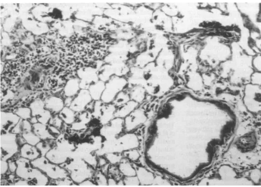
The lungs of rats and mice that received an airway instillation ofP. aeruginosa showed histologic changes of chronic pneu-monia on day 7 compared with the lungs of saline-treated animals (Figure 1). Lymphocytic infiltrates had developed around the pulmonary vasculature, and there were few neu-trophils in the alveolar spaces. Areas of the lung that did not receive the bacteria mixed with colloidal carbon did not show any accumulation of lymphocytes.
Role of CDlla in Rats
Rats that receivedP. aeruginosa showed an l l-fold increase in the number of lymphocytes per 100 p.m2of perivascular
connective tissue compared with animals that received only saline (Figure 2). This increase in lymphocyte density was inhibited by 34%when animals were treated with an anti-COlla antibody. However, a similar inhibition was seen when the animals were treated with murine IgG. Although both the anti-COlla-treated and murine IgG-treated animals showed a significant decrease in lymphocyte density com-pared with rats that received no antibody treatment, no significant differences between the anti-COlla-treated and murine IgG-treated groups were observed.
The data were also analyzed by comparing the lympho-cyte densities in vessels grouped by size (Figure 3). The perivascular infiltrates around vessels
<
2,000 p.m2incross-sectionalarea did not show significant differences in lympho-cyte densities between any of the groups. The lympholympho-cyte ac-cumulations' around vessels> 2,000 p.m2were significantly
smaller in animals treated with the anti-COlla antibody compared with rats that received no antibody. However, the rats treated with murine IgG also showed reduction in the ac-cumulation of lymphocytes around these vessels similar to the anti-COHa-treated rats.
Circulation of Anti-CDlla in Rats
516 AMERICAN JOURNAL OF RESPIRATORY CELL AND MOLECULAR BIOLOGY VOL. 12 1995
Figure 1. Histologic examination of lungs from rat 7 days after air-way instillation ofP. aeruginosa
demonstrating perivascular, lym-phocytic infiltration. (Original magnification: XlOO.)
Role of CDl1a and ICAM-l in Mice
Figure 4 shows the lymphocyte densities in perivascular con-nective tissue on day 7 ofP. aeruginosa-inducedpneumonia in wild-type mice. Animals received daily injections of the
(\1(1) 2
c
o
a-U
'e
o oT'"
en
Q)>.
1u
o
s:
c.
E
>-..J
Figure2. Role of COlla in lymphocyte accumulation duringP.
aeru-ginosa-inducedpneumonia in rats. Lymphocyte densities were de-termined from lung sections of animals that received only saline in-stillation in the airway compared with animals that received
P. aeruginosaand then had daily injections of anti-COlla (n = 4), murine IgG (n
=
4), or no antibody (n=
5). Data are mean±
SEM.anti-CDlla antibody, the anti-ICAM-l antibody, rat IgG, or no antibody on days 3 through 6 after P. aeruginosa instilla-tion. There were no significant differences in the number of lymphocytes that accumulated within the perivascular con-nective tissue of animals in any of the treatment groups.
Figure 5 shows the lymphocyte densities of the perivascu-lar connective tissue grouped by vessel size for the wild-type mice treated with either anti-CDlla, anti-ICAM-l, IgG, or no antibody. There were no significant differences in the lymphocyte accumulations between any of the treatment groups for any vessel size.
The blood lymphocyte counts on day 7 were not signif-icantly different between wild-type mice with P.
aerugi-nosa-inducedpneumonia that received daily injections of ei-ther IgG or blocking antibodies to specific adhesion molecules. The anti-CDlla-treated mice had lymphocyte counts of 6.4
±
1.3 X 10⁶cells/ml compared with 6.7±
1.8 X 10⁶cells/ml in anti-ICAM-l-treated mice and 4.2
±
0.3 X 10⁶cells/ml in IgG-treated mice.
Effect of ICAM-l Deficiency on Number and Subtypes of Accumulated Lymphocytes
The lymphocyte densities in the lungs ofICAM-l mutant and wild-type mice that received tracheal instillates of saline or
P. aeruginosaare shown in Figure 6. There was a significant increase in the number of lymphocytes in the perivascular connective tissue in the ICAM-l mutant mice that received
P. aeruginosa compared with that in the ICAM-l mutant mice that received only saline. This increase was similar to the response seen in the wild-type mice that receivedP.
aeru-ginosa.
pneumo-Figure 3. Role of CDIla in
lym-phocyte accumulation around dif-ferent vessel sizes duringP. aerugi-nasa-induced pneumonia in rats.
Lymphocyte densities were deter-mined from lung sections of animals that had P.
aeruginosa-induced pneumonia and were giv-en daily injections ofanti-Cfrl la, anti-ICAM-I, murine IgG, or no antibody. Asterisk indicates sig-nificant difference compared with the no-antibody treatment group. Data are mean
±
SEM.N
E 2.0
:;,
o
o
1.5
....
...
(I)4D
>.
1.0
o
o
s::
~
0.5
>--'
0.0
<1000
1000-2000
Vessel size (um2)
>2000
_ anti-CD 11 a _ l g G ~ No antibody
nia. The percentages of CD4 + cells, CD8+ cells, and B cells were 30.5
±
2.2%,16.1±
3.2%, and 19.6±
5.4%, respec-tively, in the ICAM-l mutant mice and 37.8±
2.8%,22.8±
7.7%, and 23.0±
2.6 %, respectively, in the wild-type mice.Discussion
By 7 days after the intratracheal instillation ofP.aeruginosa, lymphocytes accumulate in the connective tissue surround-ing the microvasculature. This accumulation occurs only in the region of the lung where organisms are deposited, sug-gesting that lymphocytes recognize the inflamed site and ac-cumulate in response to locally produced mediators. This study examined the roles of the adhesion molecules CDlla and ICAM-l in this inflammatory and immune response to
P.aeruginosa by evaluating the lymphocyte accumulation in-duced byP. aeruginosa when these adhesion molecules were inhibited or absent.
Two methods of blocking the function of these adhesion molecules were compared. Blocking monoclonal antibodies were given daily to animals with pneumonia during the period when the lymphocytic infiltrate is evolving. The ac-cumulation of lymphocytes was morphometrically quanti-tated and compared with that in animals with pneumonia that received no or control antibody. In other experiments, the lymphocyte accumulation in ICAM-l mutant mice was com-pared with that in wild-type mice.
TABLE 1
Circulating levels ofWT.l 24h after injection evaluated using immunochemistry on cytospins from circulating
leukocytes of untreated rats*
This study shows that in mice, the administration of blocking antibodies against either CDlla or ICAM-l caused no significant change in the lymphocyte accumulation during
P. aeruginosa-induced pneumonia. These results suggest that neither CDlla nor ICAM-l are necessary for the lym-phocyte accumulation to occur in this lesion. Additionally, ICAM-l mutant mice demonstrated a significant increase in lymphocyte density in the perivascular connective tissue in
re-1.5
C\I
E
::J 1.0
o o
T""
Cfj
Q)
~
os:
c. E 0.5
>-..J
0.0
Primary Antibody
Serum samples WT.I (0.02 mg/ml) Murine IgG
*Data are mean ±SEM.n = 4 for each primary antibody.
Positive Cells (%)
38 ± 3
37± 4
o
Figure4. Role ofCDlla and ICAM-I in lymphocyte accumulation during P. aeruginosa-induced pneumonia in mice. Lymphocyte
densities were determined from lung sections of animals with
P. aeruginosa-induced pneumonia that received daily injections of
518' AMERICAN JOURNAL OF RESPIRATORY CELL AND MOLECULAR BIOLOGY VOL. 12 1995
~
E 1.2
::::s
o 1.0
o
=::
0.8
UJ
Q)
>.
0.6
CJ
o
.c
0.4
a.
E
>-
0.2
..J
0.0
<1000
1000-2000
Vessel size (um
2
)
>2000
Figure5. Role of COlla and ICAM-I in lymphocyte accumu-lation around different vessel sizes during P.
aeruginosa-in-duced pneumonia in mice. Lym-phocyte densities were deter-mined from lung sections of animals that had P. aerugi-nosa-induced pneumonia and were given daily injections of anti-COlla, anti-ICAM-I, rat IgG, or no' antibody. Data are mean ± SEM.
_ .ntl-CD 11. c:::==J .nti-ICAII-1 ICJ;;.h.>j IgG
sponse toP. aeruginosathat was similar to that seen in wild-type mice receiving the bacteria. Further, there' were no significant differences in the subtypes of the accumulated lymphocytes in ICAM-l mutant mice compared with that in wild-type mice. These results are consistent with the results
2
N
E
:1
o o 'I"'"
en
CD ~ 1..o
J:a.
~
-I
o
- l - -_ _Figure6. Effect of ICAM-I deficiency on lymphocyte accumula-tion duringP. aeruginosa-induced pneumonia in mice.
Lympho-cyte densities were determined from lung sections of ICAM-I mu-tant mice that received saline airway instillates(n = 4) compared with ICAM-I mutant mice (n = 5) and ICAM-I wild-type mice
(n = 4) withP. aeruginosa-induced pneumonia. Data are mean
±
SEM.
c::==J No .ntibod,
using blocking antibodies, suggesting that ICAM-1 is not necessary for this lymphocyte response to occur in mice.
In rats, both the anti-CDlla antibody and the control IgG partially inhibited the lymphocyte accumulation during
P. aeruginosa-inducedpneumonia. This effect was observed only in vessels> 2,000 j.(.m2in cross-sectional area and not
around the smaller vessels. The partial inhibition of the lym-phocyte response in both the anti-CDlla- and the IgG-treated animals may be due to nonspecific effects of foreign proteins on cellular functions and not to inhibition of a required adhe-sion molecule.
Other adhesion pathways may be important in the devel-opment of this lesion. A likely candidate for this alternative pathway is the integrin very late activation antigen-4 (VLA-4) interacting with vascular cell adhesion molecule-l (VCAM-l) (13). VLA-4, like CDlla, is expressed on the surface of memory T cells (14), which are concentrated at sites of inflammation within the bronchial epithelium.
The role of adhesion molecules has been investigated in at least three other studies of chronic pulmonary inflamma-tion. First, Piguet and associates investigated the role of
CDIla/CD18and CDllb/CD18 in chronic inflammation and
ICAM-l inhibited antigen-induced T-cell infiltration of mice tracheae (19). Third, Denis and Bisson evaluated the contri-bution of CDlla to the inflammation and fibrosis seen in mu-rine hypersensitivity pneumonitis secondary to Faeni rec-tivirgula (20). There were no significant differences in the number or type of cells obtained by bronchoalveolar lavage between either challenged mice given control antibody (rat IgG) and challenged mice given a rat anti-mouse CD11a monoclonal antibody. However, administration of anti-CDlla significantly decreased the lung hydroxyproline levels and the morphometric lung index in mice with hypersensitivity pneumonitis. All these studies contrast with our study and show a role for CD11a/CDI8 and ICAM-l in the recruitment of lymphocytes in lung injury induced by bleomycin and hypersensitivity. Taken together, these studies suggest that the accumulation of lymphocytes, similar to the recruitment of neutrophils, occurs through at least two pathways, one that requires CDlla/ICAM-l and one that does not. The particu-lar stimulus and the ensuing response appear to be critical in determining which adhesion pathway is utilized.
The role of adhesion molecules in chronic inflammation in organs and tissues other than the lung has also been stud-ied, including experiments using ICAM-l mutant mice. Sligh and associates found that ICAM-1 mutant mice that had been sensitized and rechallenged with 2,4-dinitrofluoroben-zene (DNFB) had a 74%reduction in maximal ear swelling (a measure of delayed type hypersensitivity) compared with wild-type mice (11). Histologic studies revealed an attenua-tion of the inflammatory cell infiltrate in the ICAM-l mutant mice compared with the wild-type mice after rechallenge with DNFB. Although we do not yet understand the differ-ences in the mechanisms underlying these disparate forms of chronic inflammation in the lung and other tissues, these studies underline the importance of examining lymphocyte accumulation in chronic inflammation induced by different stimuli.
Mechanisms not requiring adhesion molecules may medi-ate the accumulation of lymphocytes inP.
aeruginosa-in-duced pneumonia. Local proliferation of lymphocytes may play a predominant role in this process. Cytokines, such as interleukin-6, that are produced by the endothelium and other cells at sites of inflammation could mediate this lym-phocyte accumulation by stimulatory effects on cell prolifer-ation and differentiprolifer-ation (21, 22).
In summary, these studies suggest that lymphocyte ac-cumulation duringP. aeruginosa-inducedpneumonia in ro-dents occurs through mechanisms that are independent of the adhesion molecules CD11a and ICAM-l. Either alternative adhesion pathways, perhaps involving VLA-4 and VCAM-l (13) or L-selectin (23), or local proliferation of lymphocytes may mediate this accumulation.
Acknowledgments:This work was supported by a grant from the Riley Memorial Association to IL.W. and PHS HL52466 to C.M.D. C.M.D. is the recipient of a Career Investigator Award from the American Lung Association. IE.S. worked in the laboratory of Arthur L. Beaudet with support of NIH Grant AI 32177.
References
1. Berman, J. S., D. J. Been, A. C. Theodore, H. Kornfeld, J. Bernardo, and D. M. Center. 1990. Lymphocyte recruitment to the lung. Am. Rev. Respir. Dis. 142:238-257.
2. Pabst, R. 1991. Compartmentalization and kinetics oflymphoid cells in the lung. Reg. Immunol. 3:62-71.
3. Abraham, E., A. A. Freitas, and A. A. Couthino. 1990. Purification and characterization of intraparenchymal lung lymphocytes. J. Immunol.
144:2117-2122.
4. Picker, L. J., and E. C. Butcher. 1992. Physiologic and molecular mecha-nisms of lymphocyte homing. Annu. Rev. Immunol. 10:561-591.
5. Stoolman, L. M. 1993. Adhesion molecules involved in leukocyte recruit-ment and lymphocyte recirculation. Chest103:79S-86S.
6. Kavanaugh, A. F., E. Lightfoot, P. E. Lipsky, and N. Oppenheimer-Marks. 1991. Role of CD11/CDI8 in adhesion and transendothelial migration of T cells.J.Immunol. 146:149-156.
7. Dustin, M. L., and T. A. Springer. 1988. Lymphocyte function-associated antigen-l (LFA-l) interaction with intercellular adhesion molecule-l (ICAM-l) is one of at least three mechanisms for lymphocyte adhesion to cultured endothelial cells. J.Cell Biol. 107:321-331.
8. Iwato, M., and S. Atsuhiko, 1991. Morphological and immunohistochemi-cal studies of the lungs and bronchus-associated lymphoid tissue in a rat model of chronic pulmonary infection with Pseudomonas aeruginosa. In-fect. Immun. 59:1514-1520.
9. Cash, H. A., D. E. Woods, B. McCullough, W. G. Johanson, and J. A. Bass. 1979. A rat model of chronic respiratory infection with Pseudomo-nas aeruginosa. Am. Rev. Respir. Dis. 119:453-459.
10. Wiebke, J. L., W. M. Quinlan, L. Graham, N. A. Doyle, and C. M. Doer-schuk. 1993. Lymphocyte recruitment in response to Pseudomonas infec-tion in the rat lung.Am. Rev. Respir. Dis. 147(Supp1.):AI2. (Abstr.) 11. Sligh, J. E., C. M. Ballantyne, S. S. Rich, H. K. Hawkins, C. W. Smith,
A. Bradley, and A. L. Beaudet. 1993. Inflammatory and immune re-sponses are impaired in mice deficient in intercellular adhesion molecule
1. Proc. Natl. Acad. Sci. USA 90:8529-8533.
12. Takei, F. 1985. Inhibition of mixed lymphocyte response by a rat monoclo-nal antibody to a murine lymphocyte activation antigen (MALA-2).J. Im-munol. 134:1403-1407.
13. Carlos, T. M., B. R. Schwartz, and N. L. Kovach. 1990. Vascular cell adhesion molecule-l mediates lymphocyte adherence to cytokine-activa-ted cultured human endothelial cells. Blood 76:965-970.
14. Saltini, c., M. Kirby, B. C. Trapnell, N. Tamura, andR.G. Crystal. 1990. Biased accumulation of T-Iymphocytes with memory-type CD45 leukocyte common antigen gene expression on the epithelial surface of the human lung. J.Exp. Med. 171:1123-1140.
15. Piguet, P. F., H. Rosen, C. Vesin, and G. E. Grau. 1993. Effective treat-ment ofthe pulmonary fibrosis elicited in mice by bleomycin or silica with anti-CD11 antibodies. Am. Rev. Respir. Dis. 147:435-441.
16. Snider, G. L., J. A. Hayes, and A. L. Korthy. 1978. Chronic interstitial pulmonary fibrosis produced in hamsters by endotracheal bleomycin.Am. Rev. Respir. Dis. 117:289-297.
17. Hesterberg, T. W.,J.E. Gerriets, K. M. Reiser, A. C. Jackson, C. E. Cross, and J. A. Last. 1981. Bleomycin-induced pulmonary fibrosis: correlation of biochemical, physiological, and histological changes.
Tox-icol. Appl. Pharmacol. 60:360-367.
18. Thrall, R. S., and R. W. Barton. 1984. A comparison of lymphocyte popu-lations in lung tissue and in bronchoalveolar lavage fluid of rats at various times during the development of bleomycin-induced pulmonary fibrosis.
Am. Rev. Respir. Dis. 129:279-283.
19. Nakajima, H., H. Sano, T. Nishimura, S. Yoshida, andI.Iwamoto. 1994. Role of vascular cell adhesion molecule-livery late activation antigen-l and intercellular adhesion molecule-l/1ymphocyte function associated antigen-l interactions in antigen induced eosinophil and T cell recruitment into the tissue. J. Exp. Med. 179: 1145-1154.
20. Denis, M., and D. Bisson. 1994. Blockade of leukocyte function-assoc-iated antigen (LFA-l) in a murine model of lung inflammation.Am.J.
Respir. Cell Mol. Bioi. 10:481-486.
21. Cavender, D. E. 1991. Interaction between endothelial cells and the cells of the immune system. Int. Rev. Exp. Pathol. 32:57-94.
22. Mantovani, A., F. Bussolino, and E. Dejana. 1992. Cytokine regulation of endothelial cell function. FASEBJ.6:2591-2599.