R E S E A R C H
Open Access
Pro-inflammatory endothelial cell dysfunction is
associated with intersectin-1s down-regulation
Sunit Singla
1, Dan Predescu
1,2, Cristina Bardita
2, Minhua Wang
2, Jian Zhang
2, Robert A Balk
1and
Sanda Predescu
1,2*Abstract
Background:The response of lung microvascular endothelial cells (ECs) to lipopolysaccharide (LPS) is central to the pathogenesis of lung injury. It is dual in nature, with one facet that is pro-inflammatory and another that is cyto-protective. In previous work, overexpression of the anti-apoptotic Bcl-XLrescued ECs from apoptosis triggered by siRNA knockdown of intersectin-1s (ITSN-1s), a pro-survival protein crucial for ECs function. Here we further characterized the cyto-protective EC response to LPS and pro-inflammatory dysfunction.
Methods and Results:Electron microscopy (EM) analyses of LPS-exposed ECs revealed an activated/dysfunctional phenotype, while a biotin assay for caveolae internalization followed by biochemical quantification indicated that LPS causes a 40% inhibition in biotin uptake compared to controls. Quantitative PCR and Western blotting were used to evaluate the mRNA and protein expression, respectively, for several regulatory proteins of intrinsic apoptosis, including ITSN-1s. The decrease in ITSN-1s mRNA and protein expression were countered by Bcl-XLand survivin upregulation, as well as Bim downregulation, events thought to protect ECs from impending apoptosis. Absence of apoptosis was confirmed by TUNEL and lack of cytochrome c (cyt c) efflux from mitochondria. Moreover, LPS exposure caused induction and activation of inducible nitric oxide synthase (iNOS) and a
mitochondrial variant (mtNOS), as well as augmented mitochondrial NO production as measured by an oxidation oxyhemoglobin (oxyHb) assay applied on mitochondrial-enriched fractions prepared from LPS-exposed ECs. Interestingly, expression of myc-ITSN-1s rescued caveolae endocytosis and reversed induction of iNOS expression. Conclusion:Our results suggest that ITSN-1s deficiency is relevant for the pro-inflammatory ECs dysfunction induced by LPS.
Background
Severe sepsis is the leading clinical cause of lung injury [1]. No specific therapy currently exists for this critical illness, and efforts to reduce its burden have been lim-ited by an incomplete understanding of the mechanisms of disease. Exposure of ECs to LPS is a widely used model for studying endothelial dysfunction in this syn-drome [2]. The endothelial response to LPS is dual in nature, with one facet that is pro-inflammatory and another that is cyto-protective [2,3]. NF-B and JNK-mediated pathways engage ECs to actively participate and regulate the pro-inflammatory process in response to LPS by expression and release of damaging
components such as iNOS, cytokines, adhesion mole-cules, and pro-coagulants [4-6]. This response has been alternatively referred to as the activated endothelial phe-notype [4,5]. Much less is known however, with respect to the exact nature, regulation and timing of the cyto-protective aspect of the ECs response. A role of Bcl
members Bcl-2 and Bcl-XLin potentially shielding the
cells from their proapoptotic environment has been described [2,3]. In our previous work, overexpression of
Bcl-XL rescued lung ECs from apoptosis after
siRNA-mediated knockdown of ITSN-1s, a novel scaffold and regulator of the general endocytic machinery, as well as a critical protein for pro-survival signaling in ECs [7,8]. ITSN-1s deficiency caused severe EC dysfunction, decrease in caveolae number, structural alterations and dysfunction of mitochondria, generation of mitochon-drial reactive oxygen species (ROS) and apoptosis [7]. * Correspondence: [email protected]
1
Pulmonary and Critical Care Medicine, Rush University Medical Center, 1750 W. Harrison Street, 297 Jelke, Chicago, IL 60612, USA
Full list of author information is available at the end of the article
Similar observations of many of these phenomena in previous studies of LPS-induced endothelial dysfunction and the suggested role of Bcl-XL in cyto-protection
dur-ing inflammatory stress led us to consider ITSN-1s as a participant in either of these events.
Numerous lines of evidence have documented the considerable oxidative and nitrosative stress associated with lung injury in human patients, and potential path-ways by which either may mediate endothelial barrier dysfunction have also been well described [9,10]. iNOS is known to be up-regulated in response to LPS in a variety of cells, including lung ECs [11]. The resultant increased NO production has the ability to potentially alter a large variety of cellular processes such as inter-endothelial junctional integrity and thereby, basal vas-cular permeability [9]. The selective inhibition of iNOS has significantly attenuated the ability of endotoxin to generate lung injury in several animal models [12,13]. Notably, loss of caveolin-1 (cav-1) expression and thus lack of caveolae endocytosis and transcytosis resulted in constitutive activation of eNOS, increased NO pro-duction and a microvascular hyperpermeability cav-1 -/- mouse phenotype [14]. Thus, in the current study, we sought to further characterize ECs dysfunction and the elements of the cyto-protective ECs response to LPS.
Methods
Cell lines and reagents
Human lung microvascular ECs were purchased from Lonza (Walkersville, Inc., MD). LPS from Escherichia coli 011:B4, calmodulin from bovine testes, tetrahydro-biopterin (BH4), protease inhibitors cocktail, L-arginine
and primers for qPCR were from Sigma-Aldrich (St. Louis, MO). Superoxide dismutase (SOD) from bovine erythrocytes was purchased from MP Biomedicals. Freeze dried Hb was from ICN Biomedicals. The In Situ Cell Death Detection Kit, Fluorescein was from Roche (Indianapolis, IN). Prolong Antifade Kit and neu-trAvidin Alexa Fluor 594 were from Molecular Probes (Eugene, OR). EZ-Link Sulfo NHS-SS-Biotin was from Fisher Scientific (Hanover Park, IL). Streptavidin-HRP conjugated, MicroBCA (bicinchoninic acid) Protein Assay Reagent and Enhanced Chemiluminescent (ECL) Western Blotting Substrate were from Pierce (Rockford, IL). Specific antibodies (Abs) were obtained from the
following sources: anti-Bcl-XL mAb, anti-NOS2 pAb
from Santa Cruz Biotechnology (Santa Cruz, CA); anti-cyt c mAb from Calbiochem; anti-ITSN-1s mAb from BD Biosciences (San Jose, CA); anti-Bim pAbs from Millipore (Billerica, MA); anti-survivin pAbs from Novus Biologicals; Horseradish peroxidase (HRP)-conju-gated reporters were from Cappel, Organon Teknika (Durham, NC).
Endothelial Cell Culture and LPS treatment
ECs were cultured at 37°C in 5% CO2 in endothelial growth medium-2 obtained from Lonza and prepared according to manufacturer’s instructions. Cells between the third and fifth passages were used for experiments. LPS (dry powder) was reconstituted in growth medium and applied over cells in a concentration of 1μg/ml for a duration of up to 48 hours.
Protein Extraction and Western blotting
Control ECs or cells exposed to 1μg/ml LPS in culture media were collected from culture dishes and washed with PBS. Cell pellets were lysed for 1 h at 4°C in 50 mM Tris HCl, pH 8.0, 150 mM NaCl, 1% NP-40, and protease inhibitors. For detection of phospho-Bim (Ser69) levels, control and LPS-treated ECs were solubi-lized in kinase buffer (20 mM Tris/HCl, pH 7.4, 150 mM NaCl, 0.1% Nonidet P-40, 1% glycerol, 0.2 mM sodium vanadate, 0.83 mM benzamidine, 0.23 mM phe-nylmethylsulfonyl fluoride, 0.5 μg/ml aprotinin, and 0.5
μg/ml leupeptin). After centrifugation at 45000 rpm for 45 min at 4°C, the supernatants were collected and pro-tein concentration determined by BCA with a bovine serum albumin standard. Equivalent protein amounts were subjected to SDS-PAGE and transferred to nitro-cellulose membranes. The membranes were probed with
anti-NOS2 pAb, anti-Bcl-XL mAb, anti-ITSN-1 mAb,
anti-Bim pAb, anti-survivin pAb, diluted in blocking buffer, (5% milk/TBS). Immunoreactive bands were visualized with the appropriate HRP-conjugated Abs and ECL detection. Densitometry was performed with ImageJ v1.37 software.
Internalization Assay
(100μl) from each lysate and normalized per mg total protein. To obviate any interference between the bioti-nylated proteins internalized via caveolae and biotin pre-sent in mitochondria [17], we evaluated the biotin content of mitochondria by ELISA applied on lysate pre-pared from control ECs, not subjected to biotinylation of cell surface proteins. Mitochondrial proteins comprise
only 0.25 × 10¹⁴ biotin molecule/mg total protein, a
value without statistical significance, when compared to the extent of biotin internalization via caveolae.
Measurement of Transendothelial Electrical Resistance and paracellular permeability
ECs were grown to confluence on gold electrode array plates available from Applied Biophysics, Inc., Troy, NY. Transendothelial electrical resistance (TER) was mea-sured by an electrical cell-substrate impedance sensing system (Applied Biophysics, Inc.) as described in [18]. Briefly, the gold electrode plates were connected to a phase-sensitive lock-in amplifier and a 1 volt 4000 Hz
AC signal was applied through a 1 MΩ resistor.
In-phase voltage was measured and used to determine TER which was normalized to initial values. A decrease in TER during the experiment reflects loss of cell-cell adhesion.
Transwell assay
Transwell chambers with .4μm pore filter inserts (BD
Bioscience) were used. The inserts were coated over-night with 0.1% gelatin, at 37°C. Cells were seeded in the upper compartment and grown for additional 3 days post-confluency. Then, cell monolayers were subjected to 1μg/ml LPS for 6 h, as an early time point, and 48 h, the time point used for biotin assay for caveolae interna-lization. The last hour of LPS treatment, 1 mg/ml dini-trophenylated (DNP)-BSA was added in the upper
chamber. To quantify the transport, 500 μl medium
from the lower chamber were removed and subjected to ELISA, via anti-DNP Ab, as previously described [19,20]. All measurements were performed in triplicate and repeated three times. A standard curve was generated by diluting known concentration of DNP-BSA.
Molecular cloning of ITSN-1s expression construct
The full-length human ITSN-1s cDNA fragment (3,660 bp) was generated by PCR amplification from ITSN-1s cDNA (gift from Suzana la Luna, Center for Genomic Regulation, UPF, and Centro de Investigacion Biomedica en Red de Enfermedades Raras, Barcelona, Spain) with high-fidelity PCR enzyme (New England Biolabs), using
the following primer pair: ITSN1s_F269-BstBI: 5’
AGTA TTCGAA CCATGGCT CAG TTT CCA ACA
CCT T 3’and ITSN1s_R3929-NheI: 5’CGTA GCTAGC
TTG CTG GCT TGG GTC CAT GTC TG 3’. The PCR
products of the full-length ITSN-1s were digested with restriction enzyme BstBI and Nhe I (New England Bio-labs) and purified (Qiagen purification kit), then cloned into a expression vector, pReiver-M10a (GeneCopoeia) at BstBI-NheI sites using T4 ligase (New England
Bio-labs). The ligation products were transformed into E.
colistrain Top10 (Invitrogen). Dozens of transforming
colonies were selected and subjected to PCR to identify the colonies with the ITSN-1s inserts using the same sequencing primer pair. The plasmid DNA was extracted from 6 selected growing clones bearing the ITSN-1s insert and was verified with correct restriction enzyme (BstBI and Nhe I) sites. Finally, one of them, CS-Z3999_ITSN1s #16 was selected for maxi-prepara-tion of plasmid DNA, sequencing confirmamaxi-prepara-tion of the entire integrity of the full-length cDNA of ITSN-1s in frame in the vector and used for transfection of ECs. All transfections were performed using FuGENE 6, accord-ing to the manufacturer’s instructions.
Mitochondrial isolation and measurement of iNOS activity Preparation of Mitochondrial fractions
EC monolayers grown on Petri dishes were washed three times with ice-cold PBS, pH 7.4, and collected by gentle scraping and centrifugation at 500 × g for 10 min. The cell pellets were resuspended in isolation buf-fer containing 250 mM sucrose, 40 mM Tris/HCl, pH
7.5, 10 mM MgCl2, 2 mM CaCl2, 1 mM
phenylmethyl-sulfonyl fluoride, 15 μg/ml leupeptin as in [7]. They
were kept on ice for 30 min and then disrupted with 40 strokes in a glass Dounce homogenizer. This was fol-lowed by centrifugation at 800 × g at 4°C for 10 minutes to remove unbroken cells, nuclei, and debris. The post-nuclear supernatant was then subjected to centrifugation at 22,000 × g at 4°C for 10 minutes to isolate the mito-chondrial pellet. The mitomito-chondrial pellet was lysed in 50 mM HEPES, pH 7.4, 1% Nonidet P-40, 10% glycerol, 1 mM EDTA, 2 mM dithiothreitol, and protease inhibitors.
Spectrophotometric determination of iNOS activity
Commercially available Hb contains a mixture of oxyHb and metHb which must be fully reduced to obtain pure oxyHb for the reaction. This was accom-plished by the Dixon and McIntosh method [22]. Briefly, 0.2 ml of 0.1 g/ml sodium dithionite was added to a prepacked Sephadex column. When all of the dithionite had completely entered the gel bed, 1 ml of 50 mg/ml Hb was added. As the Hb passed through the dithionite zone, its color changed to purple and it then changed to an orange-red color as it left the dithionite. This was collected and the concentration measured spectrophoto-metrically usingε415 nm131.0 mM-1cm-1.
Dilution or oxidation of NOS substrates or cofactors during the preparation of broken mitochondria requires the presence of 5 μg/ml calmodulin, 50μM L-arginine,
10μM tetrahydrobiopterin (BH4), and 1 KU/ml SOD in
the assay medium. These substrates were added to 1 mL of 100 mM HEPES, pH 7.1 in a cuvette followed by 4
μM oxyHb. 30 μg of mitochondrial protein was added
and the optical density continuously recorded for five minutes at a wavelength of 401 nm. NO formation was quantified usingε401(metHb-oxyHb)of 49 mM-1cm-1.
Terminal Deoxynucleotidyltransferase-mediated dUTP Nick End Labeling (TUNEL) Assay
Control ECs and cells exposed to LPS (1 μg/ml) for up
to 48 h were washed in PBS and then fixed and permea-bilized in methanol for 7 min, at -20°C. The TUNEL reaction mixtures (label and enzyme solutions) were prepared as directed by the manufacturer. It was applied over EC monolayers for 60 min, at 37°C, under dark and humid conditions. Monolayers exposed to label solution only were used as negative controls. The cells were washed and coverslips mounted using the Prolong Antifade kit. Fluorescence microscopy with an excitation wavelength of 488 nm identified TUNEL-positive cells.
qPCR studies
Cells were lysed using the Qiagen QIAShredder homo-genizer and the RNA was isolated using the Qiagen RNeasy Mini RNA isolation kit as per the manufac-turer’s instructions. cDNA was generated with the high-capacity cDNA RT kit by Applied Biosystems following the manufacturer’s protocol. Amplifications were carried
out using the following primers: ITSN-1s (5’
-CAGGCTTGAAAGTCCTCAAAG-3’, 5’
-GGTGAT-CATGCTGGAAGTCA-3’), Bim (5’-TCCCTGCTGT
CTCGATCCTC-3’, 5’
-GGTCTTCGGCTGCTTGGTAA-3’), Bcl-XL (5’-CTGTCGGTGGAAAGCGTAGA-3’, 5’
-TGCTGCATTGTTCCCATAGAG-3’), Survivin (5’
-AAGAACTGGCCCTTCTT GGA-3’, 5’
-CAACCGGAC-GAATGCTTTT-3’). mRNA levels were quantified by
real-time monitoring of the products of the cDNAs PCR
with SYBR Green fluorescent dye. Cyclophilin was used as internal control.
Electron microscopy
Control and LPS-treated cells were subjected to stan-dard EM procedure [15]. Briefly, EC monolayers were fixed in 1.5% glutaraldehyde in 0.1 M sodium cacodylate buffer, pH 7.4, containing 5% sucrose for 30 min at RT. After washing in the same buffer the cells were post-fixed in Palade’s 1% osmium tetroxide for 1 h on ice, stained with Kellenberger buffer, dehydrated in graded ethanol, and embedded in Epon at 60°C.
Results and discussion
LPS exposure causes an activated/dysfunctional ECs phenotype
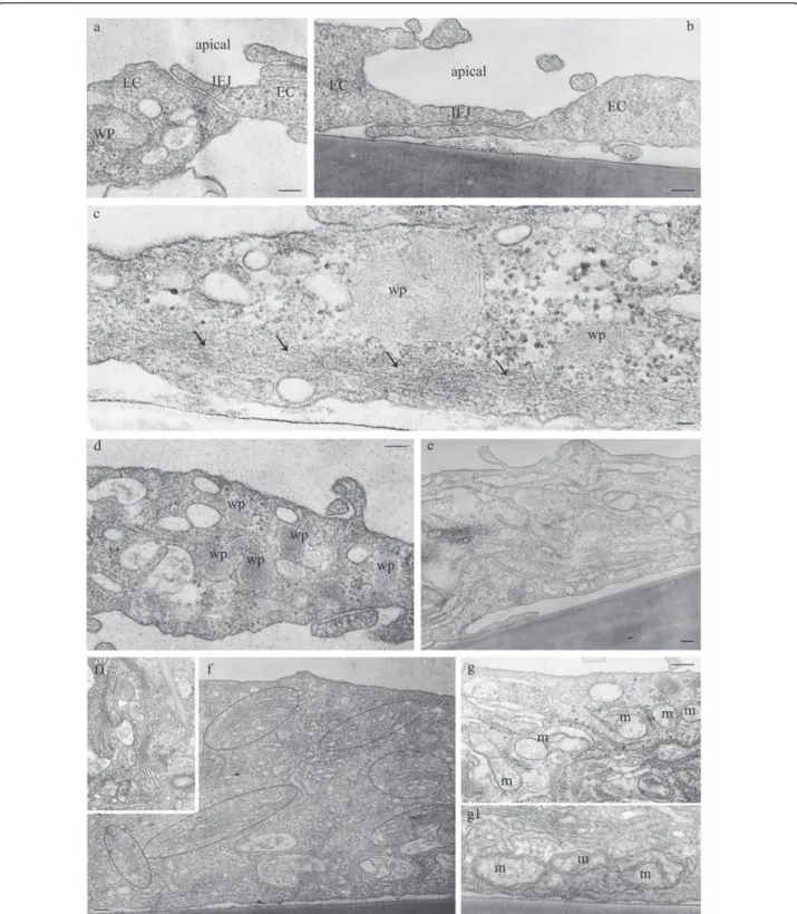
Figure 1LPS exposure causes an activated/dysfunctional ECs phenotype (I). ECs exposed to 1μg/ml LPS for 48 h show open IEJs, (a, b), increased accumulation of actin filaments at cell periphery (c, arrows), significant increase in the number of Weibel-Palade (wp) bodies (d) and Golgi (f, circled areas). An enlarged and widespread endoplasmic reticulum network and the presence of a dilated tubular system are shown in
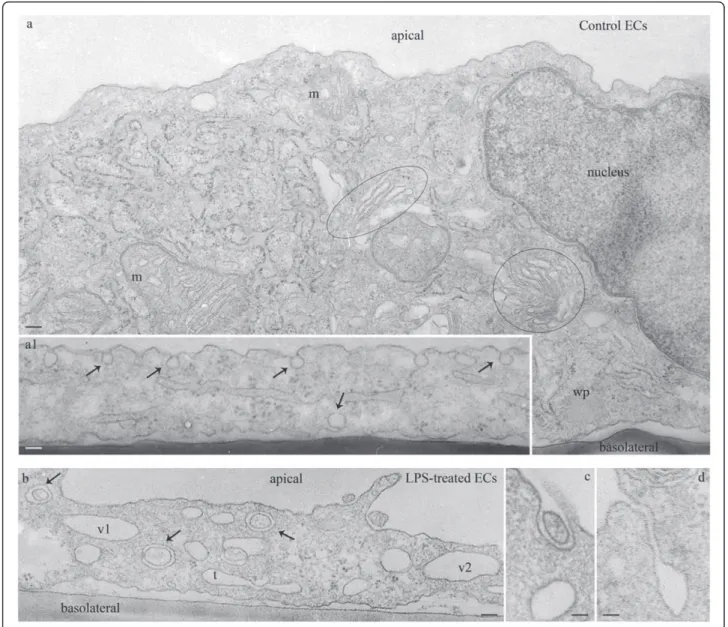
frequency of tubular structures and membraneous rings, (Figure 2b) some of them opened to the apical front of the cell (Figure 2c, d) are detected, suggesting impaired endo-cytosis and activation of alternative, non-conventional, transport pathways [24]. Altogether, these observations are consistent with an activated and dysfunctional EC phenotype.
Endocytic dysfunction and a concomitant paracellular hyperpermeability is a hallmark of the LPS-EC phenoytpe
Since LPS signaling is regulated by caveolae endocytosis [25,26] and since EM analysis revealed endocytic
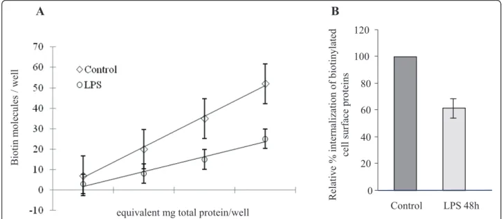
abnormalities in LPS-treated cells, we next used a biotin internalization assay as in [15,16] to address if EC expo-sure to LPS affects caveolae endocytosis. This assay is a specific and reliable approach for addressing caveolae internalization in cultured ECs [15]. Caveolae-mediated uptake of biotinylated cell surface proteins is the domi-nant mechanism of internalization in ECs; the number of clathrin-coated vesicles in ECs is relatively small and their contribution to the internalization process is minor [27]. Briefly, control and LPS-exposed ECs were sub-jected to biotinylation of cell surface proteins, at 4°C, using a cleavable biotin reagent. Then, cells were
transferred to 37°C, for 30 min, to allow internalization of biotinylated proteins. The biotinylated proteins, still on the cell surface after 30 min were reduced with glu-tathione. Then, the cells were either lysed for biochem-ical quantification of LPS effects on caveolae internalization by enzyme-linked immunosorbent assay (ELISA) or subjected to neutrAvidin-Alexa Fluor 594 staining for morphological surveys by fluorescence microscopy. Biochemical quantification of the number of biotin molecules internalized by control and LPS-exposed cells, Figure 3A indicated that in LPS-treated EC lysates, the number of biotin molecules was consid-erably smaller. A comparison of LPS-treated cells to controls shows an inhibition of 40%, (Figure 3B). Mor-phological analyses with NeutrAvidin-Alexa Fluor 594 revealed in control cells a fine punctate pattern through-out the cytoplasm, suggestive of vesicular association, (Figure 4c1). Frequently, biotin accumulation in the perinuclear area was detected. In LPS-exposed cells the staining was limited (Figure 4c2); large fluorescent puncta within the cytosol and no biotin accumulation in the perinuclear area were observed, suggestive of impaired biotin internalization. The intercellular spaces are large, consistent with disruption of IEJs. Highly mag-nified controlled and LPS-exposed cells (boxed areas), are shown, (Figure 4c1.1, c2.1).
Since 1μg/ml LPS exposure downregulates ITSN-1s
expression and as a result transcellular transport is impaired, we next evaluated the status of paracellular
permeability of ECs monolayers by measurements of TER, under the same conditions of LPS exposure. Monolayers of ECs grown on gold microlectrodes, three days post-confluency, were first stabilized for two hours to establish a linear TER baseline. Then, The stabilized monolayers were stimulated with LPS (Figure 5A, arrow) and TER was monitored over 24 hours, Figure 5A. A gradual decrease in the baseline barrier resistance was recorded for the first hours after LPS stimulation with a maximal decrease after 8 h and remaining in a plateau for the next 16 h hours. These data strongly suggest that LPS disrupts endothelial barrier and signifi-cantly increases the paracellular permeability of lung microvascular ECs. To get more insights on the extent of endothelial barrier dysfunction, we quantified the paracellular transport of dinitrophenylated albumin (DNP-BSA) using a transwell assay, as described under Methods. In brief, cells were seeded in the upper cham-ber and 3 days post-confluency, endothelial monolayers were subjected to 1μg/ml LPS for 6 h, as an early time point, and 48 h, the time point used for biotin assay for caveolae internalization. In the last hour of LPS treat-ment, BSA-DNP was layered on top of EC monolayers to a final concentration of 1 mg/ml. To quantify the
transport, 500μl medium from the lower chamber were
removed and subjected to ELISA, via DNP Ab, as pre-viously described [19,20]. As shown in Figure 5B, the permeability of the endothelial monolayer for the DNP-BSA tracer were determined at a series of decreasing
equivalent mg total protein/well
Biotin m
olecules
/ well
A
B
Relative % internalization of biotinylated
cell surface
proteins
0 20 40 60 80 100 120
Control LPS 48h
Figure 3Effects of 1μg/ml LPS exposure causes significant inhibition of caveolae internalization.A. Control and LPS-treated cells were subjected to biotinylation of cell surface proteins as described under Materials and Methods, followed by internalization of biotinylated proteins for 30 min, at 37°C. The number of biotin molecules present in lysates of control and LPS-treated cells was quantified by ELISA using
concentration from the linear part of the curve obtained by serial dilution of a standard volume of growth media removed from the lower chamber. In the presence of LPS, the amounts of DNP-BSA crossing the monolayer were considerably higher than control levels. Measure-ments of DNP-BSA amounts, using a series of concen-trations on the straight part of the curve, indicated that at 6 hours after LPS exposure, the lower chamber
con-tains 118.10 ± 7.3 ng DNP-BSA/100 μl medium, while
at 48 h, 164.63 ± 12.4 ng DNP-BSA/100μl medium,
sig-nificant increase over the control levels estimated at 10.1
± 0.9 ng DNP-BSA/100 μl medium, Figure 5C. The
findings are consistent with endothelial barrier dysfunc-tion, gap formation and DNP-BSA leakage, as result of LPS exposure.
Induction and activation of iNOS and a mitochondrial variant by LPS exposure
Expression iNOS in response to LPS has been reported for several cell types such as epithelial cells, fibroblasts,
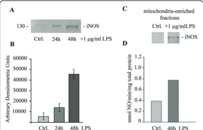
hepatocytes, ECs [28,29]. A mitochondrial NOS, mtNOS, constitutively expressed, continuously active and associated with the inner mitochondrial membrane has been reported [21,30]. LPS-induced up-regulation of mitochondrial NO is one of the specific means, however still enigmatic, by which a dysfunctional and potentially apoptotic environment is generated during the pro-inflammatory response in many cell types [31-33]. Thus, to evaluate the expression of iNOS and mtNOS in LPS-exposed human lung microvascular ECs, we first investi-gated by immunoblotting the expression of iNOS in control and LPS-treated ECs (Figure 6A). We detected weak iNOS immuno-reactivity under control conditions, and it increases by 27%, at 24 h and by 75% at 48 hours post LPS exposure (Figure 6B). Next, we investigated the effects of LPS on mtNOS expression. To this intent, mitochondria-enriched fractions prepared from these cells were lysed and subjected to SDS-PAGE. Even if mtNOS is constitutively expressed, the levels seem to be low, since no iNOS immunoreactivity was detected in
the absence of LPS and under experimental conditions used (Figure 6C). However, in the mitochondrial frac-tion prepared from LPS-exposed ECs, mtNOS protein expression is easily detected, at 48 hours of LPS expo-sure (Figure 6C). Next, we addressed if the increased expression of mtNOS resulted in increased NO produc-tion. mtNOS activity was assessed by exposing freshly prepared, unlysed mitochondrial fractions from ECs to oxyHb, as described under Methods. The rate of NO production was measured by the change in optical den-sity of the reaction mixture as generated NO quickly oxidized oxyHb to metHb. As shown in Figure 6D,
mitochondria from LPS-treated ECs exhibited a two-fold greater rate of NO formation than untreated controls (0.81 nmol/min mg total protein versus 0.40 nmol/min mg total protein).
ITSN-1s protein and mRNA levels are markedly decreased after LPS exposure
We have recently shown that specific and efficient ITSN-1s knockdown via a specific siRNA duplex tar-geting ITSN-1 gene results in severe EC dysfunction and apoptotic death, events that can be reversed by
overexpression of the anti-apoptotic Bcl-XL[7].
0.8
0.85
0.9
0.95
1
1.05
1.1
-2
0
2
4
6
8 10 12 14 16 18 20 22 24
Normalized Resistance
Time (hrs)
Control
LPS
A
B
C
Previous studies of LPS-induced endothelial dysfunc-tion suggested the ability of LPS to induce pro-apopto-tic pathways [34] as well as a role of Bcl-XL in
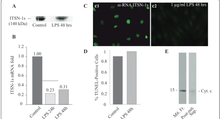
cyto-protection during inflammatory stress [3]. These obser-vations led us to consider ITSN-1s as a participant in either of these events. We therefore evaluated the levels of ITSN-1s protein in lysates prepared from LPS-treated and control ECs by immunoblotting with ITSN-1 pAb. The 140 kDa band representing ITSN-1s begins to decrease in intensity at 24 h and remains sig-nificantly decreased at 48 h after LPS exposure (Figure 7A). We also evaluated the level of ITSN-1s mRNA expression by quantitative PCR in ECs exposed to 1
μg/ml LPS for 24 or 48 hours. ITSN-1s mRNA levels
were dramatically reduced in LPS-exposed ECs com-pared to control cells (Figure 7B). ITSN-1s mRNA expression was reduced 4.3-fold at 24 hours and 3.2-fold at 48 hours of LPS treatment compared to untreated controls. These results are consistent with the idea that LPS down-regulates the expression of the pro-survival protein ITSN-1s; ITSN-1s deficiency may be relevant to ECs dysfunction induced by LPS.
Lung ECs do not undergo apoptosis in response to LPS
Since LPS-exposed ECs show decreased mRNA levels for ITSN-1s and since ITSN-1s deficiency is expected to initiate intrinsic apoptosis, we evaluated the occurrence of apoptosis in cells treated with LPS for 48 h, by TUNEL, Figure 7C, c1. Morphological surveys and mor-phometric analyzes show that the number of TUNEL-positive ECs is extremely low, comparable to untreated ECs (Figure 7D). A positive control, in which cells were subjected to ITSN-1s knockdown by siRNA as in our previous work [7], is shown in Figure 7C, c2. A comple-mentary strategy to evaluate if mitochondrial cell death occurred was the examination of cyt c efflux from mito-chondria. An enriched-mitochondrial fraction and the post-mitochondrial supernatant prepared from LPS-trea-ted ECs normalized for total protein content were sub-jected to SDS-PAGE and Western blot with cyt c mAb. No immunoreactivity for cyt c was detected in the post-mitochondrial supernatant, consistent with cyt c con-finement to mitochondria (Figure 7E). The findings sug-gest that ITSN-1s down-regulation as caused by LPS exposure of ECs did not trigger the intrinsic apoptotic pathway and suggest that LPS induces a cyto-protective response.
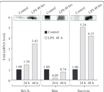
LPS directly induces Bcl-XLand survivin expression, as
well as down-regulation of the“BH3 only”Bim
expression
The protective effects of Bcl-XLoverexpression against
LPS and ITSN-1s deficiency-induced cell death were previously documented [3,7]. However, whether LPS
can directly induce Bcl-XL expression has not been
reported for human lung microvascular ECs. To
deter-mine whether LPS up-regulates the mRNA for Bcl-XL,
ECs were exposed to LPS for different time points (24
h, 48 h), and the levels of Bcl-XL protein and mRNA
were determined (Figure 8). At 24 h, the levels of Bcl-XL are increased by 1.3-fold, while at 48 h a
signifi-cant, 3.4 fold-increase, compared to controls is detected. Thus, LPS can up-regulate the mRNA for this cyto-protective protein. Levels of Bcl-XL protein
correspond to mRNA accumulation. Interestingly, ECs deficient in ITSN-1s show decreased Erk1/2 activation [7]. Erk1/2 activation is responsible for constitutive
phosphorylation of the pro-apoptotic “BH3 only”
pro-tein Bim, at Ser69 leading to proteasomal degradation
[35-37]. Thus, it is possible that down-regulation of Bim may be another possible mechanism by which ECs cyto-protection occurs in response to LPS. To deter-mine whether LPS affects the levels of phospho-Bim (Ser69), control and LPS-treated ECs were solubilized in kinase buffer; equal amounts of total protein were subjected to SDS-PAGE and Western blot with a
spe-cific phospho-Bim (Ser69) Ab. Total Bim was detected
Figure 6 Induction of iNOS and its mitochondrial variant expression by LPS exposure. Total cell lysates (70μg/lane) of control and LPS-treated ECs (24 h and 48 h) were subjected to SDS-PAGE, electrotransfer and immunoblotting with NOS2 Ab, known to recognize the iNOS and mtNOS [31]. A representative blot, documenting the increased expression of iNOS caused by LPS is shown in (A). Blots obtained from 3 different experiments performed under identical experimental conditions (70μg total protein/lane, 1:1000 NOS2 pAb dilution and 30 sec ECL exposure time) were subjected to densitometric analysis (B), Bars, ±SEC. Control and LPS-treated ECs were subjected to cell fractionation to obtain an enriched-mitochondrial fraction; mitochondria were then lysed and analyzed by SDS-PAGE and Western blot using NOS2 Ab.
with a specific anti-Bim pAb (Figure 8). Substantial
decrease in both P-Bim (Ser6 9) and total Bim was
noted. Next, qPCR for mRNA levels were performed; in response to LPS exposure, Bim mRNA was dramati-cally reduced, 11-fold compared to untreated controls at 24 hours and recovered to 1.2-fold reduction at 48 hours (Figure 8). These studies are consistent with the idea that down-regulation of Bim, as well as
up-regula-tion of Bcl-XL serve as cyto-protective pathways
against LPS-induced apoptosis in lung microvascular ECs.
Since Bim down-regulation was linked to the overex-pression of survivin, an inhibitor of apoptosis protein, [38] and since LPS-induced NF-kB signaling may regu-late survivin expression [39], we next examined by Wes-tern blot and qPCR the effects of LPS on survivin expression. As shown in Figure 8, survivin mRNA levels increased 5.2-fold in the first 24 h and remained increased by 4.2-fold at 48 h. The relative increase in survivin protein levels was confirmed by Western blot (Figure 8). The observation further documents that sur-vivin up-regulation is another anti-apoptotic effect induced by LPS in ECs.
Expression of myc-ITSN-1s in LPS-treated ECs restores endocytosis and inhibits LPS signaling
Since cav-1 is a potential molecular chaperone that directly inactivates all NOS isoforms [40], and since ITSN-1s down-regulation inhibits caveolae internaliza-tion and thereby, increases LPS signaling [25], we sought to investigate if restoring endocytosis by ectopic expression of myc-ITSN-1s can affect iNOS expression. To this intent, subconfluent ECs monolayers, exposed to LPS for 48 h, were transfected with myc tagged full-length ITSN-1s, as previously described [15]. Efficient ITSN-1s expression was detected at 48 h post-transfec-tion by immunoblotting of ECs lysates with anti-myc Ab, Figure 9C. Next, ECs grown on coverslips and exposed to LPS for 48 h, were transfected with myc-ITSN-1s and subjected to the biotin internalization assay. Fluorescent microscopy analyses of internalized biotin via neutrAvidin AlexaFluor 594 staining indicated that most of the cells display a fine punctate pattern staining (Figure 9A, a2, a3) similar to control cells (Fig-ure 9A, a1), suggestive of biotin internalization via caveolae. Biochemical quantification of biotin molecules in lysate prepared from LPS-treated/ITSN-1s-transfected
ITSN-1s
--(140 kDa) Control LPS 48 hrs
1.00
0.23 0.31
0 0.2 0.4 0.6 0.8 1.0 1.2
ITSN-1s
m
RNA
fold
1 μg/ml LPS 48 hrs
0 0.2 0.4 0.6 0.8 1
% TUNEL-Positive
Cells
si-RNA ITSN-1s
A
B
C
D
E
15 - - Cyt. c
c
1c
2cells shows that these ECs significantly recovered their ability to internalize biotin, when compared to untreated cells (Figure 9B). Less than 25% inhibition of biotin internalization, still detected after myc-ITSN-1s expres-sion, can be explained by the incomplete transfection of the entire ECs population. For the same reason, evalua-tion of the effects of ITSN-1s rescue on paracellular per-meability is methodologically limited.
To evaluate the effects of myc-ITSN-1s expression and effective endocytosis on iNOS expression, we prepared total cell lysates from control, LPS-treated and LPS-trea-ted/ITSN-1s-transfected ECs; equivalents of total protein amounts were analyzed by SDS-PAGE, electrotransfer to nitrocellulose membranes and western blot using NOS2 Ab. LPS-treated/ITSN-1s-transfected ECs show lower immunoreactivity to NOS-2 Ab and thereby, lower expression of iNOS, by comparison to LPS-treated ECs (no ITSN-1s transfection), Figure 9C. Then, the effects of myc-ITSN-1s expression on mtNOS in LPS-exposed ECs, were analyzed on mitochondrial-enriched fractions prepared from control, LPS-treated and LPS-treated/ ITSN-1s-transfected ECs; mitochondrial lysates were normalized for total protein content and analyzed by SDS-PAGE, electrotransfer to nitrocellulose membranes
and western blot using NOS2 Ab. As shown in Figure 9D, mtNOS is detected in the mitochondria fraction prepared from LPS-treated ECs. No NOS2 immunoreac-tivity was detected in control or LPS-treated/ITSN-1s-transfected ECs. The observation is consistent with the idea that ITSN-1s deficiency and the resultant impaired caveolae internalization is relevant to ECs response to inflammatory stimuli.
Conclusions
Vascular barrier dysfunction contributes to the clinical hallmark of lung injury [41]. Transport of plasma pro-teins and solutes across the endothelium involves a cross-talk between the transcellular route via caveolae and the paracellular route through IEJs, [27,42]. In a previous study, when caveolae transcytosis was disrupted by cav-1 knockdown, the IEJs opened and a protein-rich interstitial edema reminiscent of LPS-induced lung injury developed [43]. Interestingly, treatment with the NOS inhibitor, L-NAME, reversed the effects of cav-1 knockdown on junctional hyperpermeability.
Our current observations of LPS-induced ITSN-1s down-regulation and subsequent endocytic traffic dys-function and disruption of IEJs support the established cross-link between the transcellular and paracellular transport pathways and suggest a possible mechanism by which NO mediates this coupling is via NOS-2 acti-vation and its association with mitochondria.
Our data also show that during LPS-induced ITSN-1s down-regulation, ECs do not undergo apoptosis as occurs when these cells are depleted of ITSN-1s via a specific siRNA duplex targeting ITSN-1s gene [7]. Furthermore, in our study we found that ECs may be protected from apoptosis by the compensatory LPS-mediated down-regulation of pro-apoptotic Bim and up-regulation of anti-apoptotic Bcl-XL and survivin. These
observations are in line with previous studies indicating that human ECs undergo apoptosis in response to LPS only when protein synthesis is inhibited. Induced cyto-protection maintains ECs in a state of activated dysfunc-tion which may perpetuate their critical contribudysfunc-tion to the mechanisms of lung injury. Indeed, studies in animal models and human patients with lung injury have shown that apoptosis may be a prerequisite to the reso-lution of inflammation and initiation of repair [44,45]. One of the keys to advancing recovery in lung injury may therefore lie in understanding the transition between the dysfunctional, cyto-protective phenotype described here and the onset of programmed cell death.
Altogether, our studies established the role of the endocytic protein, ITSN-1s, as a novel node in LPS-EC signaling and offer interesting insights into the
0 1 2 3 4 5 6 Control LPS 48 h
Fold (m RNA level) Bcl-XL 1.00 3.43 1.39 24 h 1.00 Bim 0.09 24 h 0.74 48 h 1.00 Survivin 4.15 48 h 24 h 5.24 48 h
Figure 8Control and LPS-treated cell lysates (24 h, 48 h) were also analyzed by qPCR for mRNA expression of Bim, Bcl-XLand
significance of endocytic dysfunction, mitochondrial NO production, cyto-protection, and the potential relation-ships between these systems in ECs during LPS-induced lung injury.
List of Abbreviations
EC: human lung microvascular endothelial cell; ITSN-1s: intersectin 1-short; EM: electron microscopy; cyt c: cytochrome c; cav-1: caveolin-1; iNOS: inducible nitric oxide synthase; LPS: lipopolysaccharide; IEJ: interendothelial junction; mtNOS: mitochondrial nitric oxide synthase; DNP-BSA:
dinitrophenylated bovine serum albumin; TER: transendothelial electrical resistance.
Acknowledgements
We are grateful to Dr. Suzana de la Luna, (Center for Genomic Regulation, UPF, and Centro de Investigacion Biomedica en Red de Enfermedades Raras, Barcelona, Spain) for providing ITSN-1 cDNA. This work was supported by National Institute of Health Grants R01HL089462-01 and start-up funds from Rush University (to S.P.), the New Investigator Award, Rush University Medical Center, and a private donation from Mr. and Mrs. Richard D. Jaffee, (to S.S.)
Author details
1Pulmonary and Critical Care Medicine, Rush University Medical Center, 1750 W. Harrison Street, 297 Jelke, Chicago, IL 60612, USA.2Department of
Pharmacology, Rush University, 1735 W. Harrison Street, 1537 Jelke, Chicago, IL 60612, USA.
Authors’contributions
SS and SP conceived of the study, designed the experiments, and drafted the manuscript. DP performed the electron microscopy, TER measurements, and assisted in experimental design throughout the study. SS and CB performed the Western blotting, and immunofluorescence studies. CB carried out the ELISA for the biotin internalization and DNP-BSA assays. MW generated the ITSN-1s construct and JZ designed the primers for and carried out the quantitative PCR experiments. RAB participated in coordinating the study and helped draft the manuscript. All authors read and approved the final manuscript.
Competing interests
The authors declare that they have no competing interests.
Received: 3 January 2011 Accepted: 12 April 2011 Published: 12 April 2011
References
1. Rubenfeld GD, Caldwell E, Peabody E, Weaver J, Martin DP, Neff M, Stern EJ, Hudson LD:Incidence and outcomes of acute lung injury.N Engl J Med
2005,353:1685-1693.
3. Wang HL, Akinci IO, Baker CM, Urich D, Bellmeyer A, Jain M, Chandel NS, Mutlu GM, Budinger GR:The intrinsic apoptotic pathway is required for lipopolysaccharide-induced lung endothelial cell death.J Immunol2007,
179:1834-1841.
4. Pober JS, Sessa WC:Evolving functions of endothelial cells in inflammation.Nat Rev Immunol2007,7:803-815.
5. Bierhaus A, Chen J, Liliensiek B, Nawroth PP:LPS and cytokine-activated endothelium.Semin Thromb Hemost2000,26:571-587.
6. Ding J, Song D, Ye X, Liu SF:A pivotal role of endothelial-specific NF-kappaB signaling in the pathogenesis of septic shock and septic vascular dysfunction.J Immunol2009,183:4031-4038.
7. Predescu SA, Predescu DN, Knezevic I, Klein IK, Malik AB:Intersectin-1s regulates the mitochondrial apoptotic pathway in endothelial cells.J Biol Chem2007,282:17166-17178.
8. Knezevic I, Predescu D, Bardita C, Wang M, Sharma T, Keith B, Neamu R, Malik A, Predescu S:Regulation of Dynamin-2 Assembly-Disassembly and Function Through the SH3A Domain of Intersectin-1s.J Cell Mol Med
2010.
9. Boueiz A, Hassoun PM:Regulation of endothelial barrier function by reactive oxygen and nitrogen species.Microvasc Res2009,77:26-34. 10. Guo RF, Ward PA:Role of oxidants in lung injury during sepsis.Antioxid
Redox Signal2007,9:1991-2002.
11. Aird WC:The role of the endothelium in severe sepsis and multiple organ dysfunction syndrome.Blood2003,101:3765-3777.
12. Kristof AS, Goldberg P, Laubach V, Hussain SN:Role of inducible nitric oxide synthase in endotoxin-induced acute lung injury.Am J Respir Crit Care Med1998,158:1883-1889.
13. Wang le F, Patel M, Razavi HM, Weicker S, Joseph MG, McCormack DG, Mehta S:Role of inducible nitric oxide synthase in pulmonary microvascular protein leak in murine sepsis.Am J Respir Crit Care Med
2002,165:1634-1639.
14. Schubert W, Frank PG, Woodman SE, Hyogo H, Cohen DE, Chow CW, Lisanti MP:Microvascular hyperpermeability in caveolin-1 (-/-) knock-out mice. Treatment with a specific nitric-oxide synthase inhibitor, L-NAME, restores normal microvascular permeability in Cav-1 null mice.J Biol Chem2002,277:40091-40098.
15. Predescu SA, Predescu DN, Timblin BK, Stan RV, Malik AB:Intersectin regulates fission and internalization of caveolae in endothelial cells.Mol Biol Cell2003,14:4997-5010.
16. Klein IK, Predescu DN, Sharma T, Knezevic I, Malik AB, Predescu S:
Intersectin-2L regulates caveola endocytosis secondary to Cdc42-mediated actin polymerization.J Biol Chem2009,284:25953-25961. 17. Savage MD:Avidin-biotin chemistry: a handbookRockford, IL: Pierce
Chemical Co; 1992.
18. Xing J, Birukova AA:ANP attenuates inflammatory signaling and Rho pathway of lung endothelial permeability induced by LPS and TNFalpha.
Microvasc Res2010,79:56-62.
19. Predescu D, Horvat R, Predescu S, Palade GE:Transcytosis in the continuous endothelium of the myocardial microvasculature is inhibited by N-ethylmaleimide.Proc Natl Acad Sci USA1994,91:3014-3018. 20. Predescu SA, Predescu DN, Palade GE:Plasmalemmal vesicles function as
transcytotic carriers for small proteins in the continuous endothelium.
Am J Physiol1997,272:H937-949.
21. Ghafourifar P, Asbury ML, Joshi SS, Kincaid ED:Determination of mitochondrial nitric oxide synthase activity.Methods Enzymol2005,
396:424-444.
22. Dixon HB, McIntosh R:Reduction of methaemoglobin in haemoglobin samples using gel filtration for continuous removal of reaction products.
Nature1967,213:399-400.
23. Dauphinee SM, Karsan A:Lipopolysaccharide signaling in endothelial cells.Lab Invest2006,86:9-22.
24. Hopkins CR, Gibson A, Shipman M, Miller K:Movement of internalized ligand-receptor complexes along a continuous endosomal reticulum.
Nature1990,346:335-339.
25. Husebye H, Halaas O, Stenmark H, Tunheim G, Sandanger O, Bogen B, Brech A, Latz E, Espevik T:Endocytic pathways regulate Toll-like receptor 4 signaling and link innate and adaptive immunity.EMBO J2006,
25:683-692.
26. Shuto T, Kato K, Mori Y, Viriyakosol S, Oba M, Furuta T, Okiyoneda T, Arima H, Suico MA, Kai H:Membrane-anchored CD14 is required for
LPS-induced TLR4 endocytosis in TLR4/MD-2/CD14 overexpressing CHO cells.
Biochem Biophys Res Commun2005,338:1402-1409.
27. Predescu SA, Predescu DN, Malik AB:Molecular determinants of endothelial transcytosis and their role in endothelial permeability.Am J Physiol Lung Cell Mol Physiol2007,293:L823-842.
28. Bogdan C:Nitric oxide and the regulation of gene expression.Trends Cell Biol2001,11:66-75.
29. Geiger M, Stone A, Mason SN, Oldham KT, Guice KS:Differential nitric oxide production by microvascular and macrovascular endothelial cells.
Am J Physiol1997,273:L275-281.
30. Ghafourifar P, Richter C:Nitric oxide synthase activity in mitochondria.
FEBS Lett1997,418:291-296.
31. Lopez LC, Escames G, Tapias V, Utrilla P, Leon J, Acuna-Castroviejo D:
Identification of an inducible nitric oxide synthase in diaphragm mitochondria from septic mice: its relation with mitochondrial dysfunction and prevention by melatonin.Int J Biochem Cell Biol2006,
38:267-278.
32. Lisdero CL, Carreras MC, Meulemans A, Melani M, Aubier M, Boczkowski J, Poderoso JJ:The mitochondrial interplay of ubiquinol and nitric oxide in endotoxemia.Methods Enzymol2004,382:67-81.
33. Titheradge MA:Nitric oxide in septic shock.Biochim Biophys Acta1999,
1411:437-455.
34. Hu X, Yee E, Harlan JM, Wong F, Karsan A:Lipopolysaccharide induces the antiapoptotic molecules, A1 and A20, in microvascular endothelial cells.
Blood1998,92:2759-2765.
35. Harada H, Quearry B, Ruiz-Vela A, Korsmeyer SJ:Survival factor-induced extracellular signal-regulated kinase phosphorylates BIM, inhibiting its association with BAX and proapoptotic activity.Proc Natl Acad Sci USA
2004,101:15313-15317.
36. Ewings KE, Hadfield-Moorhouse K, Wiggins CM, Wickenden JA, Balmanno K, Gilley R, Degenhardt K, White E, Cook SJ:ERK1/2-dependent
phosphorylation of BimEL promotes its rapid dissociation from Mcl-1 and Bcl-xL.EMBO J2007,26:2856-2867.
37. Ley R, Ewings KE, Hadfield K, Howes E, Balmanno K, Cook SJ:Extracellular signal-regulated kinases 1/2 are serum-stimulated“Bim(EL) kinases”that bind to the BH3-only protein Bim(EL) causing its phosphorylation and turnover.J Biol Chem2004,279:8837-8847.
38. Romagnoli M, Seveno C, Wuilleme-Toumi S, Amiot M, Bataille R, Minvielle S, Barille-Nion S:The imbalance between Survivin and Bim mediates tumour growth and correlates with poor survival in patients with multiple myeloma.Br J Haematol2009,145:180-189.
39. Ichikawa H, Takada Y, Murakami A, Aggarwal BB:Identification of a novel blocker of I kappa B alpha kinase that enhances cellular apoptosis and inhibits cellular invasion through suppression of NF-kappa B-regulated gene products.J Immunol2005,174:7383-7392.
40. Garcia-Cardena G, Martasek P, Masters BS, Skidd PM, Couet J, Li S, Lisanti MP, Sessa WC:Dissecting the interaction between nitric oxide synthase (NOS) and caveolin. Functional significance of the nos caveolin binding domain in vivo.J Biol Chem1997,272:25437-25440.
41. Sinclair D, Braude S, Haslam P, Evans T:Pulmonary endothelial permeability in patients with severe lung injury. Clinical correlates and natural history.Chest1994,106:535-539.
42. Komarova Y, Malik AB:Regulation of endothelial permeability via paracellular and transcellular transport pathways.Annu Rev Physiol
72:463-493.
43. Miyawaki-Shimizu K, Predescu D, Shimizu J, Broman M, Predescu S, Malik AB:siRNA-induced caveolin-1 knockdown in mice increases lung vascular permeability via the junctional pathway.Am J Physiol Lung Cell Mol Physiol2006,290:L405-413.
44. Huynh MLFV, Henson PM:Phosphatidylserine-dependent ingestion of apoptotic cells promotes TGF-beta1 secretion and the resolution of inflammation.J Clin Invest2002,109:41-50.
45. Polunovsky VACB, Henke C, Snover D, Wendt C, Ingbar DH, Bitterman PB:
Role of mesenchymal cell death in lung remodeling after injury.J Clin Invest1993,92:388-397.
doi:10.1186/1465-9921-12-46