Received 19 Aug 2015|Accepted 5 May 2016|Published 13 Jun 2016
Cross-membranes orchestrate
compartmentalization and
morphogenesis in
Streptomyces
Katherine Celler
1,*, Roman I. Koning
2,*, Joost Willemse
1, Abraham J. Koster
2& Gilles P. van Wezel
1Far from being simple unicellular entities, bacteria have complex social behaviour and organization, living in large populations, and some even as coherent, multicellular entities. The filamentous streptomycetes epitomize such multicellularity, growing as a syncytial mycelium with physiologically distinct hyphal compartments separated by infrequent cross-walls. The viability of mutants devoid of cell division, which can be propagated from fragments, suggests the presence of a different form of compartmentalization in the mycelium. Here we show that complex membranes, visualized by cryo-correlative light microscopy and electron tomography, fulfil this role. Membranes form small assemblies between the cell wall and cytoplasmic membrane, or, as evidenced by FRAP experiments, large protein-impermeable cross-membrane structures, which compartmentalize the multinucleoid mycelium. All areas containing cross-membrane structures are nucleoid-restricted zones, suggesting that the membrane assemblies may also act to protect nucleoids from cell-wall restructuring events. Our work reveals a novel mechanism of controlling compartmentalization and development in multicellular bacteria.
DOI: 10.1038/ncomms11836 OPEN
1Department of Molecular Biotechnology, Institute of Biology Leiden, Leiden University, PO Box 9505, 2300 AB Leiden, The Netherlands.2Department of
B
acteria have a complex organization on both a unicellular and multicellular level, with many species demonstrating multicellular traits or behaviours analogous to those seen ineukaryotic systems1. This multicellularity can manifest itself
in different ways, from intercellular communication between individual cells in a population, to a lifecycle spent as a coherent group of cells, such as in biofilms, fruiting bodies, filaments or
mycelia2,3. A case in point are the streptomycetes, filamentous
bacteria, which grow forming highly structured multicellular
colonies composed of physiologically distinct hyphae4,5.
The streptomycetes are soil-dwelling saprophytes, well-known for their complex secondary metabolism and ability to produce an abundance of natural products including antibiotics, anticancer
agents and immunosuppressants6. After a spore has germinated, a
complex network is formed as the hyphae grow by tip extension and branching into a syncytial vegetative mycelium separated by occasional peptidoglycan-based septa, with a spacing of some 5–10mm (ref. 7). Upon nutrient depletion, the streptomycetes undergo extensive chemical and physiological differentiation, with the coordinated production of aerial hyphae and
antibiotics8,9. At this stage in the life cycle, programmed cell
death (PCD) occurs in certain parts of the vegetative mycelium and not others, which may be interpreted as a strategy to provide
nutrients to the developing aerial mycelium10. The aerial hyphae
eventually metamorphose into chains of unigenomic spores.
Though cell division is the hallmark of life, inStreptomycesit is
non-essential and the gene encoding the cell division scaffold
protein FtsZ can be deleted11. Surprisingly, although it no longer
undergoes reproductive cell division and fails to form the cross-walls that are needed to compartmentalize vegetative hyphae, the
ftsZ-deletion mutant can be propagated from fragments obtained
by mechanical maceration of a large syncytial mycelium11,12. It is
a mystery why in the absence of any physical barrier the hyphae do not ‘bleed’ to death from open hyphal ends. This strongly suggests that a yet unknown form of compartmentalization of the
hyphae exists13. Compartmentalization is also implied by the
infrequent nature of cross-wall division in the vegetative hyphae, yet prevalence of short live and dead portions of hyphae
undergoing differentiation and PCD14.
The streptomycetes grow by hyphal extension at apical sites,
with highly dynamic cell wall construction and remodelling15,16.
In the absence of a clear mid-cell reference point, growth and division in these multicellular bacteria pose an interesting problem: how can these processes take place without damaging the many chromosomes in the multinucleoid hyphae? In addition, during sporulation-specific cell division up to a hundred septa are formed in the sporogenic aerial hyphae, visualized as spectacular Z ladders over nucleoids that have not
yet segregated17–19. The canonical DNA-damage control systems
such as the SOS response or nucleoid occlusion, which mediate
division control in cells with a planktonic lifestyle20–22, have not
been identified in Streptomyces. An alternative system must
therefore exist to prevent lethal DNA damage by the cell-wall synthetic machineries.
In this work, we demonstrate the presence of a membrane
system in the vegetative hyphae of Streptomyces, existing apart
from the peptidoglycan-based cell compartmentalization
mediated by the FtsZ-guided cell division machinery. These so-called ‘cross-membranes’ form protein impermeable barriers between hyphal segments and effectively compartmentalize the multinucleoid hyphae.
Results
Intracellular membranes exist inStreptomycesvegetative hyphae.
To investigate membrane remodelling in theStreptomycesvegetative
mycelium, anionic phospholipids including phosphatidylglycerol
(PGL), were stained with the membrane dye FM5–95, cross-walls
stained with FITC-WGA (FITC-wheat germ agglutinin, or alter-natively, BODIPY-vancomycin, which binds to the terminal d-Ala
within the wall peptide of peptidoglycan23) and their localization
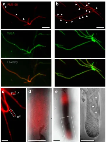
studied with fluorescence light microscopy (fLM). This not only revealed membrane structures associated with peptidoglycan cross-walls (Fig. 1a), but also in surprisingly large assemblies, several micrometres in length, spanning the width of vegetative hyphae (Fig. 1b). Lipid assemblies were present within hyphae in differing amounts, with some hyphae being devoid of membranes and others containing multiple formations, from small blebs to large structures. Quantification experiments indicated that of 218 membrane localizations counted, 75.3% localized in large or small apparently non-septal assemblies. The remaining 24.7% were at septal locations—either clearly co-localizing with BODIPY-vancomycin or in the absence of BODIPY-vancomycin—still appearing in thin, cross-wall-like form (Supplementary Table 1 and Supplementary Figs 5–8).
We applied cryo-correlative light and electron microscopy
(cryo-CLEM), combining cryo-fLM with cryo-electron
d
e/f f
a b
c d e f
Overlay WGA
FM5-95
Figure 1 | Membrane assemblies in vegetative hyphae.(a) In vegetative hyphae ofS. coelicolorM145 stained with the membrane dye FM5–95 (red) and the cell wall dye FITC-WGA (green), cross-walls are evident (arrowheads). (b) Large membrane structures/agglomerates are also found within hyphae, which do not co-localize with WGA-stained cross-walls (arrowheads). (c–f) To investigate their ultrastructure, membranes in
S. albuswere fluorescently labelled with FM5–95 and imaged with cryo-CLEM. Positions with extended lipids were observed (d–f).
tomography (cryo-ET), to study the fine structural detail of the membrane assemblies in three dimensions at nanometre resolution. Cryo-CLEM enables direct fluorescent labelling and targeting of molecules or molecular assemblies (such as intracellular membranes, DNA or cyto-structural elements) in the same cryo-immobilized samples that are then visualized by cryo-ET, avoiding the possible artefacts of chemical fixation, plastic embedding and/or metal staining. In this manner, sites of interest can be quickly located without imposing cryogenic samples to extended electron beam damage. We used
Streptomyces albusin our cryo-CLEM experiments as this strain
is morphologically very similar to model organismS. coelicoloron
agar plates, with abundant sporulation, yet has thinner hyphae in submerged cultures and therefore allows for high-resolution three-dimensional imaging by whole-cell cryo-ET. By performing
cryo-CLEM on S. albushyphae stained with FM5–95, we were
able to readily target membrane assemblies within hyphae (Fig. 1c). In young vegetative hyphae (12 h after inoculation), membranes could be seen forming bundles of tube-like structures varying from small assemblies along the cell wall to large structures completely delimiting the hyphae (Fig. 1d and Supplementary Fig. 1). The membranes were also identified at apical sites (Fig. 1e,f and Supplementary Movie 1), as seen in fLM of S. coelicolor24. Similar membrane assemblies were also found in S. coelicolor when examined by cryo-CLEM (Supplementary Fig. 2).
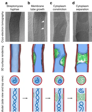
Cryo-ET and data segmentation enabled visualization and analysis of the membrane formations in three dimensions, and demonstrated that the assemblies form in the space between the cytoplasmic membrane and cell wall (Fig. 2). Membranes were observed to vary from several small tubes to larger structures, which appeared to be constricting, decreasing the connection between hyphal compartments, likely in an intermediate stage of membrane closure (Fig. 2c). Evidence of septum synthesis could also be seen associated with cross-membranes, and mostly with full cross-membrane structures, which spanned the entire hyphae (Fig. 2d). Tomograms and full 3D surface renderings can be found in Supplementary Movies 2–5.
Cross-membranes create a chromosome-free area. Excited by the novel visualization of cross-membranes that cryo-ET afforded and to specifically determine the spatial localization of the membranes relative to chromosomes, we combined membrane
staining by FM5–95 with chromosome visualization using 40,
6-diamidino-2-phenylindole (DAPI; Fig. 3a–c). It was previously demonstrated that nucleoids are absent from areas enriched in
anionic phospholipids24. We observed that all areas that showed a
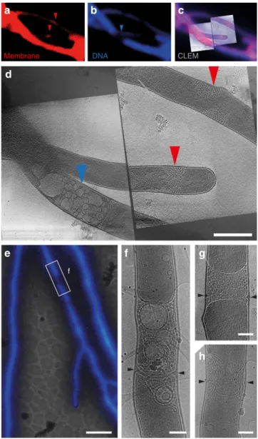
distinct gap in DAPI fluorescence in cryo-fLM, and therefore were devoid of DNA, correlated to large hyphae-spanning membrane assemblies (red arrows, Fig. 3a,d) or vesicle structures (blue arrows, Fig. 3b,d). Again, we observed evidence of septum synthesis within full cross-membrane structures, which spanned the entire hyphae (Fig. 3e–h). This correlation, as well as the frequent presence of membranes at hyphal tips, suggested that inStreptomycesvegetative hyphae membrane assemblies may act to occlude chromosomal DNA from sites of active cell wall restructuring, either for septum synthesis (Fig. 3f–h), tip growth or branching.
Membranes form independent of cell wall remodelling. To investigate the relationship between membrane formation and cell wall remodelling, we stained samples for cryo-CLEM with BODIPY-vancomycin and looked for membrane assemblies at locations of BODIPY-vancomycin staining. Experiments revealed numerous examples of BODIPY-vancomycin staining present at
septa (Fig. 4a), and to a lesser extent at growing tips (Fig. 4b), and new branch points (Fig. 4c). High-resolution transmission electron micrographs confirmed that some regions stained with BODIPY-vancomycin also contained cross-membranes (Fig. 4d). A statistical analysis on fLM images co-stained with BODIPY-vancomycin and FM5–95 indicated that of a total of 335 locations measured (containing FM5–95 staining, BODIPY-vancomycin staining or both), 117 (34.9%) demonstrated only BODIPY-vancomycin staining (and no FM5–95 staining) at septa or points of cell wall remodelling. Of the remaining locations, 153 (45.6%) demonstrated only FM5–95 staining, and 65 (19.4%) demon-strated both (Supplementary Table 1 and Supplementary Figs 5–8). This result suggests that membranes are either not essential for new cell wall formation, or that if membranes do co-localize with newly forming cell wall, the co-localization is transient. To obtain insight into the dynamics of co-localization during peptidoglycan synthesis, we therefore performed live imaging experiments. These experiments, however, repeatedly failed due to the toxicity of the immunofluorescence stains to young vegetative hyphae.
Streptomycesgrow by hyphal tip extension, with polar growth
and branching depending on DivIVA25. As we observed many
membrane-filled tips during cryo-CLEM experiments, we were Streptomyces
hyphae
Cryo-electron tomography
3D surface rendering
Model (side view and top view)
Membrane tube growth
Cytoplasm constriction
Cytoplasm separation
a b c d
Figure 2 | Electron tomography and data segmentation of membrane formations in three dimensions.Cryo-ET slices (top row), surface renderings of the tomograms (middle row) and 2D model (bottom rows, side view and top view) of intracellular membranes in vegetative hyphae of
interested to see if these membranes mark the location of DivIVA. We performed fLM studies to quantify the co-localization of this morphoprotein with membranes and found that 27.0% of DivIVA foci co-localize with membrane assemblies (Supplementary Table 2 and Supplementary Fig. 9).
Given the incomplete spatio-temporal co-localization of membranes with newly forming cell wall at cross-walls or hyphal tips, we wondered whether cross-membranes form independent of septum synthesis. We therefore quantified membrane and cell
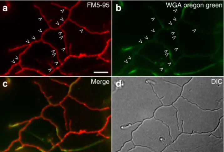
wall formation in the S. coelicolor ftsZ deletion strain, which
grows without forming septa11. fLM experiments staining the
hyphae with FM5–95 for membranes and WGA for newly
forming cell wall demonstrated that in 77 hyphae, with a total
length of roughly 2,635mm, 368 cross-membranes were present
and no cross-walls (Fig. 5). This amounts to one cross-membrane
per 7.16±0.66mm. In wild type, the number of cross-membranes
is much higher at one cross-membrane per 2.86±0.50mm
(N¼313 membranes counted), whereas the number of
cross-walls formed is one per 9.03±0.46mm (N¼99 cross-walls
counted). Cryo-electron microscopy (EM) experiments
confirmed that the cross-membranes in theftrsZ deletion strain
appear indistinguishable to those in wild-type S. coelicolor
(Supplementary Fig. 3). The complete absence of cross-walls
and abundance of cross-membranes in the ftsZ deletion strain
indicates that septum synthesis is not required for the formation of—and that septa are not necessarily present in—cross-membranes.
Cross-membranes compartmentalize hyphae. Staining of
Streptomyces mycelium with propidium iodide and SYTO 9 demonstrates alternating live and dead compartments in young vegetative mycelium, suggesting that the PCD linked to
devel-opment already occurs in very young hyphae26. We wondered if
Membrane DNA CLEM
f
a b c
d
e f g
h
Figure 3 | DNA and cross-membranes are mutually exclusive in vegetative hyphae.Vegetative hyphae ofS. albusfluorescently labelled with FM5–95 for membranes (a) and DAPI for DNA (b) were imaged by cryo-CLEM (c) and cryo-ET (d). All areas that contained membrane assemblies (red arrowheads,a,d) were devoid of DNA. Vesicularization could be seen near a completed cross-wall in an area devoid of DNA (blue arrowheads,b,d). In a second example, fluorescent labelling of vegetative hyphae ofS. albuswith DAPI (e) again revealed positions devoid of DNA (white rectangle denotedf) and filled with membranes and vesicles as shown by cryo-EM. Cell wall formation can be seen within the membranes (black arrowheads,f). Ing,h, other examples of newly forming cell wall within membranes are shown. Scale bars, 500 nma–d;e, 5mm;
f–h, 200 nm.
Cross-membranes
d
Branch point
c
Tip
b
a Septum
the large cross-membrane structures we observe (and not septa) form the cytosol impermeable barrier that enables this type of differentiation. We therefore applied fluorescence recovery after
photobleaching (FRAP) on a S. coelicolor strain constitutively
expressing enhanced green fluorescent protein (eGFP) and stained with FM5–95 for membranes, bleaching an entire compartment on one side of a cross-membrane to establish whether green fluorescent protein (GFP) molecules from the adjacent compartment can restore fluorescence. FRAP
experiments (N¼91) were performed on regions in close
proximity to membranes. These experiments demonstrated that in 71% of cases cross-membranes are cytosol permeable (Fig. 6a and Supplementary Movie 6) and in 29% of cases impermeable (Fig. 6b and Supplementary Movie 7), preventing fluorescent GFP migration into the adjacent compartment. Additional FRAP experiments were performed to distinguish cross-membranes containing cross-walls from those that do not contain any cell
wall material. In these experiments, the S. coelicolor strain
constitutively expressing eGFP was stained with both FM5–95 for membranes and far-red dye WGA-Alexa 633 to stain cross-walls. Membranes with no cell wall present were targeted to start. In these cases, cross-membranes were found to be permeable in 23% of cases (green arrows, Supplementary Fig. 4, Supplementary Movie 8) or impermeable in 77% of cases (red arrows, Supplementary Fig. 4, Supplementary Movie 8), revealing that cross-membranes alone can block diffusion within hyphae. Next, membranes with cell-wall material present were targeted. For these cross-membranes, the permeable fraction was 71%, whereas large membrane assemblies near a cross-wall completely blocked diffusion. This increased permeability of cross-membranes with cell-wall material present can be explained by the fact that
cross-walls are likely permeable13; once cross-membranes
disassemble, transport can occur over cross-walls. Taken together, our experiments show that in the absence of cross-walls, cross-membranes can act as impermeable physical barriers between adjacent compartments.
Discussion
In this work, we discovered the presence of a vast internal
membrane system in Streptomyces species that can be found
within and throughout vegetative hyphae, forming bundles of tubular structures between the cytoplasmic membrane and cell wall. Using cryo-CLEM, we observed membrane formations in small blebs along the cell wall or at hyphal tips, or as large
assemblies and hyphae-delimiting structures we termed
cross-membranes. Similar structures were observed in the 1960s (refs 27,28), although their function was unknown. Our present investigation of these membranes resolves several intriguing
questions surrounding their role in the Streptomyces lifecycle.
Although the production of antibiotics and other clinically important secondary metabolites by the streptomycetes has been linked to PCD in the vegetative mycelium, to date, little was known about how the hyphae separate into live and ‘dead’ segments.
Peptidoglycan-based septa, or cross-walls, are infrequent in the vegetative hyphae. As beautifully illustrated by live imaging
experiments, during hyphal growth of S. coelicolor cross-walls
(that is, vegetative septa) are produced seemingly randomly at sites distal from the growing tips, delimiting the hyphae into
compartments of 5–10mm (ref. 29). However, the compartments
can be up to 40mm for some other streptomycetes30, and these
compartments contain approximately one chromosome per micrometre, or tens of copies of the genome per compartment.
Cross-walls do not form in theftsZdeletion strain, which raises
the question as to how a colony can ensure its survival during PCD or following physical damage to the mycelium. It is now clear that membranes play an important role, offering a less energy-intensive compartmentalization solution than cross-wall formation.
Interestingly, although compartmentalization is rare in
bacteria, it does exist in a variety of species31–33. Recently,
a protein-mediated diffusion barrier was also discovered in the
polar stalk ofCaulobacter crescentus, acting to prevent exchange
of membrane and soluble proteins between the stalk and cell
body34. The cross-membranes in Streptomyces similarly delimit
and prevent diffusion of soluble proteins between diverse hyphal segments, as evidenced by FRAP experiments on the diffusion of molecules of GFP.
Our FRAP experiments revealed that 29% of cross-membranes are impermeable. Although unlikely, it is conceivable that laser-mediated damage to the cross-membranes may have contributed to leakage of GFP from adjacent compartments, increasing the number of permeable membranes we counted. The percentage of impermeable cross-membrane barriers may therefore be higher than what we observed. Molecules smaller than GFP may also be able to pass the cross-membranes. The high percentage of permeable membrane assemblies we observed are likely mem-brane structures that are not yet fully closed, that is, in an earlier stage of cross-membrane formation, or perhaps these membranes
serve a different purpose during theStreptomyceslifecycle.
Indeed, the abundance of lipid structures localizing in small blebs adjacent to the cell wall, in addition to the full cross-membranes delimiting hyphae, could indicate that smaller membrane assemblies may be involved in cellular functions other than compartmentalization. At locations along the hyphae, small blebs are likely mainly precursors for cross-membranes. However, membrane assemblies are also frequently observed at hyphal tips, which suggests that they may be involved in tip growth. It follows that some of the membrane blebs along the hyphae may not be precursors for cross-membranes, but rather the future sites of branching. At growing tips and new branches, it is likely that the assemblies observed have a function in creating a nucleoid-free zone in a region of high activity.
In the Streptomyces growing apex, a highly dynamic
tip-organizing complex (the so-called TIPOC) exists, consisting of many components—including various cytoskeletal elements—
FM5-95 Merge DIC a b d c V V V VV VV V V V V V V V V V V V V V VV VV V V V V V V V V V V
WGA oregon green
and ensuring that DNA replication takes place at some distance
behind the tip16,35,36. Extensive statistics using cryo-ET are
not feasible considering the high-resolution nature of these experiments, so instead we performed static fLM experiments on vegetative hyphae stained with FM5–95 and peptidoglycan marker BODIPY-vancomycin and tip growth protein DivIVA to determine the degree of membrane co-localization with new cell wall synthesis. These experiments (see Supplementary Tables 1 and 2 for details) did not indicate complete spatio-temporal co-localization. It should be noted, however, that studying dynamic events with static experiments is notoriously
difficult. Further experiments using other cell division or morphoproteins can perhaps provide more insight into the role of the membranes in providing a suitable environment for protein localization. In addition, use of an integrated system for live light microscopy and cryo-fixation (for example, the Rapid Transfer
System37or the MAVIS38) could enable the targeting of actively
growing tips or cross-walls at different stages of formation, and elucidating the role of membranes therein.
Finally, although we demonstrated that cross-membranes form independent of septum synthesis, during our experiments, we frequently observed cross-membranes containing emerging 300
250
200
150
Bleached compartment Adjacent compartment
Bleached compartment Adjacent compartment 100
–100 100 200 300 400
Time (s)
Fluorescence intensity
0 50
0
300
250
200
150
100
–100 100 200 300 400
Time (s)
Fluorescence intensity
0 50
0
–51 s –51 s
0 s Photobleaching 0 s
51 s
102 s
153 s
204 s
255 s
51 s
102 s
153 s
204 s
255 s
306 s 306 s
Permeable
membrane Impermeable membrane
Photobleaching
GFP FM5-95 Overlay GFP FM5-95 Overlay
a b
cross-walls (Fig. 3f–h). In the multicellular streptomycetes, hyphal compartments are multigenomic; it is likely that some form of subcellular organization exists that serves to separate the chromosomes. One obvious reason for such compartmentalization would be to allow cross-walls to form in a chromosome-free area.
In conclusion, we show here that cross-membranes exist in the hyphae of the streptomycetes, acting to compartmentalize the hyphae. These membranes can be cytosol permeable or impermeable, and provide a plausible answer to the question
why hyphae of the ftsZ null mutant do not ‘bleed’ to death
but rather can be fragmented and propagated. Further, the cross-membranes create DNA-free zones and offer a solution to the long-standing problem of how cell wall remodelling can take place without DNA damage in the multinucleoid hyphae.
Future technological advances in correlative microscopy,
in which fluorescently tagged proteins can be superimposed on high-resolution images of cytostructural elements in tomograms, should provide further insights into the exciting cell biology of the streptomycetes.
Methods
Bacterial strains and constructs.S. coelicolorandS. albusstrains were grown on R2YE agar or soya flour mannitol agar. Liquid cultures were grown in 2YT medium supplemented with 10% sucrose. All of the growth medium recipes used are those mentioned in the ref. 39. For fluorescence microscopy, samples from liquid cultures were spotted onto a glass microscope slide before microscopy analysis. Images of vegetative hyphae from solid growth samples were collected from samples that had been inoculated at the acute-angle junction of coverslips inserted at a 45°angle in soya flour mannitol agar plates. Strains used in this study includeS. coelicolorM145 (ref. 39), obtained from the John Innes Centre strain collection, its derivative K202 containing plasmid KF41 that expresses FtsZ-GFP17,S. albussubspeciesalbusG. ATCC 25426 and, for FRAP, aS. coelicolorstrain constitutively expressing GFP. This strain was created by transformingS. coelicolorM145 with pGreen, a pIJ8630 derivative harbouring the constitutivegap1promoter ofS. coelicolorA3(2) M145 cloned upstream of theeGFPgene (B. Zacchetti and D. Claessen, unpublished observations).
Microscopy.Fluorescence microscopy. Fluorescence and corresponding light micrographs were obtained with a Zeiss Axioscope A1 upright fluorescence microscope (with an Axiocam Mrc5 camera at a resolution of 37.5 nm per pixel), with, for the green channel, 470- to 490-nm excitation and 515 long-pass detection; and for the red channel, 530- to 550-nm excitation and 590 long-pass detection. For cryo-stage imaging, the green fluorescent images were created using 470/40-nm band-pass excitation and 525/50 band-pass detection; for the red channel, 550/25-nm band-pass excitation and 605/70 band-pass detection were used. For staining of the cell wall (peptidoglycan), we used FITC-WGA; for membrane staining, we used FM5–95 (both obtained from Molecular Probes). FRAP experiments were carried out on a Zeiss Imager system, using 488 nm excitation for eGFP with 505–530 detection and 543 nm excitation for FM5 (refs 27,28) 5–95 with LP 560 detection. Bleaching was performed with 25% laser intensity for 8 s. Images were recorded every 17 s and post-bleaching was monitored for 5 min. All images were background-corrected, setting the signal outside the hyphae to 0 to obtain a sufficiently dark background. These corrections were made using Adobe Photoshop CS5. Triple coloured FRAP experiments were performed with 488 nm excitation, which does not excite WGA-Alexa 633 and therefore the same band-passes were used for signal detection: for example, for eGFP, band-pass filter 525/50; for FM5–95, long-pass 560. WGA-Alexa 633 was imaged with 633 excitation and long-pass 650 detection.
Sample preparation for electron microscopy. A small drop (3ml) ofStreptomyces liquid culture was applied to EM grids and vitrified by plunging into a liquid ethane/propane mixture using a vitrobot Mark IV (FEI Company) operated at 22°C and 100% humidity using 1–2 s blotting. Plunge-frozen grids were stored in liquid nitrogen until further use. For cryo-electron tomography, 15 nm colloidal gold particles coupled to protein A (CMC) were added to samples as fiducial markers. For correlative light and microscopy work, holey carbon grids were used and samples were stained with FM5–95 and/or DAPI directly before sample application to the grid and subsequent plunge freezing.
Electron microscopy. Cryo-electron tomography was performed on a Tecnai 20 FEG operated at 200 kV and a Titan Krios operated at 300 keV (FEI Company). Images were recorded using Explore 3D software on a 22 k2camera mounted behind a GIF energy filter (Gatan) operated at a slit width of 20 eV. Over 100 cryo-electron tomograms of streptomycetes were recorded with 2°tilt steps between 60°and þ60°at a defocus of 15mm, at magnifications between
1,850 (6.6 nm pixels size) and8,000 (1.64 nm pixel size).
Cryo-correlative light and electron microscopy. Plunge-frozen EM grids containing fluorescently labelledStreptomyceswere imaged using a fluorescence microscope equipped with a THMS600 or CMS196 cryo-light microscope stage (Linkam), in conjunction with a Leitz DMRB (Leica), with a 100 dry objective with a working distance of 4.7 mm and a numerical aperture of 0.75. Digital images were recorded with a Leica DFC350FX CCD camera. Following cryo-fLM imaging, sample grids were stored in liquid nitrogen until they were used for cryo-EM. Over 50 cryo-CLEM images were recorded.
Image analysis and visualization.Tomographic tilt series were processed using IMOD version 4.5 (ref. 40). Projection images were pre-processed by hot pixel removal and rough alignment by cross-correlation. Final alignment was done using fiducial gold markers. The tomograms were obtained using a weighted back-projection or a simultaneous iterative reconstruction technique. Cryo-electron tomograms were Fourier filtered and denoized with a nonlinear anisotropic diffusion41to enhance the visibility of structures. For 3D surface rendering, the tomographic volumes were imported into AMIRA (FEI) for further processing and representation. Cryo-CLEM overlays were produced by manual overlay (Adobe Photoshop) of different magnification TEM and fLM images using grid bars, streptomycetes, holey carbon support film and fiducial gold markers for alignment.
Data availability.The data that support the findings of this study are available within the article or from the corresponding author upon request.
References
1. Shapiro, J. A. Thinking about bacterial populations as multicellular organisms. Annu. Rev. Microbiol.52,81–104 (1998).
2. Claessen, D., Rozen, D. E., Kuipers, O. P., Sogaard-Andersen, L. & van Wezel, G. P. Bacterial solutions to multicellularity: a tale of biofilms, filaments and fruiting bodies.Nat. Rev. Microbiol.12,115–124 (2014). 3. Lyons, N. A. & Kolter, R. On the evolution of bacterial multicellularity.Curr.
Opin. Microbiol.24,21–28 (2015).
4. Miguelez, E. M., Hardisson, C. & Manzanal, M. B. Hyphal death during colony development inStreptomycesantibioticus: morphological evidence for the existence of a process of cell deletion in a multicellular prokaryote.J. Cell Biol. 145,515–525 (1999).
5. Miguelez, E. M., Hardisson, C. & Manzanal, M. B. Streptomycetes: a new model to study cell death.Int. Microbiol.3,153–158 (2000).
6. Hopwood, D. A.Streptomyces in Nature and Medicine: the Antibiotic Makers (Oxford Univ., 2007).
7. Chater, K. F. & Losick, R. inBacteria as Multicellular Organisms (eds Shapiro, J. A. & Dworkin, M.) 149–182 (Oxford Univ., 1997). 8. Barka, E. A.et al.Taxonomy, physiology, and natural products of the
Actinobacteria.Microbiol. Mol. Biol. Rev.80,1–43 (2016). 9. Fla¨rdh, K. & Buttner, M. J.Streptomycesmorphogenetics: dissecting
differentiation in a filamentous bacterium.Nat. Rev. Microbiol.7,36–49 (2009). 10. Chater, K. F. inStreptomyces: Molecular Biology and Biotechnology
(ed. Dyson, P.) 43–86 (Caister Academic, 2011).
11. McCormick, J. R., Su, E. P., Driks, A. & Losick, R. Growth and viability of Streptomyces coelicolormutant for the cell division geneftsZ.Mol. Microbiol. 14,243–254 (1994).
12. McCormick, J. R. Cell division is dispensable but not irrelevant inStreptomyces. Curr. Opin. Biotechnol.12,689–698 (2009).
13. Jakimowicz, D. & van Wezel, G. P. Cell division and DNA segregation in Streptomyces: how to build a septum in the middle of nowhere?Mol. Microbiol. 85,393–404 (2012).
14. Manteca, A., Alvarez, R., Salazar, N., Yague, P. & Sanchez, J. Mycelium differentiation and antibiotic production in submerged cultures ofStreptomyces coelicolor.Appl. Environ. Microbiol.74,3877–3886 (2008).
15. Fla¨rdh, K. Cell polarity and the control of apical growth inStreptomyces.Curr. Opin. Microbiol.13,758–765 (2010).
16. Holmes, N. A.et al.Coiled-coil protein Scy is a key component of a multiprotein assembly controlling polarized growth inStreptomyces.Proc. Natl Acad. Sci. USA110,E397–E406 (2013).
17. Grantcharova, N., Lustig, U. & Fla¨rdh, K. Dynamics of FtsZ assembly during sporulation inStreptomyces coelicolorA3(2).J. Bacteriol.187,3227–3237 (2005). 18. Schwedock, J., McCormick, J. R., Angert, E. R., Nodwell, J. R. & Losick, R.
Assembly of the cell division protein FtsZ into ladder like structures in the aerial hyphae ofStreptomyces coelicolor.Mol. Microbiol.25,847–858 (1997). 19. Willemse, J. & van Wezel, G. P. Imaging ofStreptomyces coelicolorA3(2) with
reduced autofluorescence reveals a novel stage of FtsZ localization.PLoS ONE 4,e4242 (2009).
20. de Boer, P. A. Advances in understandingE. colicell fission.Curr. Opin. Microbiol.13,730–737 (2010).
21. Schumann, J., Mollering, H. & Jaenicke, R. Intrinsic stability and extrinsic stabilization of creatinase fromPseudomonas putida.Biol. Chem. Hoppe-Seyler 374,427–434 (1993).
23. Reynolds, P. E. Structure, biochemistry and mechanism of action of glycopeptide antibiotics.Eur. J. Clin. Microbiol. Infect. Dis.8,943–950 (1989). 24. Jyothikumar, V.et al.Cardiolipin synthase is required forStreptomyces
coelicolormorphogenesis.Mol. Microbiol.84,181–197 (2012).
25. Fla¨rdh, K., Richards, D. M., Hempel, A. M., Howard, M. & Buttner, M. J. Regulation of apical growth and hyphal branching inStreptomyces.Curr. Opin. Microbiol.15,737–743 (2012).
26. Manteca, A., Fernandez, M. & Sanchez, J. A death round affecting a young compartmentalized mycelium precedes aerial mycelium dismantling in confluent surface cultures ofStreptomyces antibioticus.Microbiology151, 3689–3697 (2005).
27. Glauert, A. M. & Hopwood, D. A. A membranous component of the cytoplasm inStreptomyces coelicolor.J. Biophys. Biochem. Cytol.6,515–516 (1959). 28. Glauert, A. M. & Hopwood, D. A. The fine structure ofStreptomyces
coelicolor. I. The cytoplasmic membrane system.J. Biophys. Biochem. Cytol.7, 479–488 (1960).
29. Jyothikumar, V., Tilley, E. J., Wali, R. & Herron, P. R. Time-lapse microscopy of Streptomyces coelicolorgrowth and sporulation.Appl. Environ. Microbiol.74, 6774–6781 (2008).
30. Kretschmer, S. Location of branches within the apical hyphal region of Streptomyces granaticolormycelia.J. Basic Microbiol.32,35–42 (1992). 31. Junglas, B.et al. Ignicoccus hospitalisandNanoarchaeum equitans:
ultrastructure, cell-cell interaction, and 3D reconstruction from serial sections of freeze-substituted cells and by electron cryotomography.Arch. Microbiol. 190,395–408 (2008).
32. Santarella-Mellwig, R., Pruggnaller, S., Roos, N., Mattaj, I. W. & Devos, D. P. Three-dimensional reconstruction of bacteria with a complex endomembrane system.PLoS Biol.11,e1001565 (2013).
33. Boekema, E. J., Scheffers, D. J., van Bezouwen, L. S., Bolhuis, H. & Folea, I. M. Focus on membrane differentiation and membrane domains in the prokaryotic cell.J. Mol. Microbiol. Biotechnol.23,345–356 (2013).
34. Schlimpert, S.et al.General protein diffusion barriers create compartments within bacterial cells.Cell151,1270–1282 (2012).
35. Celler, K., Koning, R. I., Koster, A. J. & van Wezel, G. P. Multidimensional view of the bacterial cytoskeleton.J. Bacteriol.195,1627–1636 (2013).
36. Wolanski, M.et al.Replisome trafficking in growing vegetative hyphae of Streptomyces coelicolorA3(2).J. Bacteriol.193,1273–1275 (2011). 37. Verkade, P. Moving EM: the Rapid Transfer System as a new tool for
correlative light and electron microscopy and high throughput for high-pressure freezing.J. Microsc.230,317–328 (2008).
38. Koning, R. I.et al.MAVIS: an integrated system for live microscopy and vitrification.Ultramicroscopy143,67–76 (2014).
39. Kieser, T., Bibb, M. J., Buttner, M. J., Chater, K. F. & Hopwood, D.A Practical Guide to Streptomyces Genetics(John Innes Foundation, 2000).
40. Kremer, J. R., Mastronarde, D. N. & McIntosh, J. R. Computer visualization of three-dimensional image data using IMOD.J. Struct. Biol.116,71–76 (1996). 41. Frangakis, A. S. & Hegerl, R. Noise reduction in electron tomographic
reconstructions using nonlinear anisotropic diffusion.J. Struct. Biol.135, 239–250 (2001).
Acknowledgements
We are grateful to Dennis Claessen for the gift of pGreen and for discussions.S. coelicolor M145 was obtained from the John Innes Centre strain collection, andS. albusG. ATCC 25426 was obtained from the American Type Culture Collection strain collection. This work was supported in part by Cyttron II (Life Sciences Health framework FES0908); the Netherlands Centre for Electron Nanoscopy, Leiden; the Netherlands Organization for Scientific Research (NWO VICI grant 10379; grant 175.010.2009.001) and the European Regional Development Fund of the European Commission (21Z.014). We would like to acknowledge support of R.K. from the European Union through the Horizon 2020 Programme grant iNEXT (grant agreement No. 653706).
Author contributions
K.C. and R.I.K. performed the cryo-correlative light and electron microscopy. K.C. and J.W. performed the (live) light imaging. G.P.v.W. and A.J.K. supervised the work. K.C., R.I.K. and G.P.v.W. wrote the manuscript. All authors discussed the results and commented on the manuscript.
Additional information
Supplementary Informationaccompanies this paper at http://www.nature.com/ naturecommunications
Competing financial interests:The authors declare no competing financial interests.
Reprints and permissioninformation is available online at http://npg.nature.com/ reprintsandpermissions/
How to cite this article:Celler, K.et al.Cross-membranes orchestrate compartmentalization and morphogenesis inStreptomyces.Nat. Commun.7:11836 doi: 10.1038/ncomms11836 (2016).