The effects of Functional
Electrical Stimulation on
motor-cognitive interference during gait
in people with foot drop following
stroke
Jane McAdam
School of Health Sciences
University of Salford, Salford, UK
Contents
Contents ii
List of Figures vi
List of Tables ix
Acknowledgements x
Declaration xi
Abstract xii
Chapter 1 – Introduction 1
Chapter 2 – Literature Review 4
2.1 Introduction 4
2.2 Gait and its control 5
2.2.1 Functional anatomy of the neuromuscular system 5
2.2.2 Gait kinematics and muscle activations 9
2.2.3 Aspects of behaviour believed to be associated with cognitive
control of gait 13
2.3 Stroke 18
2.3.1 Stroke overview 18
2.3.2 Impact of stroke on key motor and sensory functions 19
2.3.3 Effects of stroke on cognitive function 20
2.3.4 Effects of stroke on gait and falls 21
2.3.5 Cognition and gait post stroke 27
2.4 FES 29
2.4.1 Description of FES 29
2.4.2 Action of stimulation on nervous system 32
2.4.3 Effects of FES on gait and other recorded outcomes 36
Chapter 3 – Study of FES user reported effects 47
3.1 Introduction 47
3.2 Aims of the questionnaire study 50
3.3 Development and design 51
3.3.1 Choice of questionnaire study design 51
3.3.2 Formulation of questionnaire structure, design and content 51
3.4 Ethical approval 63
3.5 Application of questionnaire 63
3.6 Results 63
3.6.1 Questionnaire response rates 63
3.6.2 FES users 64
3.7 Discussion 71
3.7.1 Response rate 71
3.7.2 FES users 72
3.8 Conclusion 79
Chapter 4 - Dual-task methodology development and longitudinal study 80
4.1. Introduction 80
4.2. Protocol design 82
4.2.1 Study design 82
4.2.2 Previously used dual-task tests 84
4.2.3 Development of new task 96
4.2.4 Other measures 100
4.3. Pilot work to finalise protocol 103
4.3.1 Measuring cognitive task performance 103
4.3.2 Measuring dual-task performance 107
4.3.3 Discussion of results of piloting 109
4.3.4 Visual task 110
4.4 Final protocol and Ethical and R&D approval 110
4.4.1 Recruitment and screening 111
4.5. Results 115
4.5.1 Recruitment 115
4.5.2 Participant descriptors 116
4.5.3 Gait speed, task performance and other outcomes 117
4.6 Discussion 124
4.7 Conclusion 126
Chapter 5 – Cross-sectional dual-task study 128
5.1 Introduction 128
5.2 Protocol 133
5.2.1 Sheffield Protocol 134
5.2.2 Ethical and R&D approval 143
5.3 Results 143
5.3.1 Recruitment 143
5.3.2 Participant descriptors 144
5.3.3 Gait speed and visual task performance 147
5.3.4 Gait parameters 150
5.3.5 Questionnaire results 158
5.4 Discussion 164
5.4.1 Participant descriptors 164
5.4.2 The effect of FES on gait speed and gait parameters 164 5.4.3 The dual-task effect when walking without FES 168
5.4.4 Dual-task effect with FES 171
5.4.5 Questionnaire 173
5.4.6 Analysis of primary outcomes in the context of questionnaire results 174
Chapter 6 – Summary and future work 186
6.1 Summary 186
6.1.1 Overview of the thesis 186
6.1.2 Summary of each chapter 187
6.2 Limitations and future work 191
6.3 Conclusion 193
Appendix A 195
Appendix B 216
Appendix C 261
List of Figures
Figure 2.1: Pathway of upper and lower motor neurons 7 Figure 2.2: Relationship between kinaesthetic sense and proprioception 8 Figure 2.3: Timing of phases of gait during the gait cycle 10
Figure 2.4: Spatial parameters of gait 10
Figure 2.5: Activation patterns of major muscles during gait 11 Figure 2.6: Sagittal plane joint ankles during a single gait cycle of the ankle,
knee and hip joints 11
Figure 2.7: Interactions between risk factors, falls and consequences of falls
following stroke 26
Figure 2.8: Example of a surface-based FES device 30
Figure 2.9: Example of surface-based FES device in which electrodes are
housed in a cuff 31
Figure 2.10: Example of an implanted FES device 32
Figure 2.11: Typical neuron structure 33
Figure 3.1: Process of development of questionnaire content 54
Figure 3.2: Responses to questionnaires 64
Figure 3.3: Activities undertaken by FES users, both with and without FES 66 Figure 3.4: Use of walking aids by FES users, both with and without FES 66 Figure 3.5: Frequency of response to effects statements by FES users 67 Figure 3.6: The frequency (%) of most important effect chosen by FES users 68 Figure 3.7: Rating by users of concentration needed to walk with and
without FES 69
Figure 3.8: Effect of FES during dual-task conditions 70 Figure 3.9: The frequency (%) of most important dual-task effect chosen
by FES users 70
Figure 3.10: Usage of FES for current study and three published studies,
as a percentage of each study group 73 Figure 4.1: Plot of changes in gait speed over time, both with and without FES,
from published studies 83
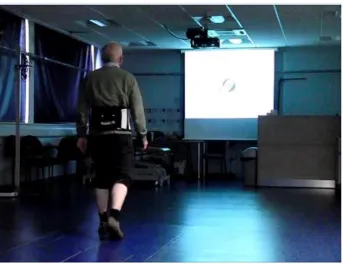
Figure 4.2: Photo of gait laboratory set-up for dual-task protocol 114 Figure 4.3: Walking speed at each visit, averaged over two trials, for each
walking condition (Participant A) 118
Figure 4.5: Walking speed at each visit, averaged over three trials,
for each walking condition (Participant B) 121 Figure 4.6: Score on visual task at each visit, averaged for three walking trials
for each condition and from one seated trial (Participant B) 122 Figure 5.1: Flowchart of participant progression through the trial 135 Figure 5.2: Gait laboratory set-up showing monitor for projection of
visual task, Vicon cameras and video cameras 138
Figure 5.3: Reflective marker placement 139
Figure 5.4: Example plot of marker position; toe marker plot, x-axis time in
seconds, y-axis vertical displacement in metres 139 Figure 5.5: Walking speed for the group, for each condition 147 Figure 5.6: Visual task performance for each condition 149 Figure 5.7: Scatterplot of speed vs task performance during dual-task
conditions, with and without FES 149
Figure 5.8: Stride time for each condition 150
Figure 5.9: Stance time of the paretic leg for all conditions 151 Figure 5.10: Stance time of the non-paretic leg for all conditions 152 Figure 5.11: Swing time of the paretic leg for all conditions 153 Figure 5.12: Swing time of the non-paretic leg for all conditions 153
Figure 5.13: Double support time for all conditions 154
Figure 5.14: Step length of the paretic leg for all conditions 155 Figure 5.15: Step length of the non-paretic leg for all conditions 155
Figure 5.16: Stride length for all conditions 156
Figure 5.17: Stride time variability for all conditions 157 Figure 5.18: Gait asymmetry index for all conditions 157 Figure 5.19: Activities undertaken by participants, both with and
without FES 158
Figure 5.20: Use of walking aids by participants, both with and without FES 159 Figure 5.21: Frequency of response to effects statements by participants 160 Figure 5.22: The frequency of the most important effects chosen by
participants 161
Figure 5.23: Frequency of scaled responses to rating by participants of
concentration needed to walk with and without FES 161 Figure 5.24: Effect of FES during dual-task conditions perceived by
participants 162
Figure 5.25: The frequency of the most important dual-task effect chosen
Figure 5.26: Plot of gait speed of each participant showing change in
speed with addition of FES 165
Figure 5.27: Task performance for each participant for each condition 176 Figure 5.28: Change in gait speed for each participant with task for
both FES and no FES 176
Figure 5.29: Task performance for each participant for each condition 177 Figure 5.30: Change in gait speed for each participant with task for
both FES and no FES 178
Figure 5.31: Change in gait speed for each participant with task for
both FES and no FES 180
List of Tables
Table 2.1: Actions of muscles innervated by two branches
of the common peroneal nerve 31
Table 3.1: Device effect statements mapped against secondary task 61 Table 3.2: Self-reported characteristics of FES users at time
of data collection 64
Table 3.3: Self-reported usage of FES by respondents 65 Table 3.4: Ranking of agreement with effect statements by frequency
(% of total group) 74
Table 3.5: Ranking of most important effect statements (% of total group) 76 Table 4.1: Task performance success (out of 10) for FES non-user
pilot participant 107
Table 4:2: Task performance success (out of 10) for FES user
pilot participants 108
Table 4.3: Speed (m/s) of FES user pilot participants 108 Table 4:4: Visual task performance success (out of 10) for FES
user pilot participant 108
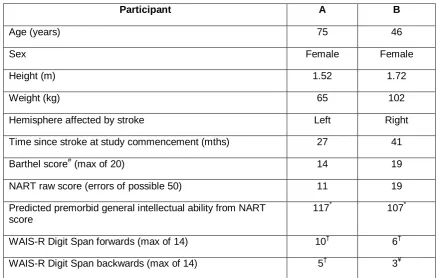
Table 4.5: Participant descriptors 116
Table 4.6: Mean speed (min, max) for each condition, over two
walking trials, collected at each visit (Participant A) 117 Table 4.7: Mean speed (min, max) for each condition, over three
walking trials, collected at each visit (Participant B) 121 Table 5.1: Calculation of sample size based on gait speed results 141
Table 5.2: Characteristics of participants (n=16) 145
Table 5.3: Usage of FES by participants (n=16) 146
Table 5.4: FRAT and FES-1 scores for the group 146
Table 5.5: Self-reported falls and near misses by participants 146 Table 5.6: Pairwise comparison results of repeated measure ANOVA, analysing differences between mean speed for each condition 148 Table 5.7: Pairwise comparison results of repeated measure ANOVA,
analysing differences between task performance for each
Acknowledgements
I would like to acknowledge the help, support and encouragement offered by several people during the course of my studies. Most importantly, my thanks go to my supervisor, Dr Laurence Kenney, who has provided unwavering support and supervision throughout the life of this thesis.
My thanks also go to Dr Sibylle Thies for her input and advice in development of the gait studies and to Dr Audrey Bowen for her advice and support, especially regarding the dual-task aspect of the thesis. I would also like to acknowledge the contribution of Dr Jane Mickleborough, who acted as co-supervisor in the initial stages of the PhD. My thanks to Dr Richard Jones and Dr Anmin Liu for their help with gait analysis. Also, thanks are extended to the Medical Statistics team based at Hope Hospital for their advice.
My special thanks go to Dr Paul Taylor and the staff at The National FES Clinic in Salisbury for their support in accessing their clinical caseload. My thanks also go to the clinicians who have facilitated recruitment of participants to the studies. Special thanks go to Alison Clarke and Jill van der Meulen at the FES clinic in Sheffield for their tremendous support for the final study of the thesis. Their contribution to recruitment and facilitating data collection by hosting the study was invaluable and very much appreciated.
I would also like to offer a special thanks to all of the FES users and pilot participants who gave of their own time to assist in the studies. Without them this work would not have been possible and I am very grateful to them.
I would like to acknowledge Salford Community Healthcare Trust for their support in the form of tuition fees and study leave. I would like to pay particular thanks to Victoria Gould who encouraged me to pursue these studies.
Declaration
Part of the work presented in Chapter 3 has been previously published as:
Abstract
A stroke can impair both motor and cognitive functioning, reducing the automaticity of walking and increasing susceptibility to motor-cognitive interference (MCI). There is also some evidence of an association between susceptibility to MCI and the increased incidence of falls in stroke. Functional Electrical Stimulation (FES) is commonly used for correction of foot drop due to stroke. At the start of the PhD, studies had shown FES increases walking speed. However, questionnaire-based studies found that users rated a reduction in effort and a reduced risk of tripping or falling as the two most important reasons for using FES. In these studies, the term ‘effort’ was not defined, but the results from a qualitative study suggested that the questionnaire respondents may have been referring to both physical and mental components. Based on this evidence the following research question was posed “Does FES reduce motor-cognitive interference during gait in people with foot drop following stroke?”
The question was first examined in a questionnaire study which collated FES user opinion from thirty current users. Respondents identified a statistically significant reduction in concentration required when walking with FES compared with walking without the device. Furthermore, the majority noted that walking without thinking about walking was easier with FES.
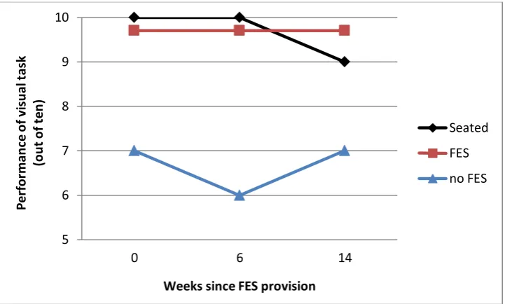
The second study developed and piloted a dual-task based methodology to assess the impact of FES on MCI during gait. Two participants with foot drop following stroke were evaluated over 14 weeks following first use of FES. In one participant, cognitive task performance was maintained at a similar level when walking with FES, compared with seated performance, and reduced without FES. The effects were less clear in the second participant. However, the study demonstrated the feasibility of the proposed methodology and provided the first quantitative evidence that FES can reduce MCI during gait.
Chapter 1 – Introduction
Functional Electrical Stimulation (FES) is the electrical stimulation of nerves to provide muscular contraction in such a way as to produce functionally useful movement. FES is a commonly used technique for correction of foot drop of central neurological origin and typically involves stimulation of the common peroneal nerve to elicit dorsiflexion and slight eversion of the foot during the swing phase of walking. The effectiveness of the intervention in terms of improving walking speed is well supported by evidence and a recent report by the National Institute for Health and Clinical Evidence (NICE) approved its use in clinical practice. However, at the start of this PhD there were few reports on the effects of FES on other components of gait, functioning and ability, and the NICE guidance of 2009 recommended that ‘further publication on the efficacy of FES would be useful, specifically including patient-reported outcomes’.
Of the limited literature describing the effects of FES on outcomes other than gait speed, there were a small number of studies that investigated user perceptions of FES. Users reported factors other than walking speed to be the most important reasons for using FES, with a reduction in effort being cited as the principal reason. A reduced risk of tripping or falling was also highly rated by users. A reduction in the effort of walking, that is clearly seen as important by FES users, could be interpreted both as a reduction in physical and mental effort and at the start of the PhD there was some support for this concept from a qualitative study of FES users.
The patient-reported effects of FES, including a reduction in effort and reduction in trips and falls required further investigation, particularly in the light of the importance placed on these effects by users. While there is some evidence that use of FES leads to a reduction in compensatory actions, such as hip hitching and circumduction, frequently used by people with foot drop to avoid foot-ground collisions during the swing phase of walking, this may not completely explain the reported reduction in effort. Similarly, although there is some evidence of improved toe clearance, this may not fully explain the reported reduction in risk of tripping and falling. There is also evidence that FES use has impact on areas of the brain serving both motor and cognitive functions. It is therefore possible that FES use may have a more complex effect than can be seen from studies of FES gait kinematics and energetics. Specifically, the user reports suggest it may reduce motor-cognitive demands during gait thus improving the ability to respond to challenges to stability and/or freeing up cognitive resources.
To explore this hypothesis, firstly, the thesis focused on building on the small number of questionnaire and qualitative studies published at the start of the PhD, by development of a questionnaire to further explore the patient-perceived effects of using FES. In particular, the questionnaire study aims were to substantiate or refute the suggestion in the literature that FES for foot drop is perceived to impact on motor-cognitive interference during gait and, if confirmed, to inform the design of subsequent experimental studies that would allow for the exploration and testing of this hypothesis.
The second aim of thesis was to investigate this hypothesis through the use of dual-task based studies. The gait laboratory studies were designed to assess the impact of FES on the cognitive control of gait. Furthermore, the aim was to collect gait parameters associated with stability to explore the contribution of FES to stability.
Thesis overview
of the most common effects, including a description of the effects on motor, sensory and cognitive systems. The effect of stroke on gait is then covered, highlighting the increased risk of falls and the circumstances of these. A discussion of the interaction of damage to motor and cognitive areas of the brain follows, including how this manifests in gait disturbances and are evident in dual-task study outcomes.
Chapter 2 then introduces FES, describing the device and its effects on nervous tissue to produce functional movement. The emerging evidence of its effect on brain plasticity and spinal mechanisms of motor control are discussed. This is followed by a review of the effect of FES, with a detailed focus on gait measures related to stability and fall risk and a critique of the literature reporting patient-centred outcomes. The chapter concludes with a discussion of the thesis aims.
Chapter 3 describes the development and implementation of the questionnaire study to explore the concept of ‘effort’ of walking. The chapter describes the process of formulation of the questionnaire content in relation to previous studies, including piloting work. Results from respondents are presented and analysed, in relation to the aims of the study. The conclusions drawn from the results, and their limitations, are discussed and their influence on the choice of tests used in the gait laboratory study is outlined
Chapter 4 describes a longitudinal gait laboratory-based of new users of FES. The study design includes a novel dual-task protocol, informed by the results of the questionnaire study reported in Chapter 3. The impact of FES on gait and performance of a cognitive task are reported.
Chapter 5 presents the results from a cross-sectional study of existing FES users, based on the same protocol as used in Chapter 4.
Chapter 2 – Literature Review
2.1 Introduction
This chapter begins by briefly describing the key components and functions of the neuromuscular system that contribute to normal gait. This is then followed by a description of normal gait outlining the gait cycle, the spatial and temporal parameters and the main events of the gait cycle, also describing the main actions of the muscles and joints. Concepts about the contribution of cognitive processes to the control of normal gait are then defined and discussed, introducing the emerging evidence to support the interaction between the motor areas of the brain and those associated with cognitive functions.
A section on stroke follows, providing a brief overview of the most common effects, including a more detailed description of the effects on motor, sensory and cognitive systems. The effect of stroke on gait is then covered, describing the typical deviations from normal gait and, in particular, highlighting the increased risk of falls and the circumstances of these amongst the stroke population. This section is concluded by discussing the interaction of damage to both the motor and cognitive areas of the brain caused by stroke, and how this manifests in gait disturbances and evidence of increased susceptibility to cognitive interference.
The next section of the chapter introduces FES, describing the device and its effects on nervous tissue to produce functional movement. The emerging evidence of its effect on brain plasticity and spinal mechanisms of motor control are also discussed. This is followed by a review of the effect of FES on traditional outcomes of gait speed and energy cost, followed by the effect on the kinematics of gait. A more detailed review of the effects on gait measures related to stability and fall risk is provided. This is followed by a critique of the literature reporting patient-centred outcomes and the conclusions drawn from this are presented.
2.2 Gait and its control
2.2.1 Functional anatomy of the Neuromuscular System
Gait is a complex behaviour, involving the coordination of many muscles and joints and processing of multiple sensory inputs for its control and adaptation (Shumway-Cook and Woollacott, 2007). Gait is controlled at various levels of the central nervous system (CNS) and the following provides an overview, based on current understanding of the functional anatomy of the nervous system, of how gait is initiated and controlled.
a) Brain
Several areas of the brain contribute to the control of gait, including the prefrontal cortex and the motor cortex; the latter comprising of the primary motor cortex, the supplementary motor cortex and the premotor cortex. Planning and preparation of coordinated, multi-joint movement is organised in the prefrontal cortex, premotor cortex and supplementary motor cortex (Kalat, 2004), with the aid of information about body position and the environment provided by the posterior parietal cortex (Rizo, 2004). Both the premotor cortex and supplementary motor cortex areas send axons (i.e. the process of an upper motor neuron that conducts nerve impulses) to the primary motor cortex, brain stem and spinal cord (Matthews, 2000). The primary motor cortex is functionally located at the end of the motor control processing scheme (Rizo, 2004) and thus outputs the motor commands, initiating movement (Kalat, 2004). Nerve fibres from the primary motor cortex form the corticospinal tract which directly controls spinal motor circuits (Matthews, 2000).
Thus, the balance of activity in the tracts will govern the equilibrium of the limbs (Matthews, 2000).
The motor cortex also sends axons to the basal ganglia. Feedback is indirectly achieved via the thalamus. The basal ganglia, thalamus and substantia nigra form three interconnected feedback loops, acting via the thalamus, allowing the basal ganglia and substantia nigra to influence the motor outputs by the motor cortex (Matthews, 2000, Rizo, 2004). The basal ganglia play a particularly important role in planning, initiating and regulating skilled movements that are normally mostly automatic (Tyldesley and Grieve, 2002), adjusts biases that allow movement to be initiated and inhibits movements that would be detrimental, thus organising sequences of movements into a smooth automatic whole (Rizo, 2004, Crossman and Neary, 2005). The basal ganglia are also involved in the acquisition of motor skills (Rizo, 2004).
The cerebellum is another motor area of the brain. It applies incoming sensory information (e.g. from vision and proprioceptors) (Rizo, 2004) and combines this with copies of motor commands sent by the motor cortex and brain stem (Matthews, 2000) to effect continual adjustments during an ongoing movement to ensure accuracy and smooth motion (Rizo, 2004) and maintain balance – by sending feedback to the brainstem and the motor cortex via the thalamus (Crossman and Neary, 2005). The cerebellum also plays a role in motor learning (Matthews, 2000).
b) Spinal cord
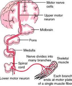
(Crossman and Neary, 2005). This means that movement on one side of the body is largely controlled by the other side of the cerebral cortex (see Figure 2.1).
[image:19.595.185.438.307.607.2]The axons in the descending tracts extend without interruption to their target neurons in the spinal cord. These are the lower motor neurons with their body in the grey matter and their axons (i.e. nerve fibres) directly innervating muscle. They are the final part of the pathway via which the nervous system controls movement (Crossman and Neary, 2005). Some motor function can be served via an indirect pathway (i.e. corticospinal fibres reach the tract by synapsing in the midbrain) but fine motor control requires intact input of the direct pathway from the primary motor cortex to the spinal cord (Rizo, 2004). Most movement requires input from both pathways.
Figure 2.1: Pathway of upper and lower motor neurons (Damjanov, 2000).
to normal walking conditions. Furthermore, the brain is the normal activator of these neural circuits (Enoka, 2008).
c) Motor units
[image:20.595.156.468.278.533.2]The functional unit of skeletal muscle is the motor unit. It consists of a lower motor neuron together with all the terminal branches of the axon and the muscle fibres that they innervate. The number of muscle fibres that are innervated by a single axon will vary, from a few to 2000, per motor unit and will depend on the size and function of the muscle. When the neuron transmits a message all of the fibres in the motor unit will contract (Watkins, 2009).
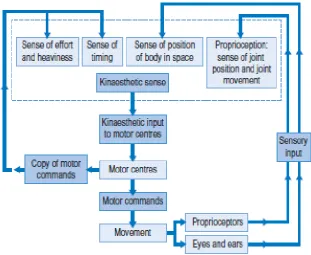
Figure 2.2: Relationship between kinaesthetic sense and proprioception (Watkins, 2009).
d) Kinaesthetic sense and proprioception
joint position and movement. Within joint capsules there are two main types of proprioceptors that signal joint angle and particular information at the end ranges of joint movement: Ruffini corpuscles which appear to be mainly responsive to tension, and Pacinian corpuscles which respond to compression (Watkins, 2009). Within skeletal muscles, a number of muscle fibres will be enclosed in a capsule to form a muscle spindle, supplied by sensory nerve endings, which respond to muscle stretch (Crossman and Neary, 2005). At the junction between skeletal muscle and its tendon, Golgi tendon organs occur and provide information about the tension exerted by the muscle (Matthews, 2000).
2.2.2 Gait kinematics and muscle activations
Human walking is a method of locomotion involving the use of two legs in an alternating pattern, to provide both support and propulsion. It is characterised by a smooth and efficient progression of the body’s centre of mass and distinguished from running by having at least one foot in contact with the ground at all times (Whittle, 2003). The components of gait (manner of walking) and the specific contribution of the lower limb muscles and joints to achieving this motion are described in the following paragraphs.
Figure 2.3: Timing of phases of gait during the gait cycle
(from http://media.lanecc.edu/users/howardc/PTA104L/104LAmbAids/104LAmbAids_print.html)
Gait can also be described in terms of spatial parameters, as shown in Figure 2.4 showing step length (i.e. the distance between two consecutive heel strikes) and stride length (i.e. the distance between two consecutive heel strikes of the same foot). Stride width is determined by the distance between the bisection of each heel (Richards, 2008).
Figure 2.4: Spatial parameters of gait.
(from http://atec.utdallas.edu/midori/Handouts/walkingGraphs.htm)
in the sagittal plane, of the ankle, knee and hip joints during the gait cycle are shown in Figure 2.6.
Figure 2.5: Activation patterns of major muscles during gait (2004).
The following describes the gait cycle in terms of the major events, the action of the major muscles and the motion at the major joints. The description is a summary of the cycle as described by several authors (Trew and Everett, 2001, Whittle, 2003, Richards, 2008). At the beginning of the gait cycle the heel strikes the ground, with the ankle close to its neutral position (i.e. 0°) with the angle between the foot and the ankle at about 90° and the heel slightly inverted with the forefoot slightly supinated. The tibialis anterior is active at this point, having maintained dorsiflexion during swing phase. The knee is almost straight just before heel strike and begins to flex immediately with the aid of contraction of the hamstrings, having aided prevention of knee hyperextension at the end of swing phase. Contraction of the hamstrings and the gluteus maximus at heel strike begins extension of the hip.
The double support phase (i.e. the period between heel strike and toe off of the opposite limb) is the period during which the foot is lowered to the ground and fully loaded. The ankle plantarflexes via eccentric contraction of the tibialis anterior and is accompanied by pronation of the forefoot and internal rotation of the tibia. The knee flexes from its almost fully extended position accompanied by eccentric contraction of the quadriceps to limit the speed and magnitude of flexion. The hip begins to extend via eccentric contraction of the hip extensors, gluteus maximus and hamstrings.
At the end of the double support phase, when the opposite toe off occurs, the first period of single support begins. At this point the foot is flat on the ground, and as soon as this occurs, the ankle motion changes from plantarflexion to dorsiflexion as the tibia moves over the stationary foot. Forefoot pronation and internal tibia rotation reach a peak at about this time and then begin to reverse. Contraction of the tibialis anterior ceases and the gastrocnemius and soleus begin to contract. The knee continues to flex and the hip continues to extend.
as its position is essentially maintained (except for a slight downward dip on the opposite side) by contraction of the hip abductors.
As the heel rises and the opposite foot strikes the ground, the toes remains on the ground and extension occurs at the metatarsophalangeal joints. The rearfoot inverts as the forefoot also becomes increasingly supinated and the tibia externally rotates, locking the midtarsal joints creating a stable foot for load bearing. The ankle moves into plantarflexion with concentric contraction of the soleus and gastrocnemius, with the latter aiding flexion of the knee, and the rectus femoris contracts eccentrically to prevent rapid flexion. At opposite foot strike the hip is in maximum extension and the motion reverses to hip flexion with the adductor longus acting as the primary hip flexor.
Toe off signals the beginning of the swing phase of the gait cycle. The forefoot remains slightly supinated during swing phase. Just after toe off, the ankle reaches its peak of plantarflexion at 25°. The tibialis anterior then begins to contract to dorsiflex the ankle contributing to toe clearance as the leg swings through, with knee flexion contributing most of the required leg shortening to achieve clearance. The knee reaches its peak of flexion during swing phase of between 60-70°, with the major part of flexion being facilitated by flexion at the hip. The knee then rapidly extends to close to full extension, in preparation for heel strike, with eccentric contraction of the hamstrings preventing hyperextension. The hip continues to flex at toe off, aided by contraction of the rectus femoris and iliopsoas. As the tibia reaches vertical during the swing phase, hip flexion ceases and the position is maintained by contraction of the hamstrings. At the end of the swing phase heel strike occurs and signals the completion of the gait cycle.
2.2.3 Aspects of behaviour believed to be associated with cognitive control of gait
involving higher level cognitive control (Sheridan and Hausdorff, 2007). This section will discuss this concept by firstly defining and describing the cognitive processes that are associated with gait control. This will be followed by a discussion of the evidence for and nature of the contribution of cognitive control of gait.
The outcome or product of all neural processes is behaviour. Cognitive control of gait refers to the contribution of neural processes that are not exclusive to the process of motor control, but also affect the everyday functioning of the individual (i.e. other aspects of behaviour). In order to explain the model, the reader is first introduced to the two aspects of behaviour believed to be involved in cognitive control of gait; cognition and executive functions (Lezak et al., 2004).
a) Cognition
Cognition is the information-handling aspect of behaviour and can be classified as;
receptive functions, memory and learning, thinking and expressive functions.
Receptive functions involve the ability to select, acquire, classify and integrate information. Thus, these functions will exploit the sensations received from the five senses (i.e. sight, hearing, touch, taste and smell), as well as those associated with movement, space, balance and effort (Berthoz, 2000). Processing of the sensations received involves perception, which is a complex process engaging activities such as awareness and recognition.
Memory and learning refer to information storage and retrieval. Memory is central to all cognitive functions (Lezak et al., 2004) and can be considered as either explicit (i.e. a conscious, intentional process) or implicit (i.e. performance of knowledge without awareness) (Squire, 2000). The first can be regarded as remembering information, objects and events and the latter as acquiring cognitive and motor skills (e.g. walking). Explicit memory engages stages of processing memory, of which one is short-term memory, which involves temporarily holding information. When this information is held in the mind, internalised and used to guide behaviour it is referred to as working memory (Lezak et al., 2004).
regarded as a function of the entire brain rather than a localised area (Lezak et al., 2004). Expressive functions are the sum of observable behaviour, from which mental activity is inferred e.g. speaking, writing and movement (Lezak et al., 2004).
The efficiency of cognition is affected by the level of consciousness, activity rate and attentional functions. Consciousness generally concerns the level at which a person is receptive to stimulation (i.e. awake) although definitions can vary. Activity rate relates to the speed at which neural processes and motor responses are performed (Lezak et al., 2004). Attention refers to several different capacities or processes, that are related aspects of how the person becomes receptive to stimuli and begins processing incoming or attended-to excitation, whether internal or external (Lezak et al., 2004). There is an agreed assumption that there is a finite capacity for attention (Woollacott and Shumway-Cook, 2002). Attention can be categorised as follows (Grieve, 2000, Lezak et al., 2004):
Focused or selective – capacity to orientate to the relevant stimuli whilst suppressing awareness of irrelevant stimuli, and is commonly referred to as concentration.
Sustained – capacity to maintain attention over a period of time.
Divided – ability to respond to more than one task at the same time or to multiple elements within one task, and is thus very sensitive to attentional capacity.
Alternating – capacity for shifting of attention from one task to another.
b) Executive functions
c) Evidence for higher level control of gait
i) Evidence from neuroimaging studies
Research in the area of brain neuroimaging supports the contribution to gait of areas of the brain, as well as those described in section 2.1.1a, that are also related to higher cognitive control. Empirical evidence from brain imaging studies have shown that during gait areas of the brain (e.g. prefrontal area) associated with higher cognitive functions are activated (Harada et al., 2009, Suzuki et al., 2004). These studies assessed changes in the haemoglobin oxygenation of the cortices, using a near-infrared spectroscopic imaging technique, whilst participants walked. Furthermore, these areas are also activated during imagined gait, with one study using positron emission tomography (PET) (Malouin et al., 2003) and another using functional magnetic resonance imaging (fMRI) (Bakker et al., 2008) to define active brain areas. Further studies of simulated gait (Francis et al., 2009, Sahyoun et al., 2004), also used fMRI, determining brain activity, in areas associated with higher cognitive function, during extension and flexion of the ankle; a movement normally associated with gait. Whilst the outcomes of the studies of imagined gait and of individual joint movements associated with gait, support the outcomes from the studies by Harada et al (2009) and Suzuki et al (2004) they should be viewed with some caution. Simulated or imagined gait will obviously not fully represent the neural processes involved in gait. For example, the study by Sahyoun et al (2004) restricted motion to the ankle joint whilst seated.
In addition, in a review of studies on the relationships between ageing and motor control by Seidler et al (2010) the authors suggested there is an age-related shift of movement control mechanisms, from automatic (lower level) control reliant upon peripheral sensorimotor systems, to attentional (higher level) control using central mechanisms. It is therefore possible that central control mechanisms are even more important than the peripheral sensorimotor system in maintaining postural stability in older adults (Seidler et al., 2010).
ii) Evidence from gait studies
used. However, there is strong evidence to support the contribution of attention, a key factor in the efficiency of cognitive functions, in gait control via numerous studies amongst both healthy and impaired populations. In particular, dual-task research methodologies have been widely used (Segev-Jacubovski et al., 2011) to assess the contribution of cognitive resources to gait. Dual-task protocols employ the simultaneous performance of two tasks (e.g. walking and cognitive task). Thus, if attentional capacity is limited and both gait and a secondary cognitive task are both demanding of attention, performance of at least one of the tasks will deteriorate when they are performed simultaneously.
There are a number of review papers covering the growing number of studies using dual-task methodologies (Woollacott and Shumway-Cook, 2002, Yogev-Seligmann et al., 2008, Al-Yahya et al., 2011, Segev-Jacubovski et al., 2011). In dual-task studies of healthy adults, often the secondary task performance declined as well as the gait speed slowing (Yogev-Seligmann et al., 2008, Seidler et al., 2010) particularly if the performance indices were sufficiently sensitive (Seidler et al., 2010). Most studies of older healthy adults elicited the same response, although there are some studies that indicate the extent of deterioration in performance of the cognitive task during dual-tasking increases with age (Yogev-Seligmann et al., 2008, Seidler et al., 2010). Furthermore, studies of gait in participants with neurological disorders have shown that the costs to gait and cognitive task performance increase in comparison to healthy controls (Woollacott and Shumway-Cook, 2002, Yogev-Seligmann et al., 2008, Segev-Jacubovski et al., 2011, Al-Yahya et al., 2011). The results of these studies generally support the widely accepted view that gait control involves attentional processes.
2.3 Stroke
2.3.1 Stroke overview
A ‘stroke’ is defined as a clinical syndrome, of presumed vascular origin, typified by rapidly developing signs of focal or global disturbance of cerebral functions lasting more than 24 hours or leading to death (WHO, 1988). A stroke will typically be the result of either a bleed (i.e. haemorrhagic stroke) or blockage (i.e. ischaemic stroke) affecting the vascular supply to the brain leading to damage and death of nerve cells within the brain. A haemorrhagic stroke, affecting approximately 20% of cases (Rudd et al., 2000), occurs when a blood vessel either within or on the surface of the brain bursts. This type of stroke tends to be more severe and is associated with higher early mortality (Mant, 2011). An ischaemic stroke is the most common, occurring in approximately 80% of cases (Rudd et al., 2000), and is caused by a blood clot forming in the main artery to the brain, a blockage transported to the brain from another blood vessel in the body or a small blood vessel deep in the brain becoming blocked.
Stroke affects between 174 – 216 people per 100,000 population in the UK each year (Mant, 2004) and its incidence is strongly associated with age, with 75% of stroke cases occurring in people over 65 years of age (DOH, 2005). There is no absolute end to recovery after stroke, however most improvement in functioning occurs within six months of onset (RCP, 2008a), although more complex aspects of physical recovery, such as speech, may improve over years (Mant, 2011). There are more than 900,000 people who have survived a stroke living in England (DOH, 2005) with approximately half of these people dependent upon on others for everyday activities, following a period of recovery. Stroke causes a greater disability impact than other chronic conditions and a greater range of disabilities than any other condition (Adamson et al., 2004).
To illustrate the complexity of stroke as a condition, below are briefly listed the common secondary problems found immediately following stroke. These include dysphagia (i.e. swallowing difficulties) (Hamdy et al., 1997); incontinence (Carr and Shepherd, 2011); shoulder pain (Fawcus, 2000); apraxia which is usually associated with left hemisphere damage and is an isolated impairment of the ability to plan and execute skilled motor tasks (RCP, 2008a); and communication and speech problems (Warlow, 2007) such as aphasia; an impairment of the ability to form and understand words (RCP, 2004), and dysarthria; characterised by slow, weak, imprecise and/or uncoordinated movements of the speech musculature (Yorkston, 1996).
It is also common for stroke to result in a disturbance of mood. In particular, depression may compound any cognitive impairments that concurrently exist (Carr and Shepherd, 2002). Anxiety is almost as common as depression, although it is frequently not recognised and can be focused on specific issues such as fear of falling and the risk of stroke recurrence (RCP, 2008a). Fatigue, an enhanced perception of effort and limited endurance for sustained physical and mental activity, is estimated to occur in 50% of stroke survivors (Harwood et al., 2011). The cause is poorly understood but may include depression, fear, loss of motivation, pain, sleep disturbance and deconditioning (Harwood et al., 2011, Duncan et al., 2012).
2.3.2 Impact of stroke on key motor and sensory functions
Loss of central control of the musculoskeletal system following stroke encompasses phenomena such as lack of coordination in movement, loss of selective movement and lack of motor control (RCP 2008). The severity can range from slight coordination problems to complete paralysis of the face and upper and lower limbs on one side of the body (i.e. hemiplegia or hemiparesis). Specific initial effects of impaired innervation to the muscles, caused by damage to the upper motor neurons, will include (Carr and Shepherd, 2011):
- muscle weakness
- reduced muscle activation and difficulty sustaining muscle activity
- reduced muscle force generation and poor timing of peak forces leading to slow movements
- poor control of synergistic muscle activity
After the initial effects of impaired motor control, other effects may emerge after several weeks. Spasticity is the most common and is typified by increased muscle tone, abnormal posturing and involuntary spasm that may cause discomfort and are particularly associated with higher levels of activity limitation (RCP 2008). Limitations to functional activity due to motor control impairments results in inactivity and this, plus weakness, leads to secondary adaptive changes to soft tissue and muscle. Furthermore, secondary neural and soft tissue changes may occur due to disuse and the weakness of certain muscle groups e.g. soft tissue contractures (Carr and Shepherd, 2011). Decreased activity will eventually result in a decline in physical fitness.
Loss or alteration of various somatic sensations is present in at least 50% of people and the severity of loss is probably associated with the extent of motor loss so the importance of sensory loss as an independent factor is unknown (RCP, 2008a). Sensory loss involving discrimination and proprioception is more often noted than loss of pain, touch and temperature sensitivity (Carey, 2006) thus joint position sense can be affected. The coexistence of sensory deficits will add to overall motor deficits, due to the inter-relationship of the function of both systems.
2.3.3 Effects of stroke on cognitive function
Some cognitive loss is thought to be present in almost all people following a stroke (RCP, 2008a) and thus affects the ability of the brain to handle information (Lezak et al., 2004). Some of the most common cognitive impairments that can occur following stroke are as follows:
Impairments of attention and concentration are probably the most pervasive cognitive deficits, especially in early stroke and when the right hemisphere is affected. This impairment may affect other unimpaired processes as attentional processing is an essential prerequisite for many cognitive and motor functions (RCP, 2004, RCP, 2008a).
Memory problems are quite common and can be affected in several ways
be diagnosed as suffering from dementia, with memory loss being a characteristic feature (RCP, 2008a).
Spatial awareness – i.e. a person’s awareness of the space around them and the space occupied by their body – can be affected by stroke. This impairment is also described as ‘neglect’, ‘visuo-spatial neglect’ or ‘inattention’ and is especially associated with right hemisphere stroke. In such cases a person may ‘neglect’ the left side of their body or fail to attend to things positioned on their left. Fatigue is particularly associated with this impairment (RCP, 2004, RCP, 2008a).
Perceptual disorders can have a varied impact. They can range from impaired
distance perception (e.g. difficulty crossing the road) to an inability to recognise an object when seen (i.e. visual agnosia) (RCP, 2004, RCP, 2008a). Apraxia or dyspraxia is a disorder of skilled voluntary movement that is not due to sensory or motor impairment. It is a conceptual inability to organise the actions required to perform the activity e.g. dressing. Some actions can be performed automatically, but not under voluntary control. It is more common after left cerebral hemisphere stroke (RCP, 2008a).
Executive functioning impairments can occur, especially when the frontal lobes are affected. Effects are seen in a person’s ability to plan a series of tasks, problem-solve and self-monitor behaviour. A striking effect on social behaviour can be associated with this impairment. It is relatively rare following stroke (RCP, 2004, RCP, 2008a).
2.3.4 Effects of stroke on gait and falls
a) Gait
Of those stroke survivors in the UK, around 20% will be affected by foot drop (Burridge et al., 1997a). It is characterised by reduced function in the muscles that serve to dorsiflex the foot (i.e. flaccid foot drop), and/or spasticity in the muscles that act to lower the foot (i.e. plantarflexors). Foot drop results in reduced ankle dorsiflexion during the swing phase of gait and at initial contact with a subsequent inability to achieve heel strike at the beginning of stance phase. Instead, at the end of swing phase, the foot lands plantarflexed, with the midfoot or forefoot contacting the ground initially instead of the heel. Furthermore, the foot can assume an equinovarus appearance, due to over-activity of the calf muscles (i.e. plantarflexors) compared to the tibialis anterior (i.e. dorsiflexors), where the foot is inverted and the forefoot is plantarflexed on the hindfoot. Evidence from one study suggests that this can occur in 18% of hemiparetic patients (Verdie et al., 2004).
Despite these common features, there are wide individual variations in the deviations from the norm. For example, in a study of 15 participants with foot drop, a number of different variations of abnormal muscle activation were identified when compared with age-matched controls. According to the authors, in many cases inappropriate calf muscle activity may contribute to foot drop as much as, if not more than the inability to activate the tibialis anterior muscles (Burridge et al., 2001). The literature on post-stroke gait predominately focuses on hemiparetic gait, and there is a lack of data defining the specific characteristics of foot drop gait. Therefore, the following description of the post-stroke gait largely concerns hemiparetic gait.
Hemiparetic gait is typically slow (Morris et al., 2010), with the average walking speed ranging from 0.23 m/s (SD = 0.11) to 0.73 m/s (SD = 0.38) (Olney and Richards, 1996). Whilst a slower speed could be the result of any of the various gait deviations evident, one study of 26 participants with mild to moderate hemiparesis, has noted that gait velocity was mainly affected by weakness of the affected hip flexors and knee extensors (Hsu et al., 2003).
normal gait (Chen et al., 2005, Morris et al., 2010). Step length asymmetry has been noted to increase but the direction of this is not consistent i.e. either limb can exhibit a shorter step length (Chen et al., 2005).
Variability in temporal and spatial gait parameters is also a characteristic of hemiparetic gait. In a large study of 94 hemiparetic participants, who were compared to healthy controls, step length and stride time variability in the post-stroke group was greater (Balasubramanian et al., 2009). Furthermore, swing time variability increased and was greatest in the paretic leg. Although stride width increases in hemiparetic gait (Chen et al., 2005), variability of this parameter does not increase when compared to healthy controls (Chen et al., 2005, Balasubramanian et al., 2009).
As one of the key requirements of gait is foot ground clearance during the swing phase, certain hemiparetic gait patterns, including foot drop gait, typically involve compensatory mechanisms at joints proximal to the ankle. A steppage gait, involving exaggerated knee and hip flexion, will lift the foot higher than usual to achieve increased ground clearance (Whittle, 2003). If there is diminished knee flexion, this strategy is not available, thus hip hitching (or hiking) or leg circumduction (Kerrigan et al., 2000, Chen et al., 2005), are required to clear the toe of the ground during the swing phase of gait. Finally, vaulting may be used to achieve foot clearance. This involves raising onto the toe of the unaffected side during stance, thus allowing the toe on the swing phase leg to clear the ground (Whittle, 2003). In common with the other compensatory mechanisms, these patterns do not contribute to forward progression of the body and are wasteful of energy (Whittle, 2003). Adoption of compensatory strategies may maintain balance and allow for the advance of the swing leg, however the energy costs can be significant (Olney and Richards, 1996, Chen et al., 2005).
In summary, foot drop gait is typically slow, unbalanced and energy inefficient. As described in the following section, a related consequence of stroke is instability and a heightened risk of falls.
b) Falls
event where the participant comes to rest on the ground, floor or lower level (Lamb et al., 2005). The healthcare cost of falls annually in the UK are estimated at US$1.6 billion, based on 2008 prices (Davis et al., 2010).
Whilst high rates are reported during inpatient episodes, falls are also very common in community-dwelling survivors. The incidence varies dependent upon the time since discharge – 23-34% for 3-4 months (Smith et al., 2006, Jorgensen et al., 2002), 40-73% for 6 months (Forster and Young, 1995, Yates et al., 2002, Hyndman and Ashburn, 2003, Hyndman and Ashburn, 2004, Soyuer and Ozturk, 2007, Harris et al., 2005, Mackintosh et al., 2005a, Mackintosh et al., 2005b, Belgen et al., 2006) and 43–70% for 1 year (Watanabe, 2005, Hyndman et al., 2002, Andersson et al., 2006, Lamb et al., 2003). Furthermore, fallers in the stroke population are more likely to become repeat fallers i.e. two or more falls in the last 12 months (Hyndman et al., 2002) with reports of incidences of between 21–57% (Forster and Young, 1995, Watanabe, 2005, Hyndman and Ashburn, 2003, Hyndman and Ashburn, 2004, Harris et al., 2005, Mackintosh et al., 2005a, Mackintosh et al., 2005b, Belgen et al., 2006, Hyndman et al., 2002, Andersson et al., 2006, Lamb et al., 2003).
The circumstances of falls amongst community-dwelling stroke survivors have been reported in several studies. The most frequently identified activity at the time of the fall was walking, most often whilst indoors, followed by transfers (Weerdesteyn et al., 2008). People have described losing their balance or getting their foot stuck whilst walking or transferring (Forster and Young, 1995). Another group of stroke survivors reported that impaired balance or other personal factors were causative factors. (Jorgensen et al., 2002). Other studies found reasons for falls to be dressing, mis-stepping, their foot getting stuck and imbalance (Belgen et al., 2006) plus self-perceived balance problems, including dizziness and spinning sensations, whilst dressing (Lamb et al., 2003).
brain into gear”. Furthermore, participants described the suspected cause of a near fall as: “I misjudged the distance of the step”; “I did not lift my foot up high enough” and “I wasn’t concentrating enough and my foot hit the curb”.
It is recognised, from the extensive literature on falls in the general population, that the circumstances and possible causes of falls are multi-factorial, and much time and effort has been spent attempting to identify specific risk factors. According to Lord et al (2007), there is consistently strong evidence to support limitations of activities of daily living (ADL), impaired gait and mobility, reduced peripheral sensation, muscle weakness and impaired cognition as important falls risk factors. These are all possible consequences of stroke and heighten its impact as a stand-alone risk factor. In particular, the contribution of cognitive impairments to falls following stroke, as also indicated by the statements of participants in the Hyndman et al (2002) study, has attracted further research.
A study of community stroke survivors (Hyndman and Ashburn, 2003) highlighted that attention deficits were common among the sample studied, and correlated with poor performance on functional measures and falls. Findings suggested that repeat fallers had greater attention deficits, plus greater balance and ADL impairments in comparison to those who did not report any instability. Therefore, results indicated that attention deficits might contribute to accident-prone behaviour and falls. In a study prior to this, general inattention was significantly associated with fall risk and impulsivity was suggested as an important factor (Rapport et al., 1993). Impulsivity, manifesting as initiating behaviours quickly and without consideration of the consequences may compound the impact of other post-stroke effects, such as poor spatial awareness. Fall risk has also been associated with the memory score on the Functional Independence Measure (FIM) (Wada et al., 2007). Finally, results from a study of stroke survivors six months after discharge noted that those with extensive involvement of the left hemisphere had a higher number of falls than those with similar involvement of the right. The authors attributed this to the greater impact on cognitive functions and reduced impact on motor functions with left, as opposed to right, hemisphere lesions (Alemdaroglu et al., 2012).
associated with reduced community reintegration (Schmid et al., 2011); one study noting that it was present in 88% of those who had fallen (Watanabe, 2005). As well as those who have fallen, fear of falling was also reported in people who had not experienced a fall (Schmid et al., 2009). It often leads to reduced physical activity through a complex process (Botner et al., 2005, Weerdesteyn et al., 2008). Fear of falling is also associated with loss of confidence and depression (Kim et al., 2012), which can contribute to a reduction in activity levels and social functioning (Weerdesteyn et al., 2008). Fear of falling has a complex relationship with the other factors associated with falls and Figure 2.7 gives an indication of its relationship with other factors for the individual. Figure 2.7 does not, however show the impact that the worries of carers of stroke survivors who have fallen may have on the range and frequency of activities performed by an individual following stroke (Forster and Young, 1995).
In summary, the risk of falls is heightened as a result of stroke, which in itself can be an obstacle to re-establishing independent function. Figure 2.7 gives an example of the interactions between risk factors and consequences of falls. It is particularly worth noting the contribution of both physical and cognitive impairments as risk factors.
2.3.5 Cognition and gait post stroke
As has been described in sections 2.2.1 and 2.2.3, gait is initiated and controlled by a combination of the motor pathways and cognitive processes. Stroke will affect both motor and cognitive processes as a result of damage to the brain. In particular, the cognitive deficits that result from stroke are various as discussed in section 2.3.3. There is a growing body of evidence to support the importance of the effect of these deficits in gait abilities post-stroke. Apart from the research evidence indicating the contribution of cognitive deficits to falls, there are numerous studies that demonstrate the interaction between cognitive deficits post stroke and gait.
Mulder (Mulder et al., 2002) proposes a persuasive model of the interaction between cognitive and motor function and its central role in functional recovery following nervous system damage. In line with much of the work described in sections 2.2.1 and 2.2.3, he proposes that the two systems are closely interrelated and that measurement of one aspect alone is insufficient to characterise functional recovery. As an example, walking speed is a commonly used outcome to measure recovery or the effect of an intervention. However, use of this alone as an outcome measure will not necessarily uncover the adaptive and compensatory strategies being used while performing the walking test and hence the level of functional recovery. In post-stroke gait, in addition to conventional measures of motor performance, a measure of the concentration or mental effort required to walk may reflect the level of functional recovery i.e. the level of reliance on a conscious mode of motor control. To assess the level of functional recovery over time or the lessening need for compensation Mulder et al (2002) proposed two approaches; measurement of cognitive involvement or visual dependency.
directions. As discussed earlier, it has been widely used to investigate the cognitive effects on gait, balance and fall risk particularly amongst elderly populations (Woollacott and Shumway-Cook, 2002, Yogev-Seligmann et al., 2008, Segev-Jacubovski et al., 2011).
There are also a growing number of studies in post-stroke gait that have used a dual-task design to assess gait. Several studies have used gait speed as an outcome, finding a deterioration in gait speed under dual-task conditions (Bowen et al., 2001, Canning et al., 2006, Hyndman et al., 2006, Plummer-D'Amato et al., 2008, Dennis et al., 2009, Pohl et al., 2011). Studies have also noted a reduction in performance of the cognitive task (Plummer-D'Amato et al., 2008, Dennis et al., 2009, Pohl et al., 2011, Hyndman et al., 2006, Kemper et al., 2006).
There are a small number of studies using other gait parameters as outcomes. The addition of a cognitive task had an adverse effect on balance with double-support time increasing in two studies (Bowen et al., 2001, Plummer-D'Amato et al., 2010). A small longitudinal study (Cockburn et al., 2003) compared stride duration initially after stroke and then 1-9 months later. Stride duration improved over time with recovery more so than cognitive performance during walking. A recent pilot study noted that paretic single limb stance time as particularly susceptible to dual-task interferences (Plummer-D'Amato and Altmann, 2012). Interestingly, in a dual-task study by Hyndman et al (2006) the participants were also assessed as fallers and non-fallers based on their fall history. Fallers coped less well with a competing cognitive task than non-fallers during walking, with a significant reduction in stride length.
2.4 FES
There are a number of treatment options for foot drop as the result of stroke. Apart from physiotherapy, and the use of Botulinum Toxin (BoTox), there are two orthotic interventions, ankle foot orthoses (AFO) and functional electrical stimulation (FES), the second of which is the focus for the thesis. This section of the literature review firstly describes the most common application of FES to address foot drop and the function of each component. Secondly, the effect on the nervous system is described both at local and central levels. Finally, a comprehensive literature review of the effects of FES is provided, with a particular focus on patient-centred outcomes.
2.4.1 Description of FES
FES can be described as the electrical stimulation of either nerves or muscles deprived of appropriate nervous control, to provide muscular contraction and thereby produce a functionally useful movement (Sujith, 2008). When used to restore movement of upper or lower limbs the FES device works as a neuroprosthesis, operating as a bypass (i.e. replacement) of the impaired sensory-motor pathway (Popovic, 2004 ) and the benefit is realised when the system is actively stimulating (Kilgore, 2004). Since this thesis is focused on foot drop of central origin, the subsequent discussion is restricted to systems used to stimulate muscle innervated by intact lower motor neurons.
FES systems comprise three basic elements; stimulator, sensors and electrodes (Lyons et al., 2002). The stimulator is used to generate and regulate stimulus pulses. The pulse and hence stimulation magnitude is adjusted by pulse width and amplitude. Sensors are used to gather command signals, typically based on the user’s motion or muscle activity, and electrodes provide the electrical interface with the body. Both implanted and surface electrodes are available, although systems based on surface electrodes are most common. A surface electrode usually comprises a wire to connect to the stimulator and a conductive woven backing mesh covered by a layer of highly conductive, adhesive hydrogel.
2004) and the initiation and termination of stimulation is controlled by one or more sensors (e.g. gyroscopes, accelerometers, foot switches) (Kilgore, 2004), the most common of which is the foot switch.
An example of a foot drop device and electrode placement is shown in Figure 2.8. Although other arrangements are possible, in this case the cathode is placed on the skin over the head of the fibula bone, where the common peroneal nerve is most superficial and just before it bifurcates into the deep and superficial branches; and the anode is placed over the motor point of the tibialis anterior muscle (Sujith, 2008). Table 2.1 illustrates the muscles that are recruited and their actions by stimulation of the common peroneal nerve. This device uses a foot switch as the sensor to trigger stimulation, with the stimulator being worn either at the waist or in a trouser pocket. Other commercially available clinical systems use an accelerometer (Sujith, 2008) and a cuff housing the surface electrodes (see Figure 2.9).
Action Superficial branch
of CPN
Deep branch of CPN
Peroneus
longus
Peroneus
brevis
Tibialis
anterior
Extensor
hallucis
longus
Extensor
digitorum
longus
Peroneus
tertius
Dorsiflexion X X X X
Plantarflexion X
Inversion X
Eversion X X X X
Table 2.1: Actions of muscles innervated by two branches of the common peroneal nerve.
Figure 2.9: Example of surface-based FES device in which electrodes are housed in a cuff. (http://www.hanger.com/orthotics/services/WalkAide/Pages/HowWalkAideWorks.aspx)
Figure 2.10: Example of an implanted FES device (Otto Bock).
2.4.2 Action of stimulation on nervous system
The stimulus generated by the FES device and transferred via electrodes to the body stimulates or excites peripheral nervous tissue. The stimulus will either be of the nerve directly or of the motor point of the nerve proximal to neuromuscular junction (Sheffler and Chae, 2007). The following section gives a background of nerve function and how FES stimulates the peripheral nervous system. There then follows a brief discussion of emerging evidence in support of the effect of FES on the CNS.
a) Functional effect
which end in a presynaptic terminal - the point at which the axon releases chemicals that cross the junction (i.e. synapse) between one neuron and the next, or to muscle fibres (Kalat, 2004).
Nerve impulses, called action potentials, are brief electrical discharges produced by an electrochemical process. When the resting membrane potential of the neuron is momentarily altered (i.e. depolarised from -70mV to 30mV) (Kalat, 2004) an action potential is generated. This is as a result of an ionic exchange between the inner part of the cell and the extracellular fluid, facilitated firstly by the permeability of the cell membrane to sodium and potassium ions, which is altered by the arrival of the stimulus, and secondly by sodium-potassium ionic pumps within the membrane (Watkins, 2009). Whenever the membrane is depolarised to the threshold value, an action potential of constant magnitude is produced (i.e. the ‘all-or-none’ phenomenon), and this travels along the cell membrane as a flow of electrical current as each adjacent region of the membrane is depolarised (Kalat, 2004). This in turn causes the release of neurotransmitters at the motor-end plate - the junction between the nerve endings and the muscle membrane (Crossman and Neary, 2005), which excites the muscle causing contraction.
Figure 2.11: Typical neuron structure