ScholarWorks @ Georgia State University
ScholarWorks @ Georgia State University
Biology Theses Department of Biology
Summer 7-12-2012
MC3R and MC4R Knockdown via RNA Interference
MC3R and MC4R Knockdown via RNA Interference
Danielle N. Mankin
Georgia State University
Follow this and additional works at: https://scholarworks.gsu.edu/biology_theses
Recommended Citation Recommended Citation
Mankin, Danielle N., "MC3R and MC4R Knockdown via RNA Interference." Thesis, Georgia State University, 2012.
https://scholarworks.gsu.edu/biology_theses/37
This Thesis is brought to you for free and open access by the Department of Biology at ScholarWorks @ Georgia State University. It has been accepted for inclusion in Biology Theses by an authorized administrator of
by
DANIELLE MANKIN
Under the Direction of Aaron Roseberry
ABSTRACT
Melanocortins (MCs) play an important role in feeding, metabolism, and energy expenditure.
While melanocortin receptor (MCR) mRNA has been found in the mesolimbic dopamine (DA)
pathway, the ability of melanocortins to regulate feeding and other behaviors through actions on
the mesolimbic DA system have not been examined. Short-hairpin RNAs (shRNAs) were
created targeting MC3R and MC4R and were tested via in vitro studies for their ability to
knockdown their target receptor. A total of three shRNAs were created targeting each receptor,
and each shRNA caused successful knockdown. These shRNAs are tools that can be used for
future in vivo studies to examine the various behavioral effects of melanocortins on the
mesolimbic DA pathway.
by
DANIELLE MANKIN
A Thesis Submitted in Partial Fulfillment of the Requirements for the Degree of
Master of Science
in the College of Arts and Sciences
Georgia State University
Copyright by Danielle N. Mankin
by
DANIELLE MANKIN
Committee Chair: Aaron Roseberry
Committee: Tim Bartness
William Walthall
Electronic Version Approved:
Office of Graduate Studies
College of Arts and Sciences
Georgia State University
ACKNOWLEDGEMENTS
This thesis would not have been possible without the guidance and the help of several
individuals. I would like to begin by thanking my peers, especially Lewis Yen and Dereka
Moore, for being an unending source of backing and encouragement. I am indebted to Dr.
Deborah Baro for allowing me to use her laboratory equipment without which none of my
experiments would have been possible. I am also grateful to Dr. Tim Bartness, for not only
acting as a part of my thesis committee, but also being a great professor and teaching me much
of the groundwork that led to my understanding of neurobiology as a science. I would like to
thanks Dr. William Walthall for giving me the opportunity to work in his laboratory and always
being a source of guidance as an adviser. Lastly, I would like to show my gratitude to my
mentor, Dr. Aaron Roseberry, whose encouragement, guidance, and support from the initial to
the final steps enabled me to develop a deeper understanding of not only the subject matter and
TABLE OF CONTENTS
ACKNOWLEDGEMENTS ... iv
LIST OF TABLES ... vii
LIST OF FIGURES ... viii
1 INTRODUCTION ... 1
1.1 The homeostatic pathway of the hypothalamus... 1
1.2 Mesolimbic dopamine pathway ... 6
1.3 Mesolimbic Dopamine System and Feeding ... 8
1.4 Reward and the dorsal striatum ... 9
1.5 Mesolimbic and homeostatic feeding pathway interactions ... 10
1.6 Mesolimbic dopamine pathway and melanocortin interactions ... 13
1.7 Research Goals ... 14
2 METHODS/EXPERIMENTAL PROTOCOL ... 15
2.1 Creation of shRNA targeting the MC3R and MC4R ... 15
2.2 ptdTomato-MC3/4R creation ... 17
2.3 shRNA testing in vitro ... 18
2.4 Data Analysis and Statistics ... 19
3 RESULTS ... 19
LIST OF TABLES
Table 1. Targeted mRNA sequences used to create pAAV short-hairpins ... 16
LIST OF FIGURES
Figure 1. Breakdown of melanocortin neuropeptides from its original state as a
preprohormone ... 2
Figure 2. Projections of POMC and AgRP neurons throughout the rat brain ... 5
Figure 3. Neuropeptides that affect dopamine neurons ... 12
Figure 4. A sample of the known relationships between the homeostatic and reward pathways ... 15
Figure 6. Design of AAV shRNAs ... 17
Figure 5. Schematic of short-hairpin creation used in RNAi... 17
Figure 7. Sample images showing effectiveness of the MC3R shRNAs on tdTomatoMC3R expression... 21
Figure 8. MC3R shRNAs decrease expression of tdTomatoMC3R in HEK-293 cells ... 22
Figure 9. Extent of knockdown of the tdTomatoMC3R by the MC3R shRNAs... 22
Figure 10. Sample images showing effectiveness of the MC4R shRNAs on tdTomatoMC4R expression... 24
Figure 11. MC4R shRNAs decrease expression of tdTomatoMC4R in HEK-293 cells ... 25
Figure 12. Extent of knockdown of the tdTomatoMC4R by the MC4R shRNAs... 25
Figure 13. Sample images showing no MC3Rsh2 effect on tdTomatoMC4R ... 26
Figure 14. MC3R shRNAs do not affect expression of tdTomatoMC4R... 27
Figure 15. Sample images showing no MC4Rsh3 effect on tdTomatoMC3R ... 27
1 INTRODUCTION
Today, it is estimated that 68% of adults and 32% of children in the United States are
considered overweight or obese (Ogden et al 2012a; 2012b; Flegal et al 2010). Obesity is shown
to have correlations with heart disease, hypertension, stroke, type II diabetes, sleep apnea,
various cancers, infertility, and overall decreased life expectancy (Haslam & James 2005).
Obesity and food intake are influenced by a variety of psychological factors such as food choice,
social interactions, and aesthetics as well as physiological factors such as energy stores,
endocrine signals, paracrine signals, and neuropeptides. It is debated how each of these factors
works to cause or cease feeding via peripheral and central control. Peripheral cues are an
important aspect of food intake control; however, the main focus of this paper will be within the
central nervous system.
1.1 The homeostatic pathway of the hypothalamus
Prior to the 1950s, scientists believed hunger was solely under gastrointestinal control;
however, the hypothalamus and brainstem gained recognition as major contributors in the role of
feeding (Kennedy 1953). Early studies describe the hypothalamus as having a main ‘feeding’
center in the lateral hypothalamus and a main ‘satiety’ center in the ventromedial hypothalamus
(Anand & Brobeck 1951; Teitelbaum & Stellar 1954; Mayer & Thomas 1967), though
subsequent research has demonstrated that multiple areas of the brain, including areas within the
hypothalamus play key roles in the regulation of feeding and body weight. One area of the
hypothalamus shown to play a key role in the regulation of food intake is the arcuate nucleus of
and many of them affect feeding such as opioids, orexins, melanin-concentrating hormone,
galanin-like peptides, galanin, and many more.
One important family of ARC neuropeptides that play a key role in regulating feeding are
the melanocortins. The melanocortins are a family of neuropeptides produced by the
proopiomelanocortin gene or POMC. Once created the POMC neuropeptide is broken down
from its preprohormone precursor to physiologically active substances such as
adrenocorticoptropic hormone (ACTH), α, β, and γ melanocyte-stimulating hormone (MSH), and
[image:12.612.99.517.318.463.2]β-endorphin (Mountjoy et al 1992; Palkovits et al 1987) as seen in Figure 1 (Cone 2006).
Figure 1. Breakdown of melanocortin neuropeptides from its original state as a preprohormone
The MSHs are commonly referred to as the melanocortins. α-MSH is considered to be the
most physiologically relevant melanocyte-stimulating hormone in the CNS because of its
anorexigenic effects (decreases feeding); however, β and γ-MSH also been shown to have some
physiological significance (Gantz et al 1999). Intraventricular injection of α-MSH is known to
cause decreased food intake, increase energy expenditure, and chronically can cause cachexia, or POMC preprohormone is broken down from its preprohormone precursor to N-terminal,
extreme weight loss and malnourishment (Panskepp et al 1976; Fan et al 1997). A second set of
neurons in the hypothalamus expresses agouti-related protein (AgRP), which is an endogenous
antagonist of melanocortin receptors. AgRP acutely increases food intake, decrease energy
expenditure, and chronically increases body weight (Fan et al 1997; Huszar et al 1997). AgRP is
an antagonist to the melanocortins (Corander et al 2011), but can also act as an inverse agonist.
This suggests that it not only blocks the receptor, but can cause the opposite effect, in this case,
increased feeding (Haskell-Luevano & Monck 2001; Nijenhuis et al 2001). ACTH is produced in
response to stress and has implications in grooming and sexual function. It is produced in the
pituitary gland and mainly affects melanocortin receptors at its site of production as well as the
adrenal cortex. β-endorphin is an endogenous opioid that has many functions, one being that it
stimulates food intake through its receptors in the ARC and other places (Appleyard et al 2003).
There are five known G-protein coupled receptors that react with MSHs, melanocortin-1
receptor (MC1R) through melanocortin-5 receptor (MC5R) (Griffon et al 1994; Labbe et al
1994). MC3R and MC4R are the only two receptors present in the central nervous system (CNS)
and therefore are the main focus of study in neurobiology (Mountjoy et al 1994; Roselli-Rehfuss
et al 1993; Gantz et al 1993; Chhaklani et al 1996). MC4R is considered the primary MCR
involved in body weight control because of its high binding affinity with α-MSH and resulting
strong physiological changes (Gantz et al 1993). MC4R plays a major role in energy homeostasis
via food consumption control, energy expenditure, changes in sympathetic nervous system
(SNS) activity. MC4R null mice are hyperphagic, gain weight, and develop insulin-resistance
induced diabetes (Gantz & Fong 2002; Sutton et al 2006; Huszar et al 1997; Garza et al 2008).
Melanocortin 4 deficiency is the largest monogenic cause for obesity in humans (Krude et al
the exact role is less clear. MC3R is involved in energy homeostasis via the manipulation of
energy metabolism, or the fat-to-mass ratio. MC3R null mice do not increase food intake, but
have a higher fat to mass body ratio than its wild-type counterparts. Research suggests this is
caused by a change in metabolism due in large part to decreases in locomotor activity (Sutton et
al 2006; Butler et al 2000; Chen et al 2000). Although MC3R has binding affinity with α, β and,
γ-MSH, it has a lack of unique agonists/antagonists that exclusively bind to the MC3R which has
made studying its effects difficult. Many still question the importance of MC3R due to
confounding research (Lee et al 2008; Fan et al 1997). Recent papers, however, show that MC3R
does in fact effect feeding, but the mechanism is still unknown (Marks et al 2006; Irani et al
2011).
Among the many neuronal subtypes in the ARC, two populations of neurons have been
shown to play an important role in feeding, the first expressing proopiomelanocortin (POMC)
and a second set of neurons expressing agouti-related peptide (AgRP) and neuropeptide Y
(NPY). POMC neurons are potent anorexigenic stimulators while AgRP/NPY neurons are potent
orexigenic stimulators (increase feeding) (Huzsar et al 1997; Fan et al 1997; Bulter et al 2000).
Recent DREADD and optogenetic technologies have shown that stimulation of POMC neurons
decreases food intake and body weight in the presence of only melanocortins and no other
neuropeptide (Aponte et al 2011). Stimulation of the AgRP/NPY neurons acutely and
dramatically increases food intake, decrease energy expenditure, and chronically increases body
weight (Aponte et al 2011). Inhibition of AgRP neurons decreases food intake to below baseline
standards independent of melanocortin stimulation suggesting AgRP is both ‘necessary and
many of the same projections as seen in Figure 2 (Cone 2005). This allows these two opposing
[image:15.612.137.469.146.369.2]systems to have control on each other.
Figure 2. Projections of POMC and AgRP neurons throughout the rat brain
Other hormones that interact with the homeostatic pathways to affect feeding and body
weight are leptin, insulin, and ghrelin. Leptin is a hormone that is largely made by white adipose
tissue, and it decreases with hunger, increases with satiety, and inhibits feeding via receptors in
the ARC (Frederich et al 1995; Maffei et al 1995; Zhang et al 1994; Halaas et al 1995; Elmquist
et al 1998). Insulin increases with blood glucose levels (Polonsky et al 1988), and causes
hypophagia and weight loss within the CNS (McGowen et al 1992). Another peptide, ghrelin,
has orexigenic effects by activating AgRP/NPY neurons in the ARC while suppressing POMC
Cowley et al 2003; Wren 2000). Although, there is no one cause for food consumption, the
melanocortins and the peptides that affect them play a vital role in feeding. Furthermore, more
recent studies focus on how the ‘reward pathway’ contributes to rewarding aspects of feeding.
1.2 Mesolimbic dopamine pathway
There are certain areas within the brain that aid in motivation, learning, and pleasure.
These circuits are responsible for the hedonics, or pleasure, of food intake without affecting the
biological necessity of it. This occurs by changing the body’s response to salient environmental
stimuli causing food intake during a time of food availability rather than for energy needs. This
can result in a pleasurable experience. Evolutionarily speaking, without these pathways, animals
with full energy stores would not always eat when food was available, and once food availability
ran out, starvation could occur. One of the major hormones responsible for hedonic feeding is
dopamine. One of its many neuronal projections extends from the ventral tegmental area (VTA)
to the nucleus accumbens (NAc) and in combination with other projections is known as the
mesolimbic dopamine (DA) system.
Researchers unknowingly began looking into this ‘reward’ area of brain while researching
the cause for drug addiction. Drugs of abuse hijack these pathways which can be evolutionarily
assumed to be useful when finding food or water. Dr. Roy A. Wise was one of the original
researchers to focus his attention on the addiction phenomena. It was found that dopamine was
important for brain self-stimulation (Yeomans 1979), and that positive drug self-administration
regions of the midbrain overlapped with areas with a high density of dopaminergic cell bodies,
specifically in the VTA and substantia nigra (Corbett & Wise 1980; Bozarth & Wise 1981; Wise
1982). At this time dopamine was the neurotransmitter believed to cause these addictions (Wise
abuse are found in the mesolimbic DA pathway, specifically from the VTA projecting to the
ventral striatum, which includes the NAc (Di Chiara & Imperato 1988; Koob 1992; McBride et
al 1999; Volkow et al 2004). Although, the VTA and NAc are often focused on, there are many
other areas involved in this system; these include the dorsal striatum (caudate putamen- CPu),
prefrontal cortex, and the amygdala. Research shows that it is the neurotransmitter dopamine in
the NAc that causes the behavior to seek the drug, or the reward, and that without dopamine, its
receptor, or its neurons, the self-administration or seeking behavior disappears (Ikemoto et al
1997; Roberts & Koob 1982). The result of increased dopamine levels from drugs of abuse
typically decreases dopamine-2 (D2) receptors (Volkow et al 2002). It is assumed that this
occurs as a reaction to an overabundance of dopamine; however, at this point it then requires
more dopamine, i.e. more drugs of abuse, to receive the same dopaminergic effect of a smaller
number of receptors. This phenomenon is thought to play a role in tolerance and addiction. This
is shown in self-administration studies where mice progressively increase the amount of cocaine
with similar resulting dopamine output (Ahmed et al 2002). The mechanisms by which dopamine
is increased depends of the specific drug of abuse (Ritz et al 1987).
Dopamine is often considered the major component of the mesolimbic DA system,
however, other transmitters play key roles in regulating the overall activity of this system.
Glutamate plays an important role in these pathways with afferent projections from areas such as
the hippocampus (O’Donnell & Grace 1995). GABA and endogenous opioids also affect the
activity of this system and compose a feedback loop within the reward pathway (Trigo et al
2010). In addition, acetylcholine (ACh), which has interneurons in the NAc and various other
(Mark et al 2011). The mesolimbic dopamine pathway has been studied for decades and is still
not completely understood.
1.3 Mesolimbic Dopamine System and Feeding
Dopamine is an important neurotransmitter with a wide range of physiological functions
including motivation, reward, and reinforcement, learning and memory, and motor control.
Conditions associated with abnormal dopamine show changes in feeding and body weight. For
example, dopamine-deficient (DD) mice are hypoactive, aphagic, and adipsic and will starve to
death without help (Zhou & Palmiter 1996). Certain atypical antipsychotics are dopamine-2 (D2)
receptor antagonists and are known to cause increased weight gain (Baptista et al 1990). D2
density has also been shown to be inversely proportional to the weight of obese patients
suggesting a potential role of D2 receptors in development of obesity (Wang et al 2002). One
study shows clozapine, a partial dopamine receptor antagonist, increases food intake in rats, and
a D2 receptor agonist reverses it (Kaur & Kulkarni 2002). Parkinsons disease is characterized by
dopamine cell death in the substantia nigra and its patients have lower body weight than their
healthy counterparts (Beyer et al 1995). Patients that are given more levodopa treatments, a
dopamine precursor, have a larger amount of weight loss suggesting the medication may be
responsible (Bachmann et al 2009).
As humans, we have the tendency to ‘finish our plate’ of food past the necessary need for
energy balance (Jansen et al 2003; Weingarten 1983). This alone suggests that some other
control systems other than basic energy needs are involved in feeding. Despite the important role
of the mesolimbic DA pathways in drugs of abuse, it is thought that these pathways were
originally designed to respond to naturally rewarding substances. Food and water increase
the overall reward that dopamine triggers, and is not specific to drugs (Hernandez & Hoebel
1988; Yoshida et al 1992). It has been shown that ‘addicted’ animals prefer highly-intense sugar
over cocaine suggesting that it is possible that food has the potency to be just as addicting as any
drug of abuse (Lenoir et al 2007). The mechanism for overeating shows similarities to drugs of
abuse. Obese patients have decreased D2 receptors similar to the decrease in drug addicts (Wang
et al 2001). In addition, patients who have undergone gastric bypass surgery have upregulated
D2 receptors, in theory, to try to restore the dopamine levels to previous pre-surgery ‘reward’
levels (Steele et al 2010). Similar findings have been shown in obese rats that are then placed on
a restricted feeding diet resulting in weight loss (Hamdi et al 1992). Rats on high fat diets are
less likely to experience ‘reward’ due to decreased self-stimulation, and presumably decreased
D2 receptors. When the D2 receptors are knocked down, feeding dramatically increases
supporting the theory that dopamine is what is responsible for giving the ‘rewarding’ feeling. It
is also possible that without these receptors animals will consume drugs or food to try to receive
the same dopamine stimulation. In evolution theory, these reward pathways were likely put in
place for species necessities such as food, water, and reproduction; however, drugs of abuse use
these pathways to cause their own rewarding effects. Although the reward pathway can
independently affect feeding, its interactions with other pathways are just as important.
1.4 Reward and the dorsal striatum
Although the ventral striatum/NAc gets a lot of attention in the mesolimbic DA pathway,
the dorsal striatum is a key target for DA output as well. The dorsal striatum which is composed
of the caudate putamen (CPu) is another key area of interest. Its inputs include the cortex,
thalamus, and the mesolimbic DA neurons which are similar areas to the ventral striatum
has domaminergic innervations via the substantia nigra. These DA neurons have been
characteristically associated with motor function and their deficiency with the neuromuscular
disorder Parkinsons Disease (PD) (Chase et al 1996). PD patients have symptoms that include
hypophagia; however, this is attributed to the associated dysphagia and depression. The CPu’s
effects on reward were shown as far back as the 80s (Hikosaka et al 1989). Research also
associates the CPu with learning, which is an important component of reward (Jog et al 1999;
Packard & White 1990).
While the CPu has known effects in drugs, learning, and PD, recent research has indicated
importance in feeding. Szczypka et al restored DA in dopamine deficient (DD)-mice only in the
CPu. This resulted in mice regaining the motivation to eat and ability to maintain an adequate
sustainable body weight which is not possible in DD-mice (Szczypka et al 2001). In addition, it
has been shown that dopamine increases in relation to drinking in rats in the NAc as well as the
CPu (Young et al 1992). This suggests that it is possible that motivation and learning, which are
components of addiction and rewarding behavior, to eat or drink is not solely contained within
the ventral striatum and its efferent projections from the VTA. The ‘reward’ circuits may lie
within the ventral as well as dorsal striatum. This also suggests that the hypophagia associated
with Parkinsons may be due to the dopamine or melanocortin neuronal changes in the dorsal
striatum.
1.5 Mesolimbic and homeostatic feeding pathway interactions
While most research has focused on the role of homeostatic or hedonic pathways of food
intake control independent of each other, it is clear that there are important interactions between
these pathways. Although this is thought of as a new area of study, researchers have been
heroine injections into the hypothalamus, a role traditionally thought of in the mesolimbic DA
pathway (Gerber et al 1981). In addition, excitation of the lateral hypothalamus via the medial
forebrain bundle causes the same dopamine release in the NAc independent of the VTA
(Hernandez & Hoebel 1988; 1988). By 1992 evidence of a direct link between the NAc shell and
the lateral hypothalamus was well documented (Heimer et al 1991; Zahm & Brog 1992), and it
was known that the NAc has role in feeding behavior (Salamone 1994).
The mechanism of how these two pathways interact and how they affect food intake is still
developing. Rada et al found glutamate levels in the lateral hypothalamus and the NAc changed
in response to food intake suggesting not only do these areas share pathways, but that they also
aid in the control of feeding (Rada et al 1997). Also, NAc-induced feeding could be attenuated
with GABA agonist injections into the lateral hypothalamus (Maldonado-Irizarry et al 1995). In
addition to the NAc the hypothalamus has been shown to have connections to the VTA.
Retrograde tracing injected into the lateral hypothalamus shows strong afferents in the VTA,
NAc shell, and many other areas (Duva et al 2005). Orexin, an orexigenic neuropeptide, has been
shown to have projections from the lateral hypothalamus to the VTA (Fadel & Deutch 2002).
Also, orexin injected into the VTA causes firing, and orexin projections to the VTA are required
for morphine-related conditioned place preference (Korotkova et al 2003; Narita et al 2006).
Recent studies have shown that orexin signaling in the VTA is required for opioid-induced food
intake in the NAc suggesting not only a connection between the lateral hypothalamus and the
VTA, but a possible relationship to rewarding food intake (Zheng et al 2007). In addition to
orexin, melanin-concentrating hormone (MCH), another orexigenic neuropeptide, also has
projections from the lateral hypothalamus to the mesolimbic DA pathway (Bittencourt et al
showed that MCH-1 receptor knockout mice, characterized by hyperactivity and hypophagia,
have increased dopamine in the VTA as well as the NAc further suggesting a functional
association between these two areas (Smith et al 2005).
In addition to the neuronal connection between the VTA, NAc, and lateral hypothalamus,
dopamine neurons in the VTA have receptors for important feeding relevant hormones similar to
the ARC. Leptin, insulin, and ghrelin not only affect feeding via the homeostatic pathways, but
they also have effects on dopamine within the mesolimbic DA system to affect feeding. Leptin
when injected in the ventricles decreases feeding-evoked dopamine release in the NAc (Krugel et
al 2003). Insulin receptors have also been found in the VTA dopaminergic neurons, (Pardini et al
2006; Figlewicz et al 2003) and intraventricular insulin was shown to reduce the self-stimulation
of sucrose (Figlewicz et al 2006). Ghrelin increases dopamine firing in the VTA and decreases
inhibitory firing and as a result causes food intake (Abizaid et al 2006; Naleid et al 2005). Figure
3 created by RD Palmiter shows the different hormones and neuropeptides that have been shown
[image:22.612.131.484.457.632.2]to affect dopaminergic neurons in the VTA (Palmiter 2001).
Figure 3. Neuropeptides that affect dopamine neurons
1.6 Mesolimbic dopamine pathway and melanocortin interactions
In addition to leptin’s, insulin’s, and ghrelin’s interaction with dopaminergic pathways,
there is also evidence indicating melanocortin involvement within the VTA and the mesolimbic
DA system. MC4R mRNA has been found in the VTA, NAc, and CPu, and the MC3R has been
found extensively in the VTA (Kishi et al 2003; Liu et al 2003; Rosseli-Rehfuss et al 1993).
α-MSH injections in the VTA have been shown to increase dopamine turnover and grooming in
rats, a typically melanocortin characterization, via the MC4R (Klusa et al 1998; Lindblom et al
2001). In addition, γ1-MSH injections into the VTA increase dopamine and its metabolites in the
NAc (Jansone et al 2004). In 1986, it was shown that α-MSH injected into the ventricles or
substantia nigra causes changes in dopamine in the caudate putamen (Torre & Celis 1986). Davis
et al showed that centrally administered AgRP, a melanocortin antagonist, increased dopamine
firing in the midbrain with associated dopamine turnover in the prefrontal cortex, a known
projection site of mesolimbic DA neurons (Davis et al 2011).
Many studies looking at drugs of abuse have also linked melanocortins with the
mesolimbic DA pathway. Alvaro et al showed that morphine down-regulated MC4R in the NAc
as well as the CPu (1996). An exogenous melanocortin receptor agonist was shown to cause an
increase in rewarding effects of amphetamines in the lateral hypothalamus (Cabeza de Vaca et al
2002). Similarly, an exogenous melanocortin receptor antagonist caused a decreased in
rewarding effects of cocaine in the NAc (Hsu et al 2005, 2009). Here, the term ‘rewarding
effects’ refers to the perceived reward received from drugs of abuse and the reinforcing
motivation to seek said drug. In addition, POMC mRNA was shown to decrease in response to
morphine (Bronstein et al 1990). This building research shows that the melanocortins interact
It seems clear that the melanocortins interact with the mesolimbic DA pathway for some
behaviors; however, there is little research referring to this interaction and its direct effects on
feeding. Xenakis and Sclafani showed that a dopamine receptor antagonist suppressed sucrose
preference (1981). In addition, melanocortin agonists or NPY antagonists can inhibit opioid
induced feeding in the NAc (Zheng et al 2010). These findings suggest that melanocortins and
AgRP/NPY have projections as well as possible feeding related interactions with the NAc.
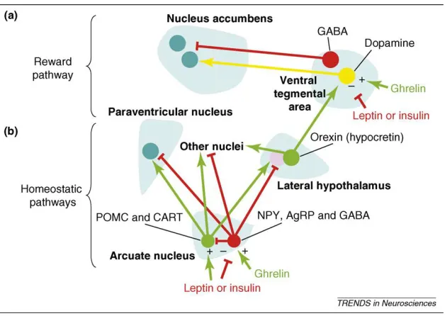
Figure 4 shows the theorized mechanism for how the homeostatic pathway interacts with the
reward pathway (Palmiter 2007). In addition, recent studies have shown that melanocortin
agonists injected directly into the VTA cause a decrease in food intake (Roseberry, personal
communication), however, more information is required to fully understand the role
melanocortins play in the mesolimbic DA system to affect feeding.
1.7 Research Goals
I will create short-hairpin RNAs (shRNAs) that target MC3R and MC4R separately. These
shRNAs will be used in in vitro co-transfections with their target receptors to examine
Figure 4. A sample of the known relationships between the homeostatic and reward pathways
2 METHODS/EXPERIMENTAL PROTOCOL
2.1 Creation of shRNA targeting the MC3R and MC4R
Three 24 base pair short hairpin RNAs (shRNAs) were designed to target the MC3R and
three to target the MC4R The shRNAs were created using previously developed methods
(Hommel et al 2003) and BLASTed to ensure specificity. The sequences of each shRNA can be
seen in Table 1.
Leptin and insulin act on the ARC on POMC/CART and AgRP/NPY neurons to decrease feeding while ghrelin acts on these areas to increase feeding in the homeostatic pathway. POMC/CART and AgRP/NPY neurons both have projections to many similar locations
Table 1. Targeted mRNA sequences used to create pAAV short-hairpins
mRNA sequence MC3R
sh1 TCCCTGACCTTGGAGGACCAATTC sh2 TGCGGCGTGATGTTCATCGTCTAC sh3 ACACGGCGCACTTCAACACCTACC MC4R
sh1 TCATAAGCCTGTTGGAGAACATTC sh2 GCAGTGGACAGGTATTTCACTATC sh3 CTACATCTCTTGTCCTCAGAATCC CONTROL
CGGAATTTAGAAACCCGGCTCCAA
The control mRNA sequence was created and tested previously and has no known target
(Hommel et al 2003). Effective shRNAs were created by using a 10 nucleotide hairpin loop
(CTTCCTGTCA) to join the antisense shRNA to the corresponding sense strand sequence as
shown in Figure 5. The shRNAs were cloned into a pAAV vector using standard methods to be
used for future creation of adeno-associated viruses (AAVs). The AAV shRNA constructs will
express the shRNAs under control of the mU6 promoter, and also contain GFP under the control
of the CMV promoter to allow for high level expression of both the shRNA and the marker GFP
Figure 6. Design of AAV shRNAs
2.2 ptdTomato-MC3/4R creation
Gene fusion constructs were created containing the MC3R or MC4R cDNA fused to the
C-terminus of the tdTomato fluorescent protein in the ptdTomato-C1 vector (Clontech
Laboratories) using EcoRI and XbaI restriction enzymes and standard molecular biology
techniques. These fusion constructs will be used for in vitro studies to test shRNA ability to
suppress tdTomatoMC3/4R. The MC4R was received as a generous gift from Dr. Xin-Yun Lu,
University of Texas Health Science Center at San Antonio, and the MC3R was synthesized de
novo by GenScript. The MC3/4R portion of the tdTomatoMC3/4R fusion constructs are not in
frame so no mature MC3/4R protein is attached to the C-terminus of the functional tdTomato
protein; however, there is full mRNA expression which allows for targeting of the tdTomato
MC3/4 R mRNA by the shRNAs.
The sense strand is the 24 bp target RNA sequence followed by a hairpin loop, the antisense strand, followed by a terminating poly T tail.
Figure 5. Schematic of short-hairpin creation used in RNAi
2.3 shRNA testing in vitro
The pAAv-shRNA constructs, as described in 2.1, were co-transfected with the
tdTomatoMC3/4R fusion constructs, as described in 2.2, into HEK-293 cells to determine the
most effective shRNA to be used for in vivo experimentation. The pAAV-shRNA (5 μg) and
ptdTomatoMC3/4R (1 μg) DNAs were co-transfected into HEK-293 cells using standard
[image:28.612.71.495.319.500.2]calcium-phosphate methods (Roseberry et al 2001). Each shRNA in vitro experiment was
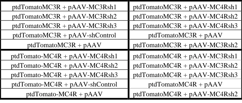
Table 2. Experimental co-tranfections into HEK-293 cells
ptdTomatoMC3R + pAAV-MC3Rsh1 ptdTomatoMC3R + pAAV-MC4Rsh1 ptdTomatoMC3R + pAAV-MC3Rsh2 ptdTomatoMC3R + pAAV-MC4Rsh2 ptdTomatoMC3R + pAAV-MC3Rsh3 ptdTomatoMC3R + pAAV-MC4Rsh3
ptdTomatoMC3R + pAAV-shControl ptdTomatoMC3R + pAAV ptdTomatoMC3R + pAAV ptdTomatoMC3R + pAAV-MC3Rsh2 ptdTomato-MC4R + pAAV-MC4Rsh1 ptdTomatoMC4R + pAAV-MC3Rsh1 ptdTomato-MC4R + pAAV-MC4Rsh2 ptdTomatoMC4R + pAAV-MC3Rsh2 ptdTomato-MC4R + pAAV-MC4Rsh3 ptdTomatoMC4R + pAAV-MC3Rsh3
ptdTomato-MC4R + pAAV-shControl ptdTomatoMC4R + pAAV ptdTomato-MC4R + pAAV ptdTomatoMC4R + pAAV-MC4Rsh2
performed 3-4 times and included an empty pAAV and a pAAV control shRNA as internal
controls. Cells were plated to polylysine covered cover-slips one day post-transfection and were
then fixed with methanol/acetone and mounted to slides three days post-transfection.
As seen in Table 2, specificity was tested by completing co-transfections with
MC3RshRNAs with tdTomatoMC4R and MC4RshRNAs with the tdTomatoMC4R. These
experiments also had an empty pAAV vector as a negative control and a receptor-specific
short-hairpin as a positive control.
2.4 Data Analysis and Statistics
Slides containing the transfected cells were viewed using a Nikon E800 upright microscope.
Slides were randomly sampled and approximately ten images were taken per transfection. Image
acquisition parameters were the same for all treatments in each individual experiment. Images
were quantified using ImageJ software (National Institute of Health, Bethesda, MD). Positive
cells were selected using brightness thresholds and cell counter functions, and all measurements
were confirmed by experimenter. Paired t-tests were used to compare the number of GFP and
tdTomato positive cells for each treatment. One-way ANOVA with Holm-Sidak post-hoc tests
were used to compare the level of tdTomato knockdown observed for each pAAV construct.
3 RESULTS
Three pAAV-shRNAs were created targeting the MC3R or MC4R, and each shRNA was
tested in vitro for its effectiveness in knocking down MC3R or MC4R. The knockdown was
measured by comparing the number of cells expressing tdTomato to the number of cells
expressing GFP, which is expressed by pAAV, and is used to mark all transfected cells. The
MC3/4Rs are fused to the C-terminus of the tdTomato and will produce one mRNA that will be
targeted by the shRNAs. In a successful knockdown, the shRNAs, co-expressed with GFP, will
knockdown the tdTomatoMC3/4R fusion reducing tdTomato fluorescence. In an unsuccessful
knockdown, both GFP and tdTomato fluorescence will be present. As mentioned previously, the
MC3/4R is not in frame with the tdTomato, and no functional receptor protein will be created;
however, these experiments will remain unaffected because the tdTomato fluorescent protein
will still be expressed. In each experiment, an empty pAAV vector and a scrambled control
Initially, the MC3R shRNAs were tested (Figure 7-9). The three shRNAs targeting MC3R
significantly decreased tdTomatoMC3R expression (Figure 7-9) to varying degrees. MC3Rsh2
had the greatest tdTomato knockdown with 85.5% followed by MC3Rsh3 with 78.9% and
MC3Rsh1 with 70.8% (Figure 9). Finally, as expected, the control shRNA had no effect on
Figure 7. Sample images showing effectiveness of the MC3R shRNAs on tdTomatoMC3R expression
Green cells indicate GFP expressed by the pAAV-shRNA, and red cells indicate
Figure 8. MC3R shRNAs decrease expression of tdTomatoMC3R in HEK-293 cells
Figure 9. Extent of knockdown of the tdTomatoMC3R by the MC3R shRNAs
* * * 0 50 100 150 200 250 300 350 400 450
Empty shControl MC3Rsh1 MC3Rsh2 MC3Rsh3
To tal n u m b e r o f Ce lls GFP tdTomato * * * 0 10 20 30 40 50 60 70 80 90 100
Empty shControl MC3Rsh1 MC3Rsh2 MC3Rsh3
% td To m ato kn o ckd o wn
Total number of GFP-positive (Light grey) and tdTomato-positive cells (Dark grey) following co-transfection of the shRNAs with the tdTomatoMC3R. ‘Empty’ refers to pAAV vector with no shRNA. *p < 0.05; n=4.
[image:32.612.112.501.423.629.2]Next, MC4R shRNAs were tested to investigate their ability to block expression of the
tdTomatoMC4R As with the MC3R, all MC4R shRNAs decreased tdTomato expression to
varying degrees (Figure 10-12). MC4Rsh3 was the most effective (90.4% knockdown) followed
by MC4Rsh2 (88.6% knockdown) and MC4Rsh1 (73.7% knockdown). Finally, similar to
MC3R, the control shRNA had no effect on expression of the tdTomatoMC4R protein (Figure
Figure 10. Sample images showing effectiveness of the MC4R shRNAs on tdTomatoMC4R expression
Figure 11. MC4R shRNAs decrease expression of tdTomatoMC4R in HEK-293 cells
Figure 12. Extent of knockdown of the tdTomatoMC4R by the MC4R shRNAs
* * * 0 50 100 150 200 250 300 350 400
Empty shControl MC4Rsh1 MC4Rsh2 MC4Rsh3
To tal n u m b e r o f c e lls GFP tdTomato * * * -20 0 20 40 60 80 100
Empty shControl MC4Rsh1 MC4Rsh2 MC4Rsh3
% t d To m ato K n o ckd o wn
Total number of GFP-positive (Light grey) and tdTomato-positive cells (Dark grey) following co-transfection of the shRNAs with the tdTomatoMC4R. ‘Empty’ refers to pAAV vector with no shRNA. * p< 0.05; n=3.
[image:35.612.128.494.427.635.2]Experiments were also performed to test the specificity of the shRNAs. In these experiments
the ability of the MC3R shRNAs to affect expression of the tdTomatoMC4R was tested, as was
the ability of the MC4R shRNAs to affect expression of the tdTomatoMC3R. As shown by
Figures 13-16, none of the shRNAs affected expression of the non-targeted receptor. This
suggests that all six of the shRNAs are specific to their own receptor. Overall, pAAV-MC3Rsh2
and pAAV-MC4Rsh3 had the greatest tdTomato knockdown, and therefore will be used for
[image:36.612.107.507.292.542.2]future in vivo experimentation.
Figure 13. Sample images showing no MC3Rsh2 effect on tdTomatoMC4R
Figure 14. MC3R shRNAs do not affect expression of tdTomatoMC4R
Figure 15. Sample images showing no MC4Rsh3 effect on tdTomatoMC3R
0 50 100 150 200 250 300
Empty MC3Rsh1 MC3Rsh2 MC3Rsh3 MC4Rsh2
Total
n
u
m
b
er
of c
el
ls
GFP
tdTomato
Total number of GFP-positive (Light grey) and tdTomato-positive cells (Dark grey) following co-transfection of the MC3R shRNAs with the tdTomato-MC4R. ‘Empty’ refers to pAAV vector with no shRNA. pAAV-MC4Rsh2 was included as a positive control; n=2.
[image:37.612.119.500.392.625.2]Figure 16. MC4R shRNAs do not affect expression of tdTomatoMC3R
4 DISCUSSION
The mesolimbic DA pathway has been shown to have physiological interactions with
melanocortins (Klusa et al 1998; Alvaro et al 1996; Bronstein et al 1990) as well as melanocortin
receptor mRNA (Roselli-Rehfuss et al 1993; Kishi et al 2003; Liu et al 2003). This system has
also been shown to affect feeding via similar mechanisms (Maldonado-Irizarry et al 1995; Wang
et al 2001; Salamone 1994). Therefore, this pathway acts as a place of interest for better
understanding melanocortin’s role in feeding behaviors. Previous studies have used a variety of
techniques (MC4R-null animals, RNAi , cell-specific receptor knockouts) to show that
melanocortins affect food intake within the hypothalamus (Huszar et al 1997; Balthasar et al
2005; Garza et al 2008). None of these relatively new techniques, however, have been used to
understand whether melanocortins affect feeding through the mesolimbic DA pathway.
0 50 100 150 200 250 300 350 400
Empty MC4Rsh1 MC4Rsh2 MC4Rsh3 MC3Rsh2
To tal n u m b e r o f c e lls GFP tdTomato
In these studies, shRNAs targeting the MC3R and MC4R were designed, tested, and
resulting in varying degrees of successful knockdown. The shRNAs with the highest knockdown
were pAAV-MC3Rsh2 with 85.5% knockdown (Figure 9) and pAAV-MC4Rsh3 with 90.4%
knockdown (Figure 12), thus making them the chosen shRNAs for future use in rats in vivo.
pAAV-MC4Rsh1 is the only short-hairpin created that could be used in rat as well as
mouse animals models because of a shared coding region, acacggcccatttcaacacctacc, found at
base pair 167 in the rat. The option of using two different animal models to investigate
melanocortin’s role in mesolimbic DA pathways could add to the evidence of a conserved
pathway between species.
Previous studies have found that the U6 promoter in vitro knockdown was found to be
~80% (Makinen et al 2006). Another similar study using the U6 promoter to target MC4R in
vitro found their knockdown ranged from 86 to 94% over three different short-hairpins (Garza et
al 2008). Therefore, the 85.4 and 90.4% knockdown for MC3R and MC4R respectively seems to
be an appropriate knockdown level in vitro. Therefore, these shRNAs would be suitable
candidates for future in vivo testing. The expression of GFP and shRNA is under the control of
different promoter regions (U6 expresses shRNA; CMV expresses GFP), and effective
knockdown still occurs as seen in current and previous studies using similar shRNA constructs
(Hommel et al 2003).
The shRNA constructs have the potential to answer many questions about the
connections between melanocortins and the mesolimbic DA pathways, and their roles in a
number of behaviors including food intake. Based on important roles of both the mesolimbic DA
system and melanocortins in feeding, a likely first set of experiments would be to examine
found in high levels in the VTA, while MC4R mRNA is found in low levels in the VTA,
moderate levels in the NAc, and high levels in the CPu (Roselli-Rehfuss et al 1993; Kishi et al
2003; Liu et al 2003). In addition to the mRNA, it is known that melanocortins have functional
or physiological effects within the same regions (Klusa et al 1998; Lindblom et al 2001; Jansone
et al 2004; Alvaro et al 1996; Hsu et al 2005, 2009). Therefore, the VTA, NAc, and CPu are all
regions of interest that could be examined. While, it is known that dopamine can affect baseline
feeding, there is evidence to suggest that it affects rewarding feeding as well. Highly palatable
food causes increased c fos in the VTA (Park & Carr 1998), and inactivation of the NAc causes
increased preference for high-fat and high-sugar foods (Zhang & Kelley 1997). Thus, normal
feeding as well as reward-related feeding, such as high fat and high sugar foods, are logical areas
to be examined.
In addition to feeding-related studies, melanocortins have also been shown to have
drug-related interactions in the mesolimbic DA system (Alvaro et al 1996; Hsu et al 2005; Bronstein
et al 1990). Thus, melanocortin shRNAs could also be used to investigate drug-related behaviors,
such as locomotion, conditioned place preference, and self-administration within the mesolimbic
DA system.
There are multiple methods that could be used to investigate melanocortin effects within
the mesolimbic DA system; however, RNA interference was the best possible for the questions
addressed in this study. Genetic knockout animals, in which knockout either occurs throughout
the brain or in specific cells, are often used to investigate similar questions. It is currently
unknown, however, which neurons MC3/4Rs are located on, and thus is a disadvantage because
it cannot be used to target overall regions, i.e. VTA. Another possible method could have been
limited by the number of microinjections allotted to one animal. This limits not only the length
of the study (acute vs. chronic), but the varying types of behaviors that can be examined within
one animal. RNA interference is not without downfall however. shRNAs are not likely to create
100% receptor knockdown. Therefore the full behavioral effects of that receptor cannot be seen.
In these studies, the ability to target a specific area over a longer period of time was important,
and thus RNA interference was chosen as the methodology.
In summary, these highly effective shRNAs can be used to target various regions of the
mesolimbic DA pathway as well as to examine various behavioral effects within these regions.
These studies will allow for investigation of melanocortin effects within the mesolimbic DA
system and for further characterization of melanocortin function in respect to rewarding feeding
REFERENCES
Abizaid A, Liu ZW, Andrews ZB, Shanabrough M, Borok E, Elsworth JD, Roth RH, Sleeman MW, Picciotto MR, Tschop MH, Gao XB, Horvath TL. Ghrelin modulates the activity and synaptic input organization of midbrain dopamine neurons while promoting appetite. J Clin Invest. 2006; 116(12): 3229-39.
Ahmed SH, Kenny PJ, Koob GF, Markou A. Neurobiological evidence for hedonic allostasis associated with escalating cocaine use. Nature Neuroscience. 2002; 5: 625-6.
Alvaro JD, Tatro JB, Quillan JM, FOgliano M, Eisenhard M, Lerner MR, Nestler EJ, Duman RS. Morphine down-regulates melanocortin-4 receptor expression in brain regions that
mediate opiate addiction. Mol. Pharmacol. 1996; 50(3): 583-91.
Anand BK, Brobeck JR. Hypothalamic control of food intake in rats and cats. Yale J. Biol. Med. 1951; 24: 123-40.
Aponte Y, Atasoy D, Sternson SM. AgRP neurons are sufficient to orchestrate feeding behavior rapidly and without training. Nat Neurosci. 2011; 14(3): 351-5.
Appleyard SM, Hayward M, Young JI, Butler AA, Cone RD, Rubinstein M, Low MJ. A role for the endogenous opioid beta-endorpin in energy homeostasis. Endocrinology. 2003; 144(5): 1753-60.
Bachmann CG, Zapf A, Brunner E, Trenkwalder C. Dopaminergic treatment is associated with decreased body weight in patients with Parkinson’s disease and dyskinesias. Eur J Neurol. 2009; 16(8): 895-901.
Balthasar N, Dalgaard LT, Lee CE, Yu J, Funahashi H, Williams T, Ferreira M, Tang V,
McGovern RA, Kenny CD, Christiansen LM, Edelstein E, Choi B, Boss O, Ashchkenasi C, Zhang CY, Mountjoy K, Kishi T, Elmquist JK, Lowell BB. Divergence of
melanocortin pathways in the control of food intake and energy expenditure. Cell. 2005; 123(3): 493-505.
Baptista T, Hernandez L, Hoebel BG. Systemic sulpride increases dopamine metabolites in the lateral hypothalamus. Pharmacol Biochem Behav. 1990; 37: 227-9.
Benoit SC, Air EL, Coolen LM, Strauss R, Jackman A, et al. The catabolic action of insulin in the brain is mediated by melanocortins. J. Neurosci. 2002; 22: 9048-52.
Beyer PL, Palarino MY, Michalek D, Busenbark K, Koller WC. Weight change and body composition in patients with Parkinson’s disease. J Am Diet Assoc. 1995; 95(9): 979-83. Bittencourt JC, Presse F, Arias C, Peto C, Vaughan J, Nahon JL, Vale W, Sawchenko PE. The
melanin-concentrating hormone system of the rat brain: an immuno- and hybridization histochemical characterization. J Comp Neurol. 1992; 319: 218-245.
Bozarth MA, Wise RA. Intracranial self-stimulation of morphine into the ventral tegmental area in rats. Life Sci. 1981; 28(5): 551-5.
Bronstein DM, Przewlocki R, Akil H. Effects of morphine treatment on pro-opiomelanocortin systems in rat brain. Brain Research. 1990; 519: 102-11.
Bulter AA, Kesterson RA, Khong T, Cullen MJ, Pelleymounter MA, Dekoning J, Baetscher M, Cone R. A unique metabolic syndrome causes obesity in the melanocortin-3 receptor-deficient mouse. Endocrinology. 2000; 141(9): 3518-21.
Carelli RM, Ijames SG, Crimling AJ. Evidence that separate neural circuits in the nucleus accumbens encode cocaine versus ‘natural’ (water and food) reward. J. Neurosci. 2000; 20: 4255-66.
Chase TN, Engber TM, Mouradian MM. Contribution of dopmainergic and glutaminergic mechanisms to the pathogensis of motor response complications in Parkonson’s disease. Adv. Neurol. 1996; 69: 497-501.
Chen AS, Marsh DJ, Trumbauer ME, Frazier EG, Guan X-M, Yu H, Rosenblum CI, Vongs A, Feng Y, Cao L, Metzger JM, Strack AM, Camacho RE, Mellin TN, Nunes CN, Min W, Fisher J, Gopal-Truter S, Macintyre DE, Chen HY, Van der Ploeg LHT. Inactivation of the mouse melanocortin-3 receptor results in increased fat mass and reduced lean body mass. Nat Genet. 2000; 26: 97-102.
Chen HY, Trumbauer ME, Chen AS, Weingarth DT, Adams JR, Frazier EG, Shen Z, Marsh DJ, Feigner SD, Gaun XM, Ye Z, Nargund RP, Smith RG, Van der Ploeg LH, Howard AD, MacNeil DJ, Qian. Orexigenic action of peripheral ghrelin is mediated by neuropeptide Y and agouti-related protein. Endocrinology. 2004; 145: 2607-12.
Chhajlani V, Muceniece R, Wikberg JE. Molecular cloning of a novel human melanocortin receptor. Biochem Biophys Res Commun. 1996; 218: 638.
Cone RD. Anatomy and regulation of the central melanocortin system. Nature Neuroscience. 2005; 8(5): 571-8.
Cone RD. Studies of the physiological function of the melanocortin system. Endocrine Reviews. 2006; 27(7): 736-49.
Corander MP, Rimmington D, Challis BG, O’Rahilly S, Coll AP. Loss of agouti-related peptide does not significantly impact the phenotype of murine POMC deficiency. Endocrinology. 2011; 152(5): 1819-28.
Corbett D, Wise RA. Intracranial self-stimulation in relation to the ascending dopaminergic systems of the midbrain: a moveable electrode mapping study. Brain Research. 1980; 285: 1-15.
Cowley MA, Smart JL, Rubinstein M, Cerdan MG, Diano S, et al. Leptin activates anorexigenic POMC neurons through a neuronal network in the arcuate nucleus. Nature. 2001; 411: 480-84.
Cowley MA, Smith RG, Diano S, Tschop M, Pronchuk N, Grove KL, Strasburger CJ,
Bidlingmaier M, Esterman M, Heiman ML, Garcia-Segura LM, Nillni EA, Mendez P, Low MJ, Sotony P, Fiedman JM, Liu H, Pinto S, Colmers WF, Cone RD, Horvarth TL. The distribution and mechanism of action of ghrelin in the CNS demonstrates a movel hypothalamic circuit regulating energy homeostasis. Neuron. 2003; 37: 649-61. Date Y, Shimbara Y, Koda S, Toshinai K, Ida T, Murakami N, Miyazato M, Kokame K,
Ishizuka Y, Ishida Y, Kageyama H, Shioda S, Kangawa K, Nakazato M. Peripheral ghrelin transmits orexigenic signals through the noradrenergic pathway from the hindbrain to the hypothalamus. Cell Metab. 2006; 4(4): 323-31.
Davis JF, Choi DL, Shurdak JD, Krause EG, Fitzgerald MF, Lipton JW, Sakai RR, Benoit SC. Central melanocortins modulate mesocorticolimbic activity and food seeking behavior in the rat. Physiol Behav. 2011; 102(5): 491-5.
Di Chiara G, Imperato A. Drugs abused by humans preferentially increase synaptic dopamine concentrations in the mesolimbic system of freely moving rats. Proc. Natl. Acad. Sci. U.S.A. 1988; 85: 5274-8.
Duva MA, Tomkins EM, Moranda LM, Kaplan R, Sukhaseum A, Stanley BG. Origins of the lateral hypothalamic afferents associated with N-methyl-d-aspartic acid-elecited eating studied using reverse microdialysis of NMDA and Fluorogold. Neurosci Res. 2005; 52(1): 95-106.
Elmquist JK, Bjorbaek C, Ahima RS, Flier JS, Saper CB. Distributions of leptin receptor mRNA isoforms in the rat brain. J Comp Neurol. 1998; 395(4): 535-47.
Fadel J, Deutch AY. Anatomical substrates of orexin-dopamine interactions: lateral
hypothalamic projections to the ventral tegmental area. Neuroscience. 2002; 111: 379-87. Fan W, Boston BA, Kesterson RA, Hruby VJ, Cone RD. Role of melanocortinergic neurons in
feeding and the agouti obesity syndrome. Nature. 1997; 385: 165-168.
Farooqi IS, Yeo GS, Keogh JM, Aminian S, Jebb SA, Butler G, Cheetham T, O’Pahilly S. Dominant and recessive inheritance of morbid obesity associated with melanocortin 4 receptor deficiency. J Clin. Invest. 2000; 106: 271-279.
Figlewicz DP, et al. Intraventricular insulin increases dopamine transporter mRNA in the rat VTA/substantia nigra. Brain Res. 1994; 644: 331-334.
Figlewicz DP, Bennett J, Evans SB, Kaiyala K, Sipols AJ, Benoit SC. Intraventricular insulin and leptin reverse place preference conditioned with high-fat diet in rats. Behav Neurosci. 2004; 118(3): 479-87.
Figlewicz DP, Evans SB, Murphy J, Hoen M, Baskin DG. Expression of receptors for insulin and leptin in ventral tegmental area/substantia nigra of the rat. Brain Res. 2003; 964: 107-15.
Figlewicz DP, Higgins MS, Ng-Evans Sb, Havel PJ. Leptin reverses sucrose-condidtioned place preference in food-restricted rats. Physiol Behav. 2001; 73(1-2): 229-34.
Flegal KM, Carroll MD, Odgen CL, Curtin LR. Prevalence and trends in obesity among US adults, 1999-2008. JAMA. 2010; 303(3): 235-41.
Flier JS. Obesity wars: molecular progress confronts an expanding epidemic. Cell. 2004; 116: 337-350.
Frederich RC, Hamann A, Anderson S, Lollmann B, Lowell BB, Flier JS. Leptin levels reflect body lipid content in mice: evidence for diet-induced resistance to leptin action. Nat Med. 1995; 1(12): 1311-14.
Fry J, Finley W. The prevalence and costs of obesity in the EU. Proceedings of the Nutrition Society. 2004; 64:987-1002.
Gantz I, Konda Y, Tashiro T, Shimoto Y, Miwa H, Munzert G, Watson SJ, DelValle J, Yamada Y. J. Biol. Chem. 1993; 268(11): 8246-50.
Gantz I, Shimoto Y, Konda Y, Miwa H, Dickinson CJ, Yamada Y. Molecular cloning and characterization of the rat fifth melanocortin receptor. Biophys Res Commun. 1994; 200: 1214-20.
Garza JC, Kim Chung Sub, Liu J, Zhang W, Lu X. Adeno-associated virus-mediated knockdown of melanocortin-4 receptor in the paraventricular nucleus of the hypothalamus promotes high-fat diet-induced hyperphagia and obesity. Journal of Endocrinology. 2008; 197(3): 471-482.
Glass MJ, Billington CJ, Levine AS. Opioid and food intake: distributed functional neural pathways? Neuropeptides. 1999; 33: 360-8.
Goncalves, MAFV. Adeno-associated virus: from defective virus to effective vector. Virology Journal. 2005; 2(43): 1-17.
Griffon N, Mignon V, Facchinetti P, Diaz J, Schwartz JC, Sokoloff P. Molecular cloning and characterization of the rat fifth melanocortin receptor. Biochem Biophys Res Commun. 1994; 200: 1007-14.
Halaas JL, Gajiwala KS, Maffei M, Cogen SL, Rabinowitz D, Lallone RL, Burley SK, Friedman JM. Weight-reducing effects of the plasma protein encoded by the obese gene. Science. 1995; 269 (5223): 543-6.
Hamdi A, Porter J, Prasad C. Decreased striatal D2 dopamine receptors in obese Zucker rats: changes during aging. Brain Research. 1992; 589: 338-40.
Haskell-Luevano C, Monck EK. Agouti-related protein functions as an inverse agonist at a constitutively active brain melanocortin-4 receptor. Regul. Pept. 2001; 99(1): 1-7. Haslam DW, James PT. Obesity. The Lancet. 2005; 366(9492): 1197-209.
Hayward MD, Pintar JE, Low MJ. Selective reward deficit in mice lacking β-endorphins and enkaphalin. J Neurosci. 2002; 22(18);8251-8.
Heimer L, Zahm DS, Churchill L, Kalivas PW, Wohltmann C. Specificity in the projection patterns of accumbal core and shell in the rat. Neuroscience. 1991; 41: 89-125.
Hernandez L, Hoebel BG. Feeding and hypothalamic stimulation increase dopamine turnover in the accumbens. Physiology & Behavior. 1988; 44(4-5): 599-606.
Hernandez L, Hoebel BG. Food reward and cocaine increase extracellular dopamine in the nucleus accumbens as measured by microdialysis. Life Sci. 1988; 42(18): 1705-12. Hervieu GJ, Cluderay JE, Harrison D, Meakin J, Maycox P, Nasir S, Leslie RA. The distribution
of the mRNA and protein products of the melanin-concentrating hormone (MCH) receptor gene, slc-1, in the central nervous system of the rat. Eur J Neurosci. 2000; 12: 1194-216.
Hikosaka O, Sakamoto M, Usui S. Functional properities of monkey caudate enruons. Acticities related to expectation of target and reward. J. Neurophysiol. 1989; 61: 814-32.
Hoebel BG. Hypothalamic self-stimulation and stimulation escape in relation to feeding and mating. Fed Proc. 1979; 38(11): 2454-61.
Hommel JD, Sears RM, Georgescu D, Simmons DL, DiLeone RJ. Local gene knockdown in the brain using viral-mediated RNA interference. Nat Med. 2003; 9(12):1539-44.
Hommel JD, Trinko R, Sears RM, Georgescu D, Liu ZW, Gao XB, Thurmon JJ, Marinelli M, DiLeone RJ. Leptin receptor signaling in midbrain dopamine neurons regulates feeding. Neuron. 2006; 51(6): 801-10.
Hsu R, Taylor JR, Newton SS, Alvaro JD, Haile C, Han G, Hruby VJ, Nestler EJ, Duman RS. Blockade of melanocortin transmission inhibits cocaine reward. Eur J Neurosci. 2005; 21(8): 2233-42.
Huszar D, Lynch CA, Fairchild-Huntress V, Dunmore JH, Fang Q, Berkemeier LR, Gu W, Kesterson RA, Boston BA, Cone RD, Smith FJ, Campfield LA, Burn P, Lee F. Targeted disruption of the melanocortin-4 receptor results in obesity in mice. Cell. 1997; 88:131-141.
melanocortin-3 receptor in the regulation of the food intake. European Journal of Pharmacology. 2011; 660(1): 80-7.
Jacobowitz DM, O’Donohue TL. Alpha-melanocyte-stimulating hormone: immunohistochemical identification and mapping in neurons of rat brain. Proc. Natl. Acad. Sci. USA. 1978; 75: 6300-04.
Jansen A, Theunissen N, Slechten K, Nederkoori C, Boon B, Mulkens S, Roefs A. Overweight children overeat after exposure to food cues. Eating behavior. 2003; 4: 197-209.
Jansone D, Bergstrom L, Svirskis S, Lindblom J, Klusa V, Wikberg JES. Opposite effects of δ1-
and δ2-melanocyte stimulating hormone on regulation of the dopaminergic mesolimbic
system in rats. Neuroscience Letters. 2004; 361(1-3): 68-71.
Jog MS, Kubota Y, Connolly CI, Hillegaart V, Graybiel AM. Building neural representations of habits. Science. 1999; 286: 1745-9.
Joseph SA, Michael GJ. Efferent ACTH-IR opiocortin projections from nucleus tractus solitaries: a hypothalamic deafferentation study. Peptides. 1988; 9: 193-201.
Kaur G, Kulkarni SK. Studies on modulation of feeding behavior by atypical antipsychotics in female mice. Prog Neuropsychopharmacol Biol Psychiatry. 2002; 26: 277-85.
Kelley AE, Berridge KC. The neuroscience of natural rewards: relevance to addictive drugs. The Journal of Neuroscience. 2002; 22(9): 3309-3311.
Kennedy GC. The role of depot fat in the hypothalamic control of food intake in the rat.
Proceedings of the Royal Society of London Series B, Containing Papers of a Biological Character Royal Society (Great Britain). 1953; 140: 578-96.
Kishi T, Aschkenasi CJ, Lee CE, Mountjoy KG, Saper CB, Elmquist JK. Expression of melanocortin 4 receptor mRNA in the central nervous system of the rat. Journal of Comparative Neurology. 2003; 457(3): 213-35.
Klusa V, Svirskis S, Opmane B, Muceniece R, Skujins A, Mutulis F, Wikberg JE, Schioth HB. Evaluation of behavioral effects of neural melanocortin receptor antagonists injected ICV and in VTA in rats. Neuropeptides. 1998; 32(6): 573-80.
Koob GF. Drugs of abuse: anatomy, pharmacology, and function of reward pathways. Trends Pharmacol. Sci. 1992; 13: 177-84.
Korotkova TM, Sergeeva OA, Eriksson KS, Haas HL, Brown RE. Excitation of ventral tegmental area dopaminergic and nondopaminergic neurons by orexins/hypocretins. J Neurosci. 2003; 23: 7-11.
Krashes MJ, Koda S, Ye CP, Rogan SC, Adams AC, Cusher DS, Maratos-Flier E, Roth BL, Lowell BB. Rapid, reversible activation of AgRP neurons drives feeding behavior in mice. J Clin Invest. 2011; 121(4): 1424-8.
Krude H, Biebermann H, Luck W, Horn R, Brabant G, Gruters A. Severe early-onset obesity, adrenal insufficiency and red hair pigmentation caused by POMC mutations in humans. Nat Genet. 1998; 19(2): 155-7.
Krugel U, Schraft T, Kittner H, Kiess W, Illes P. Basal and feeding-evoked dopamine release in the rat nucleus accumbens is depressed by leptin. Eur J Pharmacol. 2003; 482(1-3): 185-7.
Labbe O, Dsamaud F, Eggerickx D, Vassart G, Parmentier M. Molecular cloning of a mouse melanocortin 5 receptor gene widely expressed in peripheral tissues. Biochem. 33: 4543-9.
Lee M, Kim A, Conwell IM, Hruby V, MayorovA, Cai M, Wardlaw SL. Effects of selective moldulation of the central melanocortin-3-receptor on food intake and hypothalamic pomc expression. Peptides. 2008; 29: 440-7.
Lenoir M, Serre F, Cantin L, Ahmed SH. Intense sweetness surpasses cocaine reward. PLoS One. 2007; 2(8): e698.
Lindblom J, Opmane B, Mutulis F, Mutule I, Petrovsha R, Klusa V, Bergstrom L, Wikberg JES. The MC4 receptor mediates alpha-MSH induced release of nucleus accumbus dopamine. Neurochemistry. 2001; 12(10): 2155-2158.
Lindvall O, Bjorklund A. The organization of ascending catecholamine neuron systems in the rat as revealed by glyoxylic acid fluorescence method. Acta Physiol. Scand. 1974; 412: 1-48. Liu H, Kishi T, Roseberry AG, Cai X, Lee CE, Montez JM, Friedman JM, Elmquist JK.
Transgenic mice expressing green fluorescent protein under the control of the melanocortin-4 receptor promoter. J Neurosci. 2003; 23(18): 7143-54.
Luilot A, LeMoal M, Simon H. Differential reactivity of dopaminergic neurons in the nucleus accumbens in response to different behavioral situation: In in vivo voltammertric study in free moving rats. Brain Res. 1986; 397: 395-400.
Maffei M, Halaas J, Ravussin E, Pratley RE, Zhang Y, Fei H, Kim S, Lollone R, Ranganathan S et al. Leptin levels in human and rodent: measurements of plasma leptin and ob RNA in obese and weight-reduced subjects. Nat Med. 1995; 1(11): 1155-61.
Makinen PI, Koponen JK, Karkkainen AM, Malm TM, Pulkkinen KH, Koistinaho J, Turunen MP, Yla-Herttuala S. Stable RNA interference: comparison of U6 and H1 promoters in endothelial cells and in mouse brain. J Gene Med. 2006; 8: 433-41.
Maldonado-Irizarry CS, Swanson CJ, Kelley AE. Glutamate receptors in the nucleus accumbens shell control feeding behavior via the lateral hypothalamus. J Neurosci. 1995; 15: 6779-88.
Mark GP, Shabani S, Dobbs LK, Hansen ST. Cholinergic modulation of mesolimbic dopamine function and reward. Physiol. Behav. 2011; 104(1): 76-81.
Marks DL, Hruby V, Brookhart G, Cone RD. The regulation of food intake by selective stimulation of the type 3 melanocortin receptor (MC3R). Peptides. 2006; 27: 259-64. Mayer J, Thomas DW. Regulation of food intake and obesity. Science. 1967; 156: 328-37. McBride WJ, Murphy JM, Ikemoto S. Localization of the brain reinformcement mechanisms:
intracranial self-administration and intracranial place-conditioning studies. Behav. Brain Res. 1999; 101: 129-52.
McGowan MK, Andrews KM, Grossman SP. Chronic intrahypothalamic infusions of insulin or insulin antibodies alter body weight and food intake in the rat. Physiol. Behav. 1992; 51: 753-65.
Mountjoy KG, Mortrud MT, Low MJ, Simerly RB, Cone RD. Localization of the melanocortin-4 receptor (MC4R) in neuroendocrine and autonomic control circuits in the brain. Mole Endocrinol. 1994; 8: 1298-308.
Naleid AM, Grace MK, Cummings DE, Levine AS. Ghrelin induces feeding in the mesolimbic reward pathway between the ventral tegmental area and the nucleus accumbens. Peptides. 2005; 26(11): 2274-9.
Nicola SM. Dopaminergic modulation of neuronal excitability in the striatum and nucleus accumbens. Annu Rev Neurosci. 2000; 23: 185-215.
Nijenhuis WA, Oosterom J, Adan RA. AgRP (83-132) acts as an inverse agonist on the human melanocortin-4 receptor. Mol Endocrinol. 2001; 15(1): 164-71.
Nyugen DM, El-Serag HB. The epidemiology of obesity. Gastroenterology Clinics of North America. 2010; 39: 1-7.
O’Donnell P, Grace AA. Synaptic interactions among excitatory afferents to NAc neurons: hippocampal gating of prefrontal cortical input. J Neurosci. 1995; 15: 3622-3639. Ogden CL, Carroll MD, Kit BK, Flegal KM. Prevalence of obesity in the United States,
2009-2010. NCHS Data Brief. 2012; 82: 1-8.
Ogden CL, Carroll MD, Kit BK, Flegal KM. Prevalence of obesity and trends in body mass index among US children and adolescents, 1999-2000. JAMA. 2012; 307(5): 483-90. Packard MG, White NM. Lesions of the caudate nucleus selectively impair ‘reference memory’
acquisition in the radial maze. Behav Neural Biol. 1990; 53: 39-50.
Palkovits M, Mezey E, Eskay RL. Pro-opiomelanocortin-derived peptides (ACTH/
β-endorphin/α-MSH) in brainstem baroreceptor areas of the rat. Brain Res. 1987; 436: 323-8.
Palmiter RD. Is dopamine a physiologically relevant mediator of feeding behavior? Trends Neurosci. 2007; 30(8): 375-81.
Panskepp J, Reilly P, Bishop P, Meeker RB, Vilberg TR, Kastin AJ. Effects of α-MSH on motivation, vigilance, and brain respiration. Pharmacol Biochem Behav. 1976; 5: 59-64. Pardini AW, et al. Distribution of insulin receptor substrate-2 in brain areas involved in energy
homeostasis. Brain Res. 2006; 1112: 169-78.
Park TH, Carr KD. Neuroanatomical patterns of fos-like immunoreactivity induced by a
apalatable meal and meal-paired environment in saline- and naltrexone-treated rats. Brain Res. 1998; 805(1-2): 169-80.
Pecina S, Berridge KC. Opioid eating site in nucleus accumbens shell mediates food intake and hedonic ‘liking’: map based on microinjection Fos plumes. Brain Res. 2000; 863: 71-86. Rada P, Tucci S, Murzi E, Hernandez L. Extracellular glutamate increases in the lateral
hypothalamus and decreases in the nucleus accumbens during feeding. Brain. 1997; 768: 338-40.
Reynolds A, Leake, D, Boese Q, Scaringe S, Marshall WS, Khvorova A. Rational siRNA design for RNA interference. Nat Biotechnol. 2004; 22(3)L326-330.
Ritz MC, Lamb RJ, Goldberg SR, Kuher MJ. Cocaine receptors on dopamine transporters are related to self-administration of cocaine. Science. 1987; 237: 1219-23.
Roberts DC, Koob GF. Disruption of cocaine self-aministration following 6-hyproxydopamine lesions of the ventral tegmental area in rats. Pharmacol. Biochem. Behav. 1982; 17: 901-4.
Roseberry AG. Personal communication. 2012.
Roseberry AG, Bunemann M, Elavunkal J, Hosey MM. Agonst-dependent delivery of M(2) muscarinic acetylcholine receptors to the cell surface after pertussis toxin treatment. Mol Pharmacol. 2001; 59(5): 1256-68.