0095-1137/08/$08.00
⫹
0
doi:10.1128/JCM.01391-08
Copyright © 2008, American Society for Microbiology. All Rights Reserved.
Validation of 24-Hour Fluconazole MIC Readings versus the CLSI
48-Hour Broth Microdilution Reference Method: Results from a
Global
Candida
Antifungal Surveillance Program
䌤
M. A. Pfaller,
1,2* L. B. Boyken,
1R. J. Hollis,
1J. Kroeger,
1S. A. Messer,
1S. Tendolkar,
1and D. J. Diekema
1,3Departments of Pathology,
1Epidemiology,
2and Internal Medicine,
3Roy J. and Lucille A. Carver College of Medicine and
College of Public Health, University of Iowa, Iowa City, Iowa 52242
Received 21 July 2008/Accepted 3 September 2008
We performed 24- and 48-h MIC determinations and disk diffusion testing of fluconazole against more than
11,000 clinical isolates of
Candida
species. By using the reference MIC breakpoints, the categorical agreement
between the 24-h and reference 48-h broth microdilution results ranged from 93.8% (all
Candida
species) to
94.9% (all
Candida
species minus
Candida krusei
), with only 0.1% very major errors (VME). The essential
agreement (within 2 log
2dilutions) between the 24-h and 48-h results was 99.6%. The categorical agreement
between the 24-h disk diffusion results and the 24-h MIC results, using the previously established breakpoints,
was 94.4%, with 0.1% VME. Both the MIC and the disk diffusion results obtained for fluconazole after only 24 h
of incubation may be used to determine the susceptibilities of
Candida
spp. to this widely used antifungal agent.
Recent studies examining the clinical utility of “real-time”
antifungal susceptibility testing in the treatment of candidemia
have shown that when such testing is available on site,
physi-cians find the results helpful and not infrequently alter therapy
on the basis of results (2, 15, 17, 18). Collins et al. (7) found
that susceptibility testing of
Candida glabrata
isolates results in
lower overall treatment costs, based on de-escalation of
ther-apy from an expensive echinocandin to fluconazole, for
pa-tients with documented fungemia. Thus, it would appear that
routine antifungal susceptibility testing can serve as an adjunct
in the treatment of candidemia in the same way that
antibac-terial testing aids in the treatment of bacantibac-terial infections (13,
19, 40).
In light of the need to provide clinicians with useful
infor-mation sooner rather than later (14, 22) and to avoid the
potentially confounding effects of trailing growth on 48-h
flu-conazole MICs (1, 37, 39), the Clinical and Laboratory
Stan-dards Institute (CLSI; formerly NCCLS) Subcommittee for
Antifungal Testing sought to determine if reading the broth
microdilution (BMD) fluconazole MIC at 24 h would produce
valid results when they were interpreted using the existing
(48-h) breakpoints (25). Reanalysis of the MIC data set that
was used to create the original CLSI M27 48-h fluconazole
susceptibility breakpoints for
Candida
spp. (32, 38) showed
good correlation between 24-h and 48-h MICs. Furthermore,
when the 48-h fluconazole breakpoints were applied to MICs
read at 24 h, the earlier reading predicted therapeutic
out-comes as accurately as the 48-h MICs: 82% success for those
episodes in which the 24-h MIC was
ⱕ
8
g/ml (susceptible [S]
isolates), 55% success for those episodes in which the MIC was
16 to 32
g/ml (susceptible dose-dependent [SDD] isolates),
and 39% success for those episodes in which the MIC was
ⱖ
64
g/ml (resistant [R] isolates) (25). Based on these results, the
CLSI Subcommittee has included the option to read
flucon-azole MICs for
Candida
species after a 24-h incubation, using
the original interpretive breakpoints, in CLSI documents
M27-A3 and M27-S3 (5, 6).
Although earlier studies of 24-h fluconazole readings in
dif-ferent data sets and with a variety of methods versus the 48-h
reference BMD method have shown similar results and
rele-vance (8, 11, 12, 28, 31, 35, 41), further evaluation of this
concept in other data sets is warranted (25). The purpose of
the present study was to provide further documentation of the
correlation between 24-h and 48-h fluconazole BMD MICs by
assessing the essential agreement (EA; calculated as the
per-cent agreement within
⫾
2 log
2dilutions of the reference MIC)
as well as the absolute categorical agreement (CA) and error
rates obtained with a large data set of 24- and 48-h MIC results
compiled in the course of global surveillance studies (27, 29,
30, 33, 34, 36). We also provide a reassessment of the
flucon-azole disk diffusion zone diameters as they relate to the 24-h
fluconazole MIC results.
MATERIALS AND METHODS
Study design.A total of 11,654 clinical isolates ofCandidaspp. isolated from blood and other normally sterile body fluids via a global network of 105 sentinel hospital sites between January 2001 and December 2006 were included in the study. All isolates were saved on agar slants and were sent to the University of Iowa College of Medicine (Iowa City) for storage and further characterization by reference identification methods and susceptibility testing against fluconazole by BMD and disk diffusion methods (4–6, 16, 23).
Organism identification.AllCandidaspecies isolates were identified at the participating institutions by the routine method used in each laboratory. Upon receipt at the University of Iowa, the isolates were subcultured onto potato dextrose agar (Remel, Lenexa, KS) and CHROMagarCandidamedium (Hardy Laboratories, Santa Maria, CA) to ensure viability and purity. Confirmation of species identification was performed with Vitek and API products (bioMerieux, St. Louis, MO) as recommended by the manufacturer or by conventional
meth-* Corresponding author. Mailing address: Medical Microbiology
Di-vision, C606 GH, Department of Pathology, University of Iowa
Col-lege of Medicine, Iowa City, IA 52242. Phone: (319) 356-8615. Fax:
(319) 356-4916. E-mail: [email protected].
䌤
Published ahead of print on 10 September 2008.
3585
on May 16, 2020 by guest
http://jcm.asm.org/
ods as required (16). Isolates were stored as water suspensions until they were used.
Susceptibility testing.Reference antifungal susceptibility testing of all 11,654 isolates was performed by BMD exactly as described in CLSI document M27-A3 (5). Fluconazole reference powder was obtained from Pfizer Pharmaceuticals (Groton, CT). Frozen BMD panels containing serial twofold dilutions of flucon-azole (range, 0.12 to 128g/ml) in RPMI 1640 medium buffered to pH 7.0 with 0.165 M morpholinepropanesulfonic acid buffer were thawed and inoculated with an organism suspension adjusted to attain a final inoculum concentration of 1.5⫻103⫾1.0⫻103cells/ml. The panels were incubated in air at 35°C and observed for the presence or absence of growth at 24 and 48 h. The fluconazole MIC was read as the lowest concentration that produced a prominent decrease in turbidity (a ca. 50% reduction in growth) relative to that of the drug-free control (5).
Disk diffusion testing of fluconazole was performed on 11,237 of the isolates as described in NCCLS document M44-A (23). Fluconazole disks (25g) were obtained from Becton Dickinson (Sparks, MD). For disk diffusion testing, 150-mm-diameter plates containing Mueller-Hinton agar (Difco Laboratories) sup-plemented with 2% glucose and methylene blue (0.5g/ml) at a depth of 4.0 mm were used. The agar surface was inoculated by using a swab dipped in a cell suspension adjusted to the turbidity of a 0.5 McFarland standard. The plates were incubated in air at 35°C and read at 24 h. Zone diameter end points were read at 80% growth inhibition by using the BIOMIC image analysis plate reader system (version 5.9; Giles Scientific, Santa Barbara, CA). MIC interpretive cri-teria for fluconazole were those published by Pfaller et al. (32) and the CLSI (5, 6) and were as follows: S, MIC ofⱕ8g/ml; SDD, MIC of 16 to 32g/ml; R, MIC ofⱖ64g/ml. The interpretive criteria for the fluconazole disk test were those published by Pfaller et al. (32) and the NCCLS/CLSI (4, 23): S, zone diameter ofⱖ19 mm; SDD, zone diameter of 15 to 18 mm; R, zone diameter of
ⱕ14 mm.
QC.Quality control (QC) was performed for BMD in accordance with CLSI documents M27-A3 and M27-S3 (6) by usingCandida kruseiATCC 6258 and Candida parapsilosisATCC 22019. QC determinations made on each day of testing were within the 24- and 48-h control limits described by the CLSI (6). QC for disk diffusion testing was performed by usingCandida albicansATCC 90028 andC. parapsilosisATCC 22019 (4, 23).
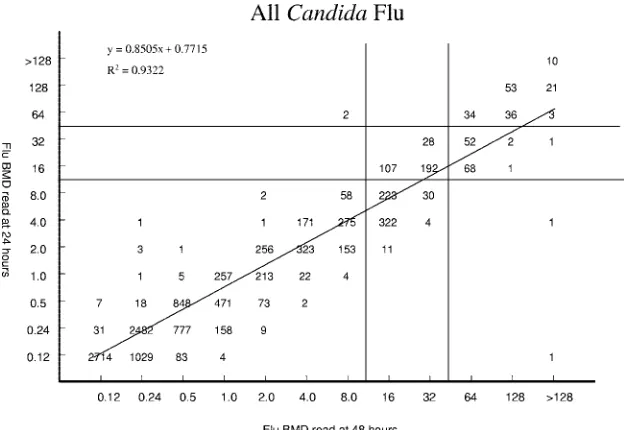
Analysis of results.The MIC results obtained for fluconazole after 24 h of incubation were compared with those obtained after 48 h of incubation by using regression statistics and a scattergram (Fig. 1). Both on-scale and off-scale results were included in the analysis. As with previous studies (31, 35), high off-scale MIC results were converted to the next highest concentration, and low off-scale MIC results were left unchanged. Discrepancies among MIC end points (24-h versus 48-h results) of more than 2 dilutions (two wells) were used to calculate the EA. The CLSI interpretive breakpoints for fluconazole were used to obtain
CA percentages between the MICs determined after 24 h of incubation and the reference 48-h BMD results. Very major errors (VME) were identified when the reference MIC indicated R and the 24-h MIC indicated S. Major errors (ME) were identified when the isolate was classified as R at 24 h of incubation and as S at 48 h. Minor errors were identified when the result of one of the readings (at 24 or at 48 h) was either S or R and that of the other was SDD.
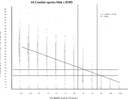
In a similar fashion, the diameters of the zones of inhibition (in millimeters) surrounding the fluconazole disks at 24 h of incubation were plotted against their respective BMD MICs read at 24 h (Fig. 2). The method of least squares was used to calculate a regression line for each comparison. The interpretive break-points defined by the CLSI (4, 6) were used to determine the CA between the disk diffusion and 24-h BMD results for fluconazole. Error rates were calculated as described above using the BMD MIC as the reference test.
RESULTS AND DISCUSSION
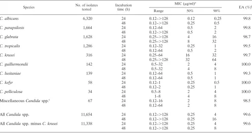
Table 1 summarizes the in vitro susceptibilities of 11,654
isolates of
Candida
spp. (14 species) to fluconazole as
deter-mined by the CLSI BMD method and read at 24 and 48 h. The
MIC results were typical of each species of
Candida
(28, 32),
with the lowest MICs at both 24 and 48 h observed for
C.
albicans
and the highest MICs observed for
C. glabrata
and
C.
krusei
. In general, the MICs read at 24 h of incubation were
twofold lower than those read at 48 h.
The overall EA between the 24-h and 48-h MIC readings
was 99.6% (91.7% were within
⫾
1 dilution). Figure 1
illus-trates the high degree of correlation between the two MIC
readings (
R
2⫽
0.9322). Of the 44 discrepancies noted between
the two readings, the MICs read after 24 h of incubation were
lower than those obtained at 48 h in 38 instances (86.4%).
Among the various species, the greatest numbers of
discrep-ancies were observed with
C. albicans
(11 discrepancies),
C.
glabrata
(21 discrepancies), and
Candida tropicalis
(6
discrep-ancies), species noted by others to exhibit the trailing
phenom-enon following incubation for 48 h (1, 24).
[image:2.585.135.447.68.283.2]Regarding the individual species of
Candida
, the EA
be-tween the 24-h and 48-h BMD MICs was
⬎
98% for each of the
14 species included in the survey (Table 1). Given the CLSI
FIG. 1. Comparison of fluconazole (Flu) BMD MICs at 24 and 48 h for 11,654
Candida
species isolates. The horizontal and vertical lines
indicate the interpretive MIC breakpoints.
on May 16, 2020 by guest
http://jcm.asm.org/
recommendation that
C. krusei
should be considered to be
intrinsically resistant to fluconazole and thus should not be
tested against this agent (5), we also determined the EA for all
isolates minus
C. krusei
; 99.6% of these results were within
⫾
2
dilution of one another.
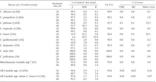
The CA between the 24-h and 48-h fluconazole MICs is
shown in Table 2. Excellent CA was observed for all
compar-isons with the exception of
C. glabrata
and
C. krusei
. The
overall CA between the 24- and 48-h results was 93.8% when
all isolates were included and 94.9% when the
C. krusei
results
were omitted. Importantly, there were only two VME
(false-susceptible results) and two ME (false-resistant results) in the
entire 11,654-isolate comparison.
Although the absolute CA for
C. glabrata
was only 67.5%,
virtually all of the errors (99.4%) were minor; they were
pre-dominantly the result of isolates determined to be S at 24 h and
SDD at 48 h (96.2% of all minor errors). This is not surprising
given the tendency of fluconazole MICs for
C. glabrata
to fall
close to the susceptible breakpoint: 59% of MICs determined
at 24 h and 78% of MICs determined at 48 h fell between 4 and
16
g/ml (data not shown). It should be noted, however, that
86% of the 156
C. glabrata
isolates that were classified as R at
the 48-h MIC determination were also R at the 24-h reading,
and only 2 isolates were S at the 24-h reading and R at the 48-h
reading (1.2% of all R
C. glabrata
isolates and 0.1% of all
C.
glabrata
isolates tested). This accuracy in detecting fluconazole
resistance among
C. glabrata
isolates is comparable or superior
to that observed with the FDA-approved commercial products
Sensititre YeastOne (TREK) and Vitek 2 yeast antifungal test
(bioMerieux) (31, 35). In view of the shift of results from SDD
(at 48 h) to S (at 24 h) for this species, the CLSI Subcommittee
has cautioned physicians and laboratorians to be aware that
when an isolate is identified as
C. glabrata
and the 24-h or 48-h
fluconazole MIC is
ⱕ
32
g/ml, patients should receive a
max-imum dosage of fluconazole (e.g., 12 mg/kg of body weight/
day) (5, 6, 25, 26).
[image:3.585.86.499.67.386.2]Disk diffusion testing of fluconazole has now been
estab-lished as a simple and inexpensive qualitative method for
de-termining the susceptibility of
Candida
to this agent, with
re-sults available within 24 h (30, 32, 34). Previously, the zone
diameter breakpoints for fluconazole disk diffusion testing
were derived by comparing the zone diameters read at 24 h
with the BMD results at 48 h by using the error rate bounded
method (21), whereby the number of discrepancies between
the zone diameter and MIC categories was minimized (32).
This process resulted in zone diameter breakpoints of
ⱖ
19 mm
(S), 15 to 18 mm (SDD), and
ⱕ
14 mm (R), with an overall CA
between the disk diffusion test results and the 48-h MIC test
results of 92.8% (2,949 isolates) and very few VME (0.1%) or
ME (0.4%) (32). Figure 2 shows the correlation between the
fluconazole disk zone diameters read at 24 h and the BMD
MIC results read at 24 h for 11,237
Candida
isolates. By using
the MIC and zone diameter breakpoints developed previously
(32), the overall CA was 94.4%, with 0.1% VME and 1.1.%
FIG. 2. Comparison of fluconazole (Flu) disk diffusion zone diameters at 24 h and BMD MICs at 24 h for 11,237
Candida
species isolates. The
horizontal and vertical lines indicate the interpretive zone diameter and MIC breakpoints, respectively.
on May 16, 2020 by guest
http://jcm.asm.org/
ME. Thus, the disk diffusion test for fluconazole performs
comparably to the 24-h MIC test without necessitating a
change in interpretive criteria.
The findings of the present study confirm and extend the
results of previous studies regarding the feasibility, accuracy,
and clinical utility of 24-h fluconazole MIC readings (8, 11, 12,
28, 31, 35, 41). Indeed, if the 24-h MIC reading were
consid-ered to be a “new test,” its performance relative to the 48-h
reference BMD test would be considered superior to those
reported for the fluconazole disk diffusion test, the Etest, the
YeastOne colorimetric method, and the Vitek 2 yeast
antifun-gal test (9, 10, 20, 35). An earlier multicenter study by
Espinel-Ingroff et al. (11) not only documented excellent EA and CA
for the comparison of 24- versus 48-h fluconazole MICs but
also found a high degree (98%) of interlaboratory
reproduc-ibility among the six participating laboratories.
Clearly, the determination of fluconazole MICs after only
24 h of incubation would provide potentially important results
in a more clinically useful time frame. Furthermore, previous
investigations have shown that the 24-h fluconazole MIC end
point correlated better than the 48-h end point with sterol
quantification (1) and with treatment outcome both clinically
(37) and in a murine model of invasive candidiasis (39). These
findings suggest that fluconazole results for isolates of
Candida
spp. with significant trailing (e.g.,
C. albicans
,
C. glabrata
, and
C. tropicalis
in the present study) should be interpreted on the
basis of the lower MIC observed at the earlier (24-h) time
point.
In addition to the data provided by Ostrosky-Zeichner et al.
(25), the clinical validity of 24-h fluconazole MICs was also
addressed in a recent study by Baddley et al. (3), in which the
authors demonstrated the association between patient
charac-teristics, MICs for
Candida
, fluconazole pharmacodynamics,
and mortality among hospitalized patients with candidemia.
These investigators confirmed our findings that fluconazole
MICs read after 24 and 48 h of incubation were very similar
(Spearman’s rank correlation coefficient, 0.91). Furthermore,
classification and regression tree (CART) analysis was used to
identify breakpoints for survival of 11.5 for a fluconazole AUC
(area under the concentration-time curve)-to-MIC ratio and of
64
g/ml for MICs read after either 24 or 48 h of incubation.
For 24-h MICs, 74% (57/77) of patients survived when the
AUC/MIC ratio or MICs were above or below these
thresh-olds, respectively (i.e.,
⬎
11.5 or
⬍
64
g/ml). Conversely, only
42.9% (3/7) of patients survived when either the AUC/MIC
ratio was less than 11.5 or the MIC exceeded 64
g/ml. Similar
results were evident for 48-h MICs. Thus, regardless of the
timing of MIC end point determination, infection with a
flu-conazole-resistant isolate was associated with increased
mor-tality. Furthermore, these studies suggest that a
clinician-con-trolled variable, fluconazole dose, may impact individual
patient survival (3). In addition to host factors, the fluconazole
dose and MICs may be helpful in managing and optimizing
outcomes for patients with candidemia (3).
[image:4.585.44.539.90.352.2]The simplicity and flexibility of disk diffusion testing makes
it a very appealing method for use in the clinical laboratory.
Although previous studies have already established the
corre-lation between the 24-h fluconazole disk zone diameter and
TABLE 1. Susceptibilities of 11,654 isolates of
Candida
spp. to fluconazole as determined by CLSI BMD methods and read after 24 and
48 h of incubation
Species No. of isolates
tested
Incubation time (h)
MIC (g/ml)a
EA (%)b
Range 50% 90%
C. albicans
6,320
24
0.12–
⬎
128
0.12
0.25
99.8
48
0.12–
⬎
128
0.25
0.5
C. parapsilosis
1,664
24
0.12–64
0.5
2
99.8
48
0.12–
⬎
128
0.5
2
C. glabrata
1,628
24
0.25–
⬎
128
4
16
98.7
48
0.25–
⬎
128
8
32
C. tropicalis
1,286
24
0.12–32
0.25
1
99.5
48
0.12–64
0.5
2
C. krusei
316
24
0.25–64
16
32
99.7
48
0.25–
⬎
128
32
64
C. guilliermondii
142
24
0.5–32
2
4
100.0
48
0.5–32
4
8
C. lusitaniae
139
24
0.12–64
0.5
1
99.3
48
0.12–64
0.5
1
C. kefyr
58
24
0.12–1
0.25
0.5
100.0
48
0.12–2
0.25
1
C. pelliculosa
34
24
0.5–8
2
4
100.0
48
1–8
4
8
Miscellaneous
Candida
spp.
c67
24
0.12–16
2
8
98.5
48
0.12–64
2
8
All
Candida
spp.
11,654
24
0.12–
⬎
128
0.25
4
99.6
48
0.12–
⬎
128
0.25
16
All
Candida
spp. minus
C. krusei
11,338
24
0.12–
⬎
128
0.25
4
99.6
48
0.12–
⬎
128
0.25
8
a
50% and 90%, MICs encompassing 50% and 90% of isolates tested, respectively.
b
Between 24- and 48-h BMD MICs.
c
IncludingC. famata(20 isolates),C. rugosa(14 isolates),C. dubliniensis(13 isolates),C. lipolytica(12 isolates), andC. zeylanoides(8 isolates).
on May 16, 2020 by guest
http://jcm.asm.org/
48-h MICs (32), the data presented here demonstrate even
better agreement between the zone diameters and 24-h
flucon-azole MICs and establish the continuing validity of the
pub-lished zone interpretive criteria.
In summary, the MICs of fluconazole can be determined
after 24 h of incubation for all species of
Candida
by using the
CLSI BMD method. The high degree of accuracy of the 24-h
reading compared to the 48-h reference method compared
favorably to those reported previously for the FDA-approved
methods Sensititre YeastOne and Vitek 2 yeast antifungal test.
Both the 24-h MIC test and the 24-h disk diffusion test reliably
identify fluconazole resistance among
Candida
spp. by using
the previously established interpretive breakpoints. The
avail-ability of fluconazole susceptibility results within a 24-h time
frame will be an important step in optimizing antifungal
ther-apy for candidiasis.
ACKNOWLEDGMENTS
Linda Elliott and Tara Schroder provided excellent support in the
preparation of the manuscript.
This work was supported in part by Pfizer, Inc., Pfizer Global
Phar-maceuticals, New York, NY.
REFERENCES
1.Arthington-Skaggs, B. A., W. Lee-Yang, M. A. Ciblak, J. P. Frade, M. E. Brandt, R. A. Hajjeh, L. H. Harrison, A. N. Sofair, and D. W. Warnock for the Candidemia Active Surveillance Group.2002. Comparison of visual and spectrophotometric methods of broth microdilution MIC end point deter-mination and evaluation of a sterol quantitation method for in vitro suscep-tibility testing of fluconazole and itraconazole against trailing and nontrailing Candidaisolates. Antimicrob. Agents Chemother.46:2477–2481. 2.Baddley, J. W., M. Patel, M. Jones, G. Cloud, A. C. Smith, and S. A. Moser.
2004. Utility of real-time antifungal susceptibility testing for fluconazole in the treatment of candidemia. Diagn. Microbiol. Infect. Dis.50:119–124. 3.Baddley, J. W., M. Patel, S. M. Bhavnani, S. A. Moser, and D. R. Andes.
2008. Association of fluconazole pharmacodynamics with mortality in pa-tients with candidemia. Antimicrob. Agents Chemother.52:3022–3028. 4.Clinical and Laboratory Standards Institute.2007. Zone diameter
interpre-tive standards, corresponding minimal inhibitory concentration (MIC) inter-pretive breakpoints, and quality control limits for antifungal disk diffusion susceptibility testing of yeasts: informational supplement, M44–S2. Clinical and Laboratory Standards Institute, Wayne, PA.
5.Clinical and Laboratory Standards Institute.2008. Reference method for broth dilution antifungal susceptibility testing of yeasts: approved standard, 3rd ed., M27–A3. Clinical and Laboratory Standards Institute, Wayne, PA. 6.Clinical and Laboratory Standards Institute.2008. Reference method for broth dilution antifungal susceptibility testing of yeasts: informational sup-plement, M27–S3. Clinical and Laboratory Standards Institute, Wayne, PA. 7.Collins, C. D., G. A. Eschenauer, S. L. Salo, and D. W. Newton.2007. To test or not to test: a cost minimization analysis of susceptibility testing for pa-tients with documentedCandida glabratafungemias. J. Clin. Microbiol.45: 1884–1888.
8.Cuenca-Estrella, M., W. Lee-Yang, M. A. Ciblak, B. A. Arthington-Skaggs, E. Mellado, D. W. Warnock, and J. L. Rodriguez-Tudela.2002. Comparative evaluation of NCCLS M27-A and EUCAST broth microdilution procedures for antifungal susceptibility testing ofCandidaspecies. Antimicrob. Agents Chemother.46:3644–3647.
9.Espinel-Ingroff, A., M. A. Pfaller, S. A. Messer, C. C. Knapp, S. Killian, H. A. Norris, and M. A. Ghannoum.1999. Multicenter comparison of the Sensi-titre YeastOne colorimetric antifungal panel with the National Committee for Clinical and Laboratory Standards M27-A reference method for testing clinical isolates of common and emergingCandidaspp.,Cryptococcusspp., and other yeast-like organisms. J. Clin. Microbiol.37:591–595.
10.Espinel-Ingroff, A., M. Pfaller, S. A. Messer, C. C Knapp, N. Holliday, and S. Killian.2004. Multicenter comparison of Sensititre YeastOne colorimet-ric antifungal panel with the NCCLS M27–A2 reference method for testing new antifungal agents against clinical isolates ofCandidaspp. J. Clin. Mi-crobiol.42:718–721.
11.Espinel-Ingroff, A., F. Barchiesi, M. Cuenca-Estrella, A. Fothergill, M. A. Pfaller, M. Rinaldi, J. L. Rodriguez-Tudela, and P. E. Verweij.2005. Com-parison of visual 24-hour and spectrophotometric 48-hour MICs to CLSI reference microdilution MICs of fluconazole, itraconazole, posaconazole, and voriconazole forCandidaspp.: a collaborative study. J. Clin. Microbiol. 43:4535–4540.
[image:5.585.43.544.80.343.2]12.Espinel-Ingroff, A., F. Barchiesi, M. Cuenca-Estrella, M. A. Pfaller, M. Rinaldi, J. L. Rodriguez-Tudela, and P. E. Verweij.2005. International and multicenter comparison of EUCAST and CLSI M27–A2 broth microdilution
TABLE 2. Categorical agreement between 24-h and 48-h CLSI BMD fluconazole MICs for 11,654 isolates of
Candida
spp.
Species (no. of isolates tested) Incubation time (h)
% of isolatesathat tested:
CA (%)
% of errors
S SDD R VME ME Minor errors
C. albicans
(6,320)
24
99.5
0.4
0.1
99.9
0.0
0.0
0.1
48
99.4
0.5
0.1
C. parapsilosis
(1,664)
24
97.5
2.3
0.2
98.2
0.0
0.0
1.8
48
96.1
3.3
0.6
C. glabrata
(1,628)
24
84.8
6.9
8.3
67.5
0.1
0.1
32.3
48
53.7
36.7
9.6
C. tropicalis
(1,286)
24
99.6
0.4
0.0
99.5
0.0
0.0
0.5
48
99.1
0.8
0.1
C. krusei
(316)
24
14.9
81.3
3.8
56.6
0.0
0.3
43.1
48
1.6
65.5
32.9
C. guilliermondii
(142)
24
97.2
2.8
0.0
95.8
0.0
0.0
4.2
48
93.0
7.0
0.0
C. lusitaniae
(139)
24
97.1
2.2
0.7
99.3
0.0
0.0
0.7
48
97.1
1.4
1.5
C. kefyr
(58)
24
100.0
0.0
0.0
100.0
0.0
0.0
0.0
48
100.0
0.0
0.0
C. pelliculosa
(34)
24
100.0
0.0
0.0
100.0
0.0
0.0
0.0
48
100.0
0.0
0.0
Miscellaneous
Candida
spp.
b(67)
24
91.0
9.0
0.0
97.0
0.0
0.0
3.0
48
89.6
9.0
1.4
All
Candida
spp. (11,654)
24
94.8
3.9
1.3
93.8
0.02
0.02
6.16
48
89.7
7.9
2.4
All
Candida
spp. minus
C. krusei
(11,338)
24
97.0
1.7
1.3
94.9
0.02
0.01
5.07
48
92.2
6.2
1.6
aIsolates were classified as S at a MIC ofⱕ8g/ml, as SDD at a MIC of 16 to 32g/ml, and as R at a MIC ofⱖ64g/ml.
bIncludingC. famata(20 isolates),C. rugosa(14 isolates),C. dubliniensis(13 isolates),C. lipolytica(12 isolates), andC. zeylanoides(8 isolates).
on May 16, 2020 by guest
http://jcm.asm.org/
methods for testing susceptibilities ofCandidaspp. to fluconazole, itracon-azole, posaconitracon-azole, and voriconazole. J. Clin. Microbiol.43:3884–3889. 13.Forrest, G.2006. Role of antifungal susceptibility testing in patient
manage-ment. Curr. Opin. Infect. Dis.19:538–543.
14.Garey, K. W., M. Rege, M. P. Pai, D. E. Mingo, K. J. Suda, R. S. Turpin, and D. T. Bearden. 2006. Time to initiation of fluconazole therapy impacts mortality in patients with candidemia: a multi-institutional study. Clin. In-fect. Dis.43:25–31.
15.Hadley, S., J. A. Martinez, L. McDermott, B. Rapino, and D. R. Snydman. 2002. Real-time antifungal susceptibility screening aids management of in-vasive yeast infections in immunocompromised patients. J. Antimicrob. Che-mother.49:415–419.
16.Hazen, K. C., and S. A. Howell.2007.Candida,Cryptococcus, and other yeasts of medical importance, p. 1762–1788.InP. R. Murray, E. J. Baron, J. H. Jorgensen, M. L. Landry, and M. A. Pfaller (ed.), Manual of clinical microbiology, 9th ed. ASM Press, Washington, DC.
17.Hospenthal, D. R., C. K. Murray, and M. G. Rinaldi.2004. The role of antifungal susceptibility in the therapy of candidiasis. Diagn. Microbiol. Infect. Dis.48:153–160.
18.Magill, S. S., C. Shields, C. L. Sears, M. Choti, and W. G. Merz.2006. Triazole cross-resistance among Candida spp.: case report, occurrence among bloodstream isolates, and implications for antifungal therapy. J. Clin. Microbiol.44:529–535.
19.Masterton, R., G. Drusano, D. L. Paterson, and G. Park.2003. Appropriate antimicrobial treatment in nosocomial infections—the clinical challenges. J. Hosp. Infect.55(Suppl. 1):1–12.
20.Matar, M. J., L. Ostrosky-Zeichner, V. L. Paetznick, J. R. Rodriguez, E. Chan, and J. H. Rex.2003. Correlation between E-test, disk diffusion, and microdilution methods for antifungal susceptibility testing of fluconazole and voriconazole. Antimicrob. Agents Chemother.47:1647–1651.
21.Metzler, C. M., and R. M. DeHaan.1974. Susceptibility tests of anaerobic bacteria: statistical and clinical considerations. J. Infect. Dis.130:588–594. 22.Morrell, M., V. J. Fraser, and M. J. Kollef.2005. Delaying empiric treatment
ofCandidabloodstream infection until positive blood culture results are obtained: a potential risk factor for mortality. Antimicrob. Agents Che-mother.49:3640–3645.
23.NCCLS.2004. Methods for antifungal disk diffusion susceptibility testing of yeasts: approved guideline, M44-A. NCCLS, Wayne, PA.
24.Ostrosky-Zeichner, L., J. H. Rex, P. G. Pappas, R. J. Hamill, R. A. Larsen, H. W. Horowitz, W. G. Powderly, N. Hyslop, C. A. Kauffman, J. Cleary, J. E. Mangino, and J. Lee.2003. Antifungal susceptibility survey of 2,000 blood-stream Candidaisolates in the United States. Antimicrob. Agents Che-mother.47:3149–3154.
25.Ostrosky-Zeichner, L., J. H. Rex, M. A. Pfaller, D. J. Diekema, B. D. Alex-ander, D. Andes, S. D. Brown, V. Chaturvedi, M. A. Ghannoum, C. C. Knapp, D. J. Sheehan, and T. J. Walsh.2008. Rationale for reading flucon-azole MICs at 24 hours rather than 48 hours when testingCandidaspp. by the CLSI M27-A2 standard method. Antimicrob. Agents Chemother., in press.
26.Pappas, P. G., J. H. Rex, J. D. Sobel, et al.2004. Guidelines for treatment of candidiasis. Clin. Infect. Dis.38:161–189.
27.Pfaller, M. A., S. A. Messer, R. J. Hollis, R. N. Jones, and D. J. Diekema. 2002. In vitro activities of ravuconazole and voriconazole compared with those of four approved systemic antifungal agents against 6,970 clinical isolates ofCandidaspp. Antimicrob. Agents Chemother.46:1723–1727. 28.Pfaller, M. A., D. J. Diekema, S. A. Messer, L. Boyken, and R. J. Hollis.2003.
Activities of fluconazole and voriconazole against 1,586 recent clinical iso-lates ofCandidaspecies determined by broth microdilution, disk diffusion,
and Etest methods: report from the ARTEMIS Global Antifungal Suscep-tibility Program, 2001. J. Clin. Microbiol.41:1440–1446.
29.Pfaller, M. A., and D. J. Diekema.2004. Twelve years of fluconazole in clinical practice: global trends in species distribution and fluconazole sus-ceptibility of bloodstream isolates of Candida. Clin. Microbiol. Infect. 10(Suppl. 1):11–23.
30.Pfaller, M. A., K. C. Hazen, S. A. Messer, L. Boyken, S. Tendolkar, R. J. Hollis, and D. J. Diekema.2004. Comparison of results of fluconazole disk diffusion testing forCandidaspecies with results from a central reference laboratory in the ARTEMIS Global Antifungal Surveillance Program. J. Clin. Microbiol.42:3607–3612.
31.Pfaller, M. A., A. Espinel-Ingroff, and R. N. Jones.2004. Clinical evaluation of the Sensititre YeastOne colorimetric antifungal plate for antifungal sus-ceptibility testing of the new triazoles voriconazole, posaconazole, and ra-vuconazole. J. Clin. Microbiol.42:4577–4580.
32.Pfaller, M. A., D. J. Diekema, and D. J. Sheehan.2006. Interpretive break-points for fluconazole andCandidarevisited: a blueprint for the future of antifungal susceptibility testing. Clin. Microbiol. Rev.19:435–447. 33.Pfaller, M. A., and D. J. Diekema.2007. Epidemiology of invasive
candidi-asis: a persistent public health problem. Clin. Microbiol. Rev.20:133–163. 34.Pfaller, M. A., D. J. Diekema, D. L. Gibbs, et al.2007. Results from the
ARTEMIS DISK Global Antifungal Surveillance Study, 1997 to 2005: an 8.5-year analysis of susceptibilities ofCandidaspecies and other yeast species to fluconazole and voriconazole determined by CLSI standardized disk dif-fusion testing. J. Clin. Microbiol.45:1735–1745.
35.Pfaller, M. A., D. J. Diekema, G. W. Procop, and M. G. Rinaldi.2007. Multicenter comparison of the VITEK 2 yeast susceptibility test with the CLSI broth microdilution reference method for testing fluconazole against Candidaspp. J. Clin. Microbiol.45:796–802.
36.Pfaller, M. A., S. A. Messer, L. Boyken, S. Tendolkar, R. J. Hollis, and D. J. Diekema.2008. Selection of a surrogate agent (fluconazole or voriconazole) for initial susceptibility testing of posaconazole againstCandidaspp.: results from a Global Antifungal Surveillance Program. J. Clin. Microbiol.46:551– 559.
37.Revankar, S. G., W. R. Kirkpatrick, R. K. McAtee, A. W. Fothergill, S. W. Redding, M. G. Rinaldi, and T. F. Patterson.1998. Interpretation of trailing endpoints in antifungal susceptibility testing by the National Committee for Clinical Laboratory Standards method. J. Clin. Microbiol.36:153–156. 38.Rex, J. H., M. A. Pfaller, J. N. Galgiani, M. S. Bartlett, A. Espinel-Ingroff,
M. A. Ghannoum, M. Lancaster, F. C. Odds, M. G. Rinaldi, T. J. Walsh, and A. L. Barry.1997. Development of interpretive breakpoints for antifungal susceptibility testing: conceptual framework and analysis of in vitro-in vivo correlation data for fluconazole, itraconazole, andCandidainfections. Clin. Infect. Dis.24:235–247.
39.Rex, J. H., P. W. Nelson, V. L. Paetznick, M. Lozano-Chiu, A. Espinel-Ingroff, and E. J. Anaissie.1998. Optimizing the correlation between results of testing in vitro and therapeutic outcome in vivo for fluconazole by testing critical isolates in a murine model of invasive candidiasis. Antimicrob. Agents Chemother.42:129–134.
40.Rex, J. H., and M. A. Pfaller.2002. Has antifungal susceptibility testing come of age? Clin. Infect. Dis.35:982–989.
41.Rodriguez-Tudela, J. L., J. P. Donnelly, M. A. Pfaller, E. Chryssantou, P. Warn, D. W. Denning, A. Espinel-Ingroff, F. Barchiesi, and M. Cuenca-Estrella.2007. Statistical analyses of correlation between fluconazole MICs forCandidaspp. assessed by standard methods set forth by the European Committee on Antimicrobial Susceptibility Testing (E.Dis. 7.1) and CLSI (M27–A2). J. Clin. Microbiol.45:109–111.