Detection in Positive-Blood-Culture Bottles: PCR–High-Resolution
Melting Analysis versus PCR-Mass Spectrometry
Kevin Jeng,a,bCharlotte A. Gaydos,aLawrence B. Blyn,cSamuel Yang,aHelen Won,aHeather Matthews,cDonna Toleno,c
Yu-Hsiang Hsieh,aKaren C. Carroll,aJustin Hardick,aBilly Masek,aAlexander Kecojevic,aRangarajan Sampath,cStephen Peterson,a and Richard E. Rothmana
Johns Hopkins University, Baltimore, Maryland, USAa; Duke University, Durham, North Carolina, USAb; and Ibis Biosciences, an Abbott Company, Carlsbad, California, USAc
Detection of pathogens in bloodstream infections is important for directing antimicrobial treatment, but current culture-based approaches can be problematic. Broad-range PCR assays which target conserved genomic motifs for postamplification amplicon analysis permit detection of sepsis-causing pathogens. Comparison of different broad-range assays is important for informing future implementation strategies. In this study, we compared positive-blood-culture bottles processed by PCR coupled to high-resolution melting curve analysis (PCR/HRMA) and PCR coupled to electrospray ionization-mass spectrometry (PCR/ESI-MS) to microbiology culture results. Genus-level concordance was 90% (confidence interval [CI], 80 to 96%) for PCR/HRMA and 94% (CI, 85 to 98%) for PCR/ESI-MS. Species-level concordance was 90% (CI, 80 to 96%) for PCR/HRMA and 86% (CI, 75 to 93%) for PCR/ESI-MS. Unlike PCR/HRMA, PCR/ESI-MS was able to resolve polymicrobial samples. Our results demonstrated that the two assays have similar overall concordance rates but may have different roles as potential adjunctive tests with stan-dard blood culture, since each method has different capabilities, advantages, and disadvantages.
D
espite ongoing research, sepsis remains a major clinical challenge. With an estimated 215,000 deaths each year in the United States, bloodstream infections cost the nation $16.7 billion annually (2). Early administration of appropriate anti-biotic therapy improves clinical outcomes for septic patients (9,11,21), but in most cases the initial clinical presentation is nonspecific, precluding targeted antibiotic selection for spe-cific pathogens. Various molecular assays which may reduce the time to detection have been developed, but standard clini-cal microbiology protocols continue to rely almost exclusively on culture-based approaches for pathogen identification, which can typically require 17 to 20 h for Gram stain results and 2 to 4 days for definitive results (17,18,22).PCR assays have relatively short turnaround times (1), can provide detailed pathogen characterization, and may detect or-ganisms missed by culture (1,7,25). By using primers that target universally conserved eubacterial regions, a wide range of species-specific internal segments can be amplified using relatively few primer pairs (19). Sequencing can be used to differentiate PCR products for pathogen diagnosis but can be slow and is prone to contamination, which can lead to false-positive results (6,16). Alternative methods that are faster may obviate complicated pre-treatment protocols to remove potential contaminants. Two broad-range PCR protocols that have been developed are PCR coupled to high-resolution melting curve analysis (HRMA) and PCR coupled to electrospray ionization-mass spectrometry (ESI-MS). Both rely on highly conserved nucleic acid targets for broad bacterial amplification. The PCR/HRMA assay relies on 3 primer targets in the universal 16S rRNA gene for panbacterial diagnosis (27), while the PCR/ESI-MS assay targets the 16S rRNA gene in addition to 23S, RNA polymerase (rpoB), and other conserved genes for bacterial detection and quantification (13,14).
HRMA measures variations in PCR product melting patterns and has the capacity to differentiate single-nucleotide differences
between similar amplicon sequences. Analyzing melting patterns unique to three hypervariable regions in the 16S rRNA gene, V1, V3, and V6, allows unambiguous species-level identification after comparing against an established library of 60 common sepsis-associated pathogens (26,27). PCR/HRMA has been shown to be relatively rapid (result turnaround time is less than 3 h) and rea-sonably simple to use. However, polymicrobial infections cannot be resolved using this method, since the presence of two distinct amplicons creates variable melting patterns. PCR/HRMA is also limited by naturally occurring genetic polymorphisms, which may create alternative melting patterns that result in organism mis-identifications.
ESI-MS measures the molecular mass of PCR products and derives unambiguous base composition data (xAxGxCxT) from amplicons to distinguish various species-specific sequences (8, 12). PCR/ESI-MS can identify more than 400 species of bacteria within a reasonable time frame of sample receipt (14). The auto-mated Plex-ID mass spectrometer allows PCR/ESI-MS to be per-formed with high-throughput capacity, permitting processing of hundreds of samples in a reasonable time frame (23). Addition-ally, PCR/ESI-MS has the capability to resolve polymicrobial in-fections, although it is more technically complex and potentially more difficult to integrate into standard care workflow. Compar-ison of broad-range PCR protocols that employ alternative meth-ods for pathogen differentiation is important for understanding
Received12 March 2012Returned for modification11 April 2012 Accepted11 June 2012
Published ahead of print1 August 2012
Address correspondence to Kevin Jeng, [email protected].
Copyright © 2012, American Society for Microbiology. All Rights Reserved.
doi:10.1128/JCM.00677-12
on May 16, 2020 by guest
http://jcm.asm.org/
the advantages of various approaches to PCR product analysis as adjunctive tests with traditional culture. The aim of this study was to compare the operational characteristics and diagnostic accu-racy of PCR/HRMA and PCR/ESI-MS for detecting microorgan-isms in blood culture bottles of positive specimens, specifically assessing concordance of each platform with reference microbiol-ogy gold standard results.
MATERIALS AND METHODS
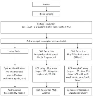
Clinical gold standard culture.This retrospective pilot study was per-formed at a tertiary-care inner city hospital using waste blood culture bottles collected between July and September 2009. Blood was drawn from patients as part of routine clinical care for suspected bloodstream infections according to treating physician orders, and 10 ml was inocu-lated into aerobic and anaerobic culture bottles and sent to the central hospital laboratory. The clinical blood culture bottles (BacT/Alert SN aer-obic, FA aeraer-obic, and SN anaerobic; bioMérieux Inc., Durham NC) used standard media and were processed using the BacT/Alert 3-D blood cul-ture system (bioMérieux, Inc.). Further identification of bacterial isolates was performed using the Phoenix microbial identification system (Bec-ton, Dickinson, Sparks, MD). None of the bottles tested contained char-coal. After reference testing was complete, the remaining waste sample was deidentified for research purposes and stored at 4°C. Pathogens iden-tified by reference testing were recorded. Explicit consent was not sought, since residual waste samples were retrospectively tested in a deidentified manner. Results from the PCR/HRMA and PCR/ESI-MS tests were com-pared to reference testing results but not used to inform clinical treat-ment. The diagnostic algorithm used by the microbiology laboratory and the study protocol, including both PCR/HRMA and PCR/ESI-MS, are
outlined inFig. 1. This study was approved by the Johns Hopkins Institu-tional Review Board.
Sample preparation and PCR for high-resolution melting curve analysis.Bacterial DNA was extracted from 500-l aliquots of blood cul-ture sample using a previously described protocol based on the Roche MagNA Pure extraction instrument (Roche Diagnostics) (26). All blood culture samples were initially stored at 4°C. Processing occurred as quickly as possible, with a maximum of 1 week of storage prior to batch DNA extraction. Three primer pairs targeting three conserved regions adjacent to hypervariable regions within the 16S rRNA gene (V1-F, V1-R, V3-F, V3-R, V6-F, and V6-R) were previously designed and validated for the PCR/HRMA assay (22,23). LCGreen dye (Idaho Technology) was added prior to PCR for high-resolution melting. PCRs involving each of the three primer pairs were performed separately and in triplicate for a total of 9 individual PCRs per sample. PCR setup, cycling conditions, and data analysis were performed as previously described (27).
High-resolution melt analysis. HRMA was performed using the
Lightscanner HR96 system (Idaho Technology, Salt Lake City, UT), uti-lizing PCR amplicons generated from DNA extracted from the blood culture samples, with fluorescence data obtained at 0.1°C intervals be-tween 60°C and 95°C. Fluorescence data from each PCR sample were analyzed in triplicate using the Light Scanner software program, version 2.0 (Idaho Technology, Salt Lake City, UT). Derivative plots of melting curves were generated and grouped by the software according to the sim-ilarity of curve shapes within a given primer set (27). By comparing curve shapes of unknown samples against those of positive-control samples, signature codes based on melting patterns across the three different primer sets utilized were generated to unambiguously identify bacterial pathogens at the species level. A previously described reference library based on these signature codes was used for a reference in this study; it
FIG 1Reference microbiology, PCR/HRMA, and PCR/ESI-MS workflow.
on May 16, 2020 by guest
http://jcm.asm.org/
[image:2.585.134.452.61.387.2]included 60 organisms commonly associated with bloodstream infections (26). For a sample to be considered a positive match, at least two of three replicates performed per region, V1, V3, and V6, were required to pro-duce melting profiles that matched those of a positive control (26,27).
Sample preparation and PCR for electrospray-ionization mass
spec-trometry.Blood culture samples (1.1 ml) were processed using a
Plex-ID bead beater, followed by a Plex-ID King Fisher instrument for bacterial DNA extraction (Abbott Molecular). PCRs were performed on the bacterial, antibiotic susceptibility, and Candida detection (BAC) assay (Ibis Biosciences, Carlsbad, CA) and were set up using a Plex-ID fluid handler (Abbott Molecular). Each assay consisted of 16 individual PCRs, 8 dedicated to broad species detection, 3 for antimi-crobial susceptibility testing, 4 forCandidadetection, and 1 for extrac-tion control. Ten microliters of DNA extract was used for each reac-tion, and an internal PCR calibrant present in every well at 100 copies per reaction served as a positive PCR control and a comparator for quantifying relative pathogen concentrations. PCR was performed un-der cycling conditions previously described (14).
Electrospray ionization mass spectrometry.Amplicons were ana-lyzed using the Plex-ID automated mass spectrometry platform (Abbott Molecular). PCR products underwent an automated weak anion ex-change protocol for purification, and accurate (61 ppm), high-resolution (M/dM.100,000 full-width half-maximum) mass spectra were measured using an established ESI-MS protocol (12). Unambiguous base composi-tion data were calculated by comparing the mass spectra of complemen-tary single-stranded PCR products. A reference library of base composi-tion data unique to over 600 pathogens has been established for previously characterized bacterial and fungal specimens (14). The amplicons from unknown samples were compared to those of organisms in the reference library, and the confidence of a match was calculated using a triangulation software package that took into account information such as the relative number of genomes per well, the number of expected PCRs agreeing with a given identification, and the base composition similarity between the reference and unknown genomic print. For these experiments, a score was calculated on a scale of 0 to 1 to indicate the relative quality of a detection. All detections with a score over 0.85 were reported; a score below 0.85 was considered indeterminate based on manufacturer recommenda-tions (Ibis Biosciences, Carlsbad, CA). BAC automated reporting was limited to different taxonomic levels (genus, group, species, etc.) for different organisms on the basis of genetic signature resolution, ex-pressed clinical needs, and/or availability of representative strains for validation. The data were derived entirely from the automated “re-search use only” reporting software, which is distinct from the soft-ware in the recently released CE-marked BAC BC assay (Ibis Biosci-ences, Carlsbad, CA).
Discordant analysis.Culture results from the microbiology labora-tory were considered the gold standard reference tests in this study. When PCR results were ambiguous or discordant, the sample was sequenced, and generated sequences were compared to known bacterial 16S rRNA sequences for definitive identification. For preparation for sequencing, the sample was initially grown on tryptic soy agar (TSA) with 5% sheep blood (TSA II; Becton, Dickinson, Sparks, MD). Single colonies were removed from each agar plate and suspended in water, and nucleic acid was extracted utilizing the Roche MagNA Pure LC robot (Roche Diagnos-tics, Indianapolis, IN) with DNA isolation kit I (Roche DiagnosDiagnos-tics, Indi-anapolis, IN). Extracted nucleic acid was amplified utilizing the V6F and V6R primers, and the PCR product was purified for sequencing utilizing the Qiagen QIAquick PCR purification kit (Qiagen, Hilden, GE). Purified PCR product was sequenced by the core facility at Johns Hopkins Univer-sity.
RESULTS
A convenience sample of 70 retrospectively collected, deidentified residual positive-blood-culture specimens was selected for PCR analysis from the study period of July to September 2009. The
blood culture specimens that were utilized for PCR analysis were the same blood culture specimens that were utilized to inform clinical care during the study period of July to September 2009. Because of the deidentified nature of our retrospective sample collection, it is unknown if duplicate positive blood cultures from the same patient were included in this study.
PCR/HRMA. Assay results were concordant with original blood culture findings for 63/70 (90.0%; 95% confidence interval, 79.9 to 95.5) samples for both genus and species identification. The identity and distribution of organisms detected by reference culture and the corresponding PCR/HRMA performance are de-tailed inTable 1and summarized inTable 2. In 5/7 discordant samples, multiple melting peaks were observed in the HRMA de-rivative plots, suggesting the presence of multiple pathogens. Fur-ther microbiologic analysis of these blood culture samples via sec-ondary culture and sequencing analysis of the discordant specimens confirmed polymicrobial growth in all five samples. The remaining 2/7 samples with discordant results were not iden-tifiable by HRMA analysis. Conventional culture testing of these 2 samples foundFusobacterium variumin one and
coagulase-nega-tiveStaphylococcus(CoNS) in the other. Although each of these
samples generated a single melting curve in HRMA, neither was associated with any specific pathogens in our reference library. The unidentified CoNS sample underwent a secondary culture and was sequenced for definitive discrepant analysis; the isolate was confirmed by both methods asStaphylococcus xylosus, an or-ganism not included in the HRMA library. Detailed results for the discrepant samples are shown inTable 3.
PCR/ESI-MS. Genus and species concordance for PCR/ ESI-MS is detailed inTable 1and summarized inTable 2. At the genus level, PCR/ESI-MS was concordant with blood culture re-sults in 66/70 (94.3%; 95% confidence interval, 85.3 to 98.1) sam-ples. In 2/4 discordant samples, conventional microbiology cul-ture was positive for Morganella morganii, while the ESI-MS reportedYersinia enterocolitica. Analysis of the mass spectrometry data revealed that the sample was a partial match forMorganella
morganiiaccording to the Plex-ID bioinformatic database, but the
Y. enterocoliticastrains in the database were a closer match.
Sub-sequent sequencing analysis confirmed the sample was positive for
M. morganii. One of four samples was positive forEnterobacter
cloacaeby culture but was reported as a polymicrobial detection
showing a near neighbor,Proteus vulgaris, and a Gram-positive organism,Enterococcus faecalis, by PCR/ESI-MS. Again, this de-tection was a partial match for Enterobacter cloacae, but the Plex-ID database recognizedProteus vulgarisas a closer match. Subsequent sequencing confirmed the presence ofE. cloacaein the sample. The final discordant sample was culture positive for group
BStreptococcus, which ESI-MS identified asKlebsiella pneumoniae.
No Gram-positive detections were made in this sample by ESI-MS; sequencing did not reveal any organisms, preventing defini-tive resolution (Table 3).
At the species level, concordance was seen in 60/70 (85.7%; 95% confidence interval, 74.8 to 92.5) samples (Table 2). Four of ten species-discordant samples were discordant at the genus level, as detailed above. In the remaining 6/10 samples, the automated Plex-ID reporting algorithm was not designed to provide species-level identification for the genera identified in the current config-uration of the assay. The genera for which species-level identifica-tion was not reported includedMicroccocus,Fusobacterium, and
on May 16, 2020 by guest
http://jcm.asm.org/
certain streptococcal groups (viridans group streptococci). De-tails of these samples are found inTable 3.
DISCUSSION
This study compared the diagnostic accuracies of two broad-range PCR assays that employ different technologies to rapidly differen-tiate unknown amplicons for pathogen identification. PCR/ HRMA and PCR/ESI-MS demonstrated similar diagnostic accu-racies in identifying pathogens in culture-positive blood samples (Genus concordance, 90% for HRMA versus 94% for ESI-MS; species concordance, 90% for HRMA versus 86% for ESI-MS). Although the study sample size was not large enough to describe species-specific concordance rates, the results of this comparison highlight some of the advantages and limitations of each assay.
Our results suggest that for pathogen identification in blood cultures, the accuracy of the PCR/HRMA platform is comparable to that of the PCR/ESI-MS approach. HRMA is an attractive ap-proach to differentiating PCR products because it is simple to perform. This methodology also utilizes relatively few consum-ables without sacrificing diagnostic performance; only 3 PCRs are required to generate data for HRMA. In contrast, 16 reactions are needed per sample for the PCR/ESI-MS BAC assay, and extracted DNA from a sample must be divided among many more reactions,
[image:4.585.45.544.77.296.2]which may be problematic for samples that already contain low numbers of an organism. PCR/HRMA can also be performed in a closed-tube fashion, reducing the risk of contamination from handling errors and further streamlining the laboratory workflow. The PCR/HRMA method has several important limitations which might restrict clinical application. First, PCR/HRMA as currently configured requires specially trained staff to be available for processing when samples become culture positive. Future technical automation could help to address this gap. In addition, the HRMA method cannot generate multiple melting curves when more than one PCR product is present in a single well, prohibiting identification of pathogens in polymicrobial samples. Clinical ap-plicability is thus limited by the prevalence of polymicrobial sam-ples under evaluation. One possible approach to addressing the inability of HRMA to differentiate polymicrobial infections is to apply PCR/HRMA analysis to subcultured bacterial isolates, al-though this would delay the time to results, offsetting the potential added value of molecular testing. The ultimate practical integra-tion of this technology into clinical practice may thus be as an adjunctive diagnostic tool, with revised testing algorithms which incorporate PCR/HRMA findings with Gram staining and other standard microbiological techniques to inform clinical decision making. Finally, the current HRMA library developed by our group is limited to only the bacterial pathogens most commonly seen in clinical settings. Thus, although the 60 organisms that the assay is formatted to detect account for more than 95% of patho-gens recovered from bloodstream infections (15), rare organisms, such as theFusobacterium variumandStaphylococcus xylosus, as were recovered from samples in this study, will still be unidentifi-able when encountered. In those cases, while the PCR will generate a signal, there will still be a reliance on traditional microbiology for definitive identification. Naturally occurring polymorphisms TABLE 2Comparison of overall performance of PCR/HRMA and PCR/
ESI-MS for both genus- and species-level concordance
Assay
No. of concordant results/no. tested (%; CIa)
Genus concordance Species concordance
[image:4.585.41.287.663.716.2]PCR/HRMA 63/70 (90.0; 79.9–95.5) 63/70 (90.0; 79.9–95.5) PCR/ESI-MS 66/70 (94.3; 85.3–98.1) 60/70 (85.7; 74.8–92.5) aCI, confidence interval.
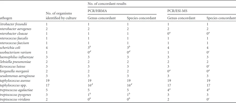
TABLE 1Numbers of organisms identified by culture and genus/species concordance of PCR/HRMA and PCR/ESI-MS
Pathogen
No. of organisms identified by culture
No. of concordant results
PCR/HRMA PCR/ESI-MS
Genus concordant Species concordant Genus concordant Species concordant
Citrobacter freundii 1 1 1 1 1
Enterobacter aerogenes 2 2 2 2 2
Enterobacter cloacae 1 1 1 0a 0a
Enterococcus faecalis 1 1 1 1 1
Enterococcus faecium 1 1 1 1 1
Escherichia coli 4 3b 3b 4 4
Fusobacterium varium 1 0d 0d 1 0c
Haemophilus influenzae 3 3 3 3 3
Klebsiella pneumoniae 2 2 2 2 2
Micrococcus luteus 3 3 3 3 0c
Morganella morganii 2 2 2 0a 0a
Pseudomonas aeruginosa 3 3 3 3 3
Staphylococcus aureus 19 19 19 19 19
Staphylococcusspp. 17 16d 16d 17 17
Streptococcus agalactiae 5 5 5 4a 4a
Streptococcus pyogenes 3 1b 1b 3 3
Streptococcus viridans 2 0b 0b 2 0c
aAll discordant samples were misidentified as an alternative organism. b
In discordant samples, HRMA melting curves suggested the presence of multiple organisms, and no individual bacterial species could be identified using HRMA alone. Samples received secondary culture, and all were found to be polymicrobial.
c
In all discordant samples, the Plex-ID automated software was not programmed to provide a species-level call based on the genus identified.
dIn all discordant samples, the organism detected by reference testing was not included in the HRMA library.
on May 16, 2020 by guest
http://jcm.asm.org/
within the regions queried by this assay also have the potential to cause organism misidentifications.
In comparison, PCR/ESI-MS is a more comprehensive diag-nostic approach, since the BAC assay employs a much more ex-tensive reference library (⬎400 species) than the PCR/HRMA as-say can and is able to resolve polymicrobial infections. Additionally, the library utilized by PCR/ESI-MS contains multi-ple, varied genetic targets, whereas the library employed by PCR/ HRMA employs only one genetic target. This makes the library for PCR/ESI-MS more effective than the PCR/HRMA library in its current configuration, since amplification, detection, and identi-fication of multiple targets from the same organism create higher confidence in a detection and identification by PCR/ESI-MS. This gives PCR/ESI-MS the ability to simultaneously characterize mul-tiple organisms and identify pathogens rarely recovered from blood samples. However, it is important to recognize that PCR/ ESI-MS assays may still be unable to make a species-level call for every genus. Misidentifications of organisms can still result from polygenic variations in closely related genera not accounted for in the library in regions queried by the assay, as was demonstrated in this study by the assay’s misidentification of samples containing
Morganella morganiiandEnterobacter cloacae.While software
up-dates that incorporate newly available genus- and species-specific sequences may improve the reporting algorithm, it also may not be possible to differentiate closely related genera using the current primers.
An additional valuable feature of the PCR/ESI-MS technology is its high-throughput capacity (8), which may be advantageous for high-volume clinical microbiology laboratories. However, more consumables are used per run, and high-throughput auto-mated processing relies on preformatted PCR plates that arrive from the manufacturer with primer pairs arranged in specific well configurations. Therefore, PCR/ESI-MS may be more complex to integrate into the clinical workflow than PCR/HRMA, since sam-ples must be batched in runs of 6 due to the preset BAC plate configurations. The current on-the-market Plex-ID instrument is
also prone to software and hardware malfunctions which could delay sample processing and result reporting. Last, as with PCR/ HRMA, PCR/ESI-MS is not a system that can be randomly ac-cessed when blood culture bottles become positive, and this is a limitation of the method and the platform. Accordingly, as with PCR/HRMA, the PCR/ESI-MS method still requires substantial investigation, investment, and evaluation before use as an adjunc-tive test or a replacement for standardized techniques, such as culture and Gram staining.
Rapid detection of bloodstream pathogens may be beneficial to clinical care. Early characterization of cultured pathogens by Gram staining is important for deescalating empirical antibiotic therapy, and knowledge of certain organisms, if identifiable through Gram staining, has a strong impact on clinical manage-ment (20,24). Broad-range PCR assays such as PCR/HRMA and PCR/ESI-MS can provide timely genus- and species-specific in-formation, allowing deescalation of empirical antibiotics based on institutional susceptibility patterns, escalation of antibiotic ther-apy if initial empirical therther-apy is found to be inadequate, or early notification of a common contaminant in the sample drawn. These data warrant more research into how these platforms may impact clinical care if employed alongside traditional testing, since the capabilities they have demonstrated in this study represent a significant improvement in time to diagnosis over current testing algorithms.
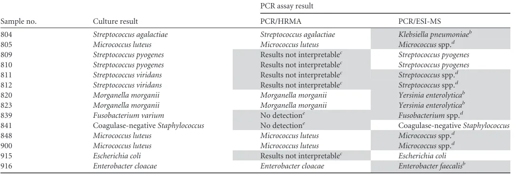
[image:5.585.40.554.79.251.2]It should also be noted that PCR/HRMA and PCR/ESI-MS are not the only molecular diagnostic methods that are in develop-ment for the detection and identification of bacterial pathogens responsible for bloodstream and other bacterial infections. Other approaches, such as matrix-assisted laser desorption ionization– time of flight mass spectrometry (MALDI-TOF/MS) and multi-plex PCR assays, could potentially influence clinical care in the future (3–5,10,13). However, as with PCR/HRMA and PCR/ESI-MS, each of these methods still requires further investigation be-fore any can be considered to have true adjunctive or replacement value for traditional microbiologic diagnostic methodologies. TABLE 3Comparison of discrepant resultsa
Sample no. Culture result
PCR assay result
PCR/HRMA PCR/ESI-MS
804 Streptococcus agalactiae Streptococcus agalactiae Klebsiella pneumoniaeb
805 Micrococcus luteus Micrococcus luteus Micrococcusspp.d
809 Streptococcus pyogenes Results not interpretablec Streptococcus pyogenes
810 Streptococcus pyogenes Results not interpretablec Streptococcus pyogenes
811 Streptococcus viridans Results not interpretablec Streptococcusspp.d
812 Streptococcus viridans Results not interpretablec Streptococcusspp.d
820 Morganella morganii Morganella morganii Yersinia enterolyticab
823 Morganella morganii Morganella morganii Yersinia enterolyticab
839 Fusobacterium varium No detectione Fusobacteriumspp.d
841 Coagulase-negativeStaphylococcus No detectione Coagulase-negativeStaphylococcus
848 Micrococcus luteus Micrococcus luteus Micrococcusspp.d
900 Micrococcus luteus Micrococcus luteus Micrococcusspp.d
915 Escherichia coli Results not interpretablec Escherichia coli
916 Enterobacter cloacae Enterobacter cloacae Enterobacter faecalisb
aDiscrepant results are shaded in gray. b
All discordant samples were misidentified as an alternative organism.
cIn discordant samples, HRMA melting curves suggested the presence of multiple organisms, and no individual bacterial species could be identified using HRMA alone. Samples
received secondary culture, and all were found to be polymicrobial.
dIn all discordant samples, the Plex-ID automated software was not programmed to provide a species-level call based on the genus identified. e
In all discordant samples, the organism detected by reference testing was not included in the HRMA library.
on May 16, 2020 by guest
http://jcm.asm.org/
Despite the potential benefits that both technologies could of-fer in diagnostic settings, culture techniques, Gram staining, and biochemical tests still offer substantial practical value. These tech-niques are simple, standard, and available in virtually all diagnos-tic settings worldwide; they are easily implemented and require little in terms of infrastructure, technical expertise, and training. Thus, although molecular method-based diagnostics, including the methodologies discussed here, offer promise for the future, their true benefits will not be realized without further investiga-tion into their capabilities, benefits, and limitainvestiga-tions.
ACKNOWLEDGMENT
L.B.B., H.M., D.T., and R.S. are full-time employees of Ibis Biosciences (a division of Abbott), the makers of the Plex-ID system.
REFERENCES
1.Andrade SS, Bispo PJ, Gales AC.2008. Advances in the microbiological diagnosis of sepsis. Shock30(Suppl. 1):41– 46.
2.Angus DC, Wax RS.2001. Epidemiology of sepsis: an update. Crit. Care Med.29:S109 –S116.
3.Banada PP, et al. 2012. Highly sensitive detection of Staphylococcus aureus directly from patient blood. PLoS One 7:e31126. doi:10.1371/ journal.pone.0031126.
4.Barczak AK, et al.2012. RNA signatures allow rapid identification of pathogens and antibiotic susceptibilities. Proc. Natl. Acad. Sci. U.S.A. doi: 10.1073/pnas.1119540109.
5.Carbonnelle E, et al.2012. Robustness of two MALDI-TOF mass spec-trometry systems for bacterial identification. J. Microbiol. Methods89: 133–136.
6.Corless CE, et al. 2000. Contamination and sensitivity issues with a real-time universal 16S rRNA PCR. J. Clin. Microbiol.38:1747–1752. 7.Dierkes C, et al.2009. Clinical impact of a commercially available
mul-tiplex PCR system for rapid detection of pathogens in patients with pre-sumed sepsis. BMC Infect. Dis.9:126. doi:10.1186/1471-2334-9-126. 8.Ecker DJ, et al. 2008. Ibis T5000: a universal biosensor approach for
microbiology. Nat. Rev. Microbiol.6:553–558.
9.Garnacho-Montero J, et al.2008. Mortality and morbidity attributable to inadequate empirical antimicrobial therapy in patients admitted to the ICU with sepsis: a matched cohort study. J. Antimicrob. Chemother.61: 436 – 441.
10. Geiss GK, et al.2008. Direct multiplexed measurement of gene expres-sion with color-coded probe pairs. Nat. Biotechnol.26:317–325. 11. Ibrahim EH, Sherman G, Ward S, Fraser VJ, Kollef MH.2000. The
influence of inadequate antimicrobial treatment of bloodstream infec-tions on patient outcomes in the ICU setting. Chest118:146 –155. 12. Jiang Y, Hofstadler SA.2003. A highly efficient and automated method of
purifying and desalting PCR products for analysis by electrospray ioniza-tion mass spectrometry. Anal. Biochem.316:50 –57.
13. Kaleta EJ, et al.2011. Comparative analysis of PCR-electrospray ioniza-tion/mass spectrometry (MS) and MALDI-TOF/MS for the identification of bacteria and yeast from positive blood culture bottles. Clin. Chem.
57:1057–1067.
14. Kaleta EJ, et al.2011. Use of PCR coupled with electrospray ionization mass spectrometry for rapid identification of bacterial and yeast blood-stream pathogens from blood culture bottles. J. Clin. Microbiol.49:345– 353.
15. Karlowsky JA, et al.2004. Prevalence and antimicrobial susceptibilities of bacteria isolated from blood cultures of hospitalized patients in the United States in 2002. Ann. Clin. Microbiol. Antimicrob.3:7. doi:10.1186/1476-0711-3-7.
16. Klaschik S, Lehmann LE, Raadts A, Hoeft A, Stuber F.2002. Compar-ison of different decontamination methods for reagents to detect low con-centrations of bacterial 16S DNA by real-time-PCR. Mol. Biotechnol.22: 231–242.
17. Mirrett S, Hanson KE, Reller LB.2007. Controlled clinical comparison of VersaTREK and BacT/ALERT blood culture systems. J. Clin. Microbiol.
45:299 –302.
18. Mirrett S, et al.2004. Controlled clinical comparison of the BacT/ALERT FN and the standard anaerobic SN blood culture medium. J. Clin. Micro-biol.42:4581– 4585.
19. Monstein HJ, Kihlstrom E, Tiveljung A.1996. Detection and identifica-tion of bacteria using in-house broad range 16S rDNA PCR amplificaidentifica-tion and genus-specific DNA hybridization probes, located within variable re-gions of 16S rRNA genes. APMIS104:451– 458.
20. Munson EL, Diekema DJ, Beekmann SE, Chapin KC, Doern GV.2003. Detection and treatment of bloodstream infection: laboratory reporting and antimicrobial management. J. Clin. Microbiol.41:495– 497. 21. Nobre V, Sarasin FP, Pugin J.2007. Prompt antibiotic administration
and goal-directed hemodynamic support in patients with severe sepsis and septic shock. Curr. Opin. Crit. Care13:586 –591.
22. Petti CA, Mirrett S, Woods CW, Reller LB.2005. Controlled clinical comparison of plastic and glass bottles of BacT/ALERT FA medium for culturing organisms from blood of adult patients. J. Clin. Microbiol.43: 1960 –1962.
23. Sampath R, et al.2007. Global surveillance of emerging influenza virus genotypes by mass spectrometry. PLoS One 2:e489. doi:10.1371/ journal.pone.0000489.
24. Uehara Y, et al.2009. Impact of reporting gram stain results from blood culture bottles on the selection of antimicrobial agents. Am. J. Clin. Pathol.132:18 –25.
25. Westh H, et al.2009. Multiplex real-time PCR and blood culture for identification of bloodstream pathogens in patients with suspected sepsis. Clin. Microbiol. Infect.15:544 –551.
26. Won H, et al.2010. Rapid identification of bacterial pathogens in positive blood culture bottles by use of a broad-based PCR assay coupled with high-resolution melt analysis. J. Clin. Microbiol.48:3410 –3413. 27. Yang S, et al.2009. Rapid identification of biothreat and other clinically
relevant bacterial species by use of universal PCR coupled with high-resolution melting analysis. J. Clin. Microbiol.47:2252–2255.