0095-1137/11/$12.00 doi:10.1128/JCM.02015-10
Copyright © 2011, American Society for Microbiology. All Rights Reserved.
Lyssavirus Detection and Typing Using Pyrosequencing
䌤
#
储
Paola De Benedictis,
1*§ Cristian De Battisti,
1§ Laurent Dacheux,
2Sabrina Marciano,
1Silvia Ormelli,
1Angela Salomoni,
1Silvia Tiozzo Caenazzo,
1Anthony Lepelletier,
2Herve
´ Bourhy,
2Ilaria Capua,
1and Giovanni Cattoli
1OIE and National Collaborating Centre for Diseases at the Animal-Human Interface, Istituto Zooprofilattico Sperimentale delle Venezie, Viale dell’Universita` 10, Legnaro, Padua, Italy,1and Institut Pasteur, Lyssavirus Dynamics and Host Adaptation Unit,
National Reference Centre for Rabies, WHO Collaborating Centre for Reference and Research on Rabies, 28 rue du Docteur Roux, 75724 Paris, Cedex 15, France2
Received 6 October 2010/Returned for modification 21 January 2011/Accepted 24 February 2011
Rabies is a fatal zoonosis caused by a nonsegmented negative-strand RNA virus, namely, rabies virus (RABV). Apart from RABV, at least 10 additional species are known as rabies-related lyssaviruses (RRVs), and some of them are responsible for occasional spillovers into humans. More lyssaviruses have also been detected recently in different bat ecosystems, thanks to the application of molecular diagnostic methods. Due to the
variety of the members of the genusLyssavirus, there is the necessity to develop a reliable molecular assay for
rabies diagnosis able to detect and differentiate among the existing rabies and rabies-related viruses. In the
present study, a pyrosequencing protocol targeting the 3ⴕterminus of the nucleoprotein (N) gene was applied
for the rapid characterization of lyssaviruses. Correct identification of species was achieved for each sample tested. Results from the pyrosequencing assay were also confirmed by those obtained using the Sanger sequencing method. A pan-lyssavirus one-step reverse transcription (RT)-PCR was developed within the framework of the pyrosequencing procedure. The sensitivity (Se) of the one-step RT-PCR assay was determined
by using in vitro-transcribed RNA and serial dilutions of titrated viruses. The assay demonstrated high
analytical and relative specificity (Sp) (98.94%) and sensitivity (99.71%). To date, this is the first case in which pyrosequencing has been applied for lyssavirus identification using a cheaper diagnostic approach than the one for all the other protocols for rapid typing that we are acquainted with. Results from this study indicate that this procedure is suitable for lyssavirus detection in samples of both human and animal origin.
Rabies is an acute encephalomyelitis caused by a nonseg-mented negative strand of RNA virus belonging to the genus Lyssavirustransmitted to humans by rabid animals. Although international organizations have recognized the disease as be-ing a high-priority zoonosis, rabies is still widely neglected in most developing countries. The World Health Organization (WHO) annually encounters at least 10 million people receiv-ing postexposure treatment and 55,000 human deaths per year, mostly children in Asia and Africa (23). This estimation cer-tainly needs a more accurate investigation, since modeling of human rabies in various countries where it is enzootic revealed a clearly higher incidence of the disease in areas under survey
(27, 28). The genus Lyssavirus includes 11 species, among
which is the classical rabies virus (RABV), which is distributed worldwide and one of the main causes of human rabies deaths. Ten additional species, generally known as rabies-related lys-saviruses (RRVs), are currently recognized by the Interna-tional Committee on the Taxonomy of Viruses (ICTV). These include Lagos bat virus (LBV), Mokola virus (MOKV),
Du-venhage virus (DUVV), European bat lyssavirus types 1 (EBLV-1) and 2 (EBLV-2), and Australian bat lyssavirus (ABLV). These species have established geographical niches, being distributed in bat populations worldwide, with the ex-ception of MOKV, an African lyssavirus for which the reser-voir species is still unresolved (31). Except for LBV, spillover transmission of RRV to humans has been demonstrated, in-ducing a fatal encephalomyelitis clinically indistinguishable from the one caused by RABV (31). Four new species have been classified recently for Aravan, Khujand, Irkut, and West Caucasian bat viruses, all of them isolated from bats in Central Asia, with only one laboratory-confirmed case of spillover to humans (25). In addition, at least 2 new lyssaviruses isolated in Africa have been described: a tentative new lyssavirus species (initially proposed as genotype 8) (13) and an unclassified lyssavirus, Shimoni bat virus, which seemed to be infecting a Commerson’s leaf-nosed bat (Hipposideros commersoni) in Ke-nya (24). These findings all suggest that further lyssaviruses remain to be detected.
Reliable and rapid diagnosis is a prerequisite for both mon-itoring lyssavirus distribution in animal reservoirs and identi-fying cases of rabies in humans. The use of a diagnostic method able to identify and distinguish among all lyssaviruses is pticularly needed when imported cases occur in rabies-free ar-eas (12) or when a lyssavirus succeeds in crossing the species barrier to infect an unexpected novel host (8). To date, the fluorescent antibody test (FAT) is the most widely adopted method for rabies diagnosis and is also the gold standard and is followed by virus isolation on cell culture (the rabies tissue * Corresponding author. Mailing address: Istituto Zooprofilattico
Sperimentale delle Venezie, Viale dell’Universita` 10, 35020 Legnaro, Padova, Italy. Phone: 39.049.8084385. Fax: 39.049.8084360. E-mail: [email protected].
§ These authors contributed equally to this work.
# Supplemental material for this article may be found at http://jcm .asm.org/.
䌤Published ahead of print on 9 March 2011.
储The authors have paid a fee to allow immediate free access to this article.
1932
on May 16, 2020 by guest
http://jcm.asm.org/
culture isolation test [RTCIT]), which should replace isolation in mice (the mouse inoculation test [MIT]) as the method recommended by international organizations (1, 2). The FAT is mainly a postmortem diagnostic tool since it is performed on brain specimens, not applicable to other kinds of samples (i.e., cerebrospinal fluid and saliva), and implemented only with difficulty to skin biopsy specimens (6). Its sensitivity (Se) is strongly affected by the freshness of the sample and by the viral strain (11). As a matter of fact, commercially available reagents for rabies diagnosis are designed for the detection of classical RABV. Molecular methods have been adopted recently as confirmatory assays or as first-choice methods in those cases in
whichintra vitamdiagnosis for humans is required. Molecular
protocols dedicated to RNA viral detection demonstrate good performances for human rabies diagnosis and are currently adopted as diagnostic tools. They are based on nested and real-time reverse transcription (RT)-PCR (5, 9, 19, 30) or nucleic acid sequence-based amplification (NASBA) (36, 37). Other protocols have also been developed to be used in animal diagnosis (3, 20, 35, 38, 39). However, all presently available methods have showed some limitations. In particular, hemi-nested RT (hnRT)-PCR methods are subjected to contamina-tion events due to a further manipulacontamina-tion of samples. The efficacy of probe-based real-time RT-PCR is influenced by mutations that arise during viral replication or in the case of an occurrence of a new strain (20, 30). Moreover, due to the
variety of the members of the genusLyssavirus, it is essential
for a reliable molecular assay to be able to detect but also differentiate among existent rabies and rabies-related viruses. However, almost all of the molecular techniques available (9, 19, 30, 35, 36, 38) require further sequencing of PCR products for lyssavirus typing, adding on an extra day for completing and interpreting results. The requirement for further genomic se-quencing in order to clearly define the species involved in the infection increases the time and final cost of the complete analysis.
Recently, microarray protocols for the detection of lyssavi-ruses have been able to demonstrate a high species-level con-cordance with standard reference assays (7, 18). Although po-tentially suitable for a diagnostic purpose, the method is still not used on a routine basis.
In the present study, we have applied pyrosequencing to
lyssavirus typing, targeting the 3⬘terminus of the nucleoprotein
(N) gene. The pyrosequencing and the one-step RT-PCR de-veloped for this purpose have also been validated with field samples from humans and animals.
MATERIALS AND METHODS
Viruses and bacterial strains.Selected viruses and bacteria were used to test the specificity and sensitivity of the one-step RT-PCR assay. A panel of 18 lyssaviruses (including 7 species) were used in order to test the specificity of the method. In addition, 2 unassigned rhabdoviridae, 2 flaviviridae, 1 paramyxoviri-dae, 9 herpesviriparamyxoviri-dae, 8 bacteria, and 4 protozoans were also tested (Table 1). Viral working stocks for the standardization of the assay were produced on BHK-21 (ATCC CCL-10) and BSR cells (a clone of BHK-21 cells), respectively, for RABV (CVS-11, batch Pd20) and EBLV-1 (8918FRA, batch Pd2). The median tissue culture infectious dose (TCID50/ml) of virus used in the sensitivity
tests was calculated according to the Spearman-Karber formula (26). Viruses other than lyssaviruses used in this study were propagated in different cell substrates: RK-13 (ATCC CCL-37) and equine derma primary cells for equine herpesviruses, Vero dogSLAM.tag cells (33) for canine distemper virus, MDCK cells (ATCC CCL-34) for canine herpesvirus, and Vero cells (ATCC CCL-81)
for flaviviruses. Human herpesviruses (cytomegalovirus, herpes simplex virus type 1 and 2, and varicella-zoster virus) were kindly provided by Jean-Claude Manuguerra, Institut Pasteur, Paris, France. Bacterial and protozoan strains were kindly provided by the Bacteriology and Parasitology Units of Istituto Zooprofilattico Sperimentale delle Venezie, Legnaro, Italy.
RNA extraction.Viral RNA was extracted from clinical samples and superna-tants of cell cultures as previously described (13) or by using the Nucleospin RNA II kit, according to the manufacturer’s instructions (Macherey-Nagel, Ger-many). For the latter, 100l of sample suspension was used for the extraction, and RNA was eluted in a final volume of 60l and stored at⫺80°C.
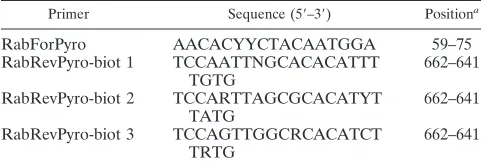
Primer set design.A lyssavirus-specific primer set was designed on a relatively conserved region of the N gene. This region was selected as the prototype for lyssavirus typing and because of the relatively large availability of N gene se-quences in the GenBank database (accession numbers of complete sese-quences used for primer design are disclosed in Table S1 in the supplemental material). We performed a multiple alignment using complete nucleotide sequences of the N gene available for historical and recent lyssaviruses., with a total of 76, 3, 4, 2, 6, 3, and 2 different sequences for RABV, LBV, MOKV, DUVV, EBLV-1, EBLV-2, and ABLV, respectively (alignment available on request). Publicly available sequences of the recently identified bat lyssaviruses, namely, Aravan, Khujand, Irkut, West Caucasian, and Shimoni bat viruses, were also aligned. The expected 603-bp amplification product comprised the 3⬘terminus of the nucleo-protein gene, which has been used widely for lyssavirus classification as the most variable region among the N genes of known species (22). Particularly, the forward primer used for both PCR amplification and pyrosequencing reactions was designed on top of the starting codon of the N gene so that the further 30-bp sequence obtained through pyrosequencing allows a rapid typing of the viral strain analyzed among the existent lyssavirus species. In order to be able to anneal with all the different species, the reverse biotinylated primer encompassed 5 different polymorphisms and was used as a mixture of three single primers, namely, Rab Rev Pyro-biot1, 2, and 3, each harboring 1, 2, and 2 distinct polymorphisms, respectively (Table 2). The reverse primer mixture was then composed by an equal concentration of all three primers, allowing for better control of the ratio of the different degenerated primers in the final mixture.
One-step RT-PCR and sequencing. The one-step RT-PCR was performed using the OneStep RT-PCR kit (Qiagen, Germany), according to the manufac-turer’s instructions. The final primer concentration applied to the PCR was 400 nM. Five microliters of isolated RNA was added to 45l of master mix, with a final volume of 50l. The following protocol was used: 30 min at 50°C and 15 min at 95°C followed by 45 cycles at 94°C for 30 s, 52°C for 30 s, and 72°C for 40 s. The pyrosequencing protocol was performed according to previously described steps (29, 32). Briefly, biotinylated amplicons were detected by gel electropho-resis using 7% silver-stained polyacrylamide gel, further captured with strepta-vidin Sepharose beads, and purified with a vacuum prep workstation according to the manufacturer’s instructions (Biotage, Uppsala, Sweden). Single-stranded biotinylated DNAs were then transferred to 40l annealing buffer containing pyrosequencing primers at a final concentration of 0.5M. Pyrosequencing reactions were performed using the Pyromark ID platform (Biotage, Uppsala, Sweden). The quality of pyrograms of 30 to 40 bp was satisfactory in all the tested samples.
[image:2.585.300.541.91.174.2]The conventional sequencing method was also applied by using the amplified product obtained by the one-step RT-PCR assay. Briefly, 603-bp sequences were generated using the BigDye Terminator, version 3.1, cycle sequencing kit (Ap-plied Biosystems, Foster City, CA) and primers used in the one-step RT-PCR assay. The products of the sequencing reactions were cleaned up using the Performa DTR ultra 96-well kit (Edge BioSystems, Gaithersburg, MD) and TABLE 2. Primer sequences targeting the lyssavirus nucleoprotein
gene 3⬘terminus
Primer Sequence (5⬘–3⬘) Positiona
RabForPyro AACACYYCTACAATGGA 59–75
RabRevPyro-biot 1 TCCAATTNGCACACATTT TGTG
662–641
RabRevPyro-biot 2 TCCARTTAGCGCACATYT TATG
662–641
RabRevPyro-biot 3 TCCAGTTGGCRCACATCT TRTG
662–641
a
Position refers to the challenge virus strain (CVS) of RABV (accession number GQ918139).
on May 16, 2020 by guest
http://jcm.asm.org/
TABLE 1. Viral and bacterial strains used in this study for specificity determination a Organism Genus Species Strain (reference no.) Host species/vector Origin Virus family Rhabdoviridae Lyssavirus RABV CVS-11 RABV GS7 1-11 Fox ( Vulpes vulpes ) France RABV Ariana 2 Dog ( Canis lupus familiaris ) Tunisia RABV Raccoon dog Raccoon dog (Nyctereutes procyonoides) Poland RABV SAG2 Vaccine France RABV SAD-B19 Vaccine Tubingen, Germany RABV Bern-C Vaccine Opava, Czech Republic RABV SAD P5 (SAD 88) Vaccine Potsdam, Germany RABV ERA Vaccine RABV HEP Vaccine RABV LEP Vaccine LBV 8619NGA Bat ( Eidolon helvum ) Nigeria MOKV 86100CAM Shrew ( Crocidura sp.) Cameroon DUVV 94286SA Bat ( Miniopterus sp.) South Africa EBLV-1 (subtype a) 122938 Bat ( Eptesicus serotinus ) France EBLV-1 (subtype b) 121411 Bat ( Eptesicus serotinus ) France EBLV-2 RV1332 Bat ( Myotis daubentonii ) United Kingdom ABLV 9810AUS Bat ( Pteropus sp.) Australia Unassigned (Le Dantec group) LDV Le Dantec virus DakHD 763 (9006SEN) Human ( Homo sapiens ) Senegal Unassigned (Ephemerovirus) KOTV Kotonkan virus Ib Ar23380 (9145NIG) Dipteran Nigeria Flaviviridae Flavivirus TBEV Tick-borne encephalitis virus, Hypr WNV West Nile virus, Eg 101 Paramyxoviridae Morbillivirus CDV Canine distemper virus, Arctic lineage Herpesviridae Varicellovirus CHV Canine herpesvirus EHV1 Equine herpesvirus 1 EHV4 Equine herpesvirus 4 SHV1 Aujeszky’s disease virus VZV Varicella-zoster virus Simplexvirus HSV1 Herpes simplex virus type 1 HSV2 Herpes simplex virus type 2 Cytomegalovirus HHV5 Human herpesvirus 5 Rhadinovirus EHV5 Equine herpesvirus 5 Bacteria Leptospira spp. Escherichia coli Brucella spp. Campylobacter spp. Salmonella spp. Rickettsia spp. Borrelia burgdorferi sensu lato Anaplasma spp. Protozoa Atoxoplasma spp. Toxoplasma spp. Plasmodium spp. Neospora spp. aLyssaviruses belonging to dif ferent species and other pathogens commonly responsible for encephalitis in mammals were tested.
on May 16, 2020 by guest
http://jcm.asm.org/
sequenced in a 16-capillary ABI Prism 3130xl genetic analyzer (Applied Biosys-tem, Foster City, CA).
Assay analytical specificity and sensitivity.The specificity of the primer set was tested on nucleic acids extracted from a panel of microorganisms which may naturally cause encephalitis or nervous signs of disease in mammals and humans (Table 1). Each strain was tested in triplicate.
The limit of detection (LoD) (14) of the one-step RT-PCR protocol was evaluated by different approaches. First, RNA was extracted from cell culture supernatant containing 10-fold serial dilutions of RABV (CVS-11) and EBLV-1 (8918FRA) titrated according to the Spearman-Karber formula (26). Second, tests were performed on brain and saliva specimens to establish whether the sample matrices could influence analytical sensitivity. Brains obtained from spe-cific pathogen-free (SPF) mice were weighed (0.1 g) and homogenized with sterile quartz sand in 1 ml (1:10, wt/vol) of phosphate-buffered saline (PBS; pH 7.4). The homogenates were then blended with the tested viruses, serially diluted, and processed for RNA extraction. Saliva was obtained from 6 healthy human volunteers, pooled, blended with the tested viruses, serially diluted, and pro-cessed for RNA extraction. Each dilution was tested in triplicate.
The LoD of the method was arbitrarily defined as the last dilution at which at least 2 out of 3 replicates of each dilution were positive. Finally, to determine the LoD of the RT-PCR protocol in terms of RNA copy numbers,in vitro -tran-scribed RNA of 7 prototype species was analyzed. Briefly, a segment of viral genome covering the entire open reading frame (ORF) of the N genes of selected viruses was generated using primers specifically designed for each spe-cies (primer sequences available on request), and the amplification products (1,480 to 1,657 bp) were cloned into the pCR-II vector using the dual promoter TOPO TA cloning kit (Invitrogen, Carlsbad, CA) according to the manufactur-er’s instructions. Recombinant plasmids were isolated from positiveE. coli col-onies using the GenElute plasmid miniprep kit (Sigma-Aldrich, St. Louis, MO). The synthetic N genes were sequenced using specific primers (sequences avail-able on request). The N-insert control plasmids were linearized using the re-striction enzyme HindIII (Roche Diagnostics, Penzberg, Germany) except for the plasmid with the MOKV insert, which was linearized with BamHI (New England BioLabs, MA). Thein vitro-transcribed RNA was obtained from the T7 promoter using the MEGAscript T7 kit (Ambion, Inc.) according to the manu-facturer’s recommendations and then quantified by a UV BioPhotometer (Ep-pendorf, Hamburg, Germany). The number of the RNA copies was calculated according to the formula reported in a previous study (17). Ten-fold dilutions of the RNA transcripts, ranging from 108
to 10⫺1
copies/l, were prepared. The LoD of the assay was determined by independent, triplicate experiments.
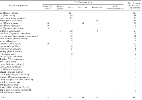
Clinical sensitivity and specificity of the assay.To evaluate whether or not the RT-PCR assay could be used as a reliable tool for rabies diagnosis, we retro-spectively analyzed 897 samples collected from animals (n⫽882) and humans (n⫽12) between 1992 and 2010 and from cell culture supernatants (n⫽3). Samples consisted of brain tissues from clinical samples (n⫽782) and from mice inoculated intracerebrally (n⫽42) with fixed strains or field samples, salivary gland specimens (n⫽1), oral swabs (n⫽24) and blood clot specimens (n⫽10) from bat surveillance, human saliva (n⫽9), skin biopsy specimens from naturally (n⫽3) and experimentally infected animals (n⫽23), and cell culture superna-tants (n⫽3). Brain samples from 28 mammalian species (including humans) were tested. The strains responsible for the infection belonged to 7 lyssavirus species, representative of 5 continents (see Table S2 in the supplemental mate-rial).
Assay performances were compared to those of the FAT, as a gold-standard test. Clinical sensitivity (Se) and specificity (Sp) and their confidence interval (CI; 95%) were calculated between the one-step RT-PCR developed and the FAT as the gold standard test. The McNemar test and Cohen’s agreement value (K) were then calculated (15).
Comparison with hnRT-PCR.A total of 45 brain samples and 23 skin biopsy specimens were analyzed in parallel by the one-step RT-PCR assay developed and the well-established hnRT-PCR previously described (9). The protocol de-scribed by Dacheux and colleagues (9) was developed and standardized in three laboratories, one in France and two in developing countries where rabies is still endemic (Cambodia and Madagascar), and it is currently used to diagnose human rabies (intra vitamand postmortem) using saliva, urine, and skin biopsy specimens.
RESULTS
Analytical laboratory evaluation. The primer set designed
was first testedin silico, using the Mega4 program (34), and
was able to anneal to all sequences analyzed, including those
from putative new species. In addition, the primer set designed was also able to detect the RNA of all 7 lyssavirus species tested. No positive results were obtained with any of the other organisms listed in Table 1, indicating high specificity.
In terms of the N gene copy number, the LoD was 10 gene
copies/l of starting material ofin vitro-transcribed RNA for
RABV, MOKV, EBLV-1, and EBLV-2, 102gene copies/l for
DUVV and ABLV, and 103gene copies/l for LBV. The LoD
of the method relative to the infectious virus titer detectable
ranged between 0.5⫻10⫺0.2and 0.5⫻101.24TCID
50/ml for
CVS-11 and EBLV-1, respectively (Table 3). The alignment between the sequences of the mixture of degenerated reverse primers and of the synthetic N genes showed a total of mis-matches less or equal to 6 for species RABV, MOKV, EBLV-1, and EBLV-2, which were detected at a concentration
of 10 gene copies/l. Total mismatches observed were 7 for
LBV, DUVV, and ABLV, for which the LoD ranged between
102and 103gene copies/l.
Clinical detection of viral RNA in samples collected from naturally and experimentally infected animals and from
hu-mans.A total of 824 samples (Table 4) was analyzed by the
FAT, as the gold standard for rabies diagnosis and by the one-step RT-PCR object of this study (Table 5). One out of the 824 analyzed samples was negative by this method and positive by FAT. Eight samples were positive by this method but were not confirmed by the standard FAT method. All of them were further confirmed as RABV by both pyrosequencing and clas-sical sequencing. The hnRT-PCR (9) was applied to those 8 samples, confirming 4 of them as positive.
The one-step RT-PCR showed high relative specificity (98.94%, CI of 97.55 to 99.65) and sensitivity (99.71%, CI of 98.40 to 99.99) values in comparison with those obtained with the FAT used as a gold-standard method. The accuracy of the method was calculated as 98.90%. The agreement between the one-step RT-PCR developed and the gold-standard method (FAT) was calculated as 98.91%, with a Cohen’s kappa coef-ficient of 0.977, which corresponds to an almost perfect agree-ment between the two methods. The difference between them calculated by applying the McNemar test was considered
sta-tistically significant (2⫽5.44), as it indicated that the
differ-ent results obtained were due to intrinsic characteristics of the two methods.
[image:4.585.301.540.99.220.2]A total of 68 samples was tested in parallel by the one-step RT-PCR assay (including the 9 previous specimens for which TABLE 3. Limit of detection of the one-step RT-PCR tested for
RABV and EBLV-1 using cell culture supernatants, brain homogenates, and saliva as biological matrices
Species Strain
Titer of virus stock (TCID50/ml)
Biological matrix
Sensitivity (TCID50/ml)
RABV CVS-11 105.8 Cell culture
supernatant
0.5⫻10⫺0.2
Brain 0.5⫻10⫺0.2
Saliva 0.5⫻10⫺0.2
EBLV-1 8918FRA 106.24 Cell culture
supernatant
0.5⫻101.24
Brain 0.5⫻101.24
Saliva 0.5⫻101.24
on May 16, 2020 by guest
http://jcm.asm.org/
discrepant results were obtained with the FAT) and the pre-viously described hnRT-PCR (9). A total of 62 of them (91.18%) showed complete agreement between the two meth-ods (Table 5).
Rapid differentiation of seven lyssavirus species through
pyrosequencing.Pyrosequencing was applied to all 375
sam-ples that tested positive by the one-step RT-PCR. Using the pyrosequencing approach, taxonomic classification of all seven lyssavirus species was possible (see Table S2 in the
supplemen-tal material). Results obtained by using pyrosequencing were directly analyzed by the software and clearly reported as an Identifire summary report (Identifire Software; Biotage, Upp-sala, Sweden). All pyrosequencing results showed complete agreement with those obtained using the Sanger method of sequencing.
DISCUSSION
In the present study, a one-step RT-PCR assay followed by pyrosequencing was developed to potentially and
simultane-ously detect and rapidly type all known lyssaviruses. The 3⬘
terminus of the viral genome as a target for the amplified product allowed us to properly identify the virus involved in the infection, either by applying pyrosequencing or classical se-quencing. We demonstrated that the pyrosequencing protocol could easily differentiate among lyssavirus species, and results were in complete agreement with those obtained using the Sanger sequencing method.
[image:5.585.47.543.81.432.2]To date, real-time and heminested RT (hnRT)-PCRs have been favored in the diagnosis of rabies, as they provide a better performance if compared to endpoint, not nested, protocols. However, the one-step RT-PCR developed in this study dem-onstrated high sensitivity and specificity levels and a high value TABLE 4. Summary of samples analyzed in this study
Species or supernatant
No. of samples from: No. of samples
per species or supernatant Saliva/oral
swab
Salivary
glands Brain Blood clot Skin biopsy
Cell culture/supernatant
Fox (Vulpes vulpes) 521 521
Cat (Felis catus) 89 89
Dog (Canis lupus familiaris) 67 67
Mouse (Mus musculus) 42 23 65
Bat(Myotis myotis) 10 10 20
Bat (Myotis capaccinii) 9 9
Bat (Eptesicus serotinus) 5 5
Badger (Meles meles) 1 20 3 24
Roe deer (Capreolus capreolus) 18 18
Raccoon dog (Nyctereutes procyonoides) 16 16
Stone marten (Martes foina) 10 10
Bovine (Bos taurus) 9 9
Human (Homo sapiens) 9 3 12
Caprine (Capra hircus) 6 6
Deer (Cervus elaphus) 5 5
Donkey (Equus asinus) 2 2
Ovine (Ovis aries) 2 2
Horse (Equus caballus) 1 1
Mouflon (Ovis musimon) 1 1
Ibex (Capra ibex) 1 1
Squirrel (Sciurus vulgaris) 1 1
Hare (Lepus europaeus) 1 1
Rat (Rattus norvegicus) 1 1
Polecat (Mustela putorius) 1 1
Rabbit (Oryctolagus cuniculus) 1 1
Chamois (Rupicapra rupicapra) 1 1
Honey badger (Mellivora capensis) 1 1
Jackal (Canis aureus) 1 1
Lion (Panthera leo) 1 1
Wildcat (Felis silvestris silvestris) 1 1
Prairie dog (Cynomys gunnisoni) 1 1
Cells/cell supernatants 3 3
Total 33 1 824 10 26 3 897
TABLE 5. Summary of samples analyzed by the one-step RT-PCR and FAT, as the gold standard, or the one-step RT-PCR and the
previously published hnRT-PCR methoda
No. of samples
One-step RT-PCR result
FAT hnRT-PCR
⫺ ⫹ Total ⫺ ⫹ Total
⫺ 468 1b 469 0 1b 1
⫹ 8c 347 355 5c 62 67
Total 476 348 824 5 63 68
a
See reference 9. b
Identical samples. c
Four of them were identical samples.
on May 16, 2020 by guest
http://jcm.asm.org/
[image:5.585.37.285.615.701.2]of accuracy here as well as in previously published studies (5, 9, 19, 20, 35). The method was in fact capable of detecting less
than 1 TCID50/ml and 10 gene copies/l of the synthetic RNA
of RABV.
Several advantages characterize this newly developed method, such as the rapidity in obtaining a final diagnosis, the lower cost, and the possibility to directly identify the lyssavirus species involved in the infection. A maximum time span of about 5 h from the submission to the final typing of a positive field sample is needed to analyze a suspect sample in labora-tory conditions. Starting from the amplified products, a pyro-sequencing analysis requires only 2 h to obtain the final result in almost half the time of that for classical Sanger sequencing. Pyrosequencing analysis does not need any terminator en-zymes or prior purification steps, thus resulting in limited costs per analysis if compared to classical sequencing. In fact, we have estimated the average cost (calculation made according to European standards) for the developed pyrosequencing pro-tocol between 3.87 to 6.03 euros/sample (for 96 or 25 samples analyzed/run, respectively), compared to 9.86 euros/reaction for the classical sequencing method.
We found a total of only 9 discrepant results out of the analyzed 897 samples using multiple diagnostic tests (Table 5). This occurrence is a rather expected event when a large set of clinical samples are included in a validation process, and this could have several explanations, such as the different targets and sensitivity levels of the applied protocols. Molecular meth-ods are able to reveal low infection levels, as well as low levels of contamination potentially occurring during the diagnostic process, from the collection of samples to the final step of amplification.
Although the occurrence of detecting failures could not be completely excluded, the use of the one-step RT-PCR in as-sociation with pyrosequencing presents the advantage of being a mutation-resistant method, since it is based on primers de-signed on conserved regions, while the most variable region is pyrosequenced. Any unexpected polymorphisms occurring in the target sequence could be easily identified. This is in con-trast with probe-based methods, such as real-time PCR, for which results can be negatively affected by the occurrence of viral mutations since the design of the probe is on the most variable region. In the case of pyrosequencing, the 30- to 40-nt fragment is sequenced more efficiently than that obtained by the Sanger method. The purpose of applying pyrosequencing is to give rapid information on the species responsible for the infection. Although further molecular analyses and phylogeny are beyond the aims of the method, results obtained can be easily integrated by conventional sequencing. Thus, the use of PCR products previously obtained and primers used for the one-step RT-PCR optimize and reduce time of testing. Further details about the lineage of the virus and its phylogeny will be obtained by sequencing the region amplified (603 bp).
The method that we developed has the advantage of being applicable to a variety of samples of human and animal origins and represents the main diagnostic tool when standard
proto-cols are not applicable, i.e.,intra vitamdiagnosis. The majority
of samples analyzed were brain specimens, and only 70/897 samples were from different matrices (Table 4). However, the analytical sensitivity of the method has been tested on saliva, showing the same sensitivity values as those for brain
speci-mens and cell culture supernatants (Table 3). In addition, we have analyzed a panel of skin biopsy specimens in parallel, comparing this method to the previously described hnRT-PCR (9) and obtaining an almost perfect agreement (see Table S2 in the supplemental material). We acknowledge that further eval-uation of the method on a larger panel of matrices is advisable before applying it routinely on these types of clinical speci-mens. Only 33 saliva specimens/oral swabs and 26 skin biopsy samples were tested in this study (Table 4). However, it should also be taken into account that these matrices are not regularly submitted to the laboratories, and therefore, they are not easily available for validation purposes.
We analyzed over 300 RABV representatives from 4 differ-ent contindiffer-ents, and strains belonging to LBV, MOKV, DUVV,
EBLV-1, EBLV-2, and ABLV. Moreover, results fromin silico
analyses clearly indicate that the method has the potential to detect all known lyssaviruses, including those belonging to pu-tative new species. A further screening of a wider panel rep-resentative of the worldwide diversity of known and emerging lyssaviruses should be applied in order to assess the perfor-mances of the method developed in this study. Improvements and subsequent revalidation of molecular diagnostic protocols should always be taken into consideration, not only for rabies but also in the cases of other viral diseases (4, 5, 20, 30, 40).
In the last decades, the use of molecular methods has largely been adopted as an alternative to diagnose viral diseases, prov-ing to be a valid aid to a diagnostic virological technique (10, 16). The lack of standardization and interlaboratory reproduc-ibility and quality issues, such as cross-contamination events or false-negative results, have been recognized as major con-straints to the use of molecular methods. International organ-izations are therefore extremely cautious in suggesting stan-dard molecular protocols for rabies diagnosis. The OIE and WHO recommend applying RT-PCR as a typing method only in specialized laboratories, while following, however, recom-mended validation guidelines (1, 2), and not as a routine tech-nique for postmortem diagnosis. From a more comprehensive point of view, a diagnostic approach based on the application of good laboratory practices, interlaboratory standardization, and the complementary use of both classical and molecular protocols will help overcome the intrinsic limitations of diag-nostic methods currently available for rabies (10). The use of an internal amplification control (IAC) is recommended to eventually identify false-negative samples resulting from the presence of PCR inhibitors or the degradation of the nucleic acid (21). In this regard, endogenous (i.e., a housekeeping gene) or exogenous nucleic acid (i.e., a microorganism which is usually not found in the sample type to be tested) can be used as an internal control. Such internal controls can be applied to many different tests, running independently of the specific vi-rus assay and currently available in molecular diagnostic lab-oratories. Alternatively, a competitive IAC (i.e., the target and the IAC are amplified with one common set of primers and under the same conditions in the same PCR tube) can be developed, bearing in mind that all these approaches have both advantages and limitations. For example, if a competitive IAC approach is applied, the different amplification assays will be competing for the same reagents in the same tube; thus, target detection sensitivity may be adversely affected (14, 21). Al-though at the moment molecular methods cannot completely
on May 16, 2020 by guest
http://jcm.asm.org/
replace standard techniques, they should be taken into account
as confirmatory tests in the case of inconclusive results forintra
vitamdiagnosis of human rabies and for lyssavirus character-ization. The one-step RT-PCR developed in this study, fol-lowed by pyrosequencing, was validated in compliance with international guidelines for diagnostic molecular techniques (1, 2). The method can be notably used in combination with standard methods as an early-warning detection tool, since it is capable of rapidly revealing the emergence or the introduction of a novel lyssavirus species in a given susceptible population or geographical area.
ACKNOWLEDGMENTS
This study was partially funded by the Italian Ministry of Health (RC 29/2007) and by the European Union through the FP6/ RABMEDCONTROL program.
We thank the following: Juan Echevarrìa Majo, ISC-III-Spain, Kris-tina Sajute, Lithuanian Veterinary Academy-Lithuania, and Jean-Claude Manuguerra, Institut Pasteur-Paris, France, for providing some of the samples analyzed in this study; Rachel Lavenir, Institut Pasteur-Paris, Paris, France, for her help and technical contribution; and Marzia Mancin, IZSVe-Italy, for statistical analyses. Francesca Ellero, IZSVe-Italy, is gratefully acknowledged for the revision of the manuscript.
REFERENCES
1.Anonymous.2005. WHO expert consultation on rabies: first report. World Health Organization, Geneva, Switzerland.
2.Anonymous.2010. OIE manual of diagnostic tests and vaccines for terrestrial animals. OIE, Paris, France
3.Black, E. M., J. P. Lowings, J. Smith, P. R. Heaton, and L. M. McElhinney. 2002. A rapid RT-PCR method to differentiate six established genotypes of rabies and rabies-related viruses using TaqMan technology. J. Virol. Meth-ods105:25–35.
4.Cattoli, G., et al.2009. False-negative results of a validated real-time PCR protocol for diagnosis of Newcastle disease due to genetic variability of the matrix gene. J. Clin. Microbiol.47:3791–3792. doi:10.1128/JCM.00895-09. 5.Coertse, J., J. Weyer, L. H. Nel, and W. Markotter.2010. Improved PCR
methods for the detection of African rabies and rabies-related lyssaviruses. J. Clin. Microbiol.48:3949–3955. doi:10.1128/JCM.01256-10.
6.Crepin, P., et al.1998. Intravitam diagnosis of human rabies by PCR using saliva and cerebrospinal fluid. J. Clin. Microbiol.36:1117–1121.
7.Dacheux, L., et al.2010. Application of broad-spectrum resequencing mi-croarray for genotyping rhabdoviruses. J. Virol.84:9557–9574. doi:10.1128/ JVI.00771-10.
8.Dacheux, L., et al.2009. European bat lyssavirus transmission among cats, Europe. Emerg. Infect. Dis.15:280–284.
9.Dacheux, L., et al.2008. A reliable diagnosis of human rabies based on analysis of skin biopsy specimens. Clin. Infect. Dis. 47:1410–1417. doi: 10.1086/592969.
10.Dacheux, L., et al.2010. More accurate insight into the incidence of human rabies in developing countries through validated laboratory techniques. PLoS Negl. Trop. Dis.4:e765. doi:10.1371/journal.pntd.0000765. 11.David, D., et al.2002. Rabies virus detection by RT-PCR in decomposed
naturally infected brains. Vet. Microbiol.87:111–118.
12.De Benedictis, P., et al.2008. Emergence of fox rabies in north-eastern Italy. Euro Surveill.13:pii:19033.
13.Delmas, O., et al.2008. Genomic diversity and evolution of the lyssaviruses. PLoS One3:e2057. doi:10.1371/journal.pone.0002057.
14.EPA.2004. Quality assurance/quality control guidance for laboratories per-forming PCR analyses on environmental samples. U.S. Environmental Pro-tection Agency, Cincinnati, OH.
15.Everitt, R. S.1989. Statistical methods for medical investigation. Oxford University Press, New York, NY.
16.Fooks, A. R., et al.2009. Emerging technologies for the detection of rabies virus: challenges and hopes in the 21st century. PLoS Negl. Trop. Dis.3:e530. doi:10.1371/journal.pntd.0000530.
17.Fronhoffs, S., et al.2002. A method for the rapid construction of cRNA standard curves in quantitative real-time reverse transcription polymerase chain reaction. Mol. Cell. Probes16:99–110. doi:10.1006/mcpr.2002.0405. 18.Gurrala, R., et al.2009. Development of a DNA microarray for simultaneous
detection and genotyping of lyssaviruses. Virus Res. 144:202–208. doi: 10.1016/j.virusres.2009.04.028.
19.Heaton, P. R., et al.1997. Heminested PCR assay for detection of six genotypes of rabies and rabies-related viruses. J. Clin. Microbiol.35:2762– 2766.
20.Hoffmann, B., et al.25 August 2010. Improved safety for the molecular diagnosis of classical rabies viruses using a TaqMan real-time RT-PCR “double check” strategy. J. Clin. Microbiol. doi:10.1128/JCM.00612-10. 21.Hoorfar, J., et al.2004. Practical considerations in design of internal
ampli-fication controls for diagnostic PCR assays. J. Clin. Microbiol.42:1863–1868. 22.Kissi, B., N. Tordo, and H. Bourhy.1995. Genetic polymorphism in the rabies virus nucleoprotein gene. Virology 209:526–537. doi:10.1006/ viro.1995.1285.
23.Knobel, D. L., et al.2005. Re-evaluating the burden of rabies in Africa and Asia. Bull. World Health Organ. 83:360–368. doi:/S0042-96862005000500012.
24.Kuzmin, I. V., et al.2010. Shimoni bat virus, a new representative of the
Lyssavirusgenus. Virus Res.149:197–210. doi:10.1016/j.virusres.2010.01.018.
25.Leonova, G. N., et al.2009. A fatal case of bat lyssavirus infection in Primo-rye Territory of the Russian Far East. WHO Rabies Bull. Eur.33:5–8. 26.Lorenz, R. G., and K. Bogel.1973. Calculation of titres and their significance,
p. 321–335.InM. M. Kaplan and H. Koprowski (ed.), Laboratory techniques in rabies, 3rd ed. World Health Organization, Geneva, Switzerland. 27.Ly, S., et al.2009. Rabies situation in Cambodia. PLoS Negl. Trop. Dis.
3:e511. doi:10.1371/journal.pntd.0000511.
28.Mallewa, M., et al.2007. Rabies encephalitis in malaria-endemic area, Ma-lawi, Africa. Emerg. Infect. Dis.13:136–139.
29.Marsh, S.2007. Pyrosequencing applications. Methods Mol. Biol.373:15–24. 30.Nadin-Davis, S. A., M. Sheen, and A. I. Wandeler.2009. Development of real-time reverse transcriptase polymerase chain reaction methods for hu-man rabies diagnosis. J. Med. Virol.81:1484–1497. doi:10.1002/jmv.21547. 31.Nel, L. H., and W. Markotter.2007. Lyssaviruses. Crit. Rev. Microbiol.
33:301–324. doi:10.1080/10408410701647602.
32.Pizzuto, M. S., C. De Battisti, S. Marciano, I. Capua, and G. Cattoli.2010. Pyrosequencing analysis for a rapid classification of fowl adenovirus species. Avian Pathol.39:391–398. doi:10.1080/03079457.2010.510499.
33.Seki, F., N. Ono, R. Yamaguchi, and Y. Yanagi.2003. Efficient isolation of wild strains of canine distemper virus in Vero cells expressing canine SLAM (CD150) and their adaptability to marmoset B95a cells. J. Virol.77:9943– 9950.
34.Tamura, K., J. Dudley, M. Nei, and S. Kumar.2007. MEGA4: Molecular Evolutionary Genetics Analysis (MEGA) software version 4.0. Mol. Biol. Evol.24:1596–1599. doi:10.1093/molbev/msm092.
35.Va´zquez-Moro´n, S., A. Avellon, and J. E. Echevarria.2006. RT-PCR for detection of all seven genotypes of Lyssavirus genus. J. Virol. Methods 135:281–287. doi:10.1016/j.jviromet.2006.03.008.
36.Wacharapluesadee, S., and T. Hemachudha.2001. Nucleic-acid sequence based amplification in the rapid diagnosis of rabies. Lancet358:892–893. doi:10.1016/S0140-6736(01)06041-X.
37.Wacharapluesadee, S., and T. Hemachudha.2010. Ante- and post-mortem diagnosis of rabies using nucleic acid-amplification tests. Expert Rev. Mol. Diagn.10:207–218. doi:10.1586/erm.09.85.
38.Wacharapluesadee, S., et al.2008. Development of a TaqMan real-time RT-PCR assay for the detection of rabies virus. J. Virol. Methods151:317– 320. doi:10.1016/j.jviromet.2008.05.004.
39.Wakeley, P. R., et al.2005. Development of a real-time, TaqMan reverse transcription-PCR assay for detection and differentiation of lyssavirus genotypes 1, 5, and 6. J. Clin. Microbiol. 43:2786–2792. doi:10.1128/ JCM.43.6.2786-2792.2005.
40.Xing, Z., et al.2008. Inability of real-time reverse transcriptase PCR assay to detect subtype H7 avian influenza viruses isolated from wild birds. J. Clin. Microbiol.46:1844–1846. doi:10.1128/JCM.02426-07.