0095-1137/04/$08.00
⫹
0 DOI: 10.1128/JCM.42.7.3169–3175.2004
Copyright © 2004, American Society for Microbiology. All Rights Reserved.
Multiplex PCR Assay for Detection of
Streptococcus suis
Species and
Serotypes 2 and 1/2 in Tonsils of Live and Dead Pigs
C. Marois,
1* S. Bougeard,
2M. Gottschalk,
3and M. Kobisch
1Unite´ de Mycoplasmologie-Bacte´riologie
1and Unite´ d’Epide´miologie Porcine et Assurance Qualite´,
2Agence Franc¸aise de
Se´curite´ Sanitaire des Aliments, 22440 Ploufragan, France, and Groupe de Recherche sur les Maladies Infectieuses
du Porc, Faculte´ de Me´decine Ve´te´rinaire, Universite´ de Montre´al, St. Hyacinthe, Que´bec, Canada, J2S7C6
3Received 9 February 2004/Returned for modification 20 March 2004/Accepted 31 March 2004
A PCR assay was developed for the detection of
Streptococcus suis
serotypes 2 and 1/2. This multiplex PCR
is based on the amplification of the gene coding for 16S rRNA of
S. suis
and on the amplification of the
cps2J
gene coding for the capsule of
S. suis
serotypes 2 and 1/2. An internal control was constructed and added in this
test to monitor the efficiency of amplification in each reaction. To evaluate the specificity of the test, 31 strains
of other bacterial species related to
S. suis
or isolated from pigs and 42 strains of
S. suis
serotypes 1 and 3 to
34 were analyzed. The detection threshold of the test was 28
S. suis
CFU/ml. The specificity and the sensitivity
of the multiplex PCR test and the presence of an internal control allowed the analysis of biological samples
without a culture step. The PCR assay was then applied to the detection of 14
S. suis
serotype 1/2 strains, 88
S. suis
serotype 2 strains isolated from pigs, and 25
S. suis
serotype 2 strains isolated from humans. This test
was also applied to analyze tonsil samples of pigs experimentally infected and carrier pigs without any symptoms.
Streptococcus suis
is an important pathogen of swine, causing
meningitis, arthritis, pericarditis, polyserositis, septicemia, and
sudden death of weaning piglets as well as growing pigs (15).
Moreover,
S. suis
may be isolated from healthy pigs, and these
animals are a source of
S. suis
transmission in pig herds. This
bacterium is also a zoonotic agent responsible for meningitis,
septicemia, arthritis, and endocarditis in humans. Most cases
have involved individuals who had occupational exposure to
pigs, like butchers, slaughterhouse workers, veterinarians, and
pig farmers (12, 18, 28, 29). More recently, several cases of
human
S. suis
infection acquired from wild boars have been
reported (2, 10, 21, 22).
Thirty-five capsular serotypes of
S. suis
have been described
(types 1/2 and 1 through 34) (27). Serotypes 2, 1/2, 9, 7, and 3
are usually isolated in France from diseased or dead pigs,
mainly in cases of meningitis, arthritis, and septicemia (5).
Although serotype 2 is considered to be the most-virulent
serotype in most countries, strains belonging to other serotypes
can also cause disease in pigs (9, 15).
Currently, bacteriological techniques are routinely used to
detect
S. suis
. Recently, PCR tests were developed. Monoplex
PCR tests, based on sequences of type-specific capsular genes
of
S. suis
, were developed to detect specifically serotypes 2 and
1/2, 1 and 14, 7 and 9 (25, 26). Then, these methods were
changed into multiplex PCR tests (31). A test based on
ampli-fication of the
epf
gene encoding the extracellular factor
pro-teins of virulent serotype 2 was also described previously (30).
In 1999, Okwumabua et al. described a PCR assay based on the
gene encoding the suilysin of
S. suis
type 2, but this target was
not conserved across capsular types or pathogenic strains (19).
In 2000, Boye et al. described a method to detect
S. suis
by in
situ hybridization with a species-specific probe targeting 16S
rRNA (6). This method does not permit the detection of
serotypes 32 through 34. In 2001, tRNA intergenic length
polymorphism analysis (tDNA-PCR) combined with capillary
electrophoresis was described by Baele et al. to identify
strep-tococci, including three
S. suis
strains (1). In 2003, a multiplex
PCR test based on
S. suis cps
genes specific to serotypes 2 (and
1/2), 1 (and 14), 7, and 9 and on the
gdh
gene encoding the
glutamate dehydrogenase of
S. suis
serotype 2 was developed
by Okwumabua et al. (20). This PCR assay allowed the
ampli-fication of all serotypes of
S. suis
with the target based on the
gdh
gene. However, this method was only applied to detect or
characterize
S. suis
from pure cultures.
In the present study, we report the development of a
mul-tiplex PCR test to detect
S. suis
species and serotypes 2 and 1/2
from tonsillar specimens, sampled from live or dead animals,
without a culture step. An internal control was constructed and
added in the multiplex PCR to monitor the efficiency of
am-plification in each reaction. Compared to bacteriological
meth-ods, the PCR assay was fast and sensitive. This PCR assay was
used to study
S. suis
infections using two
S. suis
serotype 2
strains and one
S. suis
serotype 1/2 strain in experimentally
infected specific-pathogen-free (SPF) piglets.
MATERIALS AND METHODS
Bacterial strains.The specificity of the PCR assay was tested with a collection
of 203 strains representing 172S. suisstrains belonging to one of the 35 capsular types described as well as 25 bacterial species other thanS. suis(Table 1). For some experiments, the reference strain S735 was also used. In addition, three French porcine field strains ofS. suiswere used for experimental infections:
S. suiscapsular serotype 2 strains 332 and 347, isolated from septicemia and from palatine tonsils of clinically healthy pigs, respectively, andS. suiscapsular sero-type 1/2 (strain 353) isolated from tonsils of a clinically healthy pig.
Streptococcussp. andActinobacillus lignieresiiwere cultivated on Columbia agar base supplemented with 5% sheep blood (AES Laboratories, Combourg, France).Mycoplasma hyosynoviae,Mycoplasma hyopneumoniae, andMycoplasma hyorhiniswere cultivated on Friis medium (13).Campylobacter coliand Campy-lobacter jejuniwere cultivated as previously described (17). The other strains were cultivated on pleuropneumoniae-like organism agar (PPLO agar; Difco, Cergy Pontoise, France) supplemented with nicotinamide dinucleotide (10g/
* Corresponding author. Mailing address: Agence Franc¸aise de
Se´-curite´ Sanitaire des Aliments, Unite´ de
Mycoplasmologie-Bacte´riolo-gie, BP53, 22440 Ploufragan, France. Phone: (33) 296010172. Fax: (33)
296016273 E-mail: [email protected].
3169
on May 15, 2020 by guest
http://jcm.asm.org/
ml), glucose (1 mg/ml), and 5% decomplemented horse serum. All strains were incubated at 37°C in 5% CO2.
DNA preparation.Samples were prepared for PCR as described by Kellog
and Kwok (16). Briefly, 1 ml of each initial suspension (IS) were centrifuged (12,000⫻g, 4°C, 20 min) and the pellets were resuspended in a mixture of 250l of 10 mM Tris HCl (pH 8.3), 100 mM KCl, 2.5 mM MgCl2, and 250l of 10 mM
Tris HCl (pH 8.3), 2.5 mM MgCl2, 1% (vol/vol) Tween 20 (Sigma-Aldrich
Chimie, Saint Quentin Fallavier, France), 1% (vol/vol) Triton X-100 (Sigma-Aldrich Chimie), 0.01% (vol/vol) Nonidet P-40 (Sigma-(Sigma-Aldrich Chimie), and proteinase K (120g/ml; Sigma-Aldrich Chimie). Samples were incubated for 1 h at 60°C prior to proteinase K heat inactivation at 95°C for 10 min, allowed to cool at room temperature, and kept at⫺20°C.
When inhibition of the PCR was observed, DNA was reextracted as follows. Four hundred microliters of lysate was placed with 400l of phenol–chloroform– isoamyl(ic) alcohol (25:24:1), vortexed, and centrifuged at 10,000⫻gfor 30 s. Then, the supernatant was mixed with 400l of chloroform-isoamyl(ic) alcohol (24:1), vortexed, and centrifuged, and the supernatant was mixed with 50l of 3 M sodium acetate buffer (pH 5.5) and 400l of isopropanol for 30 min at 4°C to precipitate the DNA. After centrifugation at 10,000⫻gfor 15 min, the DNA pellet was washed with 70% ethanol, dried, and resuspended in 50l of double-distilled water.
Construction of the PCR IPC.To check for the presence of inhibitors within
the PCR mixture, an internal positive control (IPC) was constructed. IPC was synthesized in one PCR. The primers used in this reaction (CI 6-s and CI 7-as) possessed 5⬘overhanging ends which were identical to the primers used in the
PCR specific forS. suisserotype 2 (cps2J-s and cps2J-as), whereas their 3⬘ends were complementary to a predetermined DNA sequence (16S ribosomal DNA [rDNA]) ofS. suisof defined length and sequence (Table 2) (AF009477). The IPC sequence was different from the 16S rDNA sequence amplified by the 16S-195(s) and 16S-489(as) primers used in the multiplex PCR test.
The IPC was a 16S rDNA fragment of 620 bp fromS. suisgenerated by PCR. The PCR mixture contained PCR buffer II (20 mM Tris-HCl [pH 8.4], 50 mM KCl, 1% glycerol, Thermostable AccuPrime protein, 1.5 mM MgCl2, 200M
[each] deoxynucleoside triphosphate), 400 nM (each) CI 6-s and CI 7-as primers, 1 U of AccuPrimeTaqDNA polymerase (Invitrogen, Cergy Pontoise, France), and 5l of a cell lysate of pure culture ofS. suisS735 reference strain. Ampli-fication was performed in a Perkin-Elmer Cetus (Courtaboeuf, France) Gene-Amp PCR system 9600. The reaction procedure consisted of 40 cycles of dena-turation at 94°C for 30 s, primer annealing at 65°C for 25 s, and extension at 72°C for 10 s. The PCR product was purified using a commercially available kit (Life Technologies, Cergy Pontoise, France). This IPC was stored in double-distilled water at⫺20°C. DNA concentration was determined spectrophotometrically.
Multiplex PCR conditions.The multiplex PCR developed in this study
[image:2.603.43.539.80.409.2]per-mitted the simultaneous detection of theS. suisspecies and serotypes 2 and 1/2. The 294-bp PCR product, specific toS. suis, was obtained with the forward primer 16S-195(s) and the reverse primer 16S-489(as2) (Table 2) defined on the 16S rDNA sequence (AF009477) (8). The second primer set, detecting serotypes 2 and 1/2, was composed of a forward primer, cps2J-s, and a reverse primer, cps2J-as (Table 2), and enabled the amplification of 459-bp products. These
TABLE 1. Bacterial strains used in the PCR specificity test
Species Strain(s) No. of strains tested(n⫽203)
Streptococcus suis
serotype 1
Reference strain 5428
a1
Streptococcus suis
serotype 1/2
Reference strain 2651
aand field strains
b14
Streptococcus suis
serotype 2
ATCC 43765, ATCC 700794, ATCC 700796
c3
Field strains isolated from pigs
d88
Field strains isolated from humans
e25
Streptococcus suis
serotypes 3 to 34
Reference strains
aand field strains
f41
Streptococcus agalactiae
ATCC 13813
1
Streptococcus acidominimus
NCDO 2025
g1
Streptococcus alactolyticus
ATCC 43077
1
Streptococcus anginosus
ATCC 33397
1
Streptococcus bovis
ATCC 33317
1
Streptococcus constellatus
ATCC 27823
1
Streptococcus difficilis
ATCC 51487
1
Streptococcus gordonii
ATCC 10558
1
Streptococcus hyointestinalis
CCUG 27888
h1
Streptococcus intestinalis
ATCC 43492
1
Streptococcus pneumoniae
ATCC 33400
1
Streptococcus porcinus
ATCC 43138 and field strain
2
Streptococcus pyogenes
ATCC 12344
1
Escherichia coli
Field strains
2
Campylobacter jejuni
Field strain
1
Campylobacter coli
Field strain
1
Mycoplasma hyopneumoniae
ATCC 25934 and field strain
2
Mycoplasma hyosynoviae
ATCC 25591 and field strain
2
Mycoplasma hyorhinis
ATCC 17981 and field strain
2
Mycoplasma flocculare
ATCC 27399
1
Actinobacillus pleuropneumoniae
iATCC 27088 and field strain
2
Actinobacillus lignieresii
ATCC 49236
1
Actinobacillus rossi
iATCC 27072
1
Pasteurella multocida
Field strain
1
Staphylococcus aureus
Field strain
1
aGroupe de Recherche sur les Maladies Infectieuses du Porc, Faculte´ de Me´decine Ve´te´rinaire, Universite´ de Montre´al, St. Hyacinthe, Que´bec, Canada (8). bField strains isolated in France, including strain 353 used in the experimental infection.
cATCC, American Type Culture Collection, Manassas, Va.
dField strains isolated in France, including strains 332 and 347 used in the experimental infection. eField strains isolated in France, The Netherlands, Canada, and the United Kingdom.
fField strains isolated in France.
gNCDO, National Collection of Dairy Organisms, Shinfield, Reading, United Kingdom. hCCUG, Culture Collection University of Go¨teborg, Go¨teborg, Sweden.
iA. pleuropneumoniaeserotype 1.
on May 15, 2020 by guest
http://jcm.asm.org/
primers were defined on the capsular genecps(AF118389), and they also am-plified the IPC (620 bp).
The multiplex PCR mixture contained PCR buffer [67 mM Tris-HCl, 16 mM (NH4)2SO4, 0.01% Tween 20, 2.5 mM MgCl2(pH 8.8)], a 600M concentration
of each deoxynucleoside triphosphate (Pharmacia Biotech, Orsay, France), 1.1 M cps2J-s and cps2J-as primers, 600 nM 16S-195(s) and 16S-489(as2) primers, 2.5 U ofTaqDNA polymerase (Eurobio, Les Ulis, France), 3 fg of IPC, and 5 l of the DNA template. The DNA template was replaced by double-distilled water for the negative control. Amplification was performed in a Perkin-Elmer Cetus GeneAmp PCR system 9600. The reaction procedure consisted of 40 cycles of denaturation at 94°C for 30 s, primer annealing at 60°C for 30 s, and extension at 72°C for 60 s. The amplified products were separated in a 2% agarose gel in TBE buffer (90 mM Tris, 90 mM borate, 2.5 mM EDTA [pH 8]) for 1 h at a constant voltage of 125 V. Amplified products were stained with ethidium bromide and detected by UV transillumination. The Smart Ladder was used as a molecular size standard (Eurogentec, Angers, France).
Sensitivity of the multiplex PCR.The sensitivity of the PCR test was evaluated
using 10-fold dilutions of a culture ofS. suisreference strain serotype 2 (S735) at a titer of 2.8⫻105CFU/ml. Then, each dilution (1 ml) was placed on tonsil
biopsy specimens (6 mm3) obtained fromS. suis-free animals and reduced into
small pieces with a scalpel, and DNA was prepared as mentioned above (16).
Experimental infection.All bacterial strains were prepared under the same
conditions: one colony, isolated from an overnight culture on Columbia blood agar base, supplemented with 5% sheep blood, was resuspended in 5 ml of Todd-Hewitt broth (THB) (Difco) and incubated 18 h at 37°C, in 5% CO2. The
bacterial cultures were then diluted in THB with 10% inactivated bovine serum, further incubated 6 h, and adjusted to 108CFU/ml. For each inoculum, the
bacterial concentration was confirmed by plating on Columbia blood agar base supplemented with 5% sheep blood.
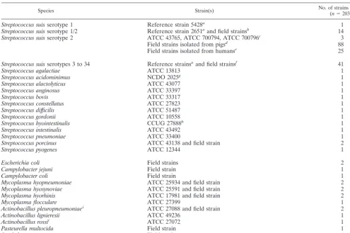
Forty-eight SPF Large White pigs, obtained from the experimental swine herd of the Agence Franc¸aise de Se´curite´ Sanitaire des Aliments (Ploufragan, France) (7), 10 weeks of age, were divided into four experimental units (units A to D) (Fig. 1). Very strict biosecurity measures were implemented in order to avoid contamination of the pigs: existence of an air filtration system and airlocks for each unit, unit-specific clothes, and compulsory showering after visiting the pigs. In unit A, six negative-control pigs received 2 ml of THB supplemented with 10% inactivated bovine serum by the intravenous route (group 1), and six animals (direct contact pigs) did not receive THB (group 2). In unit B, six pigs were infected intravenously with 1.81⫻108CFU ofS. suisstrain 332 (group 3) and six
contact pigs were not infected (group 4). In unit C, six pigs were inoculated intravenously with 3.23⫻108CFU ofS. suisstrain 347 (group 5) and six contact
pigs were not infected (group 6). In unit D, six pigs were inoculated intravenously with 1.29⫻108CFU ofS. suisstrain 353 (group 7) and six contact pigs were not
infected (group 8) (Fig. 1).
Daily clinical examinations consisted of taking rectal temperature and looking for symptoms such as lameness, tremors, opisthotonos, nystagmus, or convul-sions. Body weight was also recorded each week during the trial. Blood samples were collected 6 days before infection; on days 8, 16, and 21 postinfection (p.i.); and on sacrifice day (16 to 30 days p.i.) for serological and bacteriological analysis. Swabs from palatine tonsils were performed on day 16 p.i., placed in 2 ml of sterile water supplemented with NaCl (8.5 g/liter) (SW), and analyzed by classical bacteriological analysis and by PCR.
In each unit, 6 days before infection and on days 8, 16, and 21 p.i., 7 g of dehydrated granulated food, 15 g of feces, and 25 ml of drinking water were collected in four distant places. In each unit, four drag swabs (Sodibox, La Foreˆt Fouesnant, France), previously humidified with 5 ml of SW, were rubbed on the pen, on the air inlet system, and on the window. Food, feces, and dust samples were placed in 20 ml of SW, vortexed, and centrifuged at 4,000⫻gfor 15 min.
The pellet was resuspended in 2 ml of SW (IS). All environmental samples were analyzed by bacteriological analysis and PCR.
Pigs were randomly sampled in each group, and necropsies were planned at intervals during 2 weeks (on days 16 to 30 p.i.). After euthanasia, the thoracic organs, peritoneum, brain, and joints were examined and swabs were collected from liver, heart, lung, joints, and muscle sampled beside two femur-ilium joints (swabs and biopsy). In addition, and to compare different tonsil samples, external swabs, biopsy specimens, and the whole tonsils were compared. Samples were taken even in the cases where no gross lesions were observed and placed in 2 ml of SW (IS). Biopsy specimens, of approximately 6 mm3, were reduced into small
pieces with a scalpel and added to 2 ml of SW. All these ISs were analyzed by bacteriological analysis and by PCR.
Bacteriological analysis.Ten microliters of each sample (or IS) was placed
onto selective based Columbia medium supplemented with 5% sheep blood, 15 mg of nalidixic acid/liter, and 10 mg of colistin/liter. Then, the plates were incubated overnight at 37°C in 5% CO2.S. suis-like colonies were subcultivated
on Columbia medium supplemented with 5% sheep blood, identified by PCR, and serotyped by slide agglutination using a type-specific hyperimmune serum (14).
Serological analysis.All sera were tested with an indirect enzyme-linked
[image:3.603.47.543.80.175.2]immunosorbent assay (ELISA) using a protein extract from sonicated bacteria as antigen. This ELISA test was not attempted to be used as a diagnostic tool but rather was used to monitor the kinetics of the antibodies’ response against proteins of the homologous strain. Three ELISA tests were developed, one for each strain (332, 347, and 353) as previously described by Cloutier et al. (9). Each serum was diluted at 1/160 before analysis. The positive control consisted of a serum from a pig experimentally infected withS. suistype 2, whereas the negative
TABLE 2. Primers used to construct the PCR internal control and for multiplex PCR
Primer Sequence (5⬘-3⬘) PCR product size (bp)
16S-195(s)
CAGTATTTACCGCATGGTAGATAT
294
16S-489(as2)
GTAAGATACCGTCAAGTGAGAA
cps2J-s
GTTGAGTCCTTATACACCTGTT
459
cps2J-as
CAGAAAATTCATATTGTCCACC
CI 6-s
GTTGAGTCCTTATACACCTGTTACTCAGTGCCGCAGCTAACGCATT
620
CI 7-as
CAGAAAATTCATATTGTCCACCCGACTTCACCCCAATCATCTATCC
FIG. 1. Experimental design. Negative control animals were in unit
A and were divided into two groups: group 1 (six noninfected pigs that
received sterile THB) and group 2 (six noninfected pigs that were in
direct contact). In unit B, six pigs were infected with
S. suis
strain 332
(group 3) and six pigs were in direct contact (group 4). In unit C, six
pigs were infected with
S. suis
strain 347 (group 5) and six pigs were in
direct contact (group 6). In unit D, six pigs were infected with
S. suis
strain 353 (group 7) and six pigs were in direct contact (group 8).
on May 15, 2020 by guest
http://jcm.asm.org/
[image:3.603.305.537.478.648.2]control corresponded to a serum from an uninfected SPF pig (tested in quadru-plicate).o-Phenylenediamine at 0.4 mg/ml(Sigma-Aldrich Chimie) dissolved in 0.05 M citrate buffer (pH 5.5) with 0.5 M H2O2was used as a substrate. After 15
min at 37°C in the dark, 25l of HCl was added in each well and optical density (OD) was measured at 492 nm on a kinetic microplate reader (Labsystems, Cergy-Pontoise, France). Results were reported asS/Pratios, which were defined as the OD obtained for each serum minus the mean OD of the negative control divided by the mean OD of the positive control.
Statistical analysis of data.The data obtained in experimental study from pig
groups were compared simultaneously by Kruskall-Wallis test and in two-by-two tables by Kolmogorov-Smirnov test. Associations between strain used for the assay, tonsillar carriership (as determined by external swab, biopsy sample, and whole tonsil), and method applied (PCR and bacteriological analysis) were assessed by Fisher exact test (nⱕ5) or chi-square test (n⬎5) on independence in two-by-two tables. These tests were carried out with Systat 9.0 program for Windows. Differences were estimated to be significant when probabilities (P) were lower than 0.05.
RESULTS
Specificity and sensitivity of the multiplex PCR.
In order to
assess the specificity of the multiplex PCR, the different
mi-croorganisms listed in Table 1 were used as DNA templates.
All
S. suis
strains, including reference strains as well as field
strains, showed a fragment of 294 bp corresponding to a part of
the 16S rDNA gene. None of the other bacterial species
de-scribed in Table 1 showed any amplification product. A
frag-ment of 489 bp corresponding to a part of the
cps
gene coding
for the capsule was only obtained with
S. suis
serotypes 2 and
1/2 strains as expected.
Sensitivity results are shown in Fig. 2. The assay was carried
out with 10-fold dilutions of
S. suis
DNA extracts obtained
from a culture of reference strain S735 at a titer of 280 CFU/
l
and mixed with a tonsil palatine biopsy specimen from an SPF
pig (1 ml of dilution by biopsy). Under the conditions
de-scribed in the trials (5
l of DNA extract per assay), the
detection limit of the PCR test was 1.4 CFU/assay,
correspond-ing to 280 CFU/ml of tonsil sample for the product specific to
serotypes 2 or 1/2 and 0.14 CFU/assay (28 CFU/ml of tonsil
sample) for the product shared between all
S. suis
serotypes.
Moreover, the IPC of 620 bp was also noticeable in Fig. 2.
Clinical signs and macroscopic lesions.
Data on clinical
signs and macroscopic lesions are summarized in Table 3.
(i) Groups 1 and 2.
Negative control animals did not exhibit
[image:4.603.45.281.68.148.2]any clinical signs of
S. suis
infection. Postmortem examinations
FIG. 2. Evaluation of detection limit of the multiplex PCR.
Aga-rose electrophoresis of the PCR products obtained from serial
dilu-tions of
S. suis
S735 strain culture at a titer of 2.8
⫻
10
5CFU/ml (lane
2) and placed in tonsil biopsy specimens (1 ml of each dilution per
biopsy specimen). The positions of the specific fragments of internal
control,
S. suis
, and serotype 2 or 1/2 are indicated. Lanes: M,
molec-ular mass marker (Smart Ladder; Eurogentec); 1, negative control of
the PCR (only IPC was amplified); 3 to 8, PCR products at dilutions of
10, 10
2, 10
3, 10
4, 10
5, and 10
6.
TABLE
3.
Clinical,
pathological,
bacteriological,
PCR,
and
serological
results
observed
for
infected
and
contact
pigs
Unit Group Hyperthermia (days p.i.) Lameness (days p.i.) Macroscopic lesions a No. of pigs b in which the following was observed in indicated samples collected at necropsy: ELISA results at necropsy (Sp mean) S. suis S. suis DNA Joints Muscle Liver Heart Lung Tonsils Joints Muscle Liver Heart Lung Tonsils A (THB) 1 No No No 0 0 0 0 0 0 0 0 0 0 0 0 0.015 ⫾ 0.013 2 No No No 0 0 0 0 0 0 0 0 0 0 0 0 0.027 ⫾ 0.011 B (strain 332, type 2) 3 Yes (1 –2) Yes (1 –22) Arthritis (4), pneumonia (1) 0 1 0 0 0 6 0 0 1 0 0 6 0.333 ⫾ 0.099 4 No No Arthritis (4) 2 2 1 1 1 6 1 2 1 1 1 6 0.179 ⫾ 0.035 C (strain 347, type 2) 5 Yes (1 –2) Yes (1 –10) Arthritis (4) 1 2 0 3 2 5 0 2 0 2 2 6 0.644 ⫾ 0.045 6 No No No 1 0 0 1 1 6 1 0 0 0 0 6 0.399 ⫾ 0.086 D (strain 353, type 1/2) 7 No No Arthritis (3) 1 0 0 0 0 0 0 0 0 0 0 4 0.053 ⫾ 0.012 8 No No Arthritis (1) 0 0 0 0 0 0 0 0 0 0 0 5 0.047 ⫾ 0.013 a Data in parentheses are numbers of pigs (out of a group of six) in which lesions were observed. b Data are numbers of pigs out of a group of six.on May 15, 2020 by guest
http://jcm.asm.org/
[image:4.603.354.485.92.726.2]did not reveal any lesions in these pigs. The average daily
weight gains (ADGs) were 981 g (group 1) and 993 g (group 2).
(ii) Groups 3 and 4 (S. suis
type 2).
Rectal temperatures of
three piglets infected with strain 332 (group 3) were moderate
(40.2
⫾
0.3°C) during 24 to 48 h p.i. Under the conditions of
this trial (level three biosecurity), the normal temperature of
SPF piglets is 39.5°C. All the piglets presented lameness, 24 h
p.i. and during 1 to 22 days. Four of them developed arthritis
associated with pneumonia in one animal. Contact pigs (group
4) did not develop clinical signs, but four animals showed
arthritis at necropsy. The body growth of animals in group 3
was retarded. Differences in ADG between group 3 (395 g) and
group 4 (921 g) were significant (
P
⬍
0.05), although no
dif-ferences were noted between negative control pigs and contact
pigs (
P
⬎
0.05).
(iii) Groups 5 and 6 (S. suis
type 2).
Two piglets infected
with strain 347 (group 5) developed hyperthermia (40
⫾
0.3°C)
during 24 to 48 h, and lameness was observed in four pigs
(during 1 to 10 days). Postmortem examinations revealed
ar-thritis in these animals, whereas contact pigs (group 6) had
neither symptoms nor lesions. The body growth was affected by
the experimental infection (group 5). The ADG was 550 g,
significantly different (
P
⬍
0.05) from those observed for
pig-lets in negative control groups and group 6 (ADG
⫽
970 g).
(iv) Groups 7 and 8 (S. suis
type 1/2).
No clinical signs were
noticeable in groups 7 and 8, but moderate arthritis was
ob-served at necropsy in three infected and one contact pigs. The
ADG were, respectively, 927 and 875 g (groups 7 and 8), and
no difference was obtained between these two groups and
negative control groups (
P
⬎
0.05).
Detection of
S. suis.
All samples collected from animals in
negative control groups and analyzed 16 days p.i. (live pigs) and
after euthanasia (16 to 30 days p.i.) were negative by PCR and
bacteriological analysis. Data are summarized in Tables 3 and 4.
(i) Environmental samples.
All environmental samples were
negative by PCR and bacteriological analysis.
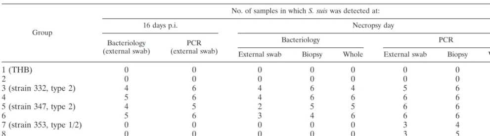
(ii) Results obtained with live pigs (16 days p.i.) (Table 4).
The comparison of PCR as well as bacteriological results,
ob-tained from tonsils in infected or in contact pigs did not show
any differences between groups 3, 4, 5, and 6 (
P
⬎
0.05).
Results from groups 7 and 8 were negative.
(iii) Results obtained in euthanized piglets (16 to 30 days
p.i.).
The results, obtained by PCR and bacteriological analysis
from 24 dead animals (Tables 3 and 4), confirmed the presence
of
S. suis
type 2 (strains 332 and 347) infection, respectively, in
14 and 19 samples from joints, muscle, liver, heart, and lungs,
but results by the two techniques were not significantly
differ-ent (
P
⬎
0.05).
S. suis
type 1/2 was isolated in only one pig
(group 7) from the joints. Sixteen to thirty days p.i.,
S. suis
type
2 was detected from the tonsils of 24 and 23 pigs by PCR and
culture, respectively, whereas nine pigs were identified as
pos-itive by PCR and negative by culture in groups 7 and 8 (
S. suis
type 1/2). In these groups, the difference between the two tests
was significant (
P
⬍
0.05). The results were not significantly
different between the three samples of tonsils (external swab,
biopsy sample, and whole tonsil) (Table 4). No significant
difference was observed between infected and contact pigs and
between groups 3, 4, 5, and 6 (
P
⬎
0.05).
Serological results.
The ELISA results obtained from
neg-ative controls but also from groups 7 and 8 were negneg-ative. Pigs
infected with
S. suis
type 2 (strains 332 and 347) developed
humoral antibodies detectable by ELISA from 16 days p.i. The
level of antibodies increased until the end of the experiment.
Contact pigs of both groups were also seropositive with a
moderate level of antibodies.
DISCUSSION
The multiplex PCR assay described in this study was based
on the amplification of a gene fragment coding for 16S rRNA
of
S. suis
and on the amplification a
cps2J
gene fragment
encoding
S. suis
serotype 2 and 1/2 capsular biosynthesis (24).
Furthermore, our test contained an internal PCR control to
eliminate false-negative samples due to inhibitors of
polymer-ization. All
S. suis
strains tested in this study were detected by
our multiplex PCR test, while none of the other bacterial
species showed a positive reaction.
The 35 capsular types of
S. suis
were recognized by this PCR
test, which was also able to detect
S. suis
in specimens from live
pigs experimentally infected.
S. suis
isolates belonging to any
serotype isolated from tonsil could be potentially virulent.
Re-cently, in our laboratory,
S. suis
serotype 5 was isolated from a
subject with septicemia without any other bacterial isolation.
This serotype was also isolated from nursery pigs with serious
cases of meningitis on a Canadian farm (9). Some
S. suis
strains isolated from healthy carrier pigs (particularly in
ton-TABLE 4. Detection of
S. suis
serotype 2 or 1/2 by bacteriological analysis and PCR
aGroup
No. of samples in whichS. suiswas detected at:
16 days p.i. Necropsy day
Bacteriology
(external swab) (external swab)PCR
Bacteriology PCR
External swab Biopsy Whole External swab Biopsy Whole
1 (THB)
0
0
0
0
0
0
0
0
2
0
0
0
0
0
0
0
0
3 (strain 332, type 2)
4
6
4
6
4
5
6
6
4
5
6
4
6
6
6
6
6
5 (strain 347, type 2)
4
5
2
5
5
6
6
6
6
5
6
3
4
6
6
6
6
7 (strain 353, type 1/2)
0
0
0
0
0
3
4
4
8
0
0
0
0
0
3
5
5
aTonsil samples (from external swab, biopsy, and whole tonsil) from live pigs (16 days p.i.) and pigs after euthanasia (16 to 30 days p.i.) were studied.
on May 15, 2020 by guest
http://jcm.asm.org/
[image:5.603.43.539.80.218.2]sils) are able to induce transmission of
S. suis
infection
be-tween pigs or bebe-tween herds. These pigs should be carefully
checked, and the detection of
S. suis
species in tonsils is the
first step of the diagnosis. Thus, the PCR assay described in
this study, based on the 16S rDNA region conserved across
capsular types, allowed the detection of
S. suis
species and
would be very useful for epidemiological studies. Other PCR
tests were previously developed to detect
S. suis
species (6, 19,
31). Okwumabua et al. and Wisselink et al. have also reported
two PCR tests to detect two of the
S. suis
virulence factors, the
suilysin and the extracellular factor, respectively (19, 30).
How-ever, the absence of these proteins in some virulent strains may
preclude the routine use of these tests. Moreover, in a previous
study we showed that these proteins were present neither in all
S. suis
strains nor in all virulent European strains (5, 19). In
2003, Okwumabua et al. developed a multiplex PCR based on
the
gdh
gene, encoding the glutamate deshydrogenase of
S. suis
serotype 2, allowing the amplification of all
S. suis
serotypes
(20). This method was very attractive, but it was only applied to
detect or characterize
S. suis
from pure cultures.
Our multiplex PCR assay allowed also the detection of all
S.
suis
serotypes and more specifically serotypes 2 and 1/2. A
preliminary study was performed to develop a PCR test able to
detect serotype 2 alone, because this serotype is the major
cause of disease in France and because it is a zoonotic agent
that causes septicemia, endocarditis, and meningitis in humans
(2, 5, 10, 21, 22, 29). The first step was to show genomic
specificities in serotype 2 strains. We sequenced the four
po-tential target genes (
cps2F
,
cps2H
,
cps2I
, and
cp
s
2J
) that code
for
S. suis
capsule, in serotypes 2 and 1/2 reference strains. In
the original work describing the sequencing of these genes, the
serotype 1/2 reference strain was not analyzed (23). The
align-ment of these nucleotide sequences showed a perfect identity
for the
cps2H
,
cps2I
, and
cps2J
genes of the two reference
strains. However, a substitution (T to C) was detected in the
cps2F
gene of serotype 2. Since this mutation leads to the
disappearance of an SspI restriction site in the serotype 1/2, a
PCR-restriction fragment length polymorphism was
devel-oped. However, this substitution was not detected in all
sero-type 2 strains isolated from subjects in the field (data not
shown). In this context, we decided to develop a PCR test able
to detect simultaneously serotypes 2 and 1/2. These serotypes
are the most frequently detected in France (77.3%) (5).
Sev-eral PCR tests, targeted on the
cps
locus, were previously
described to detect these two serotypes (20, 26, 31). We
de-signed original primers from the
cps
locus in order to (i) have
a hybridization temperature compatible with the primers based
on 16S rDNA, (ii) avoid the appearance of dimers, and (iii)
have a PCR product differing in size from the product of
16S-195s and 16S-489 as primer pair.
The sensitivity of our multiplex PCR test was evaluated in
vitro with SPF pig tonsils experimentally infected with
S. suis
type 2 reference strain and was found to be 28 CFU of
S.
suis
/ml of tonsil sample. In the in vitro conditions described in
our study, the PCR test was evaluated to be 20 times more
sensitive than culture-positive results (28 versus 500 CFU of
S.
suis
/ml). Our assay is more sensitive than other PCR tests
previously described: the detection threshold of the multiplex
test developed by Wisselink et al. (31) was 10 fg of
chromo-somal DNA in 25
l of clinical sample (approximately 200
CFU/ml according to our reckoning). This estimate probably
lacks accuracy because the test was performed not with tonsils
experimentally infected in vitro but with purified DNA. The
sensitivity of the PCR tests described by Smith et al. and
Okwumabua et al. were not evaluated (20, 25, 26).
The comparison of PCR and bacteriological isolation
ob-tained from tonsils of live pigs (healthy carriers) as well as in
other organs in dead pigs, with or without macroscopic lesions,
did not show any significant differences, with the exception of
groups 7 and 8 infected with serotype 1/2 (
P
⬍
0.05). It is
possible that the number of organisms present in samples from
groups 3 to 6 is above the detection limit for both assays. Our
multiplex PCR assay, with an internal control and without a
culture step, is easy to perform. Ninety-six samples could be
analyzed simultaneously in 6 h, whereas bacteriological
isola-tions require at least 4 days (including primary isolation,
clon-ing, biochemical identification, and serotyping). The major
dif-ficulty of bacteriological isolation is to locate
S. suis
colonies
from multi-infected samples such as tonsils. On the other hand,
tonsil specimens appeared to be helpful to detect
S. suis
in live
pigs. Our PCR assay used samples directly and was able to
detect and identify
S. suis
serotype 2 and 1/2 among suspicious
␣
-hemolytic colonies on blood agar medium. In practice, the
main advantages of this test are its abilities (i) to detect
S. suis
from multi-infected samples, (ii) to reduce the time required to
identify the bacteria, and (iii) to increase the number of
colo-nies analyzed at the same time.
The comparison of tonsil biopsy specimens, external swabs,
and whole tonsils did not show any significant differences.
These results differ from those published by Fittipaldi et al. in
2003 (11), with
Actinobacillus pleuropneumoniae
infection.
These authors showed that the PCR detection rate was higher
with whole tonsils than with tonsil biopsy specimens. However,
in that study, conventional naturally infected pigs were tested.
External tonsil swabs will be chosen in the future because
external tonsil swabs are less traumatizing for pigs.
In this experimental study, clinical signs and macroscopic
lesions induced in SPF piglets were moderate except for
ar-thritis (especially for pigs infected with
S. suis
serotype 1/2).
These results are different from those of our previous trials
carried out under these standardized conditions with different
strains of
S. suis
type 2 (3, 4). There was a transmission from
infected to contact pigs, since we detected
S. suis
in contact
pigs during the trial (on day 16) and at necropsy. At the end of
the assay, all contact pigs in units B and C were healthy carriers
pigs with no clinical symptoms of the disease (
P
⬎
0.05).
In conclusion, the assay in the present work may be used
routinely to identify pigs carrying
S. suis
serotypes 2 and 1/2
and all other serotypes. It may also be applicable for
epidemi-ological studies and transmission studies of
S. suis
and can
contribute to the control of
S. suis
infection.
ACKNOWLEDGMENTS
We thank Roland Cariolet, Bernard Beaurepaire, Ge´rard Be´ne´vent,
Pierre Ecobichon, and Jean-Claude Rault (Service de production de
porcs assainis et d’expe´rimentation, AFSSA-Ploufragan) for skilled
technical assistance and Anne Gautier-Bouchardon for critical reading
of the manuscript.
on May 15, 2020 by guest
http://jcm.asm.org/
REFERENCES
1. Baele, M., V. Storms, F. Haesebrouck, L. A. Devriese, M. Gillis, G.
Ver-schraegen, T. de Baere, and M. Vaneechoutte.2001. Application and
eval-uation of the interlaboratory reproducibility of tRNA intergenic length poly-morphism analysis (tDNA-PCR) for identification ofStreptococcusspecies. J. Clin. Microbiol.39:1436–1442.
2. Bensaid, T., B. Bonnefoi-Kyriacou, C. Dupel-Pottier, O. Bellon, E. Lagier,
and H. Chardon.2003. Streptococcus suis meningitis following wild boar
hunting. Presse Med.32:1077–1078.
3. Berthelot-He´rault, F., R. Cariolet, A. Labbe, M. Gottschalk, J. Y. Cardinal,
and M. Kobisch.2001. Experimental infection of specific pathogen free
piglets with French strains ofStreptococcus suiscapsular type 2. Can. J. Vet. Res.65:196–200.
4. Berthelot-He´rault, F., M. Gottschalk, A. Labbe, R. Cariolet, and M.
Ko-bisch.2001. Experimental airborne transmission ofStreptococcus suis
cap-sular type 2 in pigs. Vet. Microbiol.82:69–80.
5. Berthelot-He´rault, F., H. Morvan, A. M. Ke´ribin, M. Gottschalk, and M.
Kobisch.2000. Production of muramidase released protein (MRP),
extra-cellular factor (EF) and haemolysin by field isolates ofStreptococcus suis
capsular type 2, 1/2, 9, 7 and 3 isolated from swine in France. Vet. Res. 31:473–479.
6. Boye, M., A. A. Feenstra, C. Tegtmeier, L. O. Andresen, S. R. Rasmussen,
and V. Bille-Hansen.2000. Detection ofStreptococcus suisbyin situ
hybrid-ization, indirect immunofluorescence, and peroxidase-antiperoxidase assays in formalin-fixed, paraffin-embedded tissue sections from pigs. J. Vet. Diagn. Investig.12:224–232.
7. Cariolet, R., P. Marie, G. Moreau, and H. Robert.1994. Rappel des
diffe´r-entes me´thodes d’obtention de porcelets assainis: conditions de maintien du statut sanitaire et valorisation de ces animaux. Journe´es Rech. Porcine France26:1–12.
8. Chatellier, S., J. Harel, Y. Zhang, M. Gottschalk, R. Higgins, L. A. Devriese,
and R. Brousseau.1998. Phylogenetic diversity ofStreptococcus suisstrains
of various serotypes as revealed by 16S rRNA gene sequence comparison. Int. J. Syst. Bacteriol.48:581–589.
9. Cloutier, G., S. D’Allaire, G. Martinez, C. Surprenant, S. Lacouture, and M.
Gottschalk.2003. Epidemiology ofStreptococcus suisserotype 5 infection in
a pig herd with and without clinical disease. Vet. Microbiol.97:135–151.
10. Durand, F., C. L. Pe´rino, C. Recule, J. P. Brion, M. Kobisch, F. Guerber, and
J. Croize´.2001. Bacteriological diagnosis ofStreptococcus suismeningitis.
Eur. J. Clin. Microbiol.20:519–521.
11. Fittipaldi, N., A. Broes, J. Harel, M. Kobisch, and M. Gottschalk.2003.
Evaluation and field validation of PCR tests for detection ofActinobacillus pleuropneumoniaein subclinically infected pigs. J. Clin. Microbiol.41:5085– 5093.
12. Franc¸ois, B., V. Gissot, M. C. Ploy, and P. Vignon.1998. Recurrent septic
shock due toStreptococcus suis. J. Clin. Microbiol.36:2395.
13. Friis, N. F.1975. Some recommendations concerning primary isolation of
Mycoplasma suispneumoniaeandMycoplasma flocculare: a survey. Nord. Vet. Med.27:337–339.
14. Gottschalk, M., R. Higgins, M. Jacques, K. R. Mittal, and J. Henrichsen.
1989. Description of 14 new capsular types ofStreptococcus suis. J. Clin. Microbiol.27:2633–2636.
15. Higgins, R., and M. Gottschalk.1999. Streptococcal diseases, p. 563–578.In
B. E. Straw, S. D’Allaire, W. L. Mengeling, and D. J. Taylor (ed.), Diseases of swine, 8th ed. Iowa University Press, Ames.
16. Kellog, D. E., and S. Kwok.1990. Detection of human immunodeficiency
virus, p. 339–343.InM. A. Innis, D. H. Gelfand, J. J. Sninsky, and T. J. White (ed.), PCR protocols: a guide to methods and applications. Academic Press, San Diego, Calif.
17. Le Minor, L., and M. Ve´ron.1990. Bacte´riologie me´dicale.
Me´decine-Sci-ences, Flammarion, Paris, France.
18. Matsuo, H., and S. Sakamoto.2003. Purulent meningitis caused by
Strepto-coccus suisin a pig breeder. Kansenshogaku Zasshi77:340–342.
19. Okwumabua, O., O. Abdelmagid, and M. M. Chengappa.1999.
Hybridiza-tion analysis of the gene encoding a hemolysin (suilysin) ofStreptococcus suis
type 2: evidence for the absence of the gene in some isolates. FEMS Micro-biol. Lett.181:113–121.
20. Okwumabua, O., M. O’Connor, and E. Shull.2003. A polymerase chain
reaction (PCR) assay specific forStreptococcus suisbased on the gene en-coding the glutamate dehydrogenase. FEMS Microbiol. Lett.218:79–84.
21. Pedroli, S., M. Kobisch, O. Beauchet, J. P. Chaussinand, and F. Lucht.2003.
Streptococcus suis bacteriemia. Presse Med.32:599–601.
22. Rosenkranz, M, H. A. Elsner, H. J. Sturenburg, C. Weiller, J. Rother, and I.
Sobottka.2003. Streptococcus suis meningitis and septicemia contracted
from a wild boar in Germany. J. Neurol.250:869–870.
23. Smith, H. E., M. Damman, J. van der Velde, F. Wagenaar, H. J. Wisselink,
N. Stockhofe-Zurwieden, and M. A. Smits.1999. Identification and
charac-terization of the cps locus ofStreptococcus suisserotype 2: the capsule protects against phagocytosis and is an important virulence factor. Infect. Immun.67:1750–1756.
24. Smith, H. E., R. de Vries, R. van’t Slot, and M. A. Smits.2000. The cps locus
ofStreptococcus suis serotype 2: genetic determinant for the synthesis of sialic acid. Microb. Pathog.29:127–134.
25. Smith, H. E., L. van Bruijnsvoort, H. Buijs, H. J. Wisselink, and M. A. Smits.
1999. Rapid PCR test forStreptococcus suisserotype 7. FEMS Microbiol. Lett.178:265–270.
26. Smith, H. E., V. Veenbergen, J. van der Velde, M. Damman, H. J. Wisselink,
and M. A. Smits.1999. Thecpsgenes ofStreptococcus suisserotypes 1, 2, and
9: development of rapid serotype-specific PCR assays. J. Clin. Microbiol. 37:3146–3152.
27. Staats, J. J., I. Feder, O. Okwumabua, and M. M. Chengappa.1997.
Strep-tococcus suis: past and present. Vet. Res. Commun.21:381–387.
28. Strangmann, E., H. Froleke, and K. P. Kohse.2002. Septic shock caused by
Streptococcus suis: case report and investigation of a risk group. Int. J. Hyg. Environ. Health.205:385–392.
29. Trottier, S., R. Higgins, G. Brochu, and M. Gottschalk.1991. A case of
human endocarditis due toStreptococcus suisin North America. Rev. Infect. Dis.13:1251–1252.
30. Wisselink, H. J., F. H. Reek, U. Vecht, N. Stockhofe-Zurwieden, M. A. Smits,
and H. E. Smith.1999. Detection of virulent strains ofStreptococcus suistype
2 and highly virulent strains ofStreptococcus suistype 1 in tonsillar specimens of pigs by PCR. Vet. Microbiol.67:143–157.
31. Wisselink, H. J., J. J. Joosten, and H. E. Smith.2002. Multiplex PCR assays
for simultaneous detection of six major serotypes and two virulence-associ-ated phenotypes ofStreptococcus suisin tonsillar specimens from pigs. J. Clin. Microbiol.40:2922–2929.