The molecular basis of hereditary complement
factor I deficiency.
T J Vyse, … , A D Webster, M J Walport
J Clin Invest.
1996;
97(4)
:925-933.
https://doi.org/10.1172/JCI118515
.
The molecular basis of hereditary complement factor I deficiency is described in two
pedigrees. In one pedigree, there were two factor I-deficient siblings, one of whom was
asymptomatic and the other suffered from recurrent pyogenic infections. Their factor I mRNA
was analyzed by reverse transcription of fibroblast RNA followed by amplification using the
polymerase chain reaction. Both siblings were homozygous for the same transversion
(adenine to thymine) at nucleotide 1282 in the cDNA. This mutation causes histidine-400 to
be replaced by leucine. The altered histidine is a semi-conserved residue within the serine
proteinase family, although no function has been ascribed to it. The proband of the second
pedigree studied was found to be a compound heterozygote. One allele had the same
mutation as the first family, the second allele had a donor splice site mutation that resulted
in the deletion of the mRNA encoded in the fifth exon (a low-density lipoprotein receptor
domain) from its transcript.
Research Article
Find the latest version:
J. Clin. Invest.
© The American Society for Clinical Investigation, Inc. 0021-9738/96/02/0925/09 $2.00
Volume 97, Number 4, February 1996, 925–933
The Molecular Basis of Hereditary Complement Factor I Deficiency
Timothy J. Vyse,* Bernard J. Morley,* István Bartók,* Efstathios L. Theodoridis,* Kevin A. Davies,* A. David B. Webster,‡
and Mark J. Walport *
*Rheumatology Unit, Department of Medicine, RPMS, Hammersmith Hospital, London W12 0NN, United Kingdom; and ‡Royal Free
Hospital, London NW3, United Kingdom
Abstract
The molecular basis of hereditary complement factor I defi-ciency is described in two pedigrees. In one pedigree, there were two factor I–deficient siblings, one of whom was asymp-tomatic and the other suffered from recurrent pyogenic in-fections. Their factor I mRNA was analyzed by reverse transcription of fibroblast RNA followed by amplification using the polymerase chain reaction. Both siblings were ho-mozygous for the same transversion (adenine to thymine) at nucleotide 1282 in the cDNA. This mutation causes histi-dine-400 to be replaced by leucine. The altered histidine is a semi-conserved residue within the serine proteinase family, although no function has been ascribed to it. The proband of the second pedigree studied was found to be a compound heterozygote. One allele had the same mutation as the first family, the second allele had a donor splice site mutation that resulted in the deletion of the mRNA encoded in the fifth exon (a low-density lipoprotein receptor domain) from its transcript. (J. Clin. Invest. 1996. 97:925–933.) Key words:
complement • factor I • mutation • genetics •
immuno-deficiency
Introduction
Human complement factor I (FI)1 is a serine proteinase that
acts in the regulation of the complement cascade. Hereditary deficiency of FI has been reported in 23 individuals from 19 different pedigrees (1). It is associated with a propensity to pyogenic infection and follows an autosomal recessive pattern of inheritance.
FI is a plasma glycoprotein that circulates in an active form at a concentration of 35 mg/ml. Its relative molecular mass (Mr)
is estimated to be 88,000, and it is composed of two disulfide-linked polypeptide chains (Mr 50,000 and 38,000). The protein
is synthesized as a single chain precursor of 565 amino acids,
predominantly in the liver (2). Four basic amino acids are then excised from the precursor prior to secretion of the het-erodimer. FI synthesis has also been demonstrated in vitro by primary monocyte cultures (3), endothelial cells (4), and by the Raji B cell line (5).
The cDNA structure of FI has been published (6, 7) and more recently, the genomic organization determined (8). Like many of the complement proteins, FI has a modular structure. The heavy chain contains two low-density lipoprotein receptor (LDLr) domains, a CD5 domain, and a module found only in FI and complement proteins C6 and C7. The FI gene is located on chromosome 4q25 (9) and spans 63 kb. It comprises 13 ex-ons and there is a strong correlation between the exonic orga-nization of the gene and the modular structure of the protein (8). The light chain of FI, which is the serine proteinase region of the molecule, is encoded in five exons. The genomic organi-zation of the enzymic part of FI is similar to that of trypsin. The gene structure is unusual in that the first exon is small, 86 bp, and it is separated from the rest of the gene by a large first intron of 36 kb.
FI cleaves the a9 chains of C4b and C3b and thereby is in-volved in the regulation of both the classical and alternative complement pathways. The function of FI is dependent on var-ious cofactors; the cleavage of C4b requires C4-binding pro-tein (10), and the cleavage of C3b is dependent on comple-ment factor H (11). By its action on C3b, FI impedes the formation of the alternative pathway C3 convertase (C3bBb). Thus FI has an important role in inhibiting the amplification loop of the alternative pathway which generates C3 convertase from C3b. In hereditary deficiency of FI, the alternative path-way is not regulated, so there is consumptive depletion of C3. In addition, patients with FI deficiency have consumptive loss of factor B and reduced levels of factor H (reviewed in refer-ence 1). Indeed, it was the investigation of the physiology of the complement system in the first reported case of FI defi-ciency (12–15) that generated the early evidence for the exist-ence of the alternative pathway amplification loop (16).
The clinical manifestations of FI deficiency are similar to those of hereditary C3 deficiency, the most common feature being recurrent pyogenic infections, which reflects the role of C3 as an opsonin. The infections usually begin in early child-hood, their median age of onset in FI deficiency is 17 mo. There is also an increased susceptibility to Neisseria meningiti-dis infection. Patients with FI deficiency may also have “im-mune complex” type illness. There are three such examples re-ported (1, 17, 18) in which a multisystem vasculitic illness occurred, which in one case was fatal (17). There have been two reports of asymptomatic individuals with complete FI defi-ciency (1, 19). In both instances they were identified as mem-bers of a pedigree in which there was a symptomatic case. One of these asymptomatic cases (1) and her younger brother, who had recurrent infections, belong to the first family studied in this paper.
Address correspondence to Mark J. Walport, Rheumatology Unit, Department of Medicine, RPMS, Hammersmith Hospital, Du Cane Road, London W12 0NN, UK. Phone: 181 740 3276; FAX: 181 743 3109. Timothy J. Vyse’s current address is Division of Basic Sciences, National Jewish Center for Immunology and Respiratory Medicine, 1400 Jackson Street, Denver, CO 80206.
Received for publication 19 September 1995 and accepted in re-vised form 28 November 1995.
The molecular pathology of FI deficiency has not previ-ously been described. In this paper the molecular basis of FI deficiency in two pedigrees is analyzed.
Methods
Patients. Two families in which there was an inherited deficiency of FI were studied. In family 1, the proband presented at the age of 18 mo with Staphylococcus epidermidis septic arthritis of the left shoul-der. He subsequently had recurrent sinusitis and otitis media and at the age of 13 yr a N. meningitidis meningitis. The patient had a sister who is five years older. She has no history of increased susceptibility to pyogenic infections. The history of this patient and his sister and their complement investigations are described in reference 1. Briefly, they both had no detectable circulating FI or factor B, and a very low complement C3 level (most of which was in the form of C3b) mea-sured by radial immunodiffusion using ovine polyclonal antisera (The Binding Site, Birmingham, U.K.). In addition, no detectable alterna-tive pathway activity was detected when determined by hemolytic as-say with guinea pig erythrocytes in the presence of 7 mM MgCl2 and 10 mM ethyleneglycol tetraacetic acid (20). Both parents, who were well and had no history to suggest a susceptibility to infection, had z 50% of normal circulating FI, but factor B and C3 concentrations were within normal limits. The parents were both from western Scot-land, there was no history of consanguinity. In family 2, the proband was the second case of FI deficiency to be reported (21). She pre-sented with a history of recurrent infections that began at the age of four months with bacterial meningitis (Streptococcus pneumoniae) and otitis media. Between the ages of 7 and 15 years she had four fur-ther episodes of meningitis. The causative agent was identified on two occasions and found to be N. meningitidis. She had no detectable cir-culating FI and low C3 (30% normal human serum). Her mother had z 50% normal circulating FI. Her father was not available for study. There was no known relation between family 1 and family 2.
Southern blotting. Genomic DNA was extracted from whole blood as described (22). A total of 10 mg of DNA was digested overnight with 50U of restriction endonuclease. The fragments were separated on a 0.8% agarose gel and transferred onto nylon membranes (Dur-alon; Stratagene Ltd., Cambridge, U.K.) by capillary action (22, 23). The DNA was cross-linked to the membrane by exposure to ultravio-let radiation (254 nm) in a 1800UV Stratalinker (Stratagene Ltd., Cambridge, U.K.). Hybridization was carried out in 53 Denhardt’s solution, 1% SDS, 53 SSPE, and 150 mg/ml Homomix (24) overnight at 658C for 24 hours. The DNA probes were radiolabeled with [a-32P]dCTP by random priming (25). The membranes were hybrid-ized with two separate probes. The first was a 1.9-kb probe containing FI coding cDNA (the cDNA was kindly provided by Dr. R.B. Sim, Oxford, UK [7]), the second was a 343-bp 59 probe that encompassed the first exon; it was obtained by PCR of genomic DNA using oligo-nucleotide primers Int 1 and Pro 1. Int 1 bound to sequence in the first intron, and Pro 1 bound to sequence in the promoter (see Table II). The 59 probe was generated by PCR to include sequences flank-ing the first exon owflank-ing to the exon’s small size, 86 bp. The mem-branes were washed for 10 min in 23 SSC/0.1% SDS at room temper-ature, then at 658C for 30 minutes, and then finally washed in 0.53 SSC/0.1%SDS at 658C for 30 min (full-length cDNA probe), or in 13 SSC/0.1%SDS (59 probe). The membranes were then subject to auto-radiography by exposing to Hyperfilm (Amersham International plc, Bucks, U.K.) at 2708C for 2–7 d.
Polymerase chain reaction (PCR). The reaction was performed on 250 ng of genomic DNA in 50 ml containing 10 mM Tris pH 8.3, 50 mM KCl, 0.01% gelatin, 1.5 mM MgCl2, 200 mM dNTP, 250 nM of each primer and 1.25U of Taq DNA polymerase (Perkin Elmer Ltd., Beaconsfield, Bucks, U.K.). The PCR was run for 30 cycles as fol-lows: 958C for 1 min; 50–558C (depending on primer length) for 1 minute; 728C for 1 min. The sequence and position of the oligonucle-otide primers used in genomic PCR are listed in Table II.
Reverse transcription and polymerase chain reaction (RT-PCR). Total RNA was extracted (26) from fibroblasts grown from the shave skin biopsies. 250 ng of RNA was used as a substrate for first strand cDNA synthesis. This reaction was performed in 20 ml using 200 U of Superscript reverse transcriptase (GIBCO-BRL, Life Technologies, Paisley, Renfrewshire, Scotland) in 50 mM Tris pH 8.3, 75 mM KCl, 3 mM MgCl2, 10 mM dithiothreitol with 500 mM dNTP. The priming oligonucleotide (250 nM) was oligo dT12–18 (Pharmacia Biotech Ltd., Milton Keynes, U.K.). The reaction was incubated at 428C for 45 min-utes, and then 998C for 5 min to inactivate the enzyme. PCR was then performed on the whole RT reaction. The conditions used were as described above except that the reaction volume was 100 ml. The cDNA was amplified in 35 cycles as follows: 958C for 1 min; 528C for 1 min; 728C for 1 min. The RT-PCR products were cloned into the Bluescript (pBS) SK1 vector (Stratagene Ltd., Cambridge, U.K.) us-ing standard techniques (22). This was facilitated by the inclusion of BamHI and EcoRI sites in the primers. The sequence and position of the oligonucleotide primers used in RT-PCR are shown in Table I. In each PCR and RT-PCR performed a negative control was included that contained no template.
DNA sequencing. Double-stranded dideoxy termination cycle-sequencing (27) was carried out on RT-PCR fragments cloned in pBS. Genomic PCR products and the 3A-8B RT-PCR product were sequenced directly (28), and/or were phosphorylated and then cloned into pBS cut with SmaI and sequenced. The fmol™ cycle-sequencing system (Promega, Southampton, U.K.) was used according to manu-facturer’s instructions. The oligonucleotides used to prime the reac-tions (listed in Table II) were end-labeled with [γ−32P]ATP (ICN Bio-medicals Ltd., Thame, Oxfordshire, U.K.) using standard conditions (22). Internal primers that bound cDNA were used to sequence the RT-PCR clones. Internal primers were also used for direct sequenc-ing of PCR products. The primers, T3 and T7, bind consensus se-quences flanking the polylinker of pBS. The sequencing reaction products were analyzed by electrophoresis in denaturing polyacryla-mide gels (6% polyacrylapolyacryla-mide/7 M urea).
Shave skin biopsy. From a shave biopsy a sample of skin was taken (without the use of an alcohol swab) and immediately placed into RPMI 1640 medium (ICN Biomedicals Ltd., Thame, Oxfordshire, U.K.) containing 500 iu/ml penicillin, 500 mg/ml streptomycin, and 30 mg/ml amphotericin B for 30 min in order to decontaminate the spec-imen. It was then removed from this medium and placed in RPMI with 20% FCS, 50 iu/ml penicillin, and 50 mg/ml streptomycin in which it was subsequently grown. The biopsy specimen was then cut into 5–10 pieces which were placed in 60-mm diameter tissue culture dishes under coverslips. Once confluence was reached (3–5 wk) the fi-broblasts were removed with trypsin/EDTA and grown in tissue culture flasks. The cells were harvested between the third and fourth passage.
Metabolic labeling and immunoprecipitation of secreted proteins. Proteins synthesized by primary cultures of fibroblasts were radiola-beled as previously described (4). Briefly, confluent monolayers of fi-broblasts or Hep G2 cells in 75-cm2 flasks were stimulated with gamma-interferon (500 U/ml) for 24 h. After washing twice with HBSS, cultures were incubated for 6 h with 6 ml of DME/20% FCS in the absence of glutamine, methionine or cystine (ICN Biomedicals
Ltd., Thame, Oxfordshire, U.K.) supplemented with 1 mCi [35 S]me-thionine (Tran 35S Label; ICN Biomedicals Ltd., Thame, Oxfordshire, U.K.). After incubation, the media was removed and iodoacetamide was added to a final concentration of 2mM and the cells centrifuged at 500 g for 10 min. The supernatant was removed and concentrated to 2 ml in a Minicon column (Amicon Inc., Beverly, MA). The pro-tein was separated from free radioactivity using a PD10 column (Pharmacia Biotech Ltd., Milton Keynes, U.K.) equilibrated with PBS containing 0.5 mM EDTA. A single 3.5-ml fraction of protein was collected at 2.5–6 ml elution volume. The NaCl concentration of the protein fraction was raised to 0.5 M. Human IgG was isolated on a Protein G column (Pierce & Warriner, Rockford, IL) according to manufacturer’s instructions and covalently bound to CNBr-activated Sepharose 4B (Pharmacia Biotech Ltd., Milton Keynes, U.K.) (31). This IgG-Sepharose (0.5 ml packed volume) was mixed with the sam-ple and 100 ml of normal human serum was added. After a 2-h incu-bation on a slow rotary stirrer at 2–68C, the resin was pelleted by cen-trifugation at 500 g for two min. The supernatant was transferred into a 10 ml tube containing 30 ml (packed volume) of MRC OX21-Sepharose (prepared as described for IgG-OX21-Sepharose) and incubated for 2 h at 2–68C on a slow rotary stirrer. Resin was pelleted and washed three times in 10 ml of PBS containing 0.5 M NaCl and 0.5 mM EDTA followed by one wash in water. The resin pellet was mixed with 30 ml of loading buffer, in the presence (reduced) or ab-sence (nonreduced) of 0.7 M b-mercaptoethanol, boiled for 5 min and electrophoresed on an 8% polyacrylamide gel at 100 V. The anti-factor H monoclonal antibody, MRC OX23 coupled to Sepharose was used as a negative control.
Results
Southern blotting. The two families were first investigated by
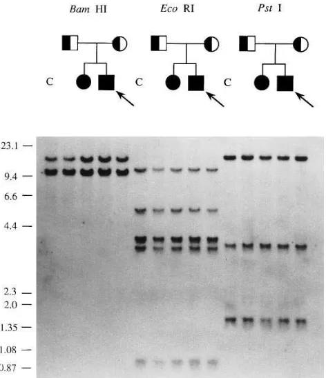
Southern blotting of genomic DNA. Neither family showed any evidence of gross rearrangement of gene structure using either the cDNA probe (Fig. 1) or the genomic DNA probe that included exon 1 (data not shown).
Family 1: investigation of cDNA. Since the Southern blot
data indicated that in both pedigrees there was no large dele-tion or inserdele-tion within the FI gene, the cDNA from the ho-mozygous FI deficient individuals was amplified by RT-PCR. Initially EBV-transformed B cells were used as the RNA sub-strate, but FI cDNA could not be reliably amplified from this source. Although fibroblasts have not been reported to
[image:4.612.59.295.58.332.2]synthe-Figure 1. Analysis of genomic DNA from all four members of family 1 (designated in pedigree form above their respective lanes) and a normal control (C). In each lane, 10 mg of DNA was digested with BamHI, EcoRI, or PstI. On the left of the autoradiograph l/HindIII and fX-174/HaeIII size markers are given (in kb). The probe used was FI coding cDNA (1.9 kb).
Table I. Location and Sequence of Oligonucleotide Primers Used in RT-PCR
Oligo Sequence Position
Primers used in RT-PCR
1R TCTAGAATTCTGTGCTTCCACTTAAGG (58–77)
2H CATGGATCCCACAGGCTTTCATCTGAG (736–753)
3R GTAGAATTCGATGACTTCTTTCAGTGT (699–716)
4H AATCGGATCCAAGGCCTTCCTACATGG (1742–1758)
5H ATTAGGATCCTGATTCCACTGGCATCC (1103–1119)
6R ACTGAATTCCTAAACTATCTTGTGGAG (996–1014)
7H TCTCGGATCCATACATTGTACTGAGA (1764–1780)
3A GCATGGAAATACAGATTCAG (380–399)
8B GCATCCATGTCAGCAGTC (950–967)
[image:4.612.54.564.539.683.2]size FI, they are known to secrete a large number of comple-ment proteins, including those of the alternative pathway (33). When fibroblast RNA was used in RT-PCR, FI cDNA was amplified after 35 cycles of PCR. Shave skin biopsies were taken from the FI deficient individuals from both families to provide an RNA source. When the two siblings from family 1 were studied by RT-PCR, the cDNA (excluding the 39 un-translated region) was amplified using oligonucleotide primers 1R and 2H, and 3R and 4H (Table I). The RT-PCR generated normal-sized products of 711 and 1078 bp, respectively from each primer pair. These products were cloned into Bluescript and sequenced. Six clones containing each RT-PCR product were sequenced, the RT-PCR was repeated and a further six clones isolated and sequenced from the second reaction. Since the sequence of the first exon (which encodes the 59 untrans-lated region and leader peptide) was not examined by RT-PCR, the first exon was amplified from genomic DNA using primers Int 1 and Pro 1. The product was then sequenced di-rectly with primers, 1A and 1B (Table II). By this combined approach, only two sequence differences were found between the cDNA sequence and the promoter sequence of the FI defi-cient homozygotes in family 1 and the published FI cDNA se-quence (6, 7) and promoter sese-quence (8). One difference was silent (see below) and the other difference was functional, an adenine to thymine transversion at nucleotide 1282,
[image:5.612.65.552.70.392.2]nomencla-ture as in (7) (Fig. 2). This nucleotide is the second base in the histidine codon (CAT) and the effect of the transversion is to alter histidine-400 to a leucine residue. The mutation occurred in the eleventh exon of the FI gene within the serine protein-ase region of the protein. The presence of this transversion was Table II. Location and Sequence of Oligonucleotides Used in PCR and Sequencing
Oligo Sequence Position
Primers used in amplifying genomic DNA
Pro1 TCTGGATTTCAGCCAAATTC (2132 to 2113)
Int1 TGGCATTGTTGTAACTGAAC (106 to 125)
Int5 ACCTCATTCAACTTTAAG (237 to 254)
5A GTAGAATTCGATGACTTCTTTCAGTGT (699–716)
6B9 CCTGCACAGCCAACTTCATC (894–913)
Int4 TGTATGAGTCATATGTAC (2137 to 2154)
2H CATGGATCCCACAGGCTTTCATCTGAG (736–753)
11A GTAGTAGACTGGATACAC (1215–1232)
11B AGCCAGAAACGATGCATG (1426–1443)
Primers used in sequencing the RT-PCR products
T3 ATTAACCCTCACTAAAG
T7 AATACGACTCACTATAG
3B TTTACTTCAACTATTCCCTC (399–418)
4B AATCCTCGGCAATGCACATG (576–595)
7A GCTTTGCATCTGTGGCTC (913–930)
8B GCATCCATGTCAGCAGTC (950–967)
11A GTAGTAGACTGGATACAC (1215–1232)
Primers used in sequencing PCR products directly
1A GAGACAAAGACCCCGAACAC (3–22)
1B CCTTGCAAAACCTTAAGTGG (68–87)
4A AGAACTATGGGTTACCAG (633–650)
6B9.seq TGTAATGCAGTCCACCTC (870–887)
11B.seq TGGAATAGGTAAGGAGAC (1400–1417)
Int4 seq CTTTCTCTTAATGACTCC (2109 to 2126)
Oligonucleotides used in genomic DNA PCR and in cycle sequencing are listed. They are named as follows: the number refers to the exon to which the primer binds, the letter, A, shows that it is complementary to the negative strand, and the letter, B, that it is the positive strand to which the primer binds. Oligonucleotides termed, Int, bind to intronic sequence, their position is indicated by their distance from the nearest exon, negative numbers denote distance upstream (59), and positive numbers distance downstream from the exon. Oligonucleotides T3 and T7 are complementary to unique sequences in the vector, pBS.
[image:5.612.317.555.534.693.2]confirmed by two means. Firstly, the eleventh exon was ampli-fied from genomic DNA by PCR using primers, 11A and 11B (see Table II). The PCR product was then sequenced directly with an internal primer, 11B.seq (see Table II). This indicated that the two FI deficient siblings in family 1 were both ho-mozygous for the 1282-T mutation and both parents were het-erozygous (data not shown). Secondly, the 1282-T mutation was found to remove an NlaIII recognition site in the wild-type sequence. Thus an NlaIII (New England Biolabs, CP Laboratories, Bishop’s Stortford, Herts, U.K.) digest was car-ried out on the 11A-11B PCR product from all four members of family 1 and a normal control (Fig. 3). The findings were in agreement with the cDNA and genomic DNA sequence data.
To ascertain whether or not the mutation at nucleotide 1282 was a polymorphic variant, 100 normal individuals were
studied by PCR. Primers 11A and 11B were used and the product digested with NlaIII. All 100 PCR products were cut by NlaIII indicating that none had the transversion at 1282 (data not shown).
Family 2: investigation of cDNA. FI cDNA was amplified
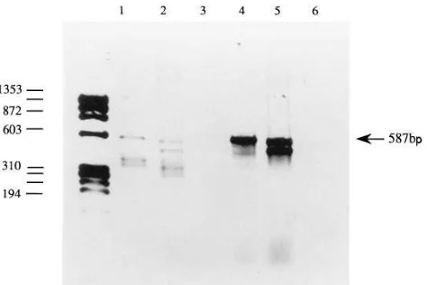
from the proband of family 2. Three oligonucleotide primer pairs 1R and 2H, 3R and 5H, and 6R and 7H were used (Table I). PCR products of 711, 439, and 802 bp were obtained respec-tively from each primer pair. There was no detectable differ-ence in size between the products from normal fibroblasts and FI deficient fibroblasts (data not shown). As in family 1, 12 clones, six from two separate RT-PCRs, were isolated and se-quenced. When 12 clones containing the PCR product of prim-ers 6R and 7H (including nucleotide 1282) were sequenced it was found that six of them were normal and the other six had the same 1282-T mutation as identified in family 1. It was con-firmed that the proband was heterozygous for the 1282 trans-version by an NlaIII digest on genomic PCR as shown in Fig. 3. The maternal FI deficient allele did not carry this mutation. No other functional changes were found when the remaining cDNA and promoter region were sequenced. However, the fact that half of the clones did not bear the 1282 mutation sug-gested that the defect on the maternal allele was compatible with RNA transcription, at least at the 39 end. It was noted that RT-PCR primers, 2H and 3R, both bound cDNA sequence that was encoded in the fifth exon of the FI gene. Thus if this exon was spliced out as a result of a splice-site mutation no PCR product would be generated from the allele with these two primers. Therefore RT-PCR was performed using primers 3A and 8B (see Table I) which lie outside the fifth exon se-quence. A single product of 587 bp was obtained from both normal and proband fibroblast RNA; a second smaller product (z 475 bp), in similar quantity to the larger one, was generated
from the proband’s RNA only (Fig. 4). The two PCR products from the proband were sequenced directly. The sequence of the smaller product revealed that the cDNA encoded by the fifth exon was completely absent (Fig. 5 a). The sequence of the larger product contained ambiguities. The RT-PCR prod-ucts were therefore cloned into pBS. Ten clones (five from one RT-PCR and five from a second reaction) containing each dif-ferent-sized RT-PCR product were isolated and sequenced. All ten clones containing the smaller RT-PCR product had the fifth exon cDNA missing, with the fourth and sixth exons cor-rectly spliced to one another (Fig. 5 b). The insert in these clones was therefore 473 bp long. All of the ten clones contain-ing the 587-bp product were the wild-type sequence (Fig. 5 c). To investigate the genomic lesion to account for this splicing defect the 39 end of the fourth intron (using primers Int 4 and 2H) and the entire fifth intron (using primers 3R and 6B9) were amplified from genomic DNA and sequenced directly. The intron four PCR product was sequenced with primer Int 4 seq. (Table II). The 108 bp at the 39 end of the fourth intron in the proband of family 2 and her mother were the same as a normal control. The complete fifth intron was directly se-quenced with primer 6B9.seq. The intron was the same as a normal control in the proband and her mother, however, the last base encoded by the fifth exon appeared to be heterozy-gous for a guanine to adenine transition in the proband and her mother. The genomic DNA PCR product from the proband, that spanned the fifth intron, was then cloned into pBS. Ten clones were sequenced (five from one PCR, and five from a second PCR). Seven of these clones contained
[image:6.612.58.263.58.168.2]wild-Figure 3. An ethidium bromide stained 3% agarose gel with fX-174/ HaeIII size markers (in bp) showing the genomic DNA PCR product obtained with oligos 11A and 11B (exon 11) that has been digested with NlaIII. The 229-bp PCR product is cut once by NlaIII in the nor-mal control (lane 5). The product generated by the proband from family 1 (lane 2) and his sister (lane 3) are not cut owing to the 1282-T mutation. The proband from family 2 (lane 6) has a normal and mutant allele at position 1282 as do the parents from family 1, the father (lane 1) and the mother (lane 4), though the digested products from the mother are not readily apparent in this photograph. The DNA of the mother from family 2 is cut by NlaIII (lane 7).
[image:6.612.59.296.492.650.2]type insert, whilst the other three had adenine at the 39 end of exon five (Fig. 6). This site corresponds to position 801 in the cDNA and at the genomic level it constitutes part of the donor (59) splice site recognition sequence of the fifth intron (34).
Analysis of the effect of the 1282-T mutation on FI protein
expression. To investigate the effect of the 1282-T mutation
fi-broblast lysates were prepared. These were analyzed by west-ern blotting using a sensitive detection system (enhanced chemiluminescence). A single signal of Mr 88,000 was
identi-fied in the cell lysates (106 fibroblasts) from a normal donor
and from the FI deficient proband from family 1 (see Fig. 7). No signal was found in the supernatant of either fibroblast line (data not shown), presumably due to the low level of expres-sion. However, FI was visualized in 4 ml of normal serum (z 140 ng) but not in the same volume of serum from the
proband (FI could not be detected when 40 ml of serum was blotted). Since the patient is capable of synthesizing FI pro-tein, detected intracellularly, one possible explanation for the absence of FI in the patient’s serum would be a secretion de-fect. FI was undetectable in the supernatant from fibroblasts using western blotting. Therefore, a more sensitive technique was used to investigate the possibility of a secretory defect. Af-ter metabolic labeling of synthesized proteins with [35
S]me-thionine, FI was immunoprecipitated and analyzed by poly-acrylamide gel electrophoresis. Secreted FI was detected in
fibroblast culture supernatants from both the FI deficient proband and from a normal donor (Fig. 8). In four separate ex-periments the amount of immunoprecipitated FI produced by the patient and normal fibroblasts was always very similar and considerably less than the levels produced by HepG2. The FI was analyzed under reducing and non-reducing conditions and identical results were produced by both the normal and pa-tient’s fibroblasts (Fig. 8). Under non-reducing conditions, a single chain of Mr 88,000 was identified, while under reducing
conditions, two forms were secreted; a single band of Mr
88,000, the pro FI molecule, and 10–20% as two bands of Mr
50,000 and 38,000 corresponding to the two chains of the ma-ture FI.The presence of two secreted forms of FI under
[image:7.612.60.296.60.437.2]reduc-Figure 5. The sequence of the two RT-PCR products generated from the proband of family 2. In panel (a), the smaller product was sequenced directly with primer, 6B9.seq. In panel (b), the smaller RT-PCR product was cloned into pBS and se-quenced with a vectderived primer. The or-der of the sequence is reversed (CTAG) in this panel. In c, the larger 587-bp RT-PCR product was cloned into pBS and sequenced with a vector-derived primer. The larger (587 bp) product is wild-type sequence, the other smaller product is normal until cDNA nu-cleotide 687 is reached, thereafter the sequence skips to nucleotide 802. Nucleotide 687 is the last (39) to be encoded by exon four and nucle-otide 802 the first (59) in exon six, the horizontal line demarcates cDNA derived from exon four (shown below) and exon six, or exon five (shown above).
[image:7.612.315.554.60.178.2]Figure 6. The sequence of genomic DNA that was amplified from the proband of family 2 with primers 6B9 (binds in exon six) and 2H (binds in exon five). The PCR product was cloned into pBS. When ten clones were sequenced the inserts were identical except at the 39-most base encoded by the fifth exon (shown by an asterisk). Seven of the clones were wild-type (guanine), but three of them were ade-nine at this position. The numbers adjacent to the sequence corre-sponds to the cDNA position. Exonic sequence is denoted in upper case and intronic DNA is given in lower case. The proband was con-cluded to be heterozygous for adenine and guanine at the 39 end of exon five, a region which forms part of the donor splice site consensus sequence of the fifth intron.
[image:7.612.316.559.504.637.2]ing conditions has been reported previously for a number of hepatoma cell lines (2).
Identification of a silent mutation. The cDNA amplified
from the probands of family 1 and family 2 were found to have a silent mutation at nucleotide 833. Both probands had a gua-nine to adegua-nine transition at this site which is in the serine-250 codon (TCG) (7). Serine-250 is encoded in the sixth exon (which encodes an LDLr module) of the FI gene. The genomic DNA was amplified by PCR using primers Int 5 and 6B9 and then sequenced directly using primer 6B9.seq. The nucleotide sequence indicated that the proband of family 1 is homozygous for 833-A as is the proband from family 2. Both mothers from the two families were 833-A/G heterozygotes, the father from family 1 was 833-A homozygous. Therefore both the 1282-T and 801-A mutations arose on the background of an 833-A al-lele, suggesting that the silent mutation may be common.
Discussion
The molecular basis of hereditary complement FI deficiency is described in two pedigrees. In the first pedigree, family 1, there were two siblings both of whom had no detectable circu-lating FI, and no functional alternative pathway activity as as-sessed by haemolytic assay. The younger sibling, the proband, had a history of recurrent pyogenic infections and a single epi-sode of N. meningitidis meningitis. His older (by five years) sis-ter had no history of increased susceptibilty to infection. Anal-ysis of the FI gene by Southern blotting gave no evidence of gross structural rearrangement and hence the affected siblings
were investigated by sequencing their FI cDNA after RT-PCR. The only functional change in sequence identified was a transversion (A to T) at nucleotide 1282. The effect of this mu-tation is to alter histidine-400 to leucine. It could be argued that the 1282-T mutation is not the basis for FI deficiency in family 1 but is simply a rare polymorphic variant. Two lines of evidence suggest that this is unlikely. Firstly, the transversion was not present in 100 normal DNA samples (200 alleles); and secondly, the mutation was identified on one of the FI alleles from the proband of family 2. Both siblings from family 1 were homozygous for this mutation when genomic DNA was ana-lyzed. The reasons for the disparity in clinical manifestations between the two siblings are not known. Clearly the fact that the molecular lesion is identical in both siblings indicates that the variation in phenotype is not related to genotype at the FI locus. Environmental factors may play a role, but since the on-set of infection was at 18 mo in the proband, and the infections were recurrent involving different organ systems, an additional genetic component is implicated in disease susceptibility. Fur-thermore, there was no obvious environmental difference ex-perienced by the two siblings. It may be speculated that since there is considerable redundancy in both the innate and adap-tive immune systems, then variation at other loci could modify disease expression.
The 1282-T mutation occurs within the eleventh exon of the FI gene. Exons 10 through 13 encode the serine proteinase region of FI (8). The serine proteinases are a large group of bi-ologically important enzymes that have been extensively stud-ied. This category of enzymes includes, trypsin, other comple-ment proteins, C2, C1r/s, factor D and factor B, and proteins of the fibrinolytic and coagulation cascades (35). When the serine proteinase amino acid sequences are aligned on the basis of crystallographic data and consensus secondary structure pre-dictions (36), it is apparent that the histidine-400 residue (which is mutated to leucine by 1282-T) in FI is a semi-con-served histidine residue. Within the serine proteinase family much is known of the molecular mechanism of their catalytic activity, and amino acid structure that determines substrate specificity and susceptibility to specific inhibitors (37, 38). No function has been allocated to histidine-400 in FI, or the semi-conserved histidine in the other serine proteinases, however, the presence of a semi-conserved residue in homologous pro-teins with differing specificities has been suggested to imply a structural rather than functional role for that residue (39).
The effect of the leucine substitution was investigated by western blotting of fibroblast lysates (Fig. 7). The technique al-lows the detection of less than 100 ng of protein. No circulating FI could be detected in the serum of the proband. The pres-ence of intracellular FI in the fibroblasts from the proband of family 1 raised the question as to whether the leucine substitu-tion impedes normal FI secresubstitu-tion. Therefore we compared the supernatant of primary fibroblast cultures from the patient and a normal individual with that of a hepatoma cell line (Hep G2). After metabolic labeling, we were able to precipitate newly synthesized FI from the supernatant of the patient’s cells (Fig. 8). While the amount of FI secreted was substan-tially less than produced by the reference hepatoma cell line, it was very similar to the level of FI secreted by the normal fibro-blasts. Thus the patient’s cells were capable, in vitro, of synthe-sizing and secreting a FI molecule, yet no FI was detectable in the patient’s serum. There are two possible explanations for this apparent discrepancy. Firstly, the histidine to leucine
[image:8.612.56.262.56.254.2]tation may affect the stability of the FI in serum. This would be very difficult to assess experimentally due to the very low lev-els of FI synthesized by the fibroblasts. Secondly, since the ma-jority of serum FI is synthesized by the liver (2), hepatocytes may utilize different mechanisms from fibroblasts to process and secrete FI. Indeed, there is some evidence that this is the case since different hepatoma cell lines (Hep 3B, Hep G2, and NPLC-KC) process FI differently (2). Thus, the histidine to leucine mutation may prevent secretion of the FI by the liver while it remains unaffected in fibroblasts.
The sequence data from the proband of family 2 and her mother revealed that her paternally-derived FI allele had the same transversion as occurred in family 1. The second family originate from northern England, whereas the first family are from western Scotland. There is no known connection be-tween the two families. Initially no maternally derived muta-tion was evident. When different primers were used for RT-PCR an alternatively spliced message, in similar amounts to normal-sized mRNA, was identified. The entire cDNA that encoded the fifth exon was absent from one allele in the proband. The acceptor (39) splice site of the fourth intron and the entire fifth intron were amplified from genomic DNA and sequenced directly. The only mutation identified was a gua-nine to adegua-nine transition at cDNA nucleotide 801, which is the 39-most base encoded by the fifth exon (see Fig. 6). It therefore forms part of the donor (59) splice site of the fifth in-tron. Both the mother and the proband were heterozygous for this mutation which was compatible with the data indicating that the 1282-T mutation on the other allele was not mater-nally derived.
The last base encoded by the exon at a donor splice site is not invariably guanine. In a recent large survey of 1446 splice sites (34), guanine was present at this position (denoted -1) in 78% of cases. Adenine was the next most common base with a frequency of 11%. Thus a change from guanine to adenine might be expected to leave some residual splice site activity on the 801-A mutant allele found in family 2. In a review of 101 splice site mutations (40), mutations affecting positions -1 and -2 in the donor splice were less common than would be ex-pected when compared with mutations affecting the invariant, intronic GT at positions 11 and 12. This presumably reflects a detection bias since mutations outside of the invariant GT may permit some splicing activity. Three examples of a guanine to adenine transition at position -1 of a donor splice have been re-ported in detail in the literature (41–43). In each instance there was skipping of the upstream exon and in two cases there was a second transcript owing to residual splicing activity at the mutated donor splice site. One of these was in the COL1A2 gene and resulted in type VII Ehlers-Danlos syndrome (41). The cryptically spliced product was in-frame but the transition resulted in an amino acid substitution (isoleucine for methio-nine). The other mutation was in the b-hexosamidase A gene and caused infantile Tay-Sachs disease (42). The cryptically spliced transcript was also in-frame but there was no amino acid substitution. In the example studied in this paper only one transcript was detected from the 801-A mutant allele. One similar case has been reported (43). In this example the transi-tion at -1 occurred in the porphobilinogen deaminase gene and resulted in acute intermittent porphyria.
The fifth exon of FI encodes the first of two adjacent LDLr modules in the heavy chain. The distance that separates the fifth from the sixth exon (also an LDLr module) is small, 142
bp (8). The fact that there are no other cryptic splice sites in the vicinity of the mutated donor site of the fifth intron is com-patible with the small size of the fifth intron. In addition, if the scoring system (which reflects the similarity of any given splice site to the consensus sequence) devised by Shapiro and Senap-athy (34) is applied to the wild-type sequence of the fifth exon’s donor splice site, the score (using the primate frequency distribution) is 69%. This score is low in comparison with the value of 82%, which is the mean score observed in 62 wild-type donor splice sites that have been documented as being subject to mutation (40). Thus the donor splice site of the fifth intron of the FI gene is relatively weak and the transition at -1 (which reduces the Shapiro-Senapathy score to 56%) is unlikely to al-low any significant residual splicing activity. The fact that a do-nor splice site mutation causes skipping of the upstream exon during pre-mRNA processing has implications for the mecha-nism of RNA splicing. The skipping of the upstream exon can not be explained by independent recognition of the donor and acceptor splice sites. Such a model would generate a transcript containing the intronic sequence downstream of the mutated donor splice site. In recent models of spliceosome assembly (44) the recognition of the acceptor splice site by the spliceo-some complex is followed by scanning downstream to the 39
end of the adjacent exon. A mutation at this point (as in the 801-A mutation) would impede spliceosome assembly with loss of the complex at the 59 and 39 ends of the exon, thereby leading to complete excision of the exon and its adjacent in-trons during splicing. This is the situation identified in the 801-A mutant allele in family 2.
The intron phase between the fifth and its neighboring ex-ons is the same, phase I (8). Because of the cex-onserved intron phase it is possible that the shortened mRNA is translated and processed to a smaller product of 523 (normal 561) amino ac-ids. This possibility was not investigated.
A silent mutation (guanine to adenine) at nucleotide 833 was identified. The transition at this position was present on both of the FI deficient mutant alleles described in this paper. In addition, the father from family 1 was homozygous 833-A, which suggests that the silent mutation is relatively common.
In conclusion, two different molecular lesions that give rise to FI deficiency have been identified. In one pedigree two sib-lings with FI deficiency (although one is asymptomatic) have the same missense mutation within the serine proteinase re-gion of FI. In the resulting protein leucine replaces histidine-400, the latter is a semi-conserved histidine within the serine proteinase family. No function has been ascribed to this histi-dine residue. It was demonstrated that fibroblasts from the pa-tient can synthesize and secrete the mutant FI protein in vitro but no FI was detectable in serum derived from the patient. The mechanism by which the histidine-400 mutation causes FI deficiency has not been determined. In the second pedigree, the proband was found to be a compound heterozygote. One allele carried the same mutation as identified in family 1, the second allele had an unusual donor splice site mutation that caused skipping of the sequence encoded by the fifth exon from its processed transcript. This is the first description of the genetic defect in FI deficiency.
Acknowledgments
was originally reported by Dr. R.A. Thompson, Regional Immunol-ogy Laboratory, East Birmingham Hospital, Birmingham, and Pro-fessor P.J. Lachmann, MRC Molecular Immunopathology Unit, Hills Road, Cambridge. We acknowledge Ms. Jenny Morris, Dermatology Unit, RPMS for advice concerning the initiation of fibroblast cell lines and Dr. P.K. Potter, Rheumatology Unit, RPMS for advice con-cerning Western blotting. We thank Dr. S.J. Perkins, Biochemistry Department, Royal Free Hospital Medical School, Rowland Hill St., London for helpful discussion concerning the structure of factor I and use of molecular graphics equipment. Dr. R.B. Sim, MRC Immu-nochemistry Unit, Oxford, kindly provided the factor I cloned cDNA, the anti-factor I monoclonal antibody, MRC OX21, the anti–factor H monoclonal antibody, MRC OX 23, and the purified factor I protein used in Western blotting.
Dr. T.J. Vyse was supported by an Arthritis and Rheumatism Council (ARC) Research Fellowship and Dr. K.A. Davies is an ARC Senior Research Fellow.
References
1. Vyse, T.J., P.J. Späth, K.A. Davies, B.J. Morley, P. Philippe, P. Athanas-siou, C.M. Giles, and M.J. Walport. 1994. Hereditary complement factor I defi-ciency. Quart. J. Med. 87:385–402.
2. Goldberger, G., M.A. Arnaout, D. Aden, R. Kay, M. Rits, and H.R. Col-ten. 1984. Biosynthesis and postsynthetic processing of human C3b/C4b inacti-vator (factor I) in three hepatoma cell lines. J. Biol. Chem. 259:6492–6497.
3. Whaley, K. 1980. Biosynthesis of the complement components and the regulatory proteins of the alternative complement pathway by the human pe-ripheral blood monocytes. J. Exp. Med. 151:501–516.
4. Julen, N., H. Dauchel, C. Lemercier, R.B. Sim, M. Fontaine, and J. Ri-poche. 1992. In vitro biosynthesis of complement factor I by human endothelial cells. Eur. J. Immunol. 22:213–217.
5. Vetvicka, V., W. Reed, M.L. Hoover, and G.D. Ross. 1993. Complement factors H and I synthesised by B cell lines function to generate a growth factor activity from C3. J. Immunol. 150:4052–4060.
6. Goldberger, G., G.A.P. Bruns, M. Rits, M.D. Edge, and D.J. Kwiat-kowski. 1987. Human complement factor I: analysis of cDNA-derived primary structure and assignment of its gene to chromosome 4. J. Biol. Chem. 262: 10065–10071.
7. Catterall, C.F., A. Lyons, R.B. Sim, A.J. Day, and T.J.R. Harris. 1987. Characterization of primary amino acid sequence of human complement con-trol protein factor I from an analysis of cDNA clones. Biochem. J. 242:849–856. 8. Vyse, T.J., G.P. Bates, M.J. Walport, and B.J. Morley. 1994. The organi-sation of the human complement factor I gene: a member of the serine protease gene family. Genomics. 24:90–98.
9. Shiang, R., J.C. Murray, C.C. Morton, K.H. Buetow, J.J. Wasmuth, A.H. Olney, W.G. Sanger, and G. Goldberger. 1989. Mapping of the human comple-ment factor I gene to 4q25. Genomics. 4:82–86.
10. Nagasawa, S., and R.M. Stroud. 1977. Mechanism of action of the C3b inactivator: Requirement for a high molecular weight cofactor (C3b-C4bINA cofactor) and production of a new C3b derivative (C3b9). Immunochemistry.
14:749–756.
11. Pangburn, M.K., R.D. Schreiber, and H.J. Müller-Eberhard. 1977. Hu-man complement C3b inactivator: isolation, characterization, and demonstra-tion of an absolute requirement for the serum protein b1H for cleavage of C3b and C4b in solution. J. Exp. Med. 146:257–270.
12. Alper, C.A., N. Abramson, J.B. Johnston, J.H. Jandl, and F.S. Rosen. 1970. Increased susceptibility to infection associated with abnormalities of com-plement-mediated functions and of the third component of complement (C3).
N. Engl. J. Med. 282:349–352.
13. Abramson, N., C.A. Alper, P.J. Lachmann, F.S. Rosen, and J.H. Jandl. 1971. Deficiency of C3 inactivator in man. J. Immunol. 107:19–27.
14. Alper, C.A., F.S. Rosen, and P.J. Lachmann. 1972. Inactivator of the third component of complement as an inhibitor in the properdin pathway. Proc. Natl. Acad. Sci. USA. 69:2910–2913.
15. Ziegler, J.B., C.A. Alper, R.S. Rosen, P.J. Lachmann, and L. Shering-ton. 1975. Restoration by purified C3b inactivator of complement-mediated function in vivo in a patient with C3b inactivator deficiency. J. Clin. Invest. 55: 668–672.
16. Lachmann, P.J., and P. Nicol. 1973. Reaction mechanism of the alterna-tive pathway of complement fixation. Lancet. 1:465–467.
17. Møller Rasmussen, J., B. Teisner, H.H. Jepsen, S.E. Svehag, F. Knud-sen, H. Kirstein, and M. Buhl. 1988. Three cases of factor I deficiency: the effect of treatment with plasma. Clin. Exp. Immunol. 74:131–136.
18. Solal-Celigny, P., M. Laviolette, J. Hebert, P.C. Atkins, M. Sirois, G. Brun, G. Lehner-Netsch, and J.M. Delâge. 1982. C3b inactivator deficiency with immune complex manifestations. Clin. Exp. Immunol. 47:197–205.
19. Maillet, F., L. Weiss, J. Chibani, and M. Kazatchkine. 1990. Déficit en facteur I, une proteine regulatrice du complement. Nouv. Presse. Med. 19:762. (letter).
20. Harrison, R.A., and P.J. Lachmann. 1986. Complement technology. In
Handbook of Experimental Immunology, Volume 1, Immunochemistry. D.M. Weir, L.A. Herzenberg, C. Blackwell, and Leonore A. Herzenberg, editors. Blackwell Scientific Publications, Oxford, 39.1–39.49.
21. Thompson, R.A., and P.J. Lachmann. 1977. A second case of human C3b inhibitor (KAF) deficiency. Clin. Exp. Immunol. 27:23–29.
22. Sambrook, J., E.F. Fritsch, and T. Maniatis. 1989. Molecular Cloning: A Laboratory Manual. Cold Spring Harbor Laboratory Press., Cold Spring Har-bor, New York.
23. Southern, E. 1975. Detection of specific sequences among DNA frag-ments separated by gel electrophoresis. J. Mol. Biol. 98:503–517.
24. Jay, E., R. Bambura, R. Paomanabhan, and W.U. Ray. 1974. DNA se-quence analysis: a general simple and rapid method for sequencing large oli-godeoxyribonucleotide fragments by mapping. Nucleic Acids Res. 1:331–353.
25. Feinberg, A.P., and B. Vogelstein. 1983. A technique for radiolabeling DNA restriction endonuclease fragments to high specific activity. Anal. Bio-chem. 132:6–13.
26. Chomczynski, P., and N. Sacchi. 1987. Single-step method of RNA isola-tion by acid guanidium thiocyanate-phenol-chloroform extracisola-tion. Anal. Bio-chem. 162:156–159.
27. Innis, M.A., K.B. Myambo, D.H. Gelfand, and M.A.D. Brow. 1988. DNA sequencing with Thermus aquaticus DNA polymerase and direct se-quencing of polymerase chain reaction-amplified DNA. Proc. Natl. Acad. Sci. USA. 85:9436–9440.
28. Engelke, D.R., P.A. Hoener, and F.S. Collins. 1988. Direct sequencing of enzymically amplified human DNA. Proc. Natl. Acad. Sci. USA. 85:544–548.
29. Laemmli, U.K. 1970. Cleavage of structural proteins during the assem-bly of the head of bacteriophage T4. Nature (Lond.). 277:680–685.
30. Towbin, H., T. Staehelin, and J. Gordon. 1979. Electrophoretic transfer of proteins from polyacrylamide gels to nitrocellulose sheets: procedure and source applications. Proc. Natl. Acad. Sci. USA. 76:4350–4354.
31. Sim, R.B., A.J. Day, B.E. Moffatt, and M. Fontaine. 1993. Complement factor I and cofactors in control of complement system convertase enzymes.
Methods Enzymol. 223:13–35.
32. Whitehead, T.P., L.J. Kricka, T.J.N. Carter, and G.H.G. Thorpe. 1979. Analytical chemiluminescence: its potential in the clinical laboratory. Clin. Chem. 25:1531–1546.
33. Katz, Y., and R.C. Strunk. 1988. Synthesis and regulation of comple-ment protein factor H in human skin fibroblasts. J. Immunol. 141:559–563.
34. Shapiro, M.B., and P. Senapathy. 1987. RNA splice junctions of differ-ent classes of eukaryotes: sequence statistics and functional implications in gene expression. Nucleic Acids Res. 15:7155–7174.
35. Neurath, H. 1985. Proteolytic enzymes, past and present. Fed. Proc. 44: 2907–2913.
36. Perkins, S.J., and K.F. Smith. 1993. Identity of the putative serine-pro-teinase fold in proteins of the complement system with nine relevant crystal structures. Biochem. J. 295:109–114.
37. Kraut, J. 1977. Serine proteases: structure and mechanism of catalysis.
Ann. Rev. Biochem. 46:331–358.
38. Bode, W., E. Meyer, and J.C. Powers. 1989. Human leucocyte and por-cine pancreatic elastase: X-ray crystal structures, mechanism, substrate specific-ity, and mechanism-based inhibitors. Biochemistry. 28:1951–1963.
39. Patthy, L. 1993. Modular design of proteases of coagulation, fibrinolysis, and complement activation: Implications for protein engineering and structure-function studies. Methods Enzymol. 222:10–22.
40. Krawczak, M. , J. Reiss, and D.N. Cooper. 1992. The mutational spec-trum of single base-pair substitutions in mRNA splice junctions of human genes: causes and consequences. Hum. Genet. 90:41–54.
41. Weil, D., M. D’Alessio, F. Ramirez, B. Steinmann, M.K. Wirtz, R.W. Glanville, and D.W. Hollister. 1989. Temperature-dependent expression of a collagen splicing defect in the fibroblasts of a patient with Ehlers-Danlos syn-drome type VII. J. Biol. Chem. 264:16804–16809.
42. Akli, S., J. Chelly, C. Mezard, S. Gandy, A. Kahn, and L. Poenaru. 1990. A “G” to “A” mutation at position -1 of a 59 splice site in a late infantile form of Tay-Sachs disease. J. Biol. Chem. 265:7324–7330.
43. Grandchamp, B., C. Picat, F. de Rooji, C. Beaumont, P. Wilson, J.C. Daybach, and Y. Nordmann. 1989. A point mutation G-A in exon 12 of the por-phobilinogen deaminase gene results in an exon skipping and is responsible for acute intermittent porphyria. Nucleic Acids Res. 17:6637–6649.