ScholarWorks @ Georgia State University
Kinesiology Dissertations Department of Kinesiology and Health
Spring 5-13-2016
Antioxidant Therapy Attenuates Post-Infarct
Cardiac Remodeling by Driving Expression of
Krüppel-Like Factor 15
Russell George Rogers III
Follow this and additional works at:https://scholarworks.gsu.edu/kin_health_diss
This Dissertation is brought to you for free and open access by the Department of Kinesiology and Health at ScholarWorks @ Georgia State University. It has been accepted for inclusion in Kinesiology Dissertations by an authorized administrator of ScholarWorks @ Georgia State University. For more information, please [email protected].
Recommended Citation
Rogers, Russell George III, "Antioxidant Therapy Attenuates Post-Infarct Cardiac Remodeling by Driving Expression of Krüppel-Like Factor 15." Dissertation, Georgia State University, 2016.
This dissertation, ANTIOXIDANT THERAPY ATTENUATES POST-INFARCT CARDIAC
REMODELING BY DRIVING EXPRESSION OF KRÜPPEL-LIKE FACTOR 15, by
RUS-SELL G. ROGERS III, was prepared under the direction of the candidate’s Dissertation
Advi-sory Committee. It is accepted by the committee members in partial fulfillment of the
require-ments for the degree, Doctor of Philosophy, in the College of Education and Human
Develop-ment, Georgia State University.
The Dissertation Advisory Committee and the student’s Department Chairperson, as
representa-tives of the faculty, certify that this dissertation has met all standards of excellence and
scholar-ship as determined by the faculty.
______________________________ _____________________________________ Jeffrey. S. Otis, Ph.D. Christopher P. Ingalls, Ph.D.
Committee Chair Committee Member
______________________________ ______________________________________ Brett J. Wong, Ph.D. Michael W. Hart, DVM
Committee Member Committee Member
______________________________ Date
______________________________ Mark Geil, Ph.D.
Chairperson, Department of Kinesiology and Health
______________________________ Paul A. Alberto, Ph.D.
Dean
By presenting this dissertation as a partial fulfillment of the requirements for the advanced degree
from Georgia State University, I agree that the library of Georgia State University shall make it
available for inspection and circulation in accordance with its regulations governing materials of
this type. I agree that permission to quote, to copy from, or to publish this dissertation may be
granted by the professor under whose direction it was written, by the College of Education and
Human Development’s Director of Graduate Studies, or by me. Such quoting, copying, or
pub-lishing must be solely for scholarly purposes and will not involve potential financial gain. It is
un-derstood that any copying from or publication of this dissertation which involves potential
finan-cial gain will not be allowed without my written permission.
All dissertations deposited in the Georgia State University library must be used in
accordance with the stipulations prescribed by the author in the preceding statement. The author
of this dissertation is:
Russell G. Rogers, III 2383 Akers Mill Rd., APT#F16
Atlanta, GA 30339
The director of this dissertation is:
Jeffrey S. Otis, PhD
Department of Kinesiology and Health College of Education and Human Development
Doctoral Candidate Georgia State University Department of Kinesiology & Health
125 Decatur Street Atlanta, GA 30303
Email: [email protected]
EDUCATION
Doctor of Philosophy, Muscle Biology 2016
Georgia State University, Atlanta, GA
Dissertation: "Antioxidant Therapy Attenuates Post-Infarct Cardiac Remodeling by Driving Expression of Krüppel-Like Factor 15"
Committee: Jeffrey S. Otis, PhD (Advisor) Christopher P. Ingalls, PhD Brett J. Wong, PhD
Michael W. Hart, DVM
Master of Education, Exercise Physiology 2011
Auburn University, Auburn, AL
Bachelor of Science, Exercise and Health Science 2010 Kennesaw State University, Kennesaw, GA
PROFESSIONAL CREDENTIALS
Instructor 2013 - Present
Department of Kinesiology and Health Georgia State University
Atlanta, GA
Graduate Research Assistant Muscle Biology Laboratory 2012 - Present Department of Kinesiology and Health
Refereed Journal Articles
1) Rogers, R.G., Baumann, C.W., & Otis, J.S. (2015). Recovery of Skeletal Muscle Function Following Injury is Not Augmented by Acute Resveratrol Supplementation. International Journal of Clinical and Experimental Physiology, 2, 29-33.
2) Baumann, C.W., Rogers, R.G., Lees, S.J., & Otis, J.S. (2014) Muscular Strength is Unaf-fected by Short-Term Resveratrol Supplementation in Aged Mouse Muscle. International Journal of Clinical and Experimental Physiology, 1, 253-257.
3) Baumann, C.W., Rogers, R.G., Gahlot, N., & Ingalls C.P. (2014) Eccentric Contractions Disrupt FKBP12 Content in Mouse Skeletal Muscle. Physiological Reports, 2 (7), e12081.
Published Abstracts
1) Rogers, R.G., Baumann, C.W., & Otis, J.S. (2015). Recovery of Skeletal Muscle Function is Not Augmented by Acute Resveratrol Supplementation after Injury. Medicine and Science in Sports and Exercise, 47, 960-961.
2) Baumann, C.W., Rogers, R.G., & Otis, J.S. (2015). Functional Deficits are Not Attenuated by Increasing Hsp70 Content Prior to Trauma-induced Injury. Medicine and Science in Sports and Exercise, 47, 505-510.
3) Rogers, R.G., Baumann, C.W., Santamore, W., Gorman, J.H., Gorman, R.C., & Ingalls, C.P. (2014). A Biocompatible Tissue Filler Attenuates Junctophilin 2 Loss after A Myocardial In-farction In Sheep Heart. Medicine and Science in Sports and Exercise, 46, 660-661.
4) Baumann, C.W., Rogers, R.G., Gahlot, N., & Ingalls, C.P. (2014). Loss of FKBP12 is Asso-ciated with Early Strength Deficits after Contraction Induced Skeletal Muscle Injury. Medi-cine and Science in Sports and Exercise, 46, 992.
Poster Presentations at National and International Conferences
1) Rogers, R.G., & Otis, J.S. Antioxidant Therapy Attenuates Post-Infarct Left Ventricular Re-modeling by Driving Expression of the Transcriptional Repressor Krüppel-Like Factor 15. Experimental Biology. San Diego, CA, April 2016.
2) Rogers, R.G., Baumann, C.W., & Otis, J.S. Recovery of Skeletal Muscle Function is Not Augmented by Acute Resveratrol Supplementation after Injury. American College of Sports Medicine Annual Meeting. San Diego, CA, May 2015.
3) Rogers, R.G., Baumann, C.W., & Otis, J.S. Recovery of Skeletal Muscle Function is Not Augmented by Acute Resveratrol Supplementation after Injury. American College of Sports Medicine Southeast Regional Chapter Annual Meeting. Jacksonville, FL, February 2015. 4) Rogers, R.G., Baumann, C.W., Santamore, W., Gorman, J.H., Gorman, R.C., & Ingalls, C.P.
BY DRIVING EXPRESSION OF KRÜPPEL-LIKE FACTOR 15
by
RUSSELL G. ROGERS, III
Background: Myocardial infarction (MI) results in severe biochemical, physiological, and
cellu-lar changes that lead to alterations in the structure and function of the myocardium. Oxidative
stress potentiates this remodeling response and is associated with progressive worsening of
car-diac function. Accordingly, we used a powerful antioxidant-based therapeutic strategy to
im-prove cardiac health and study redox-dependent signaling. Methods: MI was surgically induced
in rats by ligating the left anterior descending coronary artery. Subgroups of MI rats received
resveratrol (i.p., 10 mg/kg/day for 28 days beginning immediately post-MI). Cardiac histology
and biochemical analyses of genes and proteins implicated in cardiac fibrosis, hypertrophy, and
apoptosis, and redox-dependent signaling were analyzed. Results: As expected, MI resulted in
profound structural changes to the myocardium. Further, we observed a sharp reduction in
nu-clear factor-erythroid 2-related factor 2 (Nrf2) and Krüppel-like factor 15 (KLF15), factors that
are responsible for maintaining the endogenous antioxidant capacity and regulating cardiac gene
expression, respectively. It is likely that disruption of normal KLF15 signaling permitted the
ex-pression of several cardiac genes associated with progressive cardiac remodeling. Importantly,
daily treatment with resveratrol ameliorated cardiac remodeling, improved redox state, restored
Nrf2 expression, and up-regulated KLF15 expression. Further, induction of KLF15 signaling
fol-lowing resveratrol treatment is associated with attenuated expression of several genes implicated
in cardiac remodeling. Conclusions: Chronic oxidative stress potentiates cardiac remodeling
post-infarct, in part, by suppressing Nrf2 and KLF15 expression. Importantly, we demonstrate
that normal KLF15 signaling may be rescued with an antioxidant-based therapy, which may be
an attractive therapeutic target to support cardiac health post-MI.
BY DRIVING EXPRESSION OF KRÜPPEL-LIKE FACTOR 15
by
RUSSELL G. ROGERS, III
A Dissertation
Presented in Partial Fulfillment of Requirements for the
Degree of
Doctor of Philosophy
in
Kinesiology (Exercise Physiology: Muscle Biology)
in
Department of Kinesiology and Health
in
The College of Education and Human Development
Georgia State University
Copyright by Russell G. Rogers, III
I would like to dedicate this dissertation, in partial fulfillment of the Doctor of
Philoso-phy degree, to my family, friends, and colleagues that provided support during my collegiate
ACKNOWLEDGMENTS
I would like to take this opportunity to acknowledge the individuals that contributed to
my professional development. First and foremost, I would like to thank Dr. Jeffrey Otis for his
exceptional job as my doctoral mentor, and guidance throughout my PhD program and into my
post-doctoral endeavors. Without his support, my transition into the study of the diseased heart
would not have been possible. In parallel, he provided me with the skill-set necessary to succeed
as a post-doctoral scientist at Cedars-Sinai Heart Institute. In addition, I would like to thank my
dissertation committee members for their help and support during my dissertation research
TABLE OF CONTENTS
LIST OF TABLES V
LIST OF FIGURES VI
ABBREVIATIONS VII
CHAPTER ONE 1
MYOCARDIAL INFARCTION AND CARDIAC REMODELING 1
Introduction 1
Characteristics of Myocardial Infarction 3
Histopathology of Myocardial Infarction 4
Functional Evidence of Myocardial Infarction 5
Molecular and Cellular Events During Myocardial Infarction 6
Etiology of Myocardial Infarction 6
Anoxic Intracellular Pathophysiology 7
Cellular Death Pathways 8
Inflammatory Response 10
Post-Infarct Cardiac Remodeling 12
Oxidative Stress in the Myocardium 15
Oxidative Stress-Dependent Cardiac Remodeling 17
Krüppel Like Factors 21
KLFs in the Myocardium 22
General Functions of KLF15 in the Myocardium 23
KLF15 as a Negative Regulator of Cardiac Remodeling 25
Regulation of KLF15 27
Resveratrol Treatment 28
Hypotheses and Specific Aims 30
CHAPTER TWO 42
ANTIOXIDANT THERAPY ATTENUATES POST-INFARCT CARDIAC
REMODELING BY DRIVING EXPRESSION OF KLF15 42
Introduction 42
Methods 44
Results 50
Discussion 63
References 68
LIST OF TABLES
Table 1. Real-Time PCR Primer Pairs 53
LIST OF FIGURES
Figure 1. Hypotheses and Specific Aims 3
Figure 2. Photomicrographs from Mouse Hearts 5
Figure 3. Flow Chat of Ventricular Remodeling 12
Figure 4. Depiction of the CTGF Gene as a Downstream Target of TGF-β Signaling 19
Figure 5. Depiction of the Transcription Factors Mef2 and GATA4 as Downstream
Targets 21
Figure 6. Myocardial Infarct Scar Size 54
Figure 7. Cardiomyocyte Cross-Sectional Area 55
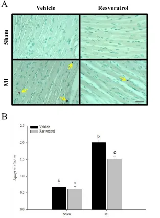
Figure 8. Cardiomyocyte Apoptosis 56
Figure 9. Gene Expressions of Anti- and Pro-Apoptotic Factors 57
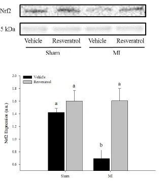
Figure 10. Nrf2 Protein Expression 58
Figure 11. Gene Expressions of Antioxidant and Oxidant Enzymes 59
Figure 12. Gene Expressions of Glutathione Synthesizing and Handling Enzymes 60
Figure 13. KLF15 Protein Expression 61
ABBREVIATIONS
ANP Atrial natriuretic peptide ATP Adenosine triphosphate ATPase Adenosine triphosphatase BNP B-type natriuretic peptide CTGF Connective tissue growth factor Cu/Zn-SOD1 Copper-zinc superoxide dismutase CVD Cardiovascular disease
DNA Deoxyribonucleic acid
Ec-SOD3 Extracellular superoxide dismutase GATA4 GATA binding protein 4
GPx Glutathione peroxidase
GSH Glutathione (reduced form) H2O2 Hydrogen peroxide
IGF-1 Insulin-like growth factor-1 IL-1β Interleukin-1beta
IL-6 Interleukin-6
iNOS Inducible nitric oxide synthase KChIP2 Kv channel-interacting protein 2
KLF Krüppel-like factor family
LAD Left anterior descending coronary artery Mef2 Myocyte enhancer factor 2
MI Myocardial infarction
Mn-SOD2 Manganese superoxide dismutase
mPTP Mitochondrial permeability transition pore NAD+ Nicotinic adenine dinucleotide (oxidized form) NADH Nicotinic adenine dinucleotide (reduced form)
NADPH Nicotinic adenine dinucleotide phosphate (reduced form) NF-κB Nuclear factor kappa-light-chain-enhancer of activated B cells Nox2 Nicotinic adenine dinucleotide phosphate oxidase 2
Nox4 Nicotinic adenine dinucleotide phosphate oxidase 4 Nrf2 Nuclear factor-erythroid 2-related factor
O2- Superoxide
OH- Hydroxyl radical
P/CAF P300/CREB-binding protein-associated factor
PGC-1 Peroxisome proliferator-activated receptor-gamma co-activator-1 PPAR-α Peroxisome proliferator-activated receptor-alpha
ROS Reactive oxygen species
SERCA Sarcoplasmic-endoplasmic reticulum calcium ATPase Smad3 Mothers against decapentaplegic homolog 3
SOD Superoxide dismutase
TGF-β Transforming growth factor-beta TLR Toll-like receptor
TNF-α Tumor necrosis factor-alpha
CHAPTER ONE
MYOCARDIAL INFARCTION AND CARDIAC REMODELING
Introduction
The high prevalence of cardiovascular disease (CVD) in the Western world is responsible
for approximately thirty-three percent of all-cause mortality in the United States. In addition,
each year nearly 1.5 million Americans experience a myocardial infarction [1, 2], which directly
contributes to mortality or secondary heart failure. The Centers for Disease Control and
Preven-tion estimate that 5.1 million people have been diagnosed with heart failure and approximately
half die within 5 years of diagnosis. Clearly, continued research investigating the molecular and
cellular mechanisms underlying the pathogenesis of CVD to develop effective treatment options
is warranted.
Importantly, myocardial infarction has been associated with a series of pathological
mo-lecular and cellular changes that result in remodeling of the myocardium. The pathologic
myo-cardium is associated with cardiac dysfunction as it progresses to secondary heart failure.
Moreo-ver, several lines of literature implicate oxidative stress as a primary feature of cardiac
remodel-ing followremodel-ing clinical and experimental myocardial infarction. Genetic studies that employ either
knockout of reactive oxygen species producing enzymes or overexpression of antioxidant
de-fense enzymes demonstrate remarkable protection of the myocardium to pathological
remodel-ing. Importantly, treatment with antioxidants has also proven beneficial to attenuate cardiac
stress in the promotion of a pathological cardiac phenotype. However, the underlying
mecha-nisms that drive oxidative stress-dependent remodeling following myocardial infarction have not
been fully elucidated.
Further, interstitial fibrosis and cardiac hypertrophy are cardinal features of the
remodel-ing myocardium in response to pathological stress. These processes appear to be under strict
con-trol of connective tissue growth factor (CTGF), myocyte enhancer factor 2 (Mef2), and GATA
binding protein 4 (GATA4), which are robustly up-regulated in response to pressure overload.
Importantly, the transcriptional repressor Krüppel-like factor 15 (KLF15) negatively regulates
the transcription of CTGF and transcriptional activity of Mef2 and GATA4. In response to in
vivo pressure overload and in vitro oxidative stress, KLF15 is dramatically down-regulated
which alleviates its inhibitory effects and thus, permits expression of pro-fibrotic and
pro-hyper-trophic genes.
While antioxidant treatment has proven beneficial in attenuating cardiac remodeling
fol-lowing myocardial infarction, the redox-sensitive mechanisms responsible for its efficacy remain
to be fully identified. To that end, no study to date has identified the expression of KLF15 in the
myocardium following infarction. Here we attempt to identify the expression profile of KLF15
and its sensitivity to the redox state in the myocardium following infarction. Importantly, we
hy-pothesize that treatment with resveratrol, a powerful antioxidant, will induce KLF15 expression
to repress cardiac gene expression associated with remodeling post-MI. For clarity, we have
Figure 1. Hypotheses and specific aims wherein myocardial infarction induces oxidative stress (SA 1), which down-regulates KLF15 and permits the expression of fibrogenic and hypertrophic genes (SA 2). Expression of these genes leads to overt structural remodeling of the myocardium (SA 3). Treatment with resveratrol induces Nrf2 signaling to restore GSH and alleviate oxidative stress, thereby driving KLF15 signaling and attenuate cardiac remodeling. Connective Tissue Growth Factor (CTGF), GATA Binding Protein 4 (GATA4), Glutathione (GSH), Krüppel-Like Factor 15 (KLF15), Myocyte Enhancer Factor 2 (Mef2), Nuclear Factor erythroid-derived 2-like factor 2 (Nrf2), Reactive Oxygen Species (ROS), Specific Aim (SA).
Characteristics of Myocardial Infarction
Myocardial infarction can be characterized by acute and chronic structural and functional
alterations that occur within minutes and manifest as overt structural remodeling weeks to
months after the initial ischemic insult. Changes in myocyte number, size, and shape manifest as
overt structural remodeling of the myocardium and are closely associated with functional
deteri-oration. The direct relationship between the structure and function of the myocardium
under-scores the essential requirement to prevent pathological cardiac remodeling and to preserve
Histopathology of Myocardial Infarction
The myocardium undergoes several morphological changes within hours after the initial
ischemic insult, which persists for several weeks to months following myocardial infarction.
During the infarction, cardiac myocytes in the ischemic zone experience necrotic cell death
char-acterized by cell swelling and disruption of the plasma and organelle membranes. Further,
car-diac myocytes in the non-infarcted region may undergo apoptotic cell death characterized by cell
shrinking and condensed nuclei [3, 4].
Additionally, excessive destruction of the extracellular matrix may destabilize the
colla-gen struts, which normally provide cellular stabilization to cardiac myocytes. As such, cardiac
myocytes in the border zone are likely to slip and potentially contribute to expansion of the
in-farct and ventricular wall thinning. Cell loss and extracellular matrix destruction increase
myo-cardial wall stress. To that end, cardiac myocytes that survive hypertrophy and the myocardium
thickens early in the remodeling process. Over time, chronic stress induces a shift from a thicker
to a longer myocyte, which coupled with infarct expansion manifest as increased ventricular
cav-ity dimension. In parallel, chronic stress likely contributes to interstitial collagen deposition and
Figure 2. Photomicrographs from mouse heart sections. Three days post-MI, the infarct zone (I) begins to form from the resultant loss of cardiomyocytes and scar tissue formation. The region adjacent to the infarct zone, termed the border zone (BZ), distinguished by the appearance of in-terstitial collagen deposition and cardiomyocyte hypertrophy appears. Adjacent to the border zone, the remote myocardium (R), experiences a lesser degree of stress than the border zone, and thus a lesser degree of remodeling. These structural derangements are augmented at 14 days post-MI, where there is apparent increase in the ventricular chamber radius. Further, the infarct zone continues to thin and expand with a dense collagen framework. In parallel, cardiomyocytes in the border zone experience hypertrophy concomitant with interstitial fibrosis [5].
Functional Evidence of Myocardial Infarction
Ischemic cardiomyopathy resulting from a myocardial infarction is commonly associated
with systolic dysfunction—a hallmark of heart failure. Intrinsic and structural alterations
mani-fest as significant deterioration to many hemodynamic parameters that define cardiac
perfor-mance. The underlying mechanisms responsible for systolic dysfunction include:
post-transla-tional modification of myofilament proteins that result in force development depression and
de-creased calcium sensitivity of the contractile apparatus, dysfunctional calcium handling, altered
ion channel function, mitochondrial and metabolic abnormalities, and pathological structural
Systolic function can be assessed with data from pressure-volume loops such as stroke
volume, ejection fraction, stroke work, end-systolic pressure-volume relation, and end-diastolic
pressure-volume relation. Relative narrowing of the pressure-volume loop accompanied by a
rightward shift is reflective of reduced stroke volume, increased end-diastolic volume, and
subse-quently reduced ejection fraction indicative of an infarcted and failing heart [6, 7]. For example,
several studies report significant depression of systolic function following experimental
myocar-dial infarction induced by LAD ligation [8-11]. Four weeks after myocarmyocar-dial infarction
ventricu-lar fractional shortening [8-11] and left ventricuventricu-lar ejection fraction [8, 11] are dramatically
re-duced. In parallel, these functional deficits were associated with pathological cardiac remodeling.
Importantly, animals that receive antioxidant interventions show remarkable improvement in
both indices of systolic function and pathological remodeling [8-11].
Molecular and Cellular Events during Acute Myocardial Infarction
The myocardium undergoes several molecular and cellular events starting with the initial
ischemic insult leading to cell death. The ensuing inflammatory response then clears the
myocar-dium of dead cells and matrix debris, and prepares the myocarmyocar-dium for structural remodeling.
Etiology of Myocardial Ischemia
Embolization or passage of an embolus (i.e., coronary plaque) within the bloodstream can
occlude one or more major coronary arteries and deprive the myocardium of sufficient oxygen
[12]. Plaque embolization is due to damage of the underlying vascular endothelium that results in
ments in the atherosclerotic lesion to the moving column of blood. Traveling thrombogenic
ele-ments trigger platelet aggregation and thrombus formation and result in occlusion of the diseased
vessel [13]. To that end, total occlusion of a major coronary artery such as the left anterior
de-scending coronary artery results in the entire thickness (i.e., the subepicardium and
subendocar-dium) of the left ventricle to become ischemic, known as transmural ischemia [13].
Alterna-tively, the partial occlusion of major arteries or significant collateral vessels results in fractional
ischemic areas of the ventricular wall. When this occurs, the under-perfused subendocardium
be-comes more susceptible to ischemic shock [13].
Anoxic Intracellular Pathophysiology
Intracellular oxygen tension within the affected myocardium falls to nearly zero within a
minute of complete cessation of blood flow [13]. As a result, reliance on oxidative
phosphoryla-tion to generate adenosine triphosphate (ATP) is significantly reduced, and ATP generaphosphoryla-tion is
relegated to anaerobic glycolysis [13]. Unfortunately, anaerobic energy production can only
pro-ceed in the presence of sufficient cytosolic nicotinamide adenine dinucleotide (NAD+). Since the
reduced form of NAD+ (NADH + H+) can only be oxidized via oxidative phosphorylation, the
concentration of NAD+ quickly falls and anaerobic glycolysis ceases. ATP consuming enzymes
such as myosin ATPase, sarcoplasmic-endoplasmic reticulum calcium ATPase (SERCA), and
membrane bound sodium-potassium ATPase pump consume the remaining ATP. The immediate
Cellular Death Pathways
Myocardial cell loss during acute myocardial infarction is a consequence of necrosis,
apoptosis, and autophagy [14-16]. Each death pathway has distinguishing features, are not
mutu-ally exclusive [14], and their contribution to cell loss is likely specific to the cellular and
extra-cellular environment. For example, different pathways may contribute variably between models
of myocardial infarction (e.g., permanent artery occlusion vs. ischemia/reperfusion) or due to the
state of cellular energy demands.
Necrosis is an unregulated, irreversible response to sustained ischemia and is the primary
driving force of cell loss during myocardial infarction [14]. Due to cellular energy failure,
so-dium and calcium ions accumulate in the cytoplasm and cause cell swelling, degeneration of
or-ganelles, loss of membrane integrity, and dissolution of the cell [17]. Upon restoration of blood
flow, necrosis continues in the infarct zone, but it may be mediated through different processes.
Until recently, reperfusion-induced necrosis was considered a passive event referred to as
“coag-ulation necrosis” where the architecture of necrotic cells remains preserved for a couple days
[14, 18]. However, it is now clear that cellular signaling pathways are capable of regulating
ne-crosis [3, 4]. Recently, this form of nene-crosis has been termed “programmed nene-crosis” and can be
initiated by tumor necrosis factor-alpha (TNF-α). Primary features of programmed necrosis are
swelling of the mitochondrial matrix, dispersion of the mitochondrial membrane potential, ATP
depletion, and opening of the mitochondrial permeability transition pore (mPTP). Activation of
caspases is not a feature of programmed necrosis; however, it may occur in parallel if matrix
swelling causes rupture of the outer mitochondrial membrane before sufficient ATP is depleted.
In this context, pro-apoptotic factors such as cytochrome c are released into the cytosol and
Cell death mediated through apoptosis requires energy to activate caspases. Therefore, it
is likely that less energy-compromised cells in the border zone are more likely to die by
apopto-sis and contribute to infarct expansion [14] However, during reperfusion (when cellular energy
level is restored) apoptosis may occur in the infarct zone [19] and is likely driven by oxidative
stress in a graded manner [20]. For example, cardiac myocytes closest to the capillaries receive
the highest level of oxidative stress and therefore, may contribute to independent initiation of
ne-crosis and apoptosis during reperfusion.
Activation of caspases is a hallmark biochemical feature of apoptosis and are activated by
two major pathways [14]. First, the extrinsic pathway is initiated as a cellular response to
inflam-mation. Plasma membrane receptors become activated by pro-inflammatory ligands such as Fas,
TNF-α, and TNF-related apoptosis-inducing ligand (TRAIL) [14]. These ligands bind death
do-main-containing receptors to form a death-inducing signaling complex, which initiates
proteo-lytic cleavage of pro-caspases and subsequently activates effector caspases [14]. Alternatively,
the intrinsic pathway requires the permeabilization of the outer mitochondrial membrane to
re-lease mitochondrial pro-apoptotic factors such as cytochrome c [14]. Importantly, elevated
cyto-solic calcium ions and reactive oxygen species (ROS) have been implicated in activating the
in-trinsic pathway [14]. To that end, the effector caspase, caspase-3 digests cellular proteins and
macromolecules, degrades DNA, and leads to cell death [14, 21].
Normally, autophagy provides a “housekeeping” function that degrades damaged
orga-nelles and macromolecules in response to stress, and promotes cell survival [22]. Alternatively,
during persistent stress autophagy can initiate a cellular death response [23]. In support of these
[16, 24]. Alternatively, reperfusion-induced autophagy activation promotes expansion of the
in-farct zone [25]. It is now clear that different pathways are responsible for the diverse roles of
au-tophagy to promote cell survival and cell death [16, 26]
Inflammatory Response
Disruption of plasma membrane integrity is a common feature of necrosis and results in
the release of intracellular contents into the extracellular and systemic compartments that initiate
a robust inflammatory response [27]. Cell surface receptors bind endogenous ligands released
from necrotic cells and activate inflammatory pathways such as Toll-like receptor
(TLR)-medi-ated pathways, the complement cascade, and the nuclear factor (NF)-κB cascade. To that end,
NF-κB plays an essential role in the induction of pro-inflammatory mediators such as TNF-α,
in-terleukin 1-beta (IL-1β), IL-6, and ROS. Importantly, NF-κB can be reciprocally activated by
TNF-α and ROS [27].
Moreover, ROS promote leukocyte chemotaxis by compliment activation , and
up-regula-tion of adhesion molecules and chemokines [28]. The first inflammatory cell type in the
periph-eral circulation to increase in numbers after the ischemic insult are neutrophils [29]. Their
pri-mary role is to release large amounts of ROS through a nicotinamide adenine dinucleotide
phos-phate (NADPH) oxidase-dependent respiratory burst, which functions to degrade damaged
parti-cles [30]. Importantly, ROS released from inflammatory infiltrates and dying cells may directly
injure healthy cardiac myocytes and vascular cells and contribute to myocardial damage [28].
As such, clearance of dead cells and debris, and inhibition of cytokine and chemokine
synthesis is crucial for the repair process. Further, optimal healing requires mechanisms that
To that end, macrophage ingestion of apoptotic cells including neutrophils and cardiac myocytes
results in powerful anti-inflammatory and immunosuppressive effects that transition to fibrous
tissue deposition to stabilize the damaged myocardium [27].
Transforming growth factor-β (TGF-β) is a key mediator in the transition from
inflamma-tion to fibrotic tissue deposiinflamma-tion [31]. Moreover, TGF-β suppresses cytokine and chemokine
ex-pression by stimulated mononuclear and endothelial cells [27]. Importantly, TGF-β inhibits
pro-liferation of most cells, modulates fibroblast behavior, stimulates synthesis of various
extracellu-lar matrix proteins [32], and suppresses matrix degradation [33]. The fibroblast conversion to a
myofibroblast phenotype is a primary feature of infarct scar formation and is characterized by
increased expression of α-smooth muscle actin, cell proliferation, and extracellular matrix
pro-tein synthesis [34].
Myofibroblasts are not normally present in high numbers in the healthy myocardium.
However, in response to mechanical stress and hormones released by inflammatory and resident
cells, myofibroblasts migrate to damaged tissue [35]. Three days following infarction, they are
the predominant cell type in the infarct zone [35]. Importantly, myofibroblasts serve two primary
functions in the infarcted myocardium: (1) provide mechanical strength to the scar by secreting
new extracellular matrix proteins and (2) synthesize factors that regulate the inflammatory and
fibrogenic responses [35]. Initially, myofibroblasts secrete a specific set of matrix proteins to
form a provisional scar to provide temporary stabilization of the ventricular myocardium.
Even-tually, the provisional scar is replaced with a more advanced scar containing a stable collagen
Post-Infarct Cardiac Remodeling
The damaged myocardium undergoes profound changes in the ventricular architecture
and geometry, referred to as “cardiac remodeling” [27]. Several molecular and cellular
altera-tions are associated with cardiac remodeling that affects both the infarct zone and non-infarct
segments of the ventricle. Over time, continued cardiac remodeling can become pathological in
nature and manifest as apoptosis of viable cardiac myocytes, interstitial fibrosis, cardiac
hyper-trophy, dilation of the left ventricle, and worsened cardiac function [27, 37, 38]. Figure 3
Figure 3. Flow chart representing the many factors involved in the pathophysiology of ventricu-lar remodeling [39]. Angiotensin II (AII), Angiotensin Converting Enzyme (ACE), Atrial Natriu-retic Peptide (ANP), B-Type NatriuNatriu-retic Peptide (BNP), Cardiac Output (CO), Extracellular Ma-trix (ECM), Endothelin-1 (ET-1), MaMa-trix Metalloproteinase (MMP), Norepinephrine (NE), Renin-Angiotensin-Aldosterone System (RAAS), Systemic Vascular Resistance (SVR), Trans-forming Growth Factor-Beta1 (TGF-β1).
Further, loss of viable cardiac myocytes is an important mechanism in the development
of pathological cardiac remodeling [40]. Shear wall stress imposed on cardiac myocytes lining
the infarct scar induce oxidative stress and activate a second inflammatory wave within these
cells. Experimental evidence document the expression of both TNF-α [41] and inducible nitric
oxide synthase (iNOS) [42] in cardiac myocytes bordering the infarct scar. Importantly,
oxida-tive and nitrosaoxida-tive stress lead to apoptosis in cardiac myocytes adjoining the infarct scar, which,
in conjunction with cell slippage, result in expansion of the infarct zone [43]. To that end, fibrous
tissue replaces dead cardiac myocytes and contributes to extension of the infarct scar [44].
The adjacent non-infarcted region defined by its proximity to the infarct zone is
com-monly referred to as the “border zone.” Surgically implanted sonomicrometers demonstrate that
the non-infarcted myocardium in the border zone can be progressively recruited into the mature
scar. In other words, there is an increased proportion of the left ventricular wall composed of scar
tissue and decreased proportion composed of viable cardiac myocytes during the remodeling
re-sponse [45]. Therefore, the border zone may not remain in a fixed position after formation of the
mature scar in the infarct zone.
In addition to infarct scar extension, oxidative stress promotes interstitial collagen
deposi-tion in the non-infarcted myocardium and contributes to restructuring of the myocardium [44].
re-sponse to a workload increase imposed on these cardiac myocytes. The magnitude of the
hyper-trophic response is dependent on several factors including: size of the initial infarct, type of
in-farct, location of the inin-farct, type of reperfusion, degree of infarct extension, ventricular preload
and afterload, and the state of inflammatory activation. Thus, it seems reasonable to presume that
the onset of the hypertrophic response will vary depending on the sum total of these and perhaps
additional factors such as oxidative stress [44].
In later stages of cardiac remodeling, the myocardium enters a state primarily driven by
chronic volume overload, which induces a characteristic dilated myocardium, in part, by
length-ening of cardiac myocytes [44]. In parallel, increased ventricular volume concomitant with
de-creased subendocardial perfusion drive elevated wall stress and result in depressed ventricular
ejection fraction [44, 46]. Importantly, ventricular dilation is associated with development of
heart failure, ventricular arrhythmias [47], and has been used to predict mortality [48].
Progression to heart failure secondary to uncomplicated myocardial infarction can be
de-fined in terms of the function, shape, and size of the left ventricle [49]. For example, infarct size
correlates well with both end-systolic volume and ejection fraction [50]. Further, infarct imaging
demonstrates a direct relationship between infarct scar size, both ventricular volumes, and
ejec-tion fracejec-tion [51]. Given that elevated end-systolic volume can be predicted from infarct size and
end-systolic volume is a major determinant of mortality following myocardial infarction [48],
extensive cardiac remodeling has been used as a surrogate endpoint for use in heart failure trails
[52, 53]. Importantly, cardiac remodeling should be considered a primary target to prevent
sec-ondary heart failure following myocardial infarction [54, 55]. Therefore, a better understanding
of the mechanisms responsible for pathological cardiac remodeling to develop effective
Oxidative Stress in the Myocardium
Oxidative stress occurs when the production of reactive oxygen species such as
superox-ide (O2-), hydrogen peroxide (H2O2), and hydroxyl radical (OH-) exceeds the cellular antioxidant
defense capacity and promotes their rapid accumulation [56]. Importantly, the unpaired electron
is an unstable free radical that will react with organic molecules such as proteins, lipids, and
nu-cleic acids and lead to disruption of cellular function [6]. ROS production occurs through
elec-tron leak from mitochondria during oxidative phosphorylation and through activation of cellular
enzymes such as NADPH oxidase, xanthine oxidase, and NOS [57].
In both the infarcted and non-infarcted myocardium, NADPH oxidases are a major
source of oxidant production [58, 59]. However, the cellular source may differ between the two
areas. For example, NADPH oxidase expression robustly increases in the infarct zone [60, 61]
and leukocytes are the primary cell type to express the enzyme [57], while cardiomyocytes are
more likely to express the enzyme in non-infarcted myocardium [62, 63]. Further, pathological
stimuli such as TNF-α, angiotensin II, norepinephrine, and mechanical stretch increase the
activ-ity of NADPH oxidase enzymes [9], which occur as a result of an ischemic insult. Therefore, an
effective oxidant scavenging system to combat oxidant production following a myocardial
in-farction is required to alleviate the accumulation of ROS.
Nuclear factor-erythroid 2-related factor 2 (Nrf2) is a highly conserved transcription
fac-tor that induces transcriptional activation of several anti-oxidant and phase II detoxifying
en-zymes that harbor the antioxidant response element (ARE) in their promoter region [64].
En-zymes under the transcriptional regulation of Nrf2 may include: NADPH dehydrogenase,
super-oxide dismutase (SOD), glutathione peroxidase (GPx), glutathione S-transferase,
Importantly, glutathione (GSH) is a major product of several Nrf2-regulated genes and is
the most abundant intracellular non-enzymatic free thiol that functions in antioxidant defense.
GSH can reduce H2O2 and lipid peroxide through a GPx catalyzed reaction and detoxify
electro-philes spontaneously or through a glutathione S-transferase catalyzed reaction [66]. Therefore,
adequate cellular GSH concentration is vital for normal cell function [67], whereas a reduced
concentration of GSH may promote accumulation of ROS and subsequent oxidative stress.
Further, mitochondrial enzymes manganese superoxide dismutase (Mn-SOD2) and GPx
appear to be the most important in controlling myocardial levels of O2- and H2O2. Approximately
90% of the SOD activity in cardiac myocytes is attributable to Mn-SOD2 [6]. For example, a
study by Li et al [68] highlighted the strict requirement of Mn-SOD2 in the regulation of
oxida-tive stress in the myocardium. Using homozygous knockout mice, they demonstrated that mice
deficient in Mn-SOD2 develop normally in utero, but die soon after birth with dilated
cardiomy-opathy [6]. In contrast, mice deficient in cytosolic superoxide dismutase (Cu/Zn-SOD1) or
extra-cellular SOD (Ec-SOD3) grow normally without a pathological cardiac phenotype [6, 69].
Im-portantly, these studies implicate the mitochondria as a significant source of ROS, which likely
contribute to pathological cardiac remodeling. In parallel, they underscore the importance of
an-tioxidant defense in attenuating pathological remodeling.
A number of clinical and experimental studies demonstrate increased generation of ROS
in heart failure [70-73]. Moreover, animal models of heart failure document a decrease in the
cel-lular antioxidant defense capacity [74, 75]. For example, Hill et al. [71, 72] provided evidence to
support a progressive reduction in SOD, GPx, and catalase activity after experimental
with acute myocardial infarction [76]. These observations indicate a common mechanism of
oxi-dative stress induction after infarction and in heart failure, which likely reflect a combination of
excessive ROS production and impaired antioxidant defense capacity. Importantly, the resultant
oxidative stress appears to be a primary feature in the infarcted and non-infarcted myocardium
during the early remodeling phase and persist in heart failure.
Oxidative Stress-Dependent Cardiac Remodeling
Reactive oxygen species production and oxidative stress are primary features in both
clin-ical and experimental myocardial infarction. Increasing evidence supports an important role for
oxidative stress in cardiac remodeling [8, 9]. Specifically, ROS have been linked to apoptosis,
interstitial fibrosis, and cardiomyocyte hypertrophy [40, 77, 78]; and the contribution of these
processes to cardiac remodeling and secondary heart failure have been well-documented [8, 9].
Importantly, experimental reduction in ROS levels can attenuate cardiac remodeling following
myocardial infarction [10, 62].
Oxidative stress driven by free radical formation in combination with reduced antioxidant
defense capacity may be an important mechanism responsible for cardiac remodeling and
pro-gression to heart failure [71]. For example, Qin et al [9] documented cardinal signs of
remodel-ing after experimental myocardial infarction, which were associated with increased NADPH
oxi-dase activity, oxidative stress, and apoptosis. Importantly, treatment with apocynin, an NADPH
oxidase inhibitor, reduced oxidative stress and apoptosis. In parallel, a study by Shiomi et al.
[10] demonstrated that cardiac remodeling and heart failure following myocardial infarction
could be attenuated with genetic overexpression of GPx. Their observations were associated with
of Nox2, a catalytic subunit of NADPH oxidase abundantly found in cardiac tissue, reduced
apoptosis, interstitial fibrosis, and myocyte hypertrophy after experimental myocardial infarction
in mice [8]. Cardiac phenotypes from both studies display similarities providing further evidence
that (1) oxidative stress drives pathological remodeling and (2) oxidative stress can occur from
excessive ROS production and/or impaired antioxidant defense capacity.
Further, treatment with O2- to induce oxidative stress has been shown to promote
apopto-sis in cardiomyocytes in vitro [79]. Moreover, oxidative stress has been demonstrated to trigger
apoptosis in several pathological conditions including: myocardial infarction, cardiomyopathies,
and heart failure [80-82]. Importantly, death of viable cardiac myocytes is an important
mecha-nism that contributes to the development of pathological remodeling [40]. Studies exploring the
mechanisms of oxidative stress-induced apoptosis have shown increased expression of the
pro-apoptotic factor Bax in the infarcted heart [83]. Further, activation of the intrinsic apoptotic
path-way was shown to be associated with oxidative stress in an animal model of dilated
cardiomyo-pathy [84]. Together, these findings suggest that oxidative stress can induce apoptosis in
cardio-myocytes. Interestingly, TGF-β-induced apoptosis is associated with oxidative stress and
antioxi-dant treatment inhibited TGF-β-dependent apoptosis [85-87]. These studies suggest that TGF-β
regulates apoptosis via mediation through oxidative stress. Importantly, both oxidative stress and
apoptosis within the non-infarcted myocardium can be abolished with chronic treatment of
anti-oxidants [88, 89].
In the infarcted heart, fibrosis is a cardinal feature of cardiac remodeling, which is
char-acterized as a scar in the infarct zone and interstitial fibrosis in the non-infarcted myocardium
my-ocardium, but also in various tissues such as the lung and liver [90, 91]. Genetic or
pharmaceuti-cal inhibition of Nox4, another catalytic subunit of NADPH oxidase that is abundantly expressed
in cardiac tissue, attenuated oxidative stress, and blocked TGF-β1stimulated ROS production
and subsequent activation of myofibroblasts [92]. These data indicate that fibrosis contributes to
cardiac remodeling and that oxidative stress drives this process. Further, oxidative stress has
been shown to directly regulate collagen synthesis and that Smad3 and CTGF are required for
TGF-β-dependent fibrosis [66]. Importantly, attenuation of oxidative stress with antioxidant
treatment inhibited fibrosis [66]. Figure 4 depicts TGF-β-dependent fibrosis mediated through
Smads and CTGF. However, the precise mechanism responsible for oxidative stress-induced
Figure 4. Depiction of the CTGF gene as a downstream target of TGF-ß signaling in fibrogene-sis [93]. Alpha-Smooth Muscle Actin (α-SMA), Connective Tissue Growth Factor (CTGF), La-tency Activated Protein (LAP), Latent TGF-β Binding Protein (LTBP), Mothers Against
Decapentaplegic Homolog (Smad), Tissue Inhibitor of Metalloproteinase (TIMP), Transforming Growth Factor-Beta (TGF-β).
In addition, ROS production has been associated with cardiac hypertrophy and secondary
heart failure [10, 94]. For example, an in vitro study exploring the mechanisms driving
patholog-ical cardiac hypertrophy documented that TNF-α signaling induced hypertrophy, which was
me-diated through NF-κB in the presence of ROS [95]. At the transcriptional level, Mef2 and
GATA4 have been implicated in pathological hypertrophy under conditions of pressure overload
and oxidative stress (Figure 5). However, the underlying mechanisms of how oxidative stress
regulates cardiac hypertrophy mediated through Mef2 and GATA4 activity following myocardial
Figure 5. Depiction of the transcription factors Mef2 and GATA4 as downstream targets in the expression of pro-hypertrophic genes [96]. Abbreviations are listed on page XIX.
Krüppel Like Factors
Krüppel-like factors (KLFs) are a subfamily of the large zinc-finger class of DNA
bind-ing transcriptional regulators. Most KLFs bind to consensus sequences such as the CACCC
ele-ment or GT box in the promoter region of target genes. Further, protein-protein interactions
regu-late trans-activation and trans-repression of target genes in non-DNA binding regions of KLFs
[97]. Moreover, KLFs are predominately expressed in the nucleus. As such, they are subject to
post-translational modification and are responsible for recruitment of transcriptional
co-activa-tor/co-repressor complexes [98]. Seventeen mammalian KLFs have been identified so far and are
these factors have a wide range of important roles including: cardiac remodeling [99, 100],
angi-ogenesis [101], monocyte activation [102], gluconeangi-ogenesis [103], and hematopoiesis [104].
KLFs in the Myocardium
Several KLFs play pivotal roles in regulating many cardiac processes including: cardiac
development, cardiac hypertrophy, cardiac metabolism, cardiac arrhythmogenesis [98], and
car-diac fibrosis [105, 106]. For example, KLF13 expression is first detected at E9.5 in the atria and
ventricles of developing embryos, which is reduced after birth. Deletion of KLF13 in Xenopus
embryos results in septal wall defects. Further, deletion in murine embryos results in
hyper-trophic hearts [107]. In addition to KLF13, KLF3 has been implicated in embryonic
cardiomyo-pathy and perinatal lethality. A missense mutation of the KLF3 gene results in embryonic
lethal-ity with hearts characterized by biventricular hypertrophy [108]. Surviving adult hearts were
fur-ther characterized by dilated cardiac chambers. Togefur-ther, these data provide a critical role for
KLF13 and KLF3 in regulating normal cardiac development.
Hypertrophic stimuli such as angiotensin II induce a robust up-regulation of KLF5
ex-pression. In contrast, targeted deletion of KLF5 blunts the angiotensin II-induced hypertrophic
response [100, 109]. Since KLF5 expression is primarily restricted to cardiac fibroblasts, Takeda
et al. [110] explored the mechanisms responsible for the interplay between cardiac fibroblasts
and cardiomyocytes. They demonstrated that transverse aortic constriction induced KLF5
ex-pression in both cardiac fibroblasts and cardiomyocytes, which was associated with cardiac
cardiomyo-cytes, were less able to drive hypertrophic and fibrogenic signaling. In a subsequent series of
ex-periments, they demonstrated that KLF5 expression in cardiac fibroblasts exerts an effect on
car-diomyocytes mediated through paracrine action of insulin-like growth factor-1
(IGF-1).
In addition, pathologic hearts from KLF10 deficient male mice were characterized by
septal wall hypertrophy, fibrosis, and myocyte disarray [111]. Interestingly, hearts from female
mice deficient in KLF10 did not display signs of hypertrophy nor fibrosis, suggesting KLF10
ex-erts its affects downstream of the estrogen receptor. In parallel, mice with cardiac-specific
dele-tion of KLF4 experienced high rates of mortality in response to pressure overload [112]. These
hearts were characterized by cardiac hypertrophy, chamber dilation, fibrosis, and apoptosis.
Taken together, these studies provide clear evidence in support of KLFs in regulating the
hyper-trophic and fibrogenic response in the myocardium.
General Functions of KLF15 in the Myocardium
Recently, KLF15 has been implicated as an independent regulator of cardiac lipid
metab-olism. In support of this, KLF15 expression in the maturing mammalian heart tracks in parallel
to the increases in lipid utilization [113]. Moreover, cardiomyopathies characterized by a
de-crease in lipid oxidation were linked to reduced KLF15 expression and this effect was reversed
with unloading of mechanical stress on the myocardium [113]. Modeling substrate flux in the
isolated heart, KLF15 deficiency resulted in a significant reduction in lipid oxidation which
tracked in parallel with increased reliance on glucose oxidation [113]. Interestingly, the alteration
in myocardial energy metabolism was associated with preserved contractile function without any
change in the expression of metabolic transcriptional regulators or co-activators such as
peroxi-some proliferator activated receptors (PPARs) and PPAR-γ co-activator 1 (PGC-1), respectively.
These data suggest that transcriptional regulators of cardiac metabolism may cooperate with
KLFs to control energy metabolism. In support of this notion, a recently published study
estab-lished the cooperative effects of PPAR-α and KLF15, wherein KLF15 binding to PPAR-α is
re-quired for PPAR-α mediated gene expression such as those involved in lipid metabolism [114].
Further, based on the expression patterns of KLF15, Jeyaraj et al. [115] speculated that
KLF15 may regulate cardiac electrophysiology. They observed that KLF15 expression in the
heart is rhythmic and that peak expression occurs during the transition from the inactive to active
phase. To support this hypothesis, the investigators provided evidence for a molecular link to
KLF15-dependent expression of a subunit required to maintain the transient outward potassium
current (Kv channel-interacting protein 2; KChIP2). To further establish a regulatory role for
KLF15 in cardiac electrophysiology, gain- and loss-of-function experiments provide additional
evidence that KLF15 excess or deficiency result in perturbations of QT intervals, abnormal
re-polarization, and increased susceptibility to ventricular arrhythmias [115].
In addition to the established functions of KLF15 in cardiac lipid metabolism and
electro-physiology, work by Fisch et al. [99] documented an expression pattern of KLF15 in the
postna-tal heart, which tracked in parallel to the time that classical hypertrophic gene markers (e.g.,
atrial natriuretic and B-type natriuretic peptides) are down-regulated. Moreover, KLF15
expres-sion is down-regulated in response to hypertrophic stimuli such as angiotensin II, phenylephrine,
and endothelin-1 [116] further establishing a regulatory role for KLF15 in cardiac hypertrophy.
patho-logical cardiac phenotype [99]. However, these mice develop severe eccentric hypertrophy in
re-sponse to pressure overload. Together, these results clearly implicate KLF15 as a negative
regu-lator of pathological cardiac hypertrophy. To that end, continuing research investigating
differen-tial roles for KLF15 in cardiac remodeling are emerging.
KLF15 as a Negative Regulator of Cardiac Remodeling
Overexpression of KLF15 potently inhibits three primary features of cardiac hypertrophy
(e.g., hypertrophic gene expression, protein synthesis, and cell growth) in rat neonatal ventricular
myocytes under both basal and stimulated conditions [100]. In contrast, KLF15 deficient mice do
not display a hypertrophic phenotype at baseline, but these animals are exquisitely sensitive to
stress and develop eccentric hypertrophy in response to pressure overload [100]. Importantly,
transcriptional activity of two well characterized activators of hypertrophic remodeling (e.g.,
my-ocyte enhancer factor 2; Mef2, and GATA binding protein 4; GATA4) are under the strict
con-trol of KLF15 [99]. Importantly, KLF15 interferes with DNA binding of these transcriptional
ac-tivators to the promoter region of their target genes, which is how they exert their
pro-hyper-trophic effects [97].
Mice overexpressing either Mef2A or Mef2C develop dilated cardiomyopathy, which is
exacerbated under conditions of pressure overload [117]. In addition, mice overexpressing
GATA4 result in severe cardiomyopathy and premature death, while cardiac myocytes in culture
develop significant hypertrophy and protein accretion [118]. Interestingly, hearts from mice with
cardiac-specific KLF15 knockout share a common phenotype with hearts from mice
Mef2 and GATA4 are known to interact and cooperate to serve as integrators for several
upstream signaling pathways. Therefore, it is not surprising that enhanced activity of these
pro-hypertrophic transcriptional activators in KLF15 deficient mice leads to marked dilated
cardio-myopathy in response to stress. Further, during postnatal cardiac development, inhibition of
Mef2 by KLF15 may serve as transcriptional “brake” for excessive Mef2 activity [99]. For
ex-ample, Molkentin and Markham [119] demonstrated that Mef2 binding and activity increase
dur-ing the postnatal period concomitant with an up-regulation of KLF15 expression [99, 120].
Moreover, gene products of Mef2 and GATA4 (e.g., atrial natriuretic and B-type natriuretic
pep-tides) are markedly down-regulated at this time [120]. These data suggest that KLF15 serves to
inhibit a pathological cardiac phenotype during maturation.
In addition to the known roles of KLF15 in regulating pathological cardiac hypertrophy,
KLF15 deficient mice display interstitial fibrosis in response to pressure overload induced by
aortic constriction when compared to wild type mice [106]. Moreover, there was a clear
associa-tion between fibrosis and expression of connective tissue growth factor (CTGF) in isolated
neo-natal rat ventricular fibroblasts. Further, in response to TGF-β1 stimulation, KLF15 expression
was markedly decreased with concomitant increased expression of CTGF. Together, this
sug-gests TGF-β stimulates the expression of CTGF and that KLF15 may negatively regulate the
ex-pression of CTGF.
In support of this hypothesis, reporter assays directly demonstrate a repressive effect of
KLF15 on the CTGF promoter under both basal and TGF-β1 stimulated conditions [106].
Inter-estingly, KLF15 did not affect DNA binding of Smad3 to the CTGF promoter. Smad3 mediates
signals from TGF-β and has been implicated in fibrosis by up-regulating the expression of CTGF
such as P/CAF are often rate limiting. Therefore, Wang et al. [106] examined whether KLF15
competes with Smad3 to bind P/CAF and subsequently inhibit transcription of CTGF. Indeed,
they demonstrated that KLF15 directly binds P/CAF competitively inhibiting Smad3 binding and
thus preventing the association of P/CAF with Smad3 on the CTGF promoter.
Taken together, KLF15 appears to act concurrently in two different cardiac cell types to
repress genes implicated in pathological cardiac remodeling. This is consistent with the notion
that cardiac fibrosis is a prominent feature of pathological cardiac hypertrophy [105]. To that
end, reports implicating KLF15 in regulating cardiac hypertrophy and fibrosis highlight an
im-portant role for KLF15 as a negative regulator of the pathological response to stress. This novel
role for KLF15 makes it an attractive target for therapeutic interventions aimed at preventing
car-diac remodeling observed following myocardial infarction.
Regulation of KLF15
KLF15 has an intriguing expression pattern in the myocardium—it is not detected during
embryonic development and its expression is very low during the early postnatal period.
How-ever, 30 days following birth, KLF15 expression is robustly up-regulated [120]. Under
condi-tions of pathological stress such as pressure overload and valvular aortic stenosis in murine
mod-els and human subjects, respectively, KLF15 expression is dramatically reduced [99, 120].
More-over, pharmacological agonists (e.g., phenylephrine and endothelin-1) known to induce a
hyper-trophic response in cardiac myocytes markedly reduce the expression of KLF15 [99].
Im-portantly, pathological conditions such as these are associated with significant oxidative stress.
In support of this, a recent report detailing expression profiles in cultured cardiomyocytes
revealed a 50% reduction of KLF15 expression when exposed to H2O2 [122]. In parallel, Vendov
et al. [123] demonstrated that oxidative stress induced by NADPH oxidase in vascular smooth
muscle cells was sufficient to drastically down-regulate of KLF15 expression. Taken together,
these studies [122-124] indicate that KLF15 is sensitive to changes in cellular redox in vitro and
that ROS-induced oxidative stress is a primary regulator of KLF15 expression in several cell
types. However, the expression pattern of KLF15 and its sensitivity to the redox state in vivo
fol-lowing a myocardial infarction are not currently known.
Resveratrol Treatment
Resveratrol (3, 5, 4-trihydroxystillbene) is a natural polyphenolic phytoalexin commonly
found in the skin of red grades and peanuts. A wide body of literature suggests a beneficial role
for this plant extract in treating many chronic diseases such as cardiovascular disease [125],
can-cer [126], and diabetes [127]. Many cardiovascular diseases such as cardiomyopathies and heart
failure secondary to myocardial infarction are associated with chronic elevations in ROS and/or
impaired cellular antioxidant defense mechanisms [6]. Therefore, treatment with resveratrol may
have beneficial effects in these redox-dependent models of disease due to its well-established
an-tioxidant capabilities.
Moreover, several studies have demonstrated that resveratrol more effectively inhibits
oxidative stress and damage when compared to conventional antioxidants [128, 129]. In parallel,
resveratrol has also been shown to directly scavenge free radicals such as O2- and OH- [130,
well-established antioxidants such as ascorbate and cysteine [132]. These studies suggest that the
po-tent antioxidant effects of resveratrol must reside in an ability to up-regulate endogenous
antioxi-dant defense mechanisms.
In support of this, data demonstrates resveratrol stimulates Nrf2 activation and induces
robust expression of numerous antioxidant enzymes in cardiac tissue [133, 134] such as
NADPH:quinone oxioreductase-1 and -2, and γ-glytamylcysteine synthase (the rate-limiting
en-zyme for GSH synthesis), glutaredoxin-1 and -2, thioredoxin-1 and -2, and heme oxygenase-1
[135]. Taken together, these data clearly demonstrate a potent antioxidant role for resveratrol and
that treatment with resveratrol may protect the myocardium from pathological remodeling driven
by oxidative stress following a myocardial infarction.
Importantly, Chen et al. [83] reported that resveratrol protects cardiac myocytes from
hy-poxia-induced apoptosis. In parallel, Soner and Şahin [136] demonstrated that resveratrol
pro-vided a protective effect against H2O2-induced myocardial contractile dysfunction and aortic
vas-oconstriction. These data suggest that resveratrol provides a cardioprotective role, which is likely
mediated by reduced oxidative stress. In agreement with previous reports, Lin et al. [137]
pro-vided additional support for a cardioprotective role of resveratrol. Their data showed that daily
treatment with resveratrol reduced infarct size and improved both systolic and diastolic function.
However, the mechanism of redox control of cardiac function and remodeling could not be
as-certained from their data. For example, their study explored the effects of daily resveratrol
treat-ment on the expression of a select few genes known to be implicated in the pathogenesis of
myo-cardial remodeling. Specifically, they quantified mRNA expression of TGF-β, ANP, and type I
collagen. They reported a significant reduction in TGF-β and ANP mRNA expression in
their data, they concluded that resveratrol exerted its beneficial effects by reducing TGF-β and
ANP mRNA levels. However, they did not explore any potential redox-dependent transcriptional
regulation.
As such, the proposed study will explore the effects of daily resveratrol treatment on
car-diac remodeling following myocardial infarction. Importantly, the redox-dependent
transcrip-tional mechanisms responsible for cardiac remodeling driven by oxidative stress will be
identi-fied. Nrf2 protein expression and mRNA transcripts of antioxidant genes will be assessed as a
surrogate marker of endogenous antioxidant capacity. Further, protein expression of KLF15 and
mRNA transcripts of its known regulatory targets associated with cardiac remodeling will be
as-sessed as an indicator of KLF15 signaling. Together, these data will provide support for the
effi-cacy of antioxidant therapy in cardiac remodeling and attempt to elucidate the redox-dependent
signaling pathways regulating cardiac remodeling.
Hypotheses and Specific Aims
The normal structure and function of a healthy heart is vital to its mechanical and
meta-bolic efficiency. Upon an ischemic insult that results in a myocardial infarction, a portion of the
contractile myocardium used to generate ventricular pressure to eject blood is lost. As such, the
myocardium undergoes a specific series of molecular and cellular changes to compensate for the
loss of viable cardiac myocytes initiated by an intense inflammatory response. The early
inflam-matory response is responsible for clearing dead cells and matrix debris from the damaged site.
Prompt resolution of the early degenerative inflammatory response is required to minimize the
Cardiac myocytes in the viable non-infarct region experience elevated mechanical stress
and chronic oxidative stress. In response to increased mechanical and oxidative stress,
myocar-dial cells express pro-hypertrophic and pro-fibrogenic gene programs. Exposure of cardiac
myo-cytes and fibroblasts to chronic pathogenic stimuli such as mechanical stress and oxidative stress
results in apoptosis of viable cardiac myocytes, excessive cardiac hypertrophy and ventricular
dilation, and interstitial fibrosis, which may progress to secondary heart failure.
Recently, KLF15 has been identified as a negative regulator of the cardiac hypertrophic
response to pathological stress. In parallel, KLF15 has been shown to attenuate the expression of
the hypertrophic gene program by the inhibition of transcriptional activators GATA4 and Mef2.
Further, KLF15 was shown to repress the transcription of the CTGF gene in response to
patho-logical stimuli. Taken together, KLF15 acts as a transcriptional regulator to support cardiac
health. However, cell culture experiments have demonstrated the KLF15 expression is
dramati-cally reduced in response to oxidative stress. If the sensitivity of KLF15 to the redox state can be
extrapolated in vivo, then presumably, its inhibition of hypertrophic and fibrogenic gene
pro-grams will be relieved to permit cardiac remodeling following myocardial infarction. To that
end, alleviating oxidative stress may rescue the expression of KLF15 and attenuate the
expres-sion of these pathological gene programs.
Therefore, in three integrated specific aims the purpose of this study will be to: (1)
determine the effect of resveratrol on oxidative stress in the myocardium following a
myo-cardial infarction, (2) determine the effect of resveratrol on the activity of KLF15 and its
downstream targets, (3) determine the effect of resveratrol in preventing pathological
car-diac remodeling following myocardial infarction (Figure 1). Together, these data will


![Figure 5. Depiction of the transcription factors Mef2 and GATA4 as downstream targets in the expression of pro-hypertrophic genes [96]](https://thumb-us.123doks.com/thumbv2/123dok_us/9114010.985060/38.612.72.537.73.380/figure-depiction-transcription-factors-downstream-targets-expression-hypertrophic.webp)