S T U D Y P R O T O C O L
Open Access
Evaluation of the clinical benefit of an
electromagnetic navigation system for
CT-guided interventional radiology procedures
in the thoraco-abdominal region compared
with conventional CT guidance (CTNAV II):
study protocol for a randomised controlled
trial
RC. Rouchy
1,2,3,4*, A. Moreau-Gaudry
3,5,6,7, E. Chipon
2,3,4, S. Aubry
8,9, L. Pazart
9, B. Lapuyade
10, M. Durand
11,12,13,
M. Hajjam
14, S. Pottier
15, B. Renard
16, R. Logier
17, X. Orry
18, A. Cherifi
19, E. Quehen
20, G. Kervio
21, O. Favelle
22,
F. Patat
23, E. De Kerviler
24, C. Hughes
2,3,4, M. Medici
2,3,4, J. Ghelfi
1,2,3,4, A. Mounier
1,2,3,4and I. Bricault
1,3,5,6,7Abstract
Background:Interventional radiology includes a range of minimally invasive image-guided diagnostic and therapeutic procedures that have become routine clinical practice. Each procedure involves a percutaneous needle insertion, often guided using computed tomography (CT) because of its availability and usability. However, procedures remain complicated, in particular when an obstacle must be avoided, meaning that an oblique trajectory is required. Navigation systems track the operator’s instruments, meaning the position and progression of the instruments are visualised in real time on the patient’s images. A novel electromagnetic navigation system for CT-guided interventional procedures (IMACTIS-CT®) has been developed, and a previous clinical trial demonstrated improved needle placement accuracy in navigation-assisted procedures. In the present trial, we are evaluating the clinical benefit of the navigation system during the needle insertion step of CT-guided procedures in the thoraco-abdominal region.
(Continued on next page)
* Correspondence:[email protected]
1Clinique Universitaire de Radiologie et Imagerie Médicale, Centre Hospitalier
Universitaire (CHU) de Grenoble-Alpes, F-38000 Grenoble, France 2Institut national de la santé et de la recherche médicale (Inserm) Centre
d’Investigation Clinique (CIC) 1406, University Grenoble-Alpes, F-38000 Grenoble, France
Full list of author information is available at the end of the article
(Continued from previous page)
Methods/design:This study is designed as an open, multicentre, prospective, randomised, controlled interventional clinical trial and is structured as a standard two-arm, parallel-design, individually randomised trial. A maximum of 500 patients will be enrolled. In the experimental arm (navigation system), the procedures are carried out using navigation assistance, and in the active comparator arm (CT), the procedures are carried out with conventional CT guidance. The randomisation is stratified by centre and by the expected difficulty of the procedure. The primary outcome of the trial is a combined criterion to assess the safety (number of serious adverse events), efficacy (number of targets reached) and performance (number of control scans acquired) of navigation-assisted, CT-guided procedures as evaluated by a blinded radiologist and confirmed by an expert committee in case of discordance. The secondary outcomes are (1) the duration of the procedure, (2) the satisfaction of the operator and (3) the irradiation dose delivered, with (4) subgroup analysis according to the expected difficulty of the procedure, as well as an evaluation of (5) the usability of the device. Discussion:This trial addresses the lack of published high-level evidence studies in which navigation-assisted
CT-guided interventional procedures are evaluated. This trial is important because it addresses the problems associated with conventional CT guidance and is particularly relevant because the number of interventional radiology procedures carried out in routine clinical practice is increasing.
Trial registration:ClinicalTrials.gov identifier: NCT01896219. Registered on 5 July 2013.
Keywords:Computed tomography, Computer-assisted medical intervention, Electromagnetic navigation, IMACTIS-CT®, Imaging guidance, Interventional radiology, Medical device, Minimally invasive
Background
Interventional radiology is a diagnostic and therapeutic specialty that includes a wide range of minimally inva-sive image-guided procedures [1, 2]. Therapeutic pro-cedures include intra-articular corticosteroid injection, abscess drainage and radiofrequency tumour ablation, whereas diagnostic procedures involve reaching a target to obtain a sample for further analysis. The common denominator of all these procedures is the percutaneous needle insertion step: the guidance of a needle to a defined target.
Different imaging modalities are used to guide the needle through the body to the target, such as radiog-raphy, computed tomography (CT), ultrasound (US) and magnetic resonance imaging (MRI) [3–6]. CT guidance provides invaluable assistance [7–9] and has been shown to be more accurate than US-guided pro-cedures [10]. Furthermore, compared with MRI, it is more accessible and has a lower associated cost and a more practical environment (magnetic field compatibil-ity, smaller machine size).
Despite the aid supplied by CT guidance, procedures remain complicated, in particular when an obstacle must be avoided (e.g., the posterior pleural cavity during adrenal gland punctures), meaning that the optimal needle path is on a plane that is oblique to the acquired axial images. An out-of-plane trajectory is associated with decreased needle placement accuracy, leading to trajectory errors which could cause perforation of neighbouring organs or insuffi-cient treatment delivery. Furthermore, radiation exposure increases, particularly in complex procedures performed by unskilled radiologists [11]. This is due mainly to the
fact that the radiologist has no means other than fluoros-copy to verify the needle’s progress in real time [12], lead-ing to increased radiation exposure for both the patient and the operator. Therefore, the operator may choose a non-optimal axial trajectory rather than the optimal, difficult oblique trajectory.
Surgical navigation systems involve first the gener-ation of an accurate 3D model of the patient. The oper-ator then uses instruments that are tracked by the navigation system, meaning that the progression of the instruments is shown on the images of the patient, and their position with respect to the patient’s anatomy can be visualised by the operator in real time [13]. Such navigation systems have demonstrated their value in the fields of orthopaedics [14, 15], urology [16] and neurosurgery [17–20], as well as for percutaneous renal punctures [21] and for use by ear, nose and throat spe-cialists [22]. In the field of interventional radiology, navigation can be achieved using CT fluoroscopy. Though the progression of the needle can be moni-tored in real time, the technique increases the radiation dose delivered [13, 23, 24], and furthermore, specific expertise is required to analyse in real time the ac-quired CT fluoroscopic images.
The start-up company IMACTIS® (Grenoble, France), in close collaboration with the Techniques de l’Ingénierie Médicale et de la Complexité – Informa-tique, Mathématiques et Applications, Grenoble (Medical Engineering and Complexity Techniques – Computer Science, Mathematics and Applications, Grenoble; TIMC-IMAG), Grenoble-Alpes University,
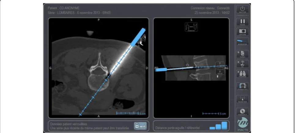
electromagnetic navigation system for CT-guided interventional radiology procedures (IMACTIS-CT®). The patient’s anatomy is visualised in 3D using a pre-viously acquired CT scan. An electromagnetic trans-mitter is attached to the patient’s skin, which enables the needle used by the operator and equipped with an electromagnetic receiver to be located. The hypo-thetical needle trajectory can therefore be displayed in real time on the images of the patient [25] (seeFig. 1). The operator can visualise the trajectory of the needle during both the planning phase (determination of the optimal route before skin penetration) and the needle insertion phase of the procedure.
Previous studies have shown that use of the naviga-tion system for CT-guided intervennaviga-tions results in high needle placement accuracy, even if the target requires an out-of-plane trajectory [26, 27]. The feasibility of oblique trajectories means that the number of possible needle trajectories is increased when using the naviga-tion system compared with convennaviga-tional CT guidance. The navigation system should therefore increase both the accuracy of the gesture and the radiologist’s confi-dence in his gesture, whereas the radiation exposure and the duration and severity of the intervention should decrease [28].
The accuracy of the IMACTIS-CT® navigation sys-tem has previously been demonstrated by the results obtained in a monocentric, prospective, randomised, controlled clinical trial (ClinicalTrials.gov identifier: NCT00828893). Among 120 patients undergoing rou-tine percutaneous CT procedures enrolled at the
Grenoble-Alpes University Hospital, half of the procedures were carried out with the assistance of the navigation system and half were carried out using conventional CT guidance. According to the intention-to-treat (ITT) analysis, the use of the navi-gation system improved the needle placement accur-acy; the median (interquartile range) distance error (in millimetres) using navigation-assisted guidance was 4.1 [2.7–9.1] compared with that using conven-tional guidance, which was 8.9 [4.9–15.1] (p< 0.001). Furthermore, according to the per-protocol (PP) ana-lysis, the use of the navigation system reduced the number of control CT scans acquired during the nee-dle insertion; using navigation-assisted guidance, a median of 2 [2, 3] scans were required, compared with conventional guidance, which required a median of 3 [2–4] scans (p= 0.01) [29].
This first clinical evaluation of the system enabled the pertinence of navigation assistance for CT-guided interventional radiology to be confirmed. However, to accurately evaluate the clinical benefit of the device, it is necessary to test the navigation system within a wider medical community. This paper describes an open, multicentre, prospective, randomised, controlled clinical trial that has been designed to evaluate the clinical benefit, in terms of safety, efficacy and performance, of using the IMACTIS-CT® system for navigation-assisted CT guidance compared with conventional CT guidance during interventional radiological procedures in the thoraco-abdominal region.
[image:3.595.57.540.475.693.2]Methods/design
Study design and setting
This study is an open, multicentre, prospective, rando-mised, controlled interventional clinical trial designed to evaluate the clinical benefit of the IMACTIS-CT® navi-gation system compared with the conventional method of CT-guided interventional radiological procedures in the thoraco-abdominal region. The nature of the proce-dures is open, including biopsies, abscess drainage, tumour ablation by radiofrequency or cryotherapy, and intra-articular corticosteroid injection procedures.
The trial is sponsored by the Grenoble-Alpes University Hospital (Isère, France). Eight further hospi-tals are participating in the study: Ambroise-Paré University Hospital (Hauts-de-Seine, France), Besançon University Hospital (Doubs, France), Bordeaux University Hospital (Gironde, France), Lille University Hospital (Nord, France), Nancy University Hospital (Meurthe-et-Moselle, France), Rennes University Hospital (Ille-et-Vilaine, France), Saint-Louis University Hospital (Paris, France) and Tours University Hospital (Indre-et-Loire, France). Also participating in this clinical trial are the eight centres of the Clinical Investi-gation Centre for Innovative Technology (CIC-IT; www.cic-it.fr) network (Besançon, Bordeaux, Garches, Grenoble, Lille, Nancy, Rennes and Tours), as well as the start-up company IMACTIS® (Grenoble, France; www.imactis.com), which developed the novel electro-magnetic navigation system for CT-guided interven-tional radiological procedures in close collaboration with the TIMC-IMAG (Grenoble-Alpes University, Grenoble, France; www-timc.imag.fr), which specialises in medical engineering, informatics and mathematics.
For this trial, a maximum of 500 patients for whom a percutaneous procedure in the thoraco-abdominal area under CT guidance has been prescribed will be enrolled in the study across the nine participating hospitals. The trial is structured as a standard two-arm, parallel-design, individually randomised trial. Patients are randomly assigned to the experimental arm of the trial in which procedures are carried out using navigation assistance (the NAV group), where the procedures are carried out using navigation assistance supplied by the medical de-vice under evaluation (IMACTIS-CT®), or to the active comparator arm of the trial in which procedures are car-ried out guided by conventional CT (CT group), where the procedures are carried out under conventional CT guidance. The randomisation is stratified by centre and by the expected difficulty of the procedure. The assign-ment is open label for both the radiologist and patient.
Because a previous clinical trial has already demon-strated improved needle placement accuracy in navigation-assisted procedures using the IMACTIS-CT® device (4.1 mm [2.7–9.1]) compared with conventional
procedures (8.9 mm [4.9–15.1], p< 0.001) according to ITT analysis [29], the aim of this trial is therefore to evaluate the clinical benefit of the navigation system. The primary outcome is a combined criterion composed of three different criteria which assess, respectively, the safety, efficacy and performance of navigation-assisted CT-guided procedures by comparing the results tained in the navigation-assisted group with those ob-tained in the conventional group. The criteria will be evaluated by a blinded expert committee. It will be de-termined that the navigation system represents a clinical benefit if non-inferiority in terms of safety, efficacy and superiority in terms of performance is obtained in the navigation-assisted group with respect to the conven-tional group. Also evaluated, in terms of secondary out-comes, are (1) the duration of the procedure, (2) the satisfaction of the operator and (3) the irradiation dose delivered, with (4) subgroup analysis according to the expected difficulty of the procedure, as well as an evalu-ation of (5) the usability of the device.
The clinical trial is overseen by the trial steering com-mittee (TSC), composed of I. Bricault (coordinating investigator, Grenoble-Alpes University Hospital), A. Moreau-Gaudry (medical coordinator of CIC-IT Grenoble), L. Carrat (president of IMACTIS®), F. Barbot (coordinator of CIC-IT Garches) and Y. Gandon (investigator, Rennes University Hospital). The TSC has responsibility for monitoring the patient enrolment rate at each centre and modifying the proposed statistical analysis framework if necessary following the interim analysis.
Materials
The medical device under evaluation in this clinical trial (IMACTIS-CT®) is an electromagnetic navigation system for CT-guided interventional radiological procedures. The device was Conformité Européenne (European Conformity; CE)-marked in 2013. Because it is an active medical device designed to be used in the field of diag-nostic and therapeutic interventional radiology, it is identified as a class IIA medical device according to the European legal framework. The device is composed of the following components:
1. A navigation station that includes:
a. A computer b. A touchscreen
c. An electromagnetic transmitter to be attached to the patient’s skin
d. An electromagnetic receiver to be attached to the needle holder
3. A packet of consumables that includes:
a. A single-use sterile needle holder
b. A single-use sterile drape to cover the receiver c. Hardware that contains the software parameters
and which enables the software to be launched
The operating principle of the navigation system is based on the localisation (position and orientation) in real time of the needle holder, equipped with an electro-magnetic receiver, with respect to the patient by means of the electromagnetic transmitter that is attached to the patient’s skin. A registration process enables a previously acquired CT scan of the patient to be positioned with re-spect to the current anatomy of the patient. The oper-ator can then visualise the position of his instrument in real time on the CT images. Therefore, during the plan-ning phase, the hypothetical trajectory of the needle, cal-culated from the current position of the needle holder, can be visualised to determine the optimal route before skin penetration, and during the needle insertion phase of the procedure, the operator can visualise the progress of the needle through the body in real time.
The president of IMACTIS® and/or an application en-gineer from the company will install the navigation sys-tem at each of the nine participating hospitals and will then provide training for the participating radiologists on how to use the system. Training is obligatory before any radiologist is authorised to carry out a navigated procedure on a patient enrolled in the trial. Training is first conducted using phantom models and is then con-tinued on one or more patients who are not enrolled in the trial. The number of training procedures carried out is noted to minimise the learning curve bias.
For this clinical trial, the foreseeable risks are those as-sociated with conventional CT-guided interventional radiology. No foreseeable supplementary risk is expected from the use of the navigation system. Contraindications are explicitly stated in the inclusion and exclusion cri-teria. Furthermore, because the navigation system is a passive medical device, the radiologist can choose to stop using the system at any moment, in particular in case of failure or malfunction of the system, and complete the procedure under conventional CT guid-ance, without any influence on the procedure or patient.
Methods
For a patient to be considered eligible for study partici-pation, the patient must answer‘yes’to all of the follow-ing inclusion criteria:
Aged≥18 years
Patients for whom a percutaneous diagnostic or therapeutic interventional procedure in the
thoraco-abdominal area under CT guidance has been pre-scribed and consensually agreed by a multidisciplin-ary team of radiologists, surgeons and clinicians
Patients affiliated with the social security (or similar) system
Patients who have signed the consent certificate of the informed consent form of the trial
Furthermore, to be considered for enrolment, the pa-tient must answer ‘no’ to all of the following exclusion criteria:
Patients with non-MRI-compatible devices or implanted material (e.g., pacemaker)
Patients with implanted ferromagnetic material in the thoraco-abdominal area that could interfere with the navigation system
Pregnant women and lactating mothers
Persons who are wards of court or under guardianship
Persons deprived of freedom by judicial or administrative decision
Persons under legal protection
Patients eligible for enrolment in the clinical trial will be informed of the study during their pre-intervention consultation with a clinical trial investigator, who will deliver clear, intelligible and objective information; an-swer questions; and verify the inclusion and exclusion criteria. A reflection period is then respected, of dura-tions varying from 2 h to 1 week before the intervention. If the patient chooses to participate in the study, the consent certificate is signed, and the patient is enrolled in the trial. To minimise the withdrawal of consent, the consent certificate is signed as closely as possible to the moment at which the procedure begins.
An initial CT scan of the patient is acquired before as-signment of the patient into the experimental or com-parator arm of the trial. First, landmarks are made on the patient’s skin using metal wire or a metal grill (re-quired for conventional CT guidance), and the electro-magnetic transmitter of the navigation system is attached to the patient’s skin (required for navigation-assisted CT guidance). The CT scan is then acquired, and it is verified that the procedure originally prescribed is maintained and that the navigation system is available and working. If the originally prescribed procedure is no longer maintained or the navigation system is not avail-able or not working, the patient is removed from the study, and the procedure is carried out under conven-tional CT guidance.
is on a plane that is oblique to the acquired axial images, the target moves with respiration, the procedure is a lung biopsy, the target is difficult to reach (due to small size or depth of location) or the needle path is very narrow. Difficult interventions are associated with an increased risk of major complications and of procedure failure and also with an increase in the number of control scans required compared with standard inter-ventions. Because these risks have a direct impact on the three criteria that constitute the primary outcome of the study, respectively, safety, efficacy and performance, the random assignment of the patient into the experimental or comparator arm of the trial is stratified by the expected difficulty of the procedure as well as by centre.
The expected difficulty of the procedure is entered into the electronic case report forms (eCRFs) provided by Medsharing (Fontenay Sous Bois, France; www.med-sharing.fr), and the patient is then randomly assigned to the experimental (NAV) or comparator (CT) arm of the trial. The randomisation lists are generated by Medshar-ing usMedshar-ing a randomisation by minimisation algorithm. By acquiring the CT scan before randomisation, it can be verified that the originally prescribed procedure is maintained and that the navigation system is available and working before the patient is assigned to the CT or NAV group, thereby minimising the later imputation of missing data for the statistical analysis.
Once the patient has been assigned to the CT or NAV group, the procedure prescribed can be carried out. For patients assigned to the CT group (procedures carried out under conventional CT guidance), the ideal needle path trajectory is defined on the acquired CT scan using the CT console. The needle entry point is identified and marked on the patient’s skin, and the sterile area is set up. A local anaesthetic is administered, and the radiolo-gist carries out the procedure, iteratively verifying the correct position of the needle by acquiring control scans until the target has been reached. A final control scan is acquired that shows the position of the needle at the tar-get before the procedure is continued to carry out the prescribed gesture (e.g., biopsy, abscess drainage). Any further control scans acquired after the ‘final control scan’are not counted for this study.
For patients assigned to the NAV group (procedures carried out using navigation assistance supplied by IMACTIS-CT®), first the navigation system is installed beside the CT scanner and connected to the network. The system can then be started up, followed by the soft-ware. The electromagnetic receiver is attached to the non-sterile needle holder, and the images of the acquired CT scan are automatically transferred to the navigation system via the network. Automatically, the arrival of the images is detected, the coherence of the series is verified and the registration of the images is carried out. The
navigation system enables the radiologist to define the ideal needle path trajectory directly on the patient by using the needle holder to navigate within the acquired CT volume. Once the ideal trajectory has been defined, the entry point is marked on the patient’s skin, and the sterile area is set up. The navigation system is then made sterile by removing the electromagnetic receiver from the non-sterile needle holder, placing the receiver in a sterile cover, and attaching it to the sterile needle holder. A local anaesthetic is then administered, and the radi-ologist carries out the procedure using navigation assist-ance until the target has been reached. Control scans can be acquired at any moment. A final control scan is acquired that shows the position of the needle at the tar-get before the procedure is continued.
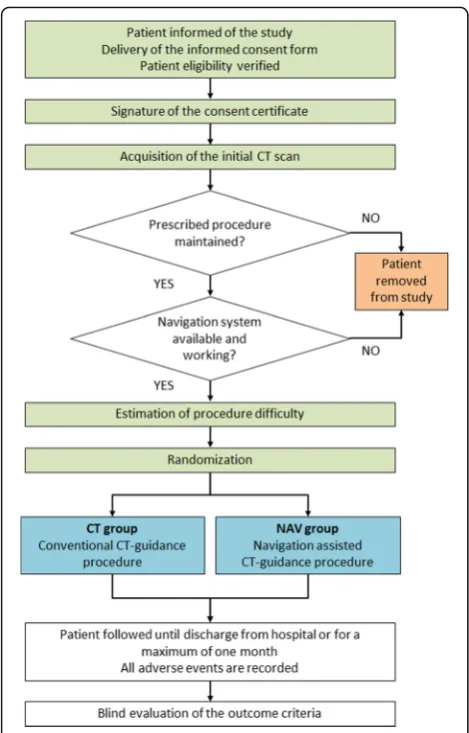
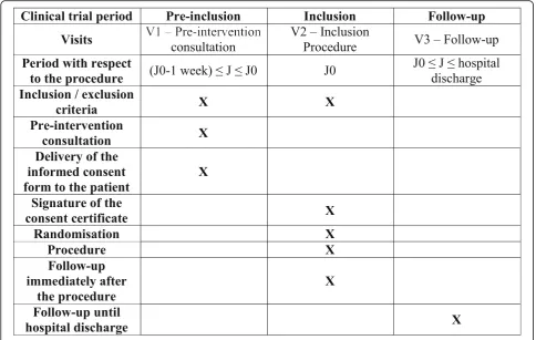
Following the procedure, patients are followed until their discharge from hospital or for a maximum of 1 month. All adverse events (AEs), serious or non-serious, that occur during or after (from immediately after the procedure until hospital discharge) the proced-ure are recorded in the eCRFs and are declared to Grenoble-Alpes University Hospital (trial sponsor), re-gardless of whether the AE is attributable to the radiolo-gist’s intervention. The AE is considered attributable if the event is directly caused by the needle insertion phase of the procedure. The AEs are classified according to the scale defined by the Society of Interventional Radiology (SIR) [30]. The study workflow and the participant time-line are presented in Figs. 2 and 3, respectively. (See Additional file 1 for information regarding the Standard Protocol Items: Recommendations for Interventional Trials [SPIRIT] checklist).
investigator of each centre has access to the list of participants enrolled in his/her centre.
The trial is evaluated according to the previously stated primary and secondary outcomes whose aim is to evaluate the clinical benefit of the navigation system. The primary outcome, a combined criterion composed of three criteria to assess the safety, efficacy and per-formance of navigation-assisted procedures, is evaluated as follows:
Safety: The number of AEs that are considered to be
major (i.e., classified as C, D, E or F according to the scale defined by the SIR) and are attributable to the needle insertion phase of the procedure
Efficacy: The number of targets reached; the target is
considered to have been reached when the needle is positioned accurately enough to allow the next step of the procedure to be carried out
Performance: The number of control scans acquired
during the needle insertion phase of the procedure; that is, the number of scans acquired between and including H1 (time of the first CT scan on which the needle is visible) and H2 (time at which the needle has reached the target)
The secondary outcomes are used to assess the time required to reach the target (H2−H1), the satisfaction of the operator with the procedure (quantitative scale) and the radiation dose delivered during the needle inser-tion phase of the procedure (radiainser-tion delivered between and including H1 and H2). Furthermore, because it is expected that the navigation system will provide a greater clinical benefit for difficult procedures compared with standard procedures, a subgroup analysis according to the expected difficulty of the procedure will be carried out. The usability of the device is also evaluated by analysis of the needle holder localisation files generated during the procedures.
To minimise ascertainment bias, the CT scans are post-processed before analysis such that it cannot be de-termined whether the scans were acquired in the experi-mental or comparator arm. For scans acquired with navigation assistance, the needle holder, the portion of the needle outside the body and the transmitter are erased. For scans acquired under conventional CT guid-ance, the portion of the needle outside the body is erased.
For each enrolled patient, the primary outcome is de-termined by a blinded evaluation of the post-processed CT scans and the patient’s medical reports (procedure report and hospital discharge summary), as previously described. The evaluation is carried out by a trained radiologist blinded to the patient group and to all evalu-ations made by the investigating radiologist who carried out the procedure. In case of discordance between the data entered by the evaluating radiologist and the inves-tigating radiologist, the discordance is presented to an expert committee by the CRA responsible for centre and data management. The expert committee is composed of two senior radiologists who are independent from the evaluating and investigating radiologists and who will provide a consensus in case of discordance.
To ensure the correct progress of the trial and to ver-ify that the proposed statistical analysis framework is ap-propriate, an interim analysis of the primary outcome will be performed once the first four participating hospi-tals have finished patient enrolment. To maintain a glo-bal cut-off of 5% for the final analysis, the interim analysis will be carried out using a cut-off of 0.1%. The results of the interim analysis will be considered by the TSC to judge whether the statistical analysis framework requires modifying. When patient enrolment is finished, Fig. 2Clinical trial overview. CT Computed tomography,CT group
[image:7.595.56.291.85.452.2]the final monitoring is completed and the coherence of the recorded data is verified, the database will be frozen, and the final statistical analysis of the data will be carried out.
Statistical analysis
The number of patients to be enrolled in the study is calculated as the maximum of the values calculated for each of the three different criteria (safety, efficacy and performance) that constitute the primary outcome, with the alpha level set to 5%. The calculations are performed as follows for each of the three criteria:
Safety: The suggested threshold for all major
complications resulting from percutaneous drainage or biopsy procedures is 10% [31,32]. The threshold is increased to 20% for lung procedures [33]. Because one of the participating hospitals is a referral centre for lung procedures, it has been decided to define the threshold for all major complications as 12% for this study (calculated as the average threshold across the centres, 12 = (10 × 8 + 20 × 1)/9, rounded up to the nearest whole number). Considering an initial complication rate of 5% in each of the groups, a sample size of 384 patients (192 in each of the experimental and
comparator arms of the trial) will be sufficient to demonstrate non-inferiority between the groups, respecting a non-inferiority cut-offδ= 7%, with 90% power and a 5% significance level.
Efficacy: The minimum threshold for intervention
success is 80%. Considering an initial success rate of 91% in each of the groups, a sample size of 380 patients (190 per group) will be sufficient to demonstrate non-inferiority between the groups in terms of success, respecting a non-inferiority cut-off
δ=−10%, with 90% power and a 5% significance level.
Performance: Considering that use of the navigation
system will decrease the number of control scans acquired by 20% compared with conventional procedures (estimated from previous clinical trial), a sample size of 222 patients (111 per group) will be sufficient to demonstrate superiority in the NAV group compared with the CT group in terms of a decrease in the number of control scans acquired, with 90% power and a 5% significance level.
[image:8.595.56.541.88.395.2]included per hospital was set at 50 ± 15. After inclusion had begun, a ninth centre requested to participate in the study. Because inclusion had already begun, the number of inclusions per centre was maintained, and the max-imum sample size was defined to be 500. The study protocol was therefore modified, and the modifications were approved by the French Health Authority (Agence Nationale de Sécurité des Médicaments et des Produits de Santé) and the relevant ethics committee (Comité de Protection des Personnes, Sud-Est V, France). The sample size calculations were carried out using nQuery Advisor® 7.0 software (Statistical Solutions, Cork, Ireland) with the method‘upper confidence limit for dif-ference in proportions (simulation)’ [34] for the safety and efficacy criteria and Noether’s method ‘sample size determination for some common nonparametric statis-tics’[35] for the performance criterion.
For the statistical analysis of the data, because non-inferiority tests are used to evaluate the safety and efficacy criteria, a PP analysis will be carried out on the data, comparing the 90% two-sided confidence interval of the difference between the proportions with the non-inferiority cut-off. Conversely, because a superiority test is used to evaluate the performance criterion, an ITT analysis will be carried out on this data, comparing the means of the two groups using Student’s t test or the non-parametric Mann-Whitney U test. For all calculations, a threshold of p< 0.05 is considered significant.
For the secondary outcome analysis, the means of the two groups will be compared using Student’s t test or the non-parametric Mann-Whitney U test for each outcome. Furthermore, subgroup analyses ac-cording to the difficulty of the procedure will be car-ried out. ITT and PP analyses will be carcar-ried out for the secondary outcomes to ensure the robustness of the results [36].
If a patient withdraws consent before random as-signment to a group, that patient is removed from the study and is excluded from the statistical ana-lysis. If other situations occur after random assign-ment to a group (consent is withdrawn, the protocol is not respected, the radiologist chooses to use conventional CT guidance rather than the navigation system for a patient in the NAV group, or the radi-ologist changes the original procedure prescribed), the patient concerned will be excluded from PP ana-lyses. For the ITT analyses, missing data in the NAV group will be filled using the worst values for proce-dures of the same difficulty rating in the same centre in the NAV group, whereas missing data in the CT group will be filled using the best values for proce-dures of the same difficulty rating in the same centre in the CT group. If after random assignment to the
NAV group the navigation system is unavailable, the patient remains in the study, and the procedure is performed under conventional CT guidance; the pa-tient will be excluded from PP analyses. For the ITT analyses, the patient remains in the NAV group, with missing data filled using the worst values for procedures of the same difficulty rating in the same centre in the NAV group.
Discussion
Navigation assistance systems have demonstrated their value in the fields of orthopaedics, urology and neuro-surgery. A previous clinical trial demonstrated that use of the IMACTIS-CT® navigation system during CT-guided interventional procedures improved needle place-ment accuracy significantly and reduced the number of control CT scans acquired. The aim of the present trial is to address the lack (to the best of our knowledge) of published high-level evidence studies involving evalu-ation of navigevalu-ation-assisted, CT-guided interventional procedures. The trial is designed to evaluate the clinical benefit, in terms of safety, efficacy and performance, of navigation-assisted, CT-guided procedures. It is expected that the more accurate needle placement achieved using the navigation system will decrease the procedure dur-ation and decrease the radidur-ation exposure delivered. This trial is important because it addresses the problems as-sociated with conventional CT guidance—inaccurate needle placement, radiation exposure—and is particu-larly relevant because the number of interventional radiological procedures carried out in routine clinical practice is increasing.
approach is therefore required for certain procedures (e.g., lung procedures). An interesting future perspective is therefore the development and inclusion of respiratory motion management into the system, which is expected to improve the system’s accuracy. Finally, because the navigation system was designed to assist CT-guided interventional procedures, the system has no multi-modal capability.
Trial status
Patient enrolment started in December 2013 and was expected to be completed before the end of June 2017.
Additional file
Additional file 1:SPIRIT 2013 checklist: recommended items to address in a clinical trial protocol and related documents. (DOC 122 kb)
Abbreviations
AE:Adverse event; CE: Conformité Européenne (European Conformity); CIC-IT: Clinical Investigation Centre for Innovative Technology; CRA: Clinical research assistant; CT: Computed tomography; CT group: Active comparator arm in which procedures are carried out guided by conventional computed tomography; eCRF: Electronic case report form; ITT: Intention to treat; MRI: Magnetic resonance imaging; NAV group: Experimental arm of the trial in which procedures are carried out using navigation assistance; PP: Per protocol; SIR: Society of Interventional Radiology; SPIRIT: Standard Protocol Items: Recommendations for Interventional Trials; TIMC-IMAG: Techniques de l’Ingénierie Médicale et de la Complexité–Informatique, Mathématiques et Applications, Grenoble (Medical Engineering and Complexity Techniques– Computer Science, Mathematics and Applications, Grenoble); TSC: Trial steering committee; US: Ultrasound
Acknowledgements
We gratefully acknowledge the Grenoble-Alpes University Hospital (Délégation à la Recherche Clinique et à l’Innovation), which sponsored the trial. As sponsor of the trial, the Grenoble-Alpes University Hospital has responsibility for the submission and amendments of the trial to the relevant authorities in order to obtain the necessary regulatory authorisations and declare adverse events to the relevant authorities, as well as the responsibility for the study design and management; the recording, monitoring, analysis and interpretation of the data; and the writing and submission of the final report. The latter responsibilities (from study design to final report) have been delegated to CIC-IT Grenoble. The authors also gratefully acknowledge the participating investigators at each recruitment centre, as well as the patients who choose to enrol in the study.
Funding
This trial is funded by the French Ministry of Health as part of the Hospital Clinical Research Program (PHRC-N 2012). The funders had no role in study design, data collection and analysis, decision to publish or preparation of the manuscript. The IMACTIS-CT® navigation system in each participating hospital is provided and installed by IMACTIS (Grenoble, France).
Availability of data and materials
Not applicable.
Authors’contributions
IB, AMG and EC designed the study. EC is a CRA responsible for centre and data management. RCR is a trained radiologist responsible for the blind evaluation of the trial outcomes and also drafted the manuscript and approved the final version. The expert committee is made up of IB and SA. AMG, in close collaboration with MM, is responsible for the statistical analysis of the data. JG and AM are responsible for post-processing the CT scans. IB, AMG, EC, RCR, CH and MM made critical revisions to the manuscript. LP, MD, SP, RL, AC, GK and FP are responsible for data management. BL, MH, BR, XO, EQ, OF and EDK are trained radiologists responsible for patient inclusion and
carrying out the interventional procedures in the different participating hospitals. All authors read, contributed to and approved the final manuscript.
Ethics approval and consent to participate
This trial is registered in the ClinicalTrials.gov database (NCT01896219) and was approved by the French Health Authority (Agence Nationale de Sécurité des Médicaments et des Produits de Santé, registered 19 June 2013 under ANSM reference number 2013-A00539-36) and the relevant ethics committee (Comité de Protection des Personnes, Sud-Est V, France, 12 June 2013, CPP reference number 13-CHUG-22). Both central ethics committees have approved this study for all eight centres involved. All patients signed a consent certificate before being enrolled in the trial, in accordance with the Declaration of Helsinki II.
Consent for publication
Not applicable.
Competing interests
IB is a member of the medical advisory board of IMACTIS® and participated in the design of the IMACTIS-CT® navigation system. The other authors declare that they have no competing interests.
Publisher’s Note
Springer Nature remains neutral with regard to jurisdictional claims in published maps and institutional affiliations.
Author details 1
Clinique Universitaire de Radiologie et Imagerie Médicale, Centre Hospitalier Universitaire (CHU) de Grenoble-Alpes, F-38000 Grenoble, France.2Institut national de la santé et de la recherche médicale (Inserm) Centre d’Investigation Clinique (CIC) 1406, University Grenoble-Alpes, F-38000 Grenoble, France.3Institut national de la santé et de la recherche médicale (Inserm) Centre d’Investigation Clinique (CIC) 1406, F-38000 Grenoble, France. 4Pole Recherche, Centre Hospitalier Universitaire (CHU) de Grenoble-Alpes,
F-38000 Grenoble, France.5Techniques de l’Ingénierie Médicale et de la Complexité–Informatique, Mathématiques et Applications, Grenoble (TIMC-IMAG), University Grenoble-Alpes, F-38000 Grenoble, France. 6Techniques de l’Ingénierie Médicale et de la Complexité–Informatique,
Mathématiques et Applications, Grenoble (TIMC-IMAG), Centre national de la recherche scientifique (CNRS), F-38000 Grenoble, France.7Pole Sante Publique, Centre Hospitalier Universitaire (CHU) de Grenoble-Alpes, F-38000 Grenoble, France.8Service de Radiologie Ostéo-Articulaire, Centre Hospitalier Universitaire (CHU) Besançon, F-25000 Besançon, France.9Institut national de la santé et de la recherche médicale (Inserm) Centre d’Investigation Clinique (CIC) 1431, F-25000 Besançon, France.10Service d’Imagerie Diagnostique et Therapeutique, Centre Hospitalier Universitaire (CHU) Bordeaux, F-33000 Bordeaux, France.11Institut national de la santé et de la recherche médicale (Inserm) Centre d’Investigation Clinique (CIC) 1401, F-33000 Bordeaux, France. 12Centre d’Investigation Clinique (CIC) 1401, University Bordeaux, F-33000
Bordeaux, France.13Centre Hospitalier Universitaire (CHU) Bordeaux, F-33000 Bordeaux, France.14Service de Radiologie, Hôpital Ambroise-Paré, Assistance Publique-Hôpitaux de Paris (AP-HP), F-92100 Boulogne-Billancourt, France. 15Institut national de la santé et de la recherche médicale (Inserm) Centre
d’Investigation Clinique (CIC) 1429, Hôpital Raymond-Poincaré, Assistance Publique-Hôpitaux de Paris (AP-HP), F-92380 Garches, France.16Service de Radiologie, Centre Hospitalier Universitaire (CHU) Lille, F-59000 Lille, France. 17Institut national de la santé et de la recherche médicale (Inserm) Centre
d’Investigation Clinique (CIC) 1403, Centre Hospitalier Universitaire (CHU) Lille, University Lille, F-59000 Lille, France.18Service de Radiologie, Centre Hospitalier Régional Universitaire (CHRU) de Nancy, F-54000 Nancy, France. 19Institut national de la santé et de la recherche médicale (Inserm) Centre
de Radiologie, Hôpital Saint Louis, Assistance Publique-Hôpitaux de Paris (AP-HP), F-75475 Paris, France.
Received: 21 December 2016 Accepted: 14 June 2017
References
1. Ikeda K, Osaki Y, Nakanishi H, Nasu A, Kawamura Y, Jyoko K, et al. Recent progress in radiofrequency ablation therapy for hepatocellular carcinoma. Oncology. 2014;87 Suppl 1:73–7.
2. Althoff CE, Bollow M, Feist E, Marticorena-Garcia SR, Eshed I, Diekhoff T, et al. CT-guided corticosteroid injection of the sacroiliac joints: quality assurance and standardized prospective evaluation of long-term effectiveness over six months. Clin Rheumatol. 2015;34:1079–84. 3. Arnolli MM, Hanumara NC, Franken M, Brouwer DM, Broeders IAMJ. An
overview of systems for CT- and MRI-guided percutaneous needle placement in the thorax and abdomen. Int J Med Robot. 2015;11:458–75. 4. Schubert T, Jacob AL, Pansini M, Liu D, Gutzeit A, Kos S. CT-guided
interventions using a free-hand, optical tracking system: initial clinical experience. Cardiovasc Intervent Radiol. 2013;36:1055–62.
5. Grasso RF, Faiella E, Luppi G, Schena E, Giurazza F, Del Vescovo R, et al. Percutaneous lung biopsy: comparison between an augmented reality CT navigation system and standard CT-guided technique. Int J Comput Assist Radiol Surg. 2013;8:837–48.
6. Grand DJ, Atalay MA, Cronan JJ, Mayo-Smith WW, Dupuy DE. CT-guided percutaneous lung biopsy: comparison of conventional CT fluoroscopy to CT fluoroscopy with electromagnetic navigation system in 60 consecutive patients. Eur J Radiol. 2011;79:e133–6.
7. Gevenois PA, Sergent G, De Myttenaere M, Beernaerts A, Rocmans P. CT-guided percutaneous drainage of an anterior mediastinal abscess with a 16 F catheter. Eur Respir J. 1995;8:869–70.
8. Kinoshita F, Kato T, Sugiura K, Nishimura M, Kinoshita T, Hashimoto M, et al. CT-guided transthoracic needle biopsy using a puncture site-down positioning technique. AJR Am J Roentgenol. 2006;187:926–32. 9. Thanos L, Poulou LS, Mailli L, Pomoni M, Kelekis DA. Image-guided
radiofrequency ablation of a pancreatic tumor with a new triple spiral-shaped electrode. Cardiovasc Intervent Radiol. 2010;33:215–8.
10. Mukhtar KN, Mahmood SN, Umair SF. CT guided percutaneous renal biopsy versus ultrasound guided for obtaining adequate tissue. J Pak Med Assoc. 2012;62:880–2.
11. Rathmann N, Haeusler U, Diezler P, Weiss C, Kostrzewa M, Sadick M, et al. Evaluation of radiation exposure of medical staff during CT-guided interventions. J Am Coll Radiol. 2015;12:82–9.
12. Sainani NI, Arellano RS, Shyn PB, Gervais DA, Mueller PR, Silverman SG. The challenging image-guided abdominal mass biopsy: established and emerging techniques“if you can see it, you can biopsy it”. Abdom Imaging. 2013;38:672–96.
13. Slotkin EM, Patel PD, Suarez JC. Accuracy of fluoroscopic guided acetabular component positioning during direct anterior total hip arthroplasty. J Arthroplasty. 2015;30(9 Suppl):102–6.
14. Brandt G, Zimolong A, Carrat L, Merloz P, Staudte HW, Lavallée S, et al. CRIGOS: a compact robot for image-guided orthopedic surgery. IEEE Trans Inf Technol Biomed. 1999;3:252–60.
15. Brandt G, Radermacher K, Zimolong A, Rau G, Merloz P, Klos TV, et al. CRIGOS: development of a compact robot for image-guided orthopedic surgery [in German]. Orthopade. 2000;29:645–9.
16. Ukimura O. Image-fusion for biopsy, intervention, and surgical navigation in urology. In: Ukimura O, editor. InTech. 2011. http://www.intechopen.com/ books/image-fusion/image-fusion-for-biopsy-intervention-and-surgical-navigation-in-urology. Accessed 1 Feb 2016
17. Uruc V, Ozden R, DogramacıY, KalacıA, Dikmen B, Yıldız OS, et al. The comparison of freehand fluoroscopic guidance and electromagnetic navigation for distal locking of intramedullary implants. Injury. 2013;44:863–6.
18. Weiner GM, Chivukula S, Chen CJ, Ding D, Engh JA, Amankulor N. Ommaya reservoir with ventricular catheter placement for chemotherapy with frameless and pinless electromagnetic surgical neuronavigation. Clin Neurol Neurosurg. 2015;130:61–6.
19. Mahan M, Spetzler RF, Nakaji P. Electromagnetic stereotactic navigation for external ventricular drain placement in the intensive care unit. J Clin Neurosci. 2013;20:1718–22.
20. Chen MJ, Gu LX, Zhang WJ, Yang C, Dong MJ. Electromagnetic navigation-guided radiofrequency thermocoagulation in trigeminal neuralgia: technical note with three case reports. J Neurol Surg A Cent Eur Neurosurg. 2013;74:251–7.
21. Ko R, Soucy F, Denstedt JD, Razvi H. Percutaneous nephrolithotomy made easier: a practical guide, tips and tricks. BJU Int. 2008;101:535–9. 22. Chang CM, Fang KM, Huang TW, Wang CT, Cheng PW. Three-dimensional
analysis of the surface registration accuracy of electromagnetic navigation systems in live endoscopic sinus surgery. Rhinology. 2013;51:343–8. 23. Soyer P, Fargeaudou Y, Boudiaf M, Hamzi L, Rymer R. Percutaneous
abdominopelvic interventional procedures using real-time CT fluoroscopy guidance at 21 mAs: an analysis of 99 consecutive cases [in French]. J Radiol. 2008;89:565–70.
24. Prosch H, Stadler A, Schilling M, Bürklin S, Eisenhuber E, Schober E, et al. CT fluoroscopy-guided vs. multislice CT biopsy mode-guided lung biopsies: accuracy, complications and radiation dose. Eur J Radiol. 2012;81:1029–33. 25. Nagel M, Schmidt G, Petzold R, Kalender WA. A navigation system for
minimally invasive CT-guided interventions. Med Image Comput Comput Assist Interv. 2005;8:33–40.
26. Bruners P, Penzkofer T, Nagel M, Elfring R, Gronloh N, Schmitz-Rode T, et al. Electromagnetic tracking for CT-guided spine interventions: phantom, ex-vivo and in-vivo results. Eur Radiol. 2009;19:990–4.
27. Wallace MJ, Gupta S, Hicks ME. Out-of-plane computed-tomography-guided biopsy using a magnetic-field-based navigation system. Cardiovasc Intervent Radiol. 2006;29:108–13.
28. Moncharmont L, Moreau-Gaudry A, Medici M, Bricault I. Phantom evaluation of a navigation system for out-of-plane CT-guided puncture. Diagn Interv Imaging. 2015;96:531–6.
29. Durand P, Moreau-Gaudry A, Silvent A-S, Frandon J, Chipon E, Médici M, et al. Computer assisted electromagnetic navigation improves accuracy in computed tomography guided interventions: A prospective randomized clinical trial. PloS One. 2017;12:e0173751.
30. Sacks D, McClenny TE, Cardella JF, Lewis CA. Society of Interventional Radiology clinical practice guidelines. J Vasc Interv Radiol. 2003;14:S199–202. 31. Bakal CW, Sacks D, Burke DR, Cardella JF, Chopra PS, Dawson SL, et al.
Quality improvement guidelines for adult percutaneous abscess and fluid drainage. J Vasc Interv Radiol. 2003;14:S223–5.
32. Cardella JF, Bakal CW, Bertino RE, Burke DR, Drooz A, Haskal Z, et al. Quality improvement guidelines for image-guided percutaneous biopsy in adults. J Vasc Interv Radiol. 2003;14:S227–30.
33. Tomiyama N, Yasuhara Y, Nakajima Y, Adachi S, Arai Y, Kusumoto M, et al. CT-guided needle biopsy of lung lesions: a survey of severe complication based on 9783 biopsies in Japan. Eur J Radiol. 2006;59:60–4.
34. Newcombe RG. Interval estimation for the difference between independent proportions: comparison of eleven methods. Stat Med. 1998;17:873–90. 35. Noether GE. Sample size determination for some common nonparametric
tests. J Am Stat Assoc. 1987;82:645–7.
36. Walker E, Nowacki AS. Understanding equivalence and noninferiority testing. J Gen Intern Med. 2011;26:192–6.
• We accept pre-submission inquiries
• Our selector tool helps you to find the most relevant journal • We provide round the clock customer support
• Convenient online submission • Thorough peer review
• Inclusion in PubMed and all major indexing services • Maximum visibility for your research
Submit your manuscript at www.biomedcentral.com/submit