4167
Introduction
Early Drosophila development is orchestrated by maternal RNAs and proteins stored within the oocyte and embryo. Coordinated translational regulation and cytoplasmic localization of specific maternal transcripts determine developmental decisions such as axis patterning and germ cell specification (reviewed by Johnstone and Lasko, 2001). During oogenesis, the TGFα signaling molecule Gurken (Grk) establishes polarity within the oocyte along both the anteroposterior and dorsoventral axes (Neuman-Silberberg and Schüpbach, 1993; González-Reyes et al., 1995; Roth et al., 1995) (reviewed by Nilson and Schüpbach, 1999). Subsequently, at the posterior end of the oocyte and early embryo, a specialized region of cytoplasm, called pole plasm, accumulates RNAs and proteins required for germline development (reviewed by Mahowald, 2001). Pole plasm assembly is initiated through the localized translation of oskar (osk) mRNA (Ephrussi et al., 1991; Kim-Ha et al., 1991). Vasa (Vas) protein and nanos (nos) mRNA are among several molecules that accumulate in the pole plasm downstream of Osk. Localized translation of nos mRNA in the pole plasm produces a Nos protein gradient that is essential to determine abdominal fate, thus directly linking germ cell development to posterior somatic patterning in the embryo (Lehmann and Nüsslein-Volhard, 1991; Wang and Lehmann, 1991).
osk, grk and nos RNAs are all under complex translational regulation in the developing oocyte, mediated through cis-acting elements in their untranslated regions (UTRs). Genetic and biochemical analyses in Drosophila ovaries and embryos have identified several translational regulatory
proteins. Bruno (Bru) is involved in repressing translation of osk and grk (Kim-Ha et al., 1995; Webster et al., 1997; Filardo and Ephrussi, 2003; Nakamura et al., 2004), and Smaug (Smg) is involved in repressing translation of nos (Smibert et al., 1996; Dahanukar et al., 1999; Nelson et al., 2004). Although recent work has linked both Bru and Smg to the cap-binding step of translation initiation (Wilhelm et al., 2003; Nakamura et al., 2004; Nelson et al., 2004), translational repression of osk, grk and nos probably targets multiple steps of translation (Lie and Macdonald, 1999; Clark et al., 2000; Nakamura et al., 2004). In general, details of the mechanisms of translational derepression and activation for specific transcripts remain obscure. In several organisms, translational activation of maternal mRNAs involves cytoplasmic polyadenylation. The activity of the Drosophila cytoplasmic polyadenylation element-binding protein, called oo18 RNA-binding protein or Orb (Lantz et al., 1992) is implicated in activating translation of osk and possibly of other mRNAs (Chang et al., 1999; Castagnetti and Ephrussi, 2003).
Our work addresses the function of the highly conserved DEAD-box RNA helicase Vas, which is required for the progression of oogenesis and for pole plasm assembly (Schüpbach and Wieschaus, 1986; Hay et al., 1988; Lasko and Ashburner, 1988; Liang et al., 1994). Based on sequence similarity with yeast Ded1p (reviewed by Linder, 2003), and the finding that expression of several proteins is reduced in vas mutants, Vas has been suggested to function in translational regulation (reviewed by Johnstone and Lasko, 2001). Females bearing hypomorphic vas mutations complete oogenesis but produce embryos lacking germ cells and lacking posterior
The DEAD-box RNA helicase Vasa (Vas) is required for germ cell development and function, as well as for embryonic somatic posterior patterning. Vas interacts with the general translation initiation factor eIF5B (cIF2, also known as dIF2), and thus may regulate translation of specific mRNAs. In order to investigate which functions of Vas are related to translational control, we have analyzed the effects of site-directed vas mutations that reduce or eliminate interaction with eIF5B. Reduction in Vas-eIF5B interaction during oogenesis leads to female sterility, with phenotypes similar to a vas null mutation. Accumulation of Gurken (Grk) protein is greatly reduced when Vas-eIF5B
interaction is reduced, suggesting that this interaction is crucial for translational regulation of grk. In addition, we show that reduction in Vas-eIF5B interaction virtually abolishes germ cell formation in embryos, while producing a less severe effect on somatic posterior patterning. We conclude that interaction with the general translation factor eIF5B is essential for Vas function during development.
Key words: Translation, Germ cells, DEAD-box, Axis-patterning, Gurken, Drosophila, cIF2, Vasa
Summary
Interaction with eIF5B is essential for Vasa function during
development
Oona Johnstone and Paul Lasko*
Department of Biology, McGill University, 1205 Avenue Docteur Penfield, Montréal, Québec H3A 1B1, Canada *Author for correspondence (e-mail: [email protected])
Accepted 27 May 2004
Development 131, 4167-4178
Published by The Company of Biologists 2004 doi:10.1242/dev.01286
segments, indicating an essential role for vas in both processes (Schüpbach and Wieschaus, 1986). An earlier function for Vas, during oogenesis, was also demonstrated through the study of null mutations that are viable but produce no embryos (Styhler et al., 1998; Tomancak et al., 1998). Although vas-null oocytes display minimal disruption of grk RNA accumulation, Grk protein levels are severely reduced, leading to the hypothesis that Vas could play a role in grk translational control (Styhler et al., 1998; Tomancak et al., 1998). Vas also appears to represent an important link between meiotic cell cycle progression and developmental events such as establishment of polarity. In response to a meiotic checkpoint, activated by a delay in DNA double-strand break (DSB) repair during oogenesis, Vas is post-translationally modified, and this corresponds to a downregulation in Grk protein accumulation (Ghabrial and Schüpbach, 1999).
In previous work, we identified a translation factor dIF2, now called eIF5B, as a Vas-binding protein (Carrera et al., 2000). A genetic interaction between null alleles of dIF2 and vas suggested a functional link between these two proteins. eIF5B/dIF2 has since been demonstrated in mammalian systems to be required for all cellular translation and to act at the 60S ribosomal subunit joining step of translation initiation (Pestova et al., 2000b). Subsequent work has indicated that translation can be regulated at the stage of subunit joining (Ostareck et al., 2001; Searfoss et al., 2001). These results suggest that Vas could function as a translational regulator of specific mRNAs through interaction with eIF5B.
To test this hypothesis, we created specific vas mutations that severely reduce its interaction with eIF5B. These mutant forms of Vas still localize correctly, allowing us to investigate which developmental functions of Vas require an interaction with eIF5B, and are therefore likely to involve a translational regulatory role. We found that the Vas-eIF5B interaction was essential for the progression of oogenesis, and for normal expression of Grk. In addition, we found that the interaction between Vas and eIF5B was crucial for germ cell specification, but we observed a much less stringent requirement for this interaction in posterior somatic segmentation. We conclude that interaction with eIF5B is essential for Vas function, and propose that Vas achieves translational regulation in the germline through eIF5B binding. The Vas-eIF5B interaction represents a significant opportunity to investigate how a tissue-specific regulator may control the ribosomal subunit joining step of translation initiation to activate the translation of specific transcripts.
Materials and methods
Plasmid construction and mutagenesis
Amino acid reference numbers used are based on the Vas protein predicted by the genomic sequence. The vas-coding region was PCR amplified from the original vas cDNA that lacks one copy of a 39 nucleotide tandem repeat, encoding amino acids 141-153 (Lasko and Ashburner, 1988), and inserted into XhoI/NotI digested pBluescript to use as a template for mutagenesis. Specific deletions in vas were generated using PCR-mediated mutagenesis, using the following primers: ∆616-618, 5′ TTTCTACGCACCTGTGGTGCC and 3′ AGTCTGGCCAGATCCCTCCAAG; ∆616, 5′ CCGGACTTTCTAC-GCACCTGTG and 3′ (same as for ∆616-618); ∆617, 5′ GACTTTCTACGCACCTGTGGTG and 3′ AACAGTCTGGCCA-GATCCCTC; ∆618, 5′ (same as for ∆616-618) and 3′
CGGAACAGTCTGGCCAGATCC. Mutagenesis was followed by blunt-end ligation, and was verified by sequencing. Each vas-coding region was then digested out of pBluescript using XhoI and NotI, and subcloned into a XhoI/NotI digested plasmid P[w+ Pvas-gfp]
(Nakamura et al., 2001), derived from pCaSpeR2. In addition, each vas-coding region was PCR amplified out of pBluescript and subcloned into a NcoI/XhoI digested pEG202 vector, used for expression in yeast.
Yeast interaction trap assays
The yeast strain EGY48 was co-transformed with ‘bait’ constructs cloned in pEG202, ‘prey’ constructs cloned in pJG4-5, and a lacZ reporter plasmid pSH18-34. β-Galactosidase activity was monitored using a plate-based assay as described previously (Golemis et al., 1997) and in liquid culture (Reynolds et al., 1997).
Protein expression and western blotting
Preparation of yeast protein extracts was performed according to the Yeast Protocols Handbook (Clontech). For every transformed strain, a 5 ml overnight culture in selective media was used to inoculate a 50 ml culture in YPD media, incubated at 30°C with shaking (220-250 rpm) until OD600=0.4-0.6. Cells were centrifuged
at 1000 g for 5 minutes at 4°C, resuspended in 50 ml cold H2O,
centrifuged at 1000 g for 5 minutes at 4°C and frozen in liquid nitrogen. Pellets were thawed in pre-warmed Cracking Buffer (8 M Urea, 5% SDS, 40 mM Tris-HCl [pH6.8], 0.1 mM EDTA, 0.4 mg/ml Bromophenol Blue), supplemented with 1 mM PMSF, 1 3 protease inhibitor cocktail (Roche Diagnostics) and 10 µl β -mercaptoethanol/ml buffer. Samples were transferred into microcentrifuge tubes containing glass beads, and heated at 70°C for 10 minutes. They were then vortexed for 1 minute, and centrifuged at 20,000 g for 5 minutes at 4°C. Supernatants were kept on ice while pellets were boiled at 100°C for 3-5 minutes, vortexed for 1 minute and centrifuged at 20,000 g for 5 minutes at 4°C, and then combined with the first supernatants. Drosophila ovarian proteins were extracted by homogenization in phosphate-buffered saline (PBS)/1 mM PMSF/1 3protease inhibitor cocktail (Roche Diagnostics). Samples were centrifuged at 18,000 g for 15 minutes at 4°C, and supernatants were combined with SDS loading buffer. For western blotting, proteins were resolved on SDS-PAGE gels and transferred onto nitrocellulose membranes, blocked overnight at 4°C in PBS/2% skim milk/0.05% Tween-20 (PBSTM). Membranes were incubated for 1 hour at room temperature with primary antibodies diluted in PBSTM, washed with PBSTM, then incubated for 1 hour at room temperature with HRP-conjugated secondary antibodies (Amersham Pharmacia) diluted 1:5000 in PBSTM. Membranes were washed with PBSTM and proteins were detected by chemiluminescence (NEN). Rabbit anti-Vas was used at 1:5000. Mouse anti-actin (ICN Biomedicals) was used at 1:5000. Mouse anti-α-Tubulin (Sigma) was used at 1:5000. Rabbit anti-4E-BP was used at 1:2000.
Immunohistochemistry and in situ hybridization
Immunostaining of ovaries and embryos with rabbit anti-Nos (1:1000), rabbit Osk (1:500), rabbit Tud (1:250) and rat anti-Vas (1:2000) was performed as described previously (Kobayashi et al., 1999). Fluorescent antibody staining was detected using goat anti-rabbit Alexa546nm, anti-rat Alexa633nm and anti-mouse Alexa568nm
for 20 minutes in 0.5 µg/ml DAPI, washed in PBST and then mounted in 70% glycerol/PBS. In situ hybridization was performed as described previously (Kobayashi et al., 1999), except that DMSO was omitted during fixation, and PBS/0.1% Tween-20 was used instead of MAB throughout the protocol.
Fly strains and techniques
yw flies were used for P element-mediated germline transformation. vas alleles used for subsequent analyses were vasPD(Schüpbach and
Wieschaus, 1986) and vasPH165(Styhler et al., 1998). To visualize
GFP in ovaries, they were dissected in PBS and fixed in 4% formaldehyde/PBS/0.2% Tween-20. Samples were washed in PBS and mounted in 70% glycerol/PBS. For live GFP visualization in embryos, they were collected on a sieve and washed with H2O, then
dechorionated with bleach and washed again with H2O. Embryos
were then mounted in Halocarbon oil (series 400) and examined immediately. For visualization of dorsal appendages, eggs were collected on a sieve and washed with H2O, then mounted in Hoyer’s
medium and incubated overnight at 60°C.
Results
The C-terminal region of Vas is required for interaction with eIF5B
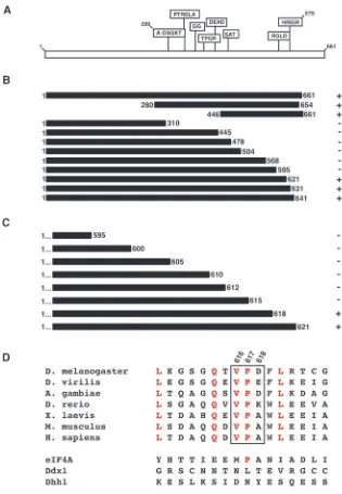
Vas contains many well-defined sequence motifs that are conserved among DEAD-box helicases (Fig. 1A) (Linder et al., 1989). Previous work has shown that interaction with eIF5B involved residues C-terminal to amino acid 310 of Vas (Carrera et al., 2000). In order to map more precisely the region of Vas required for interaction with eIF5B, we created a series of large deletions within Vas and tested these in the yeast two-hybrid system against the clone of eIF5B that we previously isolated in a yeast two-hybrid screen (Fig. 1B). We confirmed that the C-terminal half of Vas was required for interaction with eIF5B, and specifically implicated the region from residue 595 to 621 of Vas in eIF5B-interaction. We then tested a series of Vas constructs that began at the N terminus and ended between residues 595 and 621 (Fig. 1C). This analysis implicated residues 616-618 of Vas as being crucial for eIF5B interaction. We examined whether this region of Vas was conserved in homologous proteins in different species (Fig. 1D). Both V616 and P617 are identical among Vas homologs, while D618 does not appear to be conserved. Some neighboring residues are also invariant among Vas homologs. Although a proline corresponding to P617 is found in eIF4A, in general, more distant Drosophila DEAD-box proteins such as eIF4A, Ddx1 and Dhh1 are not highly conserved within this area (Fig. 1D). These results suggest that this region of Vas has been conserved within its homologous proteins for a specific function, and is not a general feature of DEAD-box proteins.
Creation of specific vas mutations that reduce or abolish eIF5B interaction
[image:3.612.46.361.68.523.2]We targeted residues 616-618 of Vas to create specific mutations in an attempt to disrupt eIF5B-interaction. We deleted all three residues (∆616-618), as well as each residue
Fig. 1. The Vas C terminus is required for eIF5B interaction. (A) Schematic of Vas, showing motifs characteristic of DEAD-box proteins. (B) Deletions in Vas were tested against eIF5B for direct interaction in the yeast two-hybrid system, using a plate-based assay for β-galactosidase activity. Four replicates were tested for each sample, and were compared to a positive control (full-length Vas, 1-661) and a negative control (Vas 1-310). Interaction with eIF5B is indicated by +, and absence of interaction by –. (C) Further deletions in Vas, ending between residues 595 and 621, were tested against eIF5B in the same system. (D) The region surrounding residues V616, P617 and D618 of Vas (indicated by a box), was compared with
individually (∆616, ∆617 and ∆618), and tested these in the yeast two-hybrid system against eIF5B, using a liquid assay for
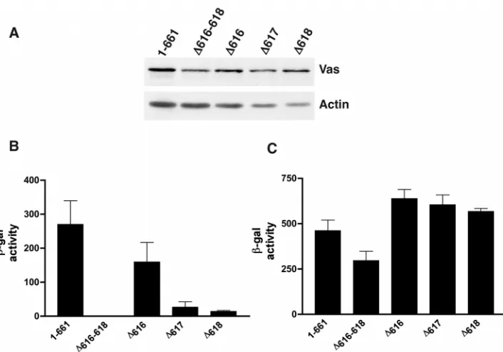
β-galactosidase activity to quantify the level of interaction. All of these mutant forms of Vas expressed protein efficiently and stably in yeast (Fig. 2A). We found that the ∆616-618 deletion completely eliminated detectable interaction with eIF5B (Fig. 2B). The single amino acid deletions also all showed a reduction in eIF5B-interaction relative to the full-length Vas protein, ~1.7-fold for ∆616, tenfold for ∆617 and 20-fold for
∆618 (Fig. 2B). In order to verify that these mutations in Vas affected eIF5B-interaction specifically and not interaction between Vas and Osk, which is crucial for pole plasm assembly (Breitwieser et al., 1996), we also tested the Vas deletions against Osk in the same assay (Fig. 2C). We found that relative to the full-length Vas protein, ∆616-618 showed a modest (less than twofold) reduction in Osk interaction. The single amino acid deletions, however, showed no reduction of this interaction (Fig. 2C). We conclude that these individual Vas deletions specifically disrupt interaction with eIF5B without affecting the interaction between Vas and Osk. Furthermore, the interaction site of a third known Vas-binding protein, Gustavus (Gus) maps to a region near the N terminus of Vas (Styhler et al., 2002), very distant from the residues we found were crucial for eIF5B interaction. Thus, the vas mutations we have generated that affect eIF5B interaction do not affect its interaction with any other known binding protein, and do not affect any conserved motif that is implicated in any function common to DEAD-box proteins, such as ATP binding, ATP hydrolysis, RNA binding and RNA unwinding (Pause and Sonenberg, 1992; Tanner et al., 2003) (reviewed by Tanner and Linder, 2001).
eIF5B interaction is not required for localization of Vas to the pole plasm
During oogenesis, Vas protein is present throughout the cytoplasm of nurse cells and is enriched in nuage particles that
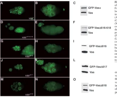
are concentrated around the nurse cell nuclei (Lasko and Ashburner, 1990). Within the oocyte, Vas is also distributed throughout the cytoplasm, and in later stages accumulates in the developing posterior pole plasm, where it remains concentrated in the early embryo. The subsequent functions of Vas all require this posterior localization, thus we wanted to establish whether specific mutations that reduce Vas-eIF5B interaction also affected Vas localization. To do this, we expressed GFP-fusions of various forms of Vas in transgenic flies under the control of the endogenous vas promoter. We tested the levels of expression of the transgenic fusion proteins on western blots, using endogenous Vas protein as an internal control (Fig. 3). Expression of the wild-type transgenic protein was comparable with that of the endogenous protein (Fig. 3C). Despite generating multiple transgenic lines for each, the
∆616-618, ∆616 and ∆618 proteins were always expressed at lower levels than the wild-type transgenic protein, when each was compared with levels of endogenous Vas (Fig. 3F,I,O). However, the ∆617 transgenic protein was expressed at levels comparable with the wild-type transgenic protein (Fig. 3L).
Using GFP fluorescence, we monitored the localization of the transgenic fusion proteins in ovaries from flies bearing two copies of each transgene in a wild-type background (Fig. 3). We found that in all of the deletion mutants, GFP-Vas protein distribution mimicked that of the endogenous protein, accumulating in the perinuclear region of nurse cells in earlier stages (Fig. 3D,G,J,M), and concentrating in the pole plasm in later stages (Fig. 3E,H,K,N). In flies expressing the ∆616-618 transgenic protein (Fig. 3D,E), the GFP signal was weak because of low protein expression, but even in this case, correct localization could be detected. From these data, we conclude that eIF5B interaction is not required to achieve Vas localization.
[image:4.612.204.557.68.315.2]To avoid complications resulting from dose effects, we chose to focus the remainder of our analysis on the vas∆617mutation because of its high level of protein expression and the strong
effect of this mutation on eIF5B interaction. Comparing the wild-type vas+ and the vas∆617 transgenes allowed us to determine the phenotypic consequences of specifically reducing eIF5B interaction, and to separate the functions of Vas that require that interaction from those that depend solely on its localization and expression.
Vas-eIF5B interaction is required for female fertility and for grk regulation
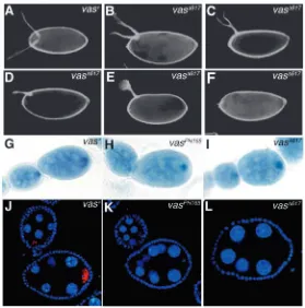
In order to investigate the requirement of the Vas-eIF5B interaction during oogenesis, we examined the vas∆617 transgene in the background of a vas null allele, vasPH165 (Styhler et al., 1998). The wild-type vas+transgene, and the vas∆617transgene express comparable levels of protein in the vas null background (Fig. 4A). Most vasPH165 egg chambers arrest early in oogenesis, producing few mature eggs, none of which hatches into embryos (Styhler et al., 1998) (Fig. 4B). Strikingly, vasPH165;P{vas∆617} females exhibit a very similar arrest in oogenesis to that of vasPH165 itself, producing few mature eggs that do not hatch (Fig. 4D). In both vasPH165and vasPH165;P{vas∆617} females of the same age (3-4 days), the number of stage 14 eggs per ovary ranges widely from 0-32 with an average of nine, roughly a third the average of wild-type. Examples of severely atrophied vasPH165 and vasPH165;P{vas∆617} ovaries are shown in Fig. 4B,D. Similar to vasPH165 (Styhler et al., 1998), eggs produced by vasPH165;P{vas∆617} females sometimes exhibit a duplicated micropyle at both the anterior and posterior ends (data not shown). By contrast, ovaries from vasPH165;P{vas+} females produce abundant mature eggs that can hatch into viable
embryos (Fig. 4C). Thus, the vas∆617transgene does not rescue the oogenesis arrest caused by the vasPH165mutation, indicating that interaction between Vas and eIF5B is crucial for the progression of oogenesis.
Dorsal appendages provide a sensitive assay for dorsoventral patterning, and in grk mutants, ventralization of the eggshell can be observed as a fusion or absence of dorsal appendages. In several vas alleles, including vasPH165, dorsal appendage defects, indicative of a ventralization phenotype, are observed (Styhler et al., 1998; Tinker et al., 1998; Tomancak et al., 1998). In agreement with previous reports (Styhler et al., 1998), we observed that 63% of eggs produced by vasPH165females exhibited one fused or semi-fused dorsal appendage, 12% had no dorsal appendages and 25% had two dorsal appendages. We examined dorsal appendages in our transgenic lines and found that in vasPH165;P{vas∆617} females, 67% of eggs exhibited one semi-fused or semi-fused dorsal appendage (Fig. 5C-E), 11% formed no dorsal appendages (Fig. 5F) and 22% had two dorsal appendages (Fig. 5B). Conversely, 82% of control vasPH165;P{vas+} eggs had two dorsal appendages (Fig. 5A). Thus, the vas∆617transgene does not rescue the ventralization phenotype of the vasPH165mutation.
[image:5.612.53.439.71.390.2]We then assessed grk RNA and protein expression in vasPH165;P{vas∆617} ovaries, and found that it also was indistinguishable in early stages of development from that observed for vasPH165(Fig. 5H,I,K,L) (Styhler et al., 1998). grk RNA is strongly enriched in the oocyte (Fig. 5H,I); however, the protein level is severely reduced (Fig. 5K,L). In control vasPH165;P{vas+} ovaries, concentration of both grk RNA and protein in the oocyte resembles wild type (Fig. 5G,J). Thus, the vas∆617transgene does not support efficient expression of Grk in a vas-null background. We conclude that the requirements for Vas for the progression of oogenesis, for dorsoventral patterning of the egg chamber, and for grk regulation, all rely on its interaction with eIF5B.
Fig. 3. Localization and expression of transgenic GFP-fusion proteins. For each transgenic genotype, localization of GFP is demonstrated in early and late stages of oogenesis: (A,B) vas+; (D,E) vas∆616-618;
(G,H) vas∆616; (J,K) vas∆617;
(M,N) vas∆618. Protein expression in
Pole plasm components assemble in vasPD;P{vas∆617} ovaries
Pole plasm assembly requires the sequential posterior localization of multiple proteins and RNAs (reviewed by Mahowald, 2001). Osk, which is at the top of a complex hierarchy of factors involved in pole plasm assembly, is required for recruitment of Vas to the posterior (Lasko and Ashburner, 1990). Downstream of Vas localization, Tud protein is recruited (Bardsley et al., 1993), and Osk, Vas and Tud are required for pole cell formation and posterior segmentation. In order to investigate the requirement for the Vas-eIF5B interaction in pole plasm assembly, we examined the vas∆617transgene in the
background of a hypomorphic vas allele, vasPD, as vasPH165; P{vas∆617} ovaries produce few late-stage egg chambers. The vasPD allele, in which Vas is detectable only in the germarial stages of oogenesis (Lasko and Ashburner, 1990), completes oogenesis normally, but the embryos produced lack pole cells and posterior segmentation. Sequencing of the vasPD allele did not reveal any alteration in the coding sequence (Liang et al., 1994), thus this mutation is believed to affect only the level of vas expression and not the nature of Vas protein.
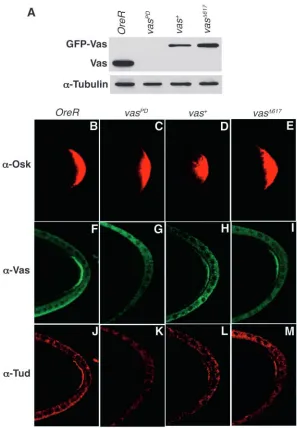
[image:6.612.245.561.74.276.2]In wild-type stage 10 egg chambers, strong posterior accumulation of Osk, Vas and Tud protein is evident (Fig. 6B,F,J), while in vasPDovaries, which have severely reduced levels of Vas protein (Fig. 6A), posterior Osk is abundant, but neither Vas nor Tud is detected at the posterior (Fig. 6C,G,K). We next compared the accumulation of these three proteins in vasPD; P{vas+} and vasPD; P{vas∆617} ovaries, which contain comparable levels of Vas protein as assayed by western blotting (Fig. 6A). We found that Osk, Vas and Tud protein can all be readily detected at the posterior of stage 10 oocytes (Fig. 6D-E,H-I,L-M), at apparently equivalent levels for both transgenic
Fig. 4. Vas-eIF5B interaction is required for the progression of oogenesis. (A) Western blot of ovarian extracts from wild-type (OreR), vasPH165,
vasPH165;P{vas+} and vasPH165;P{vas∆617} females
was probed with anti-Vas. The same blot was probed with anti-4E-BP as a loading control. (B) Ovaries from vasPH165females produce few
mature stage 14 eggs owing to a developmental arrest during oogenesis. (C) Expression of a wild-type vas+transgene rescues this defect of the
vasPH165mutation, allowing the production of
abundant stage14 eggs. (D) Ovaries from vasPH165;P{vas∆617} females resemble those of
vasPH165, exhibiting a similar developmental arrest
during oogenesis and producing few mature eggs.
Fig. 5. Vas-eIF5B interaction is important for the
establishment of dorsoventral polarity in the egg, and for grk regulation. Dark-field photographs of dorsal appendage phenotypes (A-F). Eighty-two percent of vasPH165;P{vas+}
eggs have two dorsal appendages (A). Dorsal appendages in vasPH165;P{vas∆617} eggs reveal a range of phenotypes: 22%
have two dorsal appendages (B), 67% exhibit one semi-fused or fully fused dorsal appendage (C-E) and 11% have no dorsal appendages (F). (G-L) grk RNA and Grk protein were visualized in the early stages of oogenesis, through in situ hybridization and immunostaining. (J-L) Grk protein is shown in red; DAPI staining of DNA is shown in blue. (G,J) In vasPH165;P{vas+} ovaries, both RNA and protein are enriched
in the developing oocyte. (H-L) In both vasPH165and
vasPH165;P{vas∆617} ovaries, grk RNA is enriched in the
[image:6.612.42.321.298.580.2]genotypes. Thus, GFP-Vas∆617 not only localizes correctly to the posterior, but it is also able to recruit the downstream pole plasm component Tud. We conclude that interaction with eIF5B is not required for the role of Vas in the initial assembly of the pole plasm.
Vas-eIF5B interaction is critical for germ cell formation
It has previously been impossible to differentiate whether the embryonic phenotypes produced by hypomorphic vas alleles such as vasPDwere simply due to a lack of posterior Vas protein in the oocyte, resulting in a failure of recruitment of pole plasm components, or whether Vas has an additional regulatory function within the pole plasm. Analysis of the vas∆617transgene in the vasPDbackground allowed us to address this question, as Vas protein is expressed abundantly from this transgene, localizes correctly and is able to recruit Tud, an essential factor for germ cell specification (Ephrussi and Lehmann, 1992). We examined pole cell formation in the progeny of flies bearing either the vas+transgene or the vas∆617transgene in the vasPD
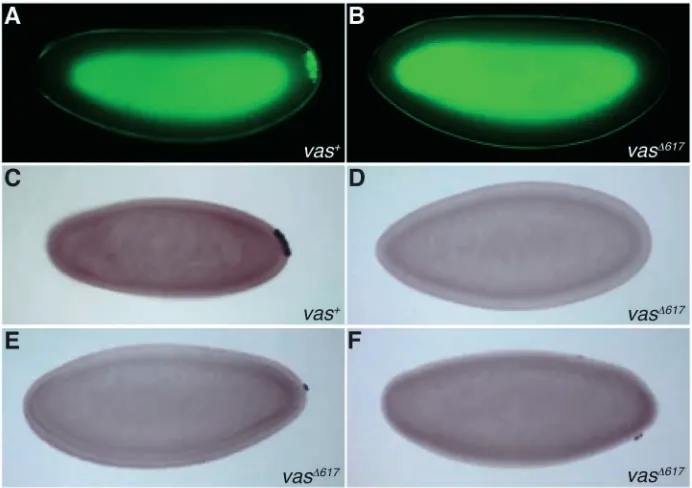
background. For simplicity, transgenic embryos will be referred to by the genotype of the mother. Using detection of GFP-Vas as a marker in live embryos at the cellular blastoderm stage, we found that the majority of vasPD;P{vas+ } embryos had formed pole cells at this stage, indicating that the wild-type transgene rescues this vas phenotype (Fig. 7A). By contrast, only one vasPD;P{vas∆617} embryo out of 216 examined exhibited pole cells at the same stage, indicating that expression of the vas∆617 transgene could not rescue this vas phenotype (Fig. 7B). We verified this result using Nos as an independent marker for pole cells. Although 58% of vasPD;P{vas+ }embryos exhibited Nos-positive cells at the posterior of the embryo at the cellular blastoderm stage (Fig. 7C), 81% of vasPD;P{vas∆617} embryos examined did not have any Nos-positive cells (Fig. 7D). Five percent of these embryos formed one to three Nos-positive cells at the posterior (Fig. 7E), while in 14% of embryos, Nos-positive cells were visible, but at inappropriate positions (Fig. 7F). These results demonstrate that, downstream of initial pole plasm assembly, the Vas-eIF5B interaction is vital for embryonic germ cell formation.
Reduction in Vas-eIF5B interaction does not abolish somatic posterior segmentation or Nos deployment
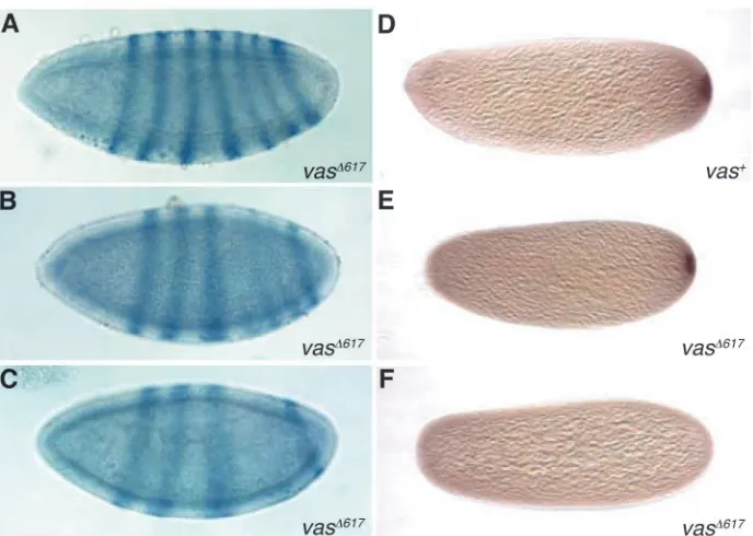
[image:7.612.47.344.68.496.2]Expression of fushi tarazu (ftz) RNA serves as a molecular marker for the incipient segmentation pattern of the embryo. The wild-type pattern of ftz expression is in seven transverse stripes (Hafen et al., 1984), and mutations in posterior group genes such as vas inhibit the expression of stripes 4-6. In contrast to vasPD, in which all embryos exhibit severe posterior segmentation defects (Schüpbach and Wieschaus, 1986), we observed that half of vasPD;P{vas∆617} embryos have a wild-type segmentation pattern, as inferred from a wild-type ftz distribution (Fig. 8A). In fact, when allowed to complete development, many vasPD;P{vas∆617} embryos hatched into viable larvae. A further 20% of the vasPD;P{vas∆617} embryos exhibited a weak posterior group phenotype in which stripes 4-6 of ftz were present but were reduced in width and intensity relative to the other stripes (Fig. 8B), although the remaining 30% exhibited a stronger phenotype (Fig. 8C), more closely resembling that of vasPD (Schüpbach and Wieschaus, 1986). Over 90% of vasPD;P{vas+} embryos expressed ftz in the normal pattern of seven stripes (data not shown). From this analysis, we conclude that the vas∆617 transgene can partially rescue the abdominal segmentation defect of the vasPD allele, suggesting that the
Fig. 6. Pole plasm assembly in the presence of reduced Vas-eIF5B interaction. (A) Western blot of ovarian extracts from wild-type (OreR), vasPD, vasPD;P{vas+}
and vasPD;P{vas∆617} females was probed with anti-Vas.
The same blot was probed with anti-α-Tubulin as a loading control. (B-M) Stage 10 ovaries from OreR, vasPD, vasPD;P{vas+} and vasPD;P{vas∆617} females were
Vas-eIF5B interaction is less crucial for posterior patterning than it is for pole cell specification.
nos translation is tightly regulated such that it is repressed outside of the pole plasm and active in the pole plasm (Gavis and Lehmann, 1994). In existing vas mutants, nos is unlocalized and untranslated, and the absence of a Nos protein gradient prevents abdominal formation (Gavis and Lehmann, 1994; Wang et al., 1994). We examined Nos protein distribution in vasPD;P{vas∆617} embryos directly. Consistent with the ftz expression data, 55% of vasPD;P{vas∆617} embryos exhibited a detectable Nos gradient (Fig. 8E,F). In control vasPD;P{vas+} embryos, a Nos protein gradient could be observed in 95% of the embryos (Fig. 8D). Thus, the vas∆617 transgene partially rescues the Nos accumulation defect of the vasPD allele, suggesting that either the Vas-eIF5B interaction is dispensable for Nos translation, or that the low level of interaction between Vas∆617 and eIF5B is sufficient to activate Nos.
Discussion
The Vas-eIF5B interaction is essential for oogenesis, dorsoventral patterning, and Grk expression
We have analyzed a mutant form of Vas, Vas∆617, which has greatly reduced ability to interact with eIF5B. As residue 617 is not involved in binding to any known Vas-interacting protein other than eIF5B, and it is outside the region of Vas that contains the well-characterized catalytic domains that are present in all DEAD-box proteins, we are confident that Vas∆617 specifically disrupts the Vas-eIF5B interaction, and that this mutation can be used to identify developmental processes that are sensitive to an association between Vas and the general translational machinery. We found that the Vas-eIF5B interaction is crucial for the progression of oogenesis, for correct dorsoventral patterning of the egg, and for expression of high levels of Grk in the developing oocyte. These results are most easily explained if grk is a target
for Vas-mediated translational activation acting through its association with eIF5B.
A role for Vas in positively regulating grk translation is consistent with previous work (Styhler et al., 1998; Tomancak et al., 1998; Ghabrial and Schüpbach, 1999). Vas-mediated regulation of grk is in turn regulated in response to a meiotic checkpoint, activated when DNA double-strand break (DSB) repair is prevented during meiotic recombination (Ghabrial et al., 1998; Ghabrial and Schüpbach, 1999). In response to this checkpoint, Vas is posttranslationally modified, and Grk accumulation is reduced. It will be important to understand the nature of the DSB-dependent modification of Vas, and to determine whether it affects the Vas-eIF5B interaction, in order to gain insight into the mechanism connecting cell cycle regulation with oocyte patterning.
The RNA-binding and unwinding activities of wild-type Vas and several mutant forms of Vas have previously been assessed through in vitro assays (Liang et al., 1994). Two mutant forms of Vas, encoded by the vasO14and vasO11alleles, were found to be severely reduced for binding to an artificial RNA substrate, and a third form, encoded by vasD5, was defective for RNA unwinding but not for binding. Although vasD5leads to defects in oogenesis, vasO11 phenotypically resembles vasPD, and vasO14is a weak temperature-sensitive allele (Lasko and Ashburner, 1990). In the light of our present results, it is surprising that a mutant form of Vas that cannot interact with RNA would nevertheless support oogenesis. Perhaps in vivo, the RNA-binding and helicase activities of Vas are stimulated or enhanced through a co-factor or through posttranslational modifications, and the in vitro assay used in our earlier study may not accurately reflect Vas activity in vivo.
Are there target RNAs for Vas-eIF5B regulation in the pole plasm?
[image:8.612.215.561.72.316.2]Reduction of the Vas-eIF5B interaction by expressing Vas∆617 severely reduces pole cell formation. This happens despite the
Fig. 7. Vas-eIF5B interaction is vital for pole cell formation. Pole cells were visualized at the cellular blastoderm stage using either GFP-Vas or immunostaining for Nos protein. The pictures shown are representative for each genotype. (A,B) Using GFP-Vas as a marker in live embryos, pole cells could be detected at the posterior of the majority of vasPD;P{vas+}
embryos (A), but in fewer than 1% of vasPD;P{vas∆617} embryos (B). (C) Using
immunostaining for Nos as a marker for pole cells, Nos-positive cells could be detected at the posterior in 58% of vasPD;P{vas+} embryos. (D,E) In 81% of
vasPD;P{vas∆617} embryos, no Nos-positive
cells were apparent at the same stage (D), whereas in 5%, one to three Nos-positive cells could be detected at the posterior (E). (F) In 14% of vasPD;P{vas∆617} embryos,
ability of vasPD;P{vas∆617} oocytes to accumulate Osk, Vas and Tud at the posterior pole, demonstrating an essential role for Vas in pole cell specification that is dependent upon its association with eIF5B, and that cannot be substituted by Osk and Tud. The simplest interpretation of these results is that Vas derepresses translation of a localized RNA required for pole cell specification, in a manner analogous to what appears to be the case for grk.
We considered the possibility that the Vas-eIF5B interaction could target osk mRNA. It has previously been shown that whereas Osk protein accumulates normally in vas mutant ovaries, Osk levels are severely reduced at the posterior of vas mutant embryos (Harris and Macdonald, 2001), suggesting a role for Vas in posterior accumulation of Osk after its initial recruitment, and/or in stabilizing Osk at the posterior. We observed comparable and substantial levels of Osk at the posterior in vasPD;P{vas∆617} and vasPD;P{vas+} embryos (data not shown), arguing against a direct role for the Vas-eIF5B interaction in activating translation of osk mRNA. A requirement for Vas in Par1-mediated phosphorylation and stabilization of Osk has been suggested (Breitwieser et al., 1996; Markussen et al., 1997; Riechmann et al., 2002). As Vas∆617 localizes normally and is able to interact with Osk, we would not expect this mutation to have any effect on this Osk modification pathway. Thus, our findings are consistent with a model whereby Vas influences Osk activity through effects on phosphorylation, anchoring and/or stability, perhaps through Par1, rather than directly regulating osk translation.
Another candidate target for the Vas-eIF5B interaction is germ cell-less (gcl), the activity of which is important for pole cell specification but not for posterior patterning (Jongens et al., 1992). Unfortunately, with current reagents, and with new antisera we have generated, we cannot reliably detect Gcl protein even in wild-type embryos prior to pole bud formation, thus we cannot presently address the effects on gcl translation of any mutation that abrogates pole cell formation. In addition,
effects on gcl cannot fully explain the severe consequences of the Vas∆617 mutation on pole cell formation, because the number of pole cells formed in maternal gcl-null embryos is somewhat higher than in vasPD;P{vas∆617} embryos (Robertson et al., 1999). This suggests that even if gcl is a target, the Vas-eIF5B interaction may regulate translation of more than one target RNA involved in pole cell formation.
Is the Vas-eIF5B interaction required for posterior patterning?
[image:9.612.223.567.69.314.2]Although the Vas-eIF5B interaction is vital for pole cell specification, it is perhaps less so for posterior patterning and establishment of the Nos gradient. Previous analysis of hypomorphic mutations in posterior-group genes, including vas has indicated that a higher level of activity is required for pole cell specification than for posterior patterning. For example, all embryos produced by females homozygous for vasO14 (Lasko and Ashburner, 1990), osk301 (Lehmann and Nüsslein-Volhard, 1986) and tudWC (Schüpbach and Wieschaus, 1986), lack pole cells, but some have normal posterior patterning and are able to hatch. Our present results suggest two alternative explanations for these observations. One possibility is that the Vas-eIF5B interaction is required for posterior patterning, but that the residual activity present in Vas∆617 is sufficient to achieve the low activity level that is necessary. Alternatively, the Vas-eIF5B interaction may be dispensable for posterior patterning, and the fact that we do not observe complete rescue of this phenotype with the vas∆617 transgene may be due to an indirect effect of this mutation, resulting from a general destabilization of the pole plasm that occurs in embryos that do not form pole cells (Iida and Kobayashi, 2000). In such embryos, pole plasm components localize initially but become fully delocalized by the blastoderm stage (Lasko and Ashburner, 1990). Consistent with this idea, all of the pole plasm components examined that are downstream of Vas, including nos RNA, could be detected
Fig. 8. Reduced Vas-eIF5B binding does not abrogate somatic patterning or Nos deployment. RNA in situ hybridization for ftz was used as an indicator of somatic segmentation at the cellular blastoderm stage. (A) Fifty percent of vasPD;P{vas∆617}
embryos revealed a normal ftz distribution of seven transverse stripes. (B,C) In 20% of vasPD;P{vas∆617} embryos, stripes 4-6 were
weaker and less defined than the others (B), and in 30%, more severe defects such as deletions and fusions of these segments were apparent (C). (D) Nos protein, visualized through immunostaining in 0- to 2-hour-old embryos, was present at the posterior of 95% of vasPD;P{vas+}
embryos. (E,F) Posterior Nos was detectable in 55% of the vasPD;P{vas∆617}
at the posterior of vas ;P{vas } embryos, although to variable degrees (data not shown).
Previous work has suggested that nos may be a target for Vas-mediated translational regulation (Gavis et al., 1996). Outside of the pole plasm, nos translation is repressed through the binding of Smg, and possibly other repressors, to its 3′ UTR (Smibert et al., 1996; Dahanukar et al., 1999; Crucs et al., 2000; Nelson et al., 2004). Smg achieves this regulation at least in part through interaction with the eIF4E-binding protein Cup, thus influencing the cap-eIF4E-binding stage of translation (Nelson et al., 2004). Within the pole plasm, in complexes with Osk and Vas, nos translational repression is overcome, potentially through a direct interaction between Osk and Smg (Dahanukar et al., 1999), which may displace Smg-Cup interaction (Nelson et al., 2004). Our analysis of Vas∆617 does not support an important role for the Vas-eIF5B interaction in activating nos translation in the pole plasm, as clearly translation of nos is far less sensitive to the level of this interaction than is translation of grk in early oocytes. The primary function of Vas in nos accumulation may therefore be in anchoring nos mRNA in complexes within the pole plasm, consistent with recent observations that nos mRNA is trapped at the posterior by complexes containing Vas (Forrest and Gavis, 2003). It of course remains possible that the low level of residual eIF5B binding provided by Vas∆617 is sufficient to fulfill a role of Vas in activating translation of this transcript.
How might Vas-eIF5B interaction regulate translation of grk and potentially other target mRNAs?
Cap-dependent translation initiation in eukaryotes requires many translation initiation factors, and involves several main steps (reviewed by Pestova et al., 2001). Most known mechanisms of translational regulation impinge on the recruitment of the cap-binding complex eIF4F to the mRNA, which represents the rate-limiting first step of initiation. mRNA circularization through proteins such as Cup serves an important role in translational control by allowing 3′ UTR-bound regulatory factors to influence translation initiation at the 5′end of the transcript. (Nakamura et al., 2004; Nelson et al., 2004; Wilhelm et al., 2003).
60S ribosomal subunit joining represents the interface between translation initiation and elongation, and the Vas-eIF5B interaction suggests a distinct mechanism of translational control occurring at this last stage of initiation. Although this step has not historically been considered a target for regulation, several examples have emerged to suggest that subunit joining may in fact be subject to regulation. Translational repression of mammalian 15-lipoxygenase (LOX) mRNA is mediated by hnRNP proteins that bind to a specific 3′UTR regulatory element, and which are thought to act by blocking the activity of either eIF5 or eIF5B (Ostareck et al., 2001). An additional link between mRNA 3′regulatory regions, and eIF5B activity, comes from analysis of two DEAD-box proteins in yeast, Ski2p and Slh1p (Searfoss et al., 2001). These proteins are required to achieve the selective translation of poly(A)+ mRNAs, relative to poly(A)-mRNAs, and genetic experiments suggest that they specifically repress the translation of poly(A)-mRNAs by acting through eIF5 and eIF5B.
Together with these studies, our work suggests that in the Drosophila germline, specific translational repression events may target eIF5B and the ribosomal subunit joining step of initiation. Vas, which potentially functions at the 3′ UTR through interaction with specific repressor proteins, may act to alleviate a translation block occurring at this step. Such a model is consistent with what is known about translational regulation of grk. For example, grk translation is repressed by Bru, which binds to a Bruno-response element within its 3′UTR (Filardo and Ephrussi, 2003). Vas interacts with Bru (Webster et al., 1997), suggesting that Vas could function as a derepressor by overcoming Bru-mediated repression of grk translation. However, the inability of a vas transgene to ameliorate the phenotype of nosGAL4VP16-driven overexpression of Bru, might argue against this model (Filardo and Ephrussi, 2003). The mechanism by which Bru regulates grk remains unclear. Translational repression of osk by Bru relies on direct interaction with Cup, linking Bru with eIF4E (Nakamura et al., 2004). However, mutations in cup that prevent interaction with Bru do not appear to affect Grk expression, suggesting that Bru may operate through a distinct mechanism to regulate grk translation (Nakamura et al., 2004). In addition, in vitro translation assays have suggested that Bru can mediate translational repression through a cap-independent mechanism (Lie and Macdonald, 1999). Thus, Bru may be capable of regulating translation at more than one stage. Based on the observations for the mammalian hnRNP proteins on the LOX mRNA, and the Ski2p and Slh1p helicases in yeast, specific translational repressors such as Bru could target the subunit joining step of initiation.
eIF5B is thought to form a molecular bridge between the two ribosomal subunits, and to play a fundamental role in stabilizing the initiator Met-tRNAiMetin the ribosomal P site (reviewed by Pestova et al., 2000a). Inhibition of eIF5B activity could occur while the factor is bound to the initiation complex, at the start codon, and block its ability to link or stabilize the ribosomal subunits. Through circularization of the mRNA, this block could be achieved by trans-acting factors at the 3′UTR, and the Vas-eIF5B interaction may be involved in alleviating these specific repression events, potentially through displacement of a repressor protein. Alternatively, Vas could play a role in recruitment of eIF5B to specific transcripts. As eIF5B is required for all cellular translation, a general mechanism must exist to recruit this factor to all transcripts. However, in a scenario where repressor proteins may be blocking the subunit joining step, either through a direct effect on eIF5B, or another mechanism, it is conceivable that eIF5B could become limiting for translation. In this situation, Vas could play a role in recruiting this factor to specific transcripts.
This work was supported by PHS research grant RO1HD36631 to P.L. We are grateful to the following people for gifts of reagents: Anne Ephrussi for anti-Osk, Elizabeth Gavis for anti-Nos, Henry Krause for the ftz cDNA, Akira Nakamura for the P[w+Pvas-gfp] plasmid, Trudi
References
Bardsley, A., McDonald, K. and Boswell, R. E. (1993). Distribution of tudor protein in the Drosophila embryo suggests separation of functions based on site of localization. Development 119, 207-219.
Breitwieser, W., Markussen, F.-H., Horstmann, H. and Ephrussi, A. (1996). Oskar protein interaction with Vasa represents an essential step in polar granule assembly. Genes Dev. 10, 2179-2188.
Carrera, P., Johnstone, O., Nakamura, A., Casanova, J., Jäckle, H. and Lasko, P. (2000). VASA mediates translation through interaction with a Drosophila yIF2 homolog. Mol. Cell 5, 181-187.
Castagnetti, S. and Ephrussi, A. (2003). Orb and a long poly(A) tail are required for efficient oskar translation at the posterior pole of the Drosophila oocyte. Development 130, 835-843.
Chang, J. S., Tan, L. and Schedl, P. (1999). The Drosophila CPEB homolog, orb, is required for oskar protein expression in oocytes. Dev. Biol. 215, 91-106.
Clark, I. E., Wyckoff, D. and Gavis, E. R. (2000). Synthesis of the posterior determinant Nanos is spatially restricted by a novel cotranslational regulatory mechanism. Curr. Biol. 10, 1311-1314.
Crucs, S., Chatterjee, S. and Gavis, E. R. (2000). Overlapping but distinct RNA elements control repression and activation of nanos translation. Mol. Cell 5, 457-467.
Dahanukar, A., Walker, J. A. and Wharton, R. P. (1999). Smaug, a novel RNA-binding protein that operates a translational switch in Drosophila. Mol. Cell 4, 209-218.
Ephrussi, A. and Lehmann, R. (1992). Induction of germ cell formation by oskar. Nature 358, 387-392.
Ephrussi, A., Dickinson, L. K. and Lehmann, R. (1991). Oskar organizes the germ plasm and directs localization of the posterior determinant nanos. Cell 66, 37-50.
Filardo, P. and Ephrussi, A. (2003). Bruno regulates gurken during Drosophila oogenesis. Mech. Dev. 120, 289-297.
Forrest, K. M. and Gavis, E. R. (2003). Live imaging of endogenous RNA reveals a diffusion and entrapment mechanism for nanos mRNA localization in Drosophila. Curr. Biol. 13, 1159-1168.
Gavis, E. R. and Lehmann, R. (1994). Translational regulation of nanos by RNA localization. Nature 369, 315-318.
Gavis, E. R., Lunsford, L., Bergsten, S. E. and Lehmann, R. (1996). A conserved 90 nucleotide element mediates translational repression of nanos RNA. Development 122, 2791-2800.
Ghabrial, A. and Schüpbach, T. (1999). Activation of a meiotic checkpoint regulates translation of Gurken during Drosophila oogenesis. Nat. Cell Biol. 1, 354-357.
Ghabrial, A., Ray, R. P. and Schüpbach, T. (1998). okra and spindle-B encode components of the RAD52 DNA repair pathway and affect meiosis and patterning in Drosophila oogenesis. Genes Dev. 12, 2711-2723. Golemis, E. A., Serebriiskii, I., Gyuris, J. and Brent, R. (1997). Interaction
trap/two-hybrid system to identify interacting proteins. In Current Protocols in Molecular Biology (ed. F. M. Ausubel, R. Brent, R. E. Kingston, D. D. Moore, J. G. Seidman, J. A. Smith and K. Struhl), unit 20.1, pp. 1-35. New York, NY: Wiley.
González-Reyes, A., Elliott, H. and St Johnston, D. (1995). Polarization of both major body axes in Drosophila by gurken-torpedo signalling. Nature 375, 654-658.
Hafen, E., Kuroiwa, A. and Gehring, W. J. (1984). Spatial distribution of transcripts from the segmentation gene fushi tarazu during Drosophila embryonic development. Cell 37, 833-841.
Harris, A. N. and Macdonald, P. M. (2001). aubergine encodes a Drosophila polar granule component required for pole cell formation and related to eIF2C. Development 128, 2823-2832.
Hay, B., Jan, L. Y. and Jan, Y. N. (1988). A protein component of Drosophila polar granules is encoded by vasa and has extensive sequence similarity to ATP-dependent helicases. Cell 55, 577-587.
Iida, T. and Kobayashi, S. (2000). Delocalization of polar plasm components caused by grandchildless mutations, gs(1)N26 and gs(1)N441, in Drosophila melanogaster. Dev. Growth Differ. 42, 53-60.
Johnstone, O. and Lasko, P. (2001). Translational regulation and RNA localization in Drosophila oocytes and embryos. Annu. Rev. Genet. 35, 365-406.
Jongens, T. A., Hay, B., Jan, L. Y. and Jan, Y. N. (1992). The germ cell-less gene product: a posteriorly localized component necessary for germ cell development in Drosophila. Cell 70, 569-584.
Kim-Ha, J., Smith, J. L. and Macdonald, P. M. (1991). oskar mRNA is localized to the posterior pole of the Drosophila oocyte. Cell 66, 23-35.
Kim-Ha, J., Kerr, K. and Macdonald, P. M. (1995). Translational regulation of oskar mRNA by bruno, an ovarian RNA-binding protein, is essential. Cell 81, 403-412.
Kobayashi, S., Amikura, R., Nakamura, A. and Lasko, P. F. (1999). Techniques for analyzing protein and RNA distribution in Drosophila ovaries and embryos at structural and ultrastructural resolution. In Advances in Molecular Biology: A Comparative Methods Approach to the Study of Oocytes and Embryos (ed. J. Richter), pp. 426-445. New York, NY: Oxford University Press.
Lantz, V., Ambrosio, L. and Schedl, P. (1992). The Drosophila orb gene is predicted to encode sex-specific germline RNA-binding proteins and has localized transcripts in ovaries and early embryos. Development 115, 75-88. Lasko, P. F. and Ashburner, M. (1988). The product of the Drosophila gene vasa is very similar to eukaryotic initiation factor-4A. Nature 335, 611-617. Lasko, P. F. and Ashburner, M. (1990). Posterior localization of vasa protein correlates with, but is not sufficient for, pole cell development. Genes Dev. 4, 905-921.
Lehmann, R. and Nüsslein-Volhard, C. (1986). Abdominal segmentation, pole cell formation, and embryonic polarity require the localized activity of oskar, a maternal gene in Drosophila. Cell 47, 141-152.
Lehmann, R. and Nüsslein-Volhard, C. (1991). The maternal gene nanos has a central role in posterior pattern formation of the Drosophila embryo. Development 112, 679-691.
Liang, L., Diehl-Jones, W. and Lasko, P. (1994). Localization of vasa protein to the Drosophila pole plasm is independent of its RNA-binding and helicase activities. Development 120, 1201-1211.
Lie, Y. S. and Macdonald, P. M. (1999). Translational regulation of oskar mRNA occurs independent of the cap and poly(A) tail in Drosophila ovarian extracts. Development 126, 4989-4996.
Linder, P. (2003). Yeast RNA helicases of the DEAD-box family involved in translation initiation. Biol. Cell 95, 157-167.
Linder, P., Lasko, P. F., Ashburner, M., Leroy, P., Nielsen, P. J., Nishi, K., Schnier, J. and Slonimski, P. P. (1989). Birth of the D-E-A-D box. Nature 337, 121-122.
Mahowald, A. P. (2001). Assembly of the Drosophila germ plasm. Int. Rev. Cytol. 203, 187-213.
Markussen, F.-H., Breitwieser, W. and Ephrussi, A. (1997). Efficient translation and phosphorylation of Oskar require Oskar protein and the RNA helicase Vasa. Cold Spring Harb. Symp. Quant. Biol. 62, 13-17.
Nakamura, A., Amikura, R., Hanyu, K. and Kobayashi, S. (2001). Me31B silences translation of oocyte-localizing RNAs through the formation of cytoplasmic RNP complex during Drosophila oogenesis. Development 128, 3233-3242.
Nakamura, A., Sato, K. and Hanyu-Nakamura, K. (2004). Drosophila Cup is an eIF4E binding protein that associates with Bruno and regulates oskar mRNA translation in oogenesis. Dev. Cell 6, 69-78.
Nelson, M. R., Leidal, A. M. and Smibert, C. A. (2004). Drosophila Cup is an eIF4E-binding protein that functions in Smaug-mediated translational repression. EMBO J. 23, 150-159.
Neuman-Silberberg, F. S. and Schüpbach, T. (1993). The Drosophila dorsoventral patterning gene gurken produces a dorsally localized RNA and encodes a TGF alpha-like protein. Cell 75, 165-174.
Nilson, L. A. and Schüpbach, T. (1999). EGF receptor signaling in Drosophila oogenesis. Curr. Top. Dev. Biol. 44, 203-243.
Ostareck, D. H., Ostareck-Lederer, A., Shatsky, I. N. and Hentze, M. W. (2001). Lipoxygenase mRNA silencing in erythroid differentiation: the
3′UTR regulatory complex controls 60S ribosomal subunit joining. Cell 104,
281-290.
Pause, A. and Sonenberg, N. (1992). Mutational analysis of a DEAD box RNA helicase: the mammalian translation initiation factor eIF-4A. EMBO J. 11, 2643-2654.
Pestova, T. V., Dever, T. E. and Hellen, C. U. T. (2000a). Ribosomal subunit joining. In Translational Control of Gene Expression (ed. N. Sonenberg, J. W. B. Hershey and M. B. Mathews), pp. 425-445. Cold Spring Harbor, NY: Cold Spring Harbor Laboratory Press.
Pestova, T. V., Lomakin, I. B., Lee, J. H., Choi, S. K., Dever, T. E. and Hellen, C. U. T. (2000b). The joining of ribosomal subunits in eukaryotes requires eIF5B. Nature 403, 332-335.
Pestova, T. V., Kolupaeva, V. G., Lomakin, I. B., Pilipenko, E. V., Shatsky, I. N., Agol, V. I. and Hellen, C. U. T. (2001). Molecular mechanisms of translation initiation in eukaryotes. Proc. Natl. Acad. Sci. USA 98, 7029-7036.
Molecular Biology (ed. F. M. Ausubel, R. Brent, R. E. Kingston, D. D. Moore, J. G. Seidman, J. A. Smith and K. Struhl), unit 13.6, pp. 1-6. New York, NY: Wiley.
Riechmann, V., Gutierrez, G. J., Filardo, P., Nebreda, A. R. and Ephrussi, A. (2002). Par-1 regulates stability of the posterior determinant Oskar by phosphorylation. Nat. Cell Biol. 4, 337-342.
Robertson, S. E., Dockendorff, T. C., Leatherman, J. L., Faulkner, D. L. and Jongens, T. A. (1999). germ cell-less is required only during the establishment of the germ cell lineage of Drosophila and has activities which are dependent and independent of its localization to the nuclear envelope. Dev. Biol. 215, 288-297.
Roth, S., Neuman-Silberberg, F. S., Barcelo, G. and Schüpbach, T. (1995). cornichon and the EGF receptor signaling process are necessary for both anterior-posterior and dorsal-ventral pattern formation in Drosophila. Cell 81, 967-978.
Schüpbach, T. and Wieschaus, E. (1986). Maternal-effect mutations altering the anterior-posterior pattern of the Drosophila embryo. Roux’s Arch. Dev. Biol. 195, 302-317.
Searfoss, A., Dever, T. E. and Wickner, R. (2001). Linking the 3′poly(A) tail to the subunit joining step of translation initiation: relations of Pab1p, eukaryotic translation initiation factor 5B (Fun12p), and Ski2p-Slh1p. Mol. Cell. Biol. 21, 4900-4908.
Smibert, C. A., Wilson, J. E., Kerr, K. and Macdonald, P. M. (1996). smaug protein represses translation of unlocalized nanos mRNA in the Drosophila embryo. Genes Dev. 10, 2600-2609.
Styhler, S., Nakamura, A., Swan, A., Suter, B. and Lasko, P. (1998). vasa
is required for GURKEN accumulation in the oocyte, and is involved in oocyte differentiation and germline cyst development. Development 125, 1569-1578.
Styhler, S., Nakamura, A. and Lasko, P. (2002). VASA localization requires the SPRY-domain and SOCS-box containing protein, GUSTAVUS. Dev. Cell 3, 865-876.
Tanner, N. K. and Linder, P. (2001). DExD/H box RNA helicases: from generic motors to specific dissociation functions. Mol. Cell 8, 251-262. Tanner, N. K., Cordin, O., Banroques, J., Doère, M. and Linder, P. (2003).
The Q motif: a newly identified motif in DEAD box helicases may regulate ATP binding and hydrolysis. Mol. Cell 11, 127-138.
Tinker, R., Silver, D. and Montell, D. J. (1998). Requirement for the Vasa RNA helicase in gurken mRNA localization. Dev. Biol. 199, 1-10. Tomancak, P., Guichet, A., Zavorszky, P. and Ephrussi, A. (1998). Oocyte
polarity depends on regulation of gurken by Vasa. Development 125, 1723-1732.
Wang, C. and Lehmann, R. (1991). Nanos is the localized posterior determinant in Drosophila. Cell 66, 637-647.
Wang, C., Dickinson, L. K. and Lehmann, R. (1994). Genetics of nanos localization in Drosophila. Dev. Dyn. 199, 103-115.
Webster, P. J., Liang, L., Berg, C. A., Lasko, P. and Macdonald, P. M. (1997). Translational repressor bruno plays multiple roles in development and is widely conserved. Genes Dev. 11, 2510-2521.