Journal of Chemical and Pharmaceutical Research, 2014, 6(3):1506-1511
Research Article
CODEN(USA) : JCPRC5
ISSN : 0975-7384
Changes detection of serum Raman spectroscopy at different stages of lung
cancer using spectral parameters and regression tree
Xiaozhou Li
a, Hong Lou
b*, Lili Jin
b, Tianyue Yang
aand Siqi Li
ca
School of Science, Shenyang Ligong University, Shenyang, China
b
School of Life Science, Liaoning University, Shenyang, China
c
Integrated Life Sciences, Kent State University, Kent, America
_____________________________________________________________________________________________
ABSTRACT
Raman spectroscopy is a vibrational spectroscopic technique that can be used to optically probe the molecular changes associated with diseased organs. The objective of our study was to explore Raman spectroscopy for detecting different stage changes of serum taken from lung cancer patients. Serum samples were obtained from 26 lung cancer patients at different periods before operation. Raman spectra differed significantly at different stages, showing molecular changes with the development of lung cancer. The ratio of Raman intensities excited by 488.0 nm and 514.5 nm laser decreased as lung cancer progress. The results of this exploratory study indicate that Raman spectroscopy provides significant potential for the noninvasive monitoring of lung cancer.
Keywords: lung cancer, serum, Raman spectroscopy, diagnosis
_____________________________________________________________________________________________
INTRODUCTION
Lung cancer is the most common cancer in humans worldwide, and is estimated to have 1.8 million newly diagnosed cases and 1.59 million death in 2014[1]. The overall 5-year survival rate of patients with lung cancer is less than 16%. The high mortality rate of lung cancer is due partly to the fact that few of lung cancers are detected at an early stage[2]. Early lung cancer detection is crucial to increasing the survival rates. However, changes caused by lung cancer are too subtle and difficult to be detected visually by conventional diagnostic methods[3].
As Raman spectroscopy can take a fingerprint of specific molecule, and will be disturbed very little by water[4]. Raman spectroscopy has been successfully used in the detection of biological tissues, cells and bio-fluids for disease diagnosis[5–7]. Advantages of Raman spectroscopy include non-invasiveness, fastness and convenience[8]. Monitoring the condition of lung cancer patients using Raman spectroscopy may help improve the treatment of lung cancer and the adjustment of the therapy[9].
In this paper, serum of lung cancer patients was measured by Raman spectroscopy for 5 weeks to see the variations of the serum components. We found that Raman spectroscopy can detect the changes of serum with the development of lung cancer. This study may provide a potential tool for the lung cancer monitoring in future clinical treatment.
EXPERIMENTAL SECTION
Serum samples were obtained from Tumor Hospital of Liaoning Province and were exactly diagnosed as terminal stages of lung cancer.
After amplified by a lock-in amplifier, spectral data were input into computer and transacted. The spectral range scanned was from 520 nm to 640 nm and from 500 nm to 620 nm at a spectral resolution of 2 cm-1. The frequency of the chopper was 700 Hz. Figure 1 shows the main parts of our instrument: an Ar-ion laser (made in 772 factory in Nanjing), a PMT (R456 model), a lock-in amplifier (391A model), and a double spectrometer (HRD-1 model). The wavelengths of 488.0 nm and 514.5 nm were chosen for excitation.
Fig.1 Instrument
We collected samples once a week, and obtained 3 sets of specimens in different period of the subject. And for each sample, two spectra were measured: (1) the spectrum from 520 nm to 640 nm excited by 514.5 nm; (2) the spectrum from 500 nm to 620 nm excited by 488.0 nm. What we recorded was relative intensity of Raman peaks in order to reduce such interference as the undulation of laser power. And for the purpose of lessening influence of other harmful factors, we sampled several data at each wavelength, and then recorded the average value. And in the process of original data transaction, method of least squares was used to smooth spectra. Due to the resolution improvement of second order derivative spectrum compared with the original, it was utilized to find the position U of Raman peak (mode C) and two nearest inflexions (Wa and Wb) (see Fig.2). Supposing that fluorescence intensity is linear with
wavelength between inflexion Wa and Wb (it is acceptable because the value of second order derivative spectrum of
fluorescence is very small), we got fluorescence intensity at location U and then Ir.
Fig.2 Several types of spectra calculated during the course of data processing (A: original spectrum; B: first derivative spectrum; C: second order derivative spectrum; D: third order derivative spectrum)
RESULTS AND DISCUSSION
Fig.5 (in the first, third and fifth week respectively). In Fig.3, three Raman peaks (mode A, B and C, mode C is the strongest one) can be observed, but not very distinct due to the strong background interference. However, there is no peak A in Fig.4, and the intensity of peaks B and C decreases compared with Fig.3. But fluorescence spectrum almost maintains its former shape. In Fig.5, we are not capable of observing any peak directly, but can find peak C by means of derivative of spectrum (only in spectrum excited by 488.0 nm, not by 514.5 nm). In the sixth week and later, even mode C cannot be detected.
Fig.3 Normalized spectrum in the first week: (a) 514.5 nm excitation; (b) 488.0 nm excitation
Fig.4 Normalized spectrum in the third week: (a) 514.5 nm excitation; (b) 488.0 nm excitation
Fig.5 Normalized spectrum in the fifth week: (a) 514.5 nm excitation; (b) 488.0 nm excitation
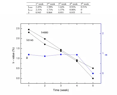
There are some differences in fluorescence spectra in different period, such as intensity, peak position. Those can be attributed to some biochemical changes occurring in serum as disease progress. For further quantitative study, we calculated Ir-value (relative intensity of Raman peak C) and â-value (Ir5145/Ir4880) of such spectra (see Table 1), and
made a graph of Ir-value, â and time (see Fig.6). It demonstrates that Ir is approximately linear with time in this
Tab. 1 Ir-value and â-value in different period
1st week 2nd week 3rd week 4th week 5th week
Ir4880 2.45% 1.98% 1.44% 0.92% 0.51%
Ir5145 2.31% 1.71% 1.37% 0.86% 0
[image:4.595.110.502.82.400.2]â 0.943 0.864 0.951 0.935 0
Fig.6 Connection between Ir-value and tumor evolution (in different period)
Classification and regression tree (CART) was employed on the three parameters (Ir4880, Ir5145, and â) for the
[image:4.595.173.426.489.658.2]classification between the first 4 weeks (the fifth week was left off because of its apparent difference with other stages). Figure 7 displays the CART tree based on the three parameters. It can be seen that by judging on the value the each parameter samples were separated finally after several steps.
Fig.7 Classification and regression tree based on the three parameters
The resubstitution error was 0.025 in our CART. Figure 8 shows the relationship of the cost of substitution and the number of terminal nodes for the cross-validation and resubstitution error through ten-fold cross validation. The misclassification cost reaches an optimal value at the point of 4 nodes. Figure 9 demonstrates the optimized CART tree. From the figure, we can see that after only 2 steps, groups were separated. The first two parameters Ir4880 and Ir5145 were
significant difference among difference stages of lung cancer is observed in parameter â.
Fig.8 Cross-validation, resubstitution error and the best choice for CART
Fig.9 Pruned CART tree
In serum spectrum, three Raman peaks are almost the same as resonance Raman spectrum of beta carotene in carbon tetrachloride solution, whether peak location or intensity distribution[10]. We assumed that they were derived from beta carotene emission. Beta carotene is a kind of carotenoids and can be translated into vitamin A. In epidemiology, studies showed that the incidence of cancer and the content of beta carotene are closely relevant. The higher the content of beta carotene, the less incidence of cancer[11]. And some researches demonstrated that beta carotene, to some extent, can restrain the growth and progression of tumor cells[12]. Our result indicated that the content of beta carotene decreased with aggravation of lung cancer. It means that beta carotene has close relationship with cancer. Such conclusion agrees well with former studies in chromatogram and epidemiology.
Due to the endogenous fluorescence background presented in blood plasma, which is of the order of a million times more intense, the relatively weak Raman signals are difficult to extract. Therefore, near-IR radiation may be used for further studies.
CONCLUSION
REFERENCES
[1] International Agency for Research on Cancer. World Cancer Report 2014, World Health Organization: Geneva,
2014.
[2] P Boyle; B Levin. World Cancer Report 2008, 1st ed., World Health Organization, 2009. [3] PB Bach. Journal of the National Comprehensive Cancer Network, 2008, 6(3), 271 –275. [4] PJ Lambert; AG Whitman; OF Dyson; SM Akula. Virol. J, 2006, 3, 51.
[5] A Mizuno; H Kitajima; K Kawauchi; S Muraishi; Y Ozaki. Journal of Raman spectroscopy, 1994, 25(1), 25–29. [6] N Uzunbajakava; A Lenferink; Y Kraan; B Willekens; G Vrensen; J Greve; C Otto. Biopolymers, 2003, 72(1), 1–9. [7] AJ Berger; TW Koo; I Itzkan; G Horowitz; MS Feld. Appl Opt, 1999, 38(13), 2916–2926.
[8]LM Moreira; L Silveira Jr.; FV Santos; JP Lyon; R Rocha; RA Zângaro; AB Villaverde; MTT Pacheco.
Spectroscopy, 2008, 22(1), 1–19.
[9] Y Oshima; H Shinzawa; T Takenaka; C Furihata; H Sato. J Biomed Opt, 2010, 15(1), 017009. [10] P Yan; L Sanren; W Lixin. Cancer Research And Clinic, 1997, 9(3), 150-153.
[11] PR Carey. Biochemical applications of Raman and resonance Raman spectroscopies, Molecular biology, Academic Press: New York, 1982.