R E S E A R C H A R T I C L E
Open Access
B cell depletion in diffuse progressive systemic
sclerosis: safety, skin score modification and IL-6
modulation in an up to thirty-six months
follow-up open-label trial
Silvia Bosello
1, Maria De Santis
1, Gina Lama
2, Cristina Spanò
2, Cristiana Angelucci
2, Barbara Tolusso
1, Gigliola Sica
2,
Gianfranco Ferraccioli
1*Abstract
Introduction:An over-expression of CD19 has been shown in B cells of systemic sclerosis (SSc) and B cells are thought to contribute to the induction of skin fibrosis in the tight skin mouse model. The aim was to define the outcome on safety and the change in skin score after rituximab therapy in SSc patients and to correlate the clinical characteristics with the levels of interleukin (IL)-6 and with the immune cell infiltrate detected by
immunohistochemistry.
Methods:Nine patients with SSc with mean age 40.9 ± 11.1 years were treated with anti-CD20, 1 g at time 0 and after 14 days. Skin biopsy was performed at baseline and during the follow-up. B-cell activating factor (BAFF) and IL-6 levels were also determined at the follow-up times.
Results:After 6 months patients presented a median decrease of the skin score of 43.3% (range 21.1-64.0%), and a decrease in disease activity index and disease severity index. IL-6 levels decreased permanently during the follow up. After treatment, a complete depletion of peripheral blood B cells was observed in all but 2 patients. Only 3 patients presented CD20 positive cells in the biopsy of the involved skin at baseline.
Conclusions:Anti-CD20 treatment has been well tolerated and SSc patients experienced an improvement of the skin score and of clinical symptoms. The clear fall in IL-6 levels could contribute to the skin fibrosis improvement, while the presence of B cells in the skin seems to be irrelevant with respect to the outcome after B cell depletion. Trial registration:ISRCTN77554566.
Introduction
Although the pathogenesis of systemic sclerosis (SSc) remains unknown, the B cell abnormalities characterized by autoantibody production [1], hyper-g-globulinemia and polyclonal B cell hyperactivity [2] are thought to play an important role in the disease. It has been pre-viously described that SSc patients have distinct abnormalities of blood homeostasis and B cell compart-ments, characterized by expanded naïve cells and acti-vated, but diminished, memory B cells [3]. Furthermore, the expression of CD19, a critical signal transduction
molecule of B cells that regulates autoantibody produc-tion, is significantly increased in memory and naïve B cells in SSc patients [3,4]. Analysis of DNA microarrays of cutaneous biopsies from diffuse SSc (dSSc) patients demonstrated a higher expression of clusters of genes of CD20-positive cells [5].
In the tight-skin mice, a genetic model of human SSc, the CD19 signaling pathway appeared to be constitu-tively activated [6,7] and the loss of CD19 expression significantly up-regulated surface IgM expression, com-pletely abrogated hyper-g-globulinemia and autoantibody production, and also inhibited IL-6 production [7]. Additionally, in this animal model, the down-regulation of B cell function led to a decrease in skin fibrosis * Correspondence: [email protected]
1Division of Rheumatology, Catholic University, Medical School, Via G.
Moscati, 31 - Rome, 00168, Italy
during the disease onset [8]. Likewise, in a bleomycin-induced SSc mouse model, another animal model that shares many characteristics with human SSc, CD19 defi-ciency inhibited the development of skin and lung fibro-sis, hyper-g-globulinemia, and autoantibody production [9]. Thus, B cells could have a relevant impact on the development of fibrotic changes as reported in the mouse scleroderma models [6-9] and also in CCl4 -induced liver injury, in an antibody- and T cell-indepen-dent manner [10].
In several studies focusing on the pathogenesis of SSc, the increased levels of IL-6 in the skin, serum, and bronchoalveolar lavage fluid of SSc patients suggest a role of this cytokine in promoting fibrosis by enhancing inflammation [11-13]. Furthermore, immunohistochem-istry data demonstrated an over-expression of IL-6 on endothelium and fibroblasts of involved skin of sclero-derma patients compared with normal skin [14]. SSc dermal fibroblasts constitutively produce about a four-fold increase in IL-6 levels with respect to healthy con-trols fibroblasts [15] and secretion of IL-6 from lung fibroblast is induced by SSc lung-derived B cells [16]. Recently, it has been reported that B-cell activating fac-tor (BAFF), an essential component of B cell homeosta-sis and a potent B-cell survival factor associated with autoimmune disease in humans, is increased in SSc patients compared with healthy controls [17]. In the tight-skin mice, BAFF antagonist augmented anti-fibro-genic cytokines and inhibited the development of skin fibrosis. Finally, after BAFF stimulation, B-cells had a significantly enhanced ability to produce IL-6 [18].
Two recent open-label studies reported the safety of anti-CD20 treatment in SSc patients; despite both studies describing a decrease in myofibroblast score on serial skin biopsies after treatment, only one reported an improvement in skin score [19,20]. In these two studies, lung function remained stable during follow up, whereas a case report suggested a possible beneficial role of rituxi-mab on lung involvement in scleroderma disease [21].
The primary aim of the current prospective study was to evaluate the changes in the skin score from baseline to at least 6 up to 36 months of follow up after anti-CD20 therapy. Secondary aims were to assess the poten-tial efficacy of rituximab on lung function, to investigate the modification in IL-6 and BAFF serum levels as bio-logical parameters of disease activity, and to correlate the clinical characteristics with the immune cell infil-trate detected by immunohistochemistry.
Materials and methods
Patients and treatment
Nine patients with progressive cutaneous SSc involve-ment, who showed a worsening of skin score higher than 10% after the conventional cyclophosphamide therapy
[22] (up to 6 g), were treated with rituximab, two infu-sions of 1000 mg, two weeks apart, together with 100 mg methylprednisolone at each infusion, after three months of wash-out. All patients fulfilled the American College of Rheumatology classification criteria for scleroderma [23] and gave their informed consent to enter the study, which was approved by our Ethics Institutional Commit-tee. All patients accepted that their biographical and clin-ical information could be eventually published.
Inclusion criteria were: age older than 18 years, a wor-sening in skin score higher than 10% after the conven-tional cyclophosphamide therapy, and a diffuse disease with trunk involvement. Exclusion criteria were: rest dyspnoea or signs and symptoms of heart failure, serious and uncontrolled coexisting diseases, infection, immuno-deficiency or a history of tuberculosis contact, or cancer. None of the patients was taking corticosteroids daily. Three patients were re-treated with rituximab 1 g × 2 (days 1 to 15): the first patient because after 18 months she presented with a reactivation of her arthritis, while the other two patients were re-treated after 12 months because they presented a precocious and quicker B cell-recovery at months 3 and 7 (CD19 >4.5%).
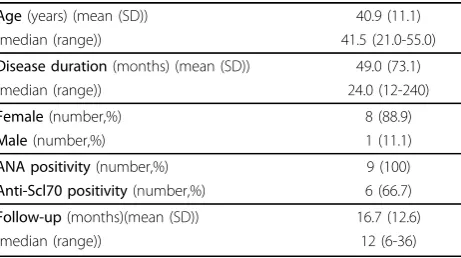
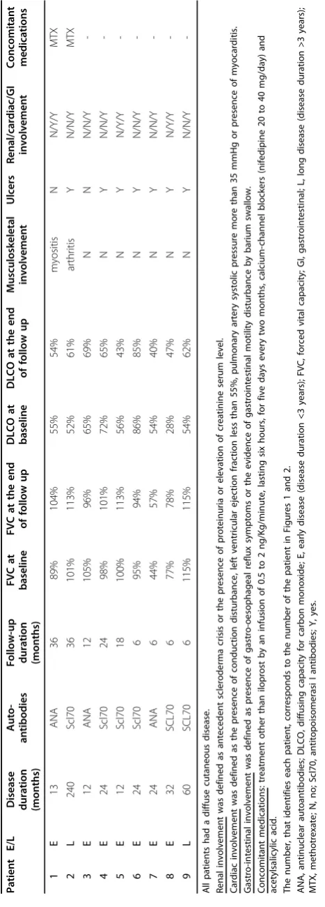
[image:2.595.307.538.585.714.2]There were eight women and one man, with a mean (standard deviation (SD))age of 40.9 ± 11.1 years, and a median disease duration of 2.0 (range:1.0 to 12.0) years. Seven patients had an early disease, defined as a disease duration less than three years since the occurrence of Raynaud’s phenomenon. All patients presented a diffuse skin disease (dSSc); moreover, six (66.7%) had antiScl70-Abs positivity and three (33.3%) only presented antinuc-lear antibodies (ANA) positivity (Table 1) [24]. All nine patients continued to receive iloprost (by an infusion of 0.5 to 2 ng/kg/minute for five days every two months), calcium-channel blockers (nifedipine 20 to 40 mg/day) and acetylsalicylic acid from the moment of medical diagnosis. One of the two patients with long disease also presented with a metacarpophalangeal and wrist arthritis
Table 1 Demographic and clinical characteristics of nine patients treated with rituximab
Age(years) (mean (SD)) 40.9 (11.1)
(median (range)) 41.5 (21.0-55.0)
Disease duration(months) (mean (SD)) 49.0 (73.1)
(median (range)) 24.0 (12-240)
Female(number,%) 8 (88.9)
Male(number,%) 1 (11.1)
ANA positivity(number,%) 9 (100)
Anti-Scl70 positivity(number,%) 6 (66.7)
Follow-up(months)(mean (SD)) 16.7 (12.6)
(median (range)) 12 (6-36)
and one patient had myositis with high creatine kinase levels. Both these patients received methotrexate 15 mg/ week after cyclophosphamide, one for treatment of arthritis and the other for myositis therapy. Both patients experienced a worsening of their skin fibrosis despite this therapy.
The extent of skin involvement was evaluated by the Rodnan skin score, performed by two observers and their results averaged [25]. Every three months, activity index [26] and severity index were assessed [27] and Global Health Status (GH) and Health Assessment Questionnaire (HAQ) were administered to patients to evaluate the influence of the disease on daily functions. At the same time intervals, blood samples were collected to determine IL-6 and BAFF levels and to count CD19-positive cells by flow cytometry.
Internal organ involvement
All nine patients underwent pulmonary function tests to define forced vital capacity (FVC) and diffusing capacity for carbon monoxide (DLCO) before treatment and every six months. High-resolution computed tomogra-phy (HRCT) was performed before treatment and every 12 months. Renal involvement was defined as a sclero-derma crisis or the presence of proteinuria or elevation in creatinine serum level. Creatinine levels and urine analysis were performed every three months. Cardiac involvement was defined as the presence of conduction disturbance, left ventricular ejection fraction (LVEF) less than 50%, pulmonary artery systolic pressure (PASP) more than 35 mmHg or presence of myocarditis; elec-trocardiography (ECG) and echocardiography were per-formed at the beginning of the treatment and every six months. Gastrointestinal involvement was defined as the presence of gastro-esophageal reflux symptoms or the evidence of gastrointestinal motility disturbance by bar-ium swallow performed before treatment.
Biological marker detection
Serum levels of IL-6 and BAFF (R&D Systems, Minnea-polis, MN, USA) were measured using an ELISA, as described by the manufacturer. Erythrocyte sedimenta-tion rate, total immunoglobulin (Ig) G, IgM and IgA were part of the routine clinical care of each patient. ANA were determined by indirect immunofluorescence using Hep-2 cells as substrates and autoantibodies speci-ficities were further assessed by ELISA (Shield, Dundee, UK). Peripheral blood CD19-positive cell count was obtained by flow cytometry every three months.
Skin biopsies and immunohistochemical analysis
Skin biopsies were performed in seven patients, who gave their informed consent, before treatment and in the five patients that achieved 12 months of follow up
from the beginning of anti-CD20 therapy. Four healthy controls gave their informed consent to undergo fore-arm skin biopsy. In dSSc patients, cutaneous specimens were taken from the distal forearm for the clinically involved skin and from the buttock for clinically unin-volved skin. The biopsies were fixed into 10% formalin for two hours followed by paraffin inclusion for histolo-gical and immunohistochemical analysis.
Immunohistochemistry was carried out on 5μm thick sections on polylysine-coated slides. After routine depar-affinization and rehydration, antigen retrieval was per-formed. Slide-mounted sections were heated in a microwave oven at 700 watt twice for four minutes in 10 mmol/L sodium citrate buffer (pH 6.0). Tissue sec-tions were allowed to cool at room temperature (RT). Quenching of endogenous peroxidase activity was per-formed with Tris-buffered saline (TBS; pH 7.6) contain-ing 2% hydrogen peroxide for 10 minutes at RT. Blocking was performed with 20% normal goat serum in TBS for 60 minutes at RT.
The sections were incubated with CD3 and anti-CD20 mouse monoclonal antibodies (mAbs, Clone PS1 and L26 respectively; IgG2a; Ylem, Rome, Italy) both 1:100 diluted in blocking solution (20% normal goat serum in TBS) for 60 minutes at RT. Then, the Super Picture Polymer detection kit (Zymed Laboratories, South San Francisco, CA, USA) was used for 30 minutes at RT. The chromogenic reaction was developed with 3,3’-diaminobenzidine tetrahydrochloride solution (Zymed Laboratories, South San Francisco, CA, USA). The nuclei were lightly counterstained with Mayer’s hematoxylin. Negative controls without primary antibo-dies were performed for all reactions. As all mAbs were of IgG2aisotype, mouse mAb IgG2aserved as an iso-type-specific control. Human tonsil specimens were used as positive controls for both antibodies. All con-trols were run under the same conditions and the same IgG concentrations were used for the respective primary antibodies. Positive cells were counted by two indepen-dent observers in six randomly selected fields (total area: 7.38 mm2) for each section at × 400 magnification. Differences between observers about staining evaluation were resolved by consensus. The total number of posi-tive cells was calculated.
Statistical analysis
Results
Skin score, activity and severity indices
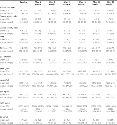
All nine SSc patients treated with rituximab experienced an improvement of the skin score, activity index, sever-ity index, HAQ and GH during the follow up if com-pared to pre-treatment values (Table 2 and Figure 1). Neither infections nor infusion reactions were observed. The only serious adverse event was the development of an occult breast cancer, which was thought to be unre-lated to the study medication. The mean follow up was 16.7 ± 12.6 months: all patients reached a six-month fol-low up, five patients reached a 12-month folfol-low up, four patients reached an 18-month follow up, three patients reached a 24-month follow up and two patients reached a 36-month follow up.
Interestingly, in all nine patients treated with rituxi-mab, the skin score improved gradually over time (Fig-ure 1 and Table 2). After six months, the skin score improved in all the patients, decreasing from 21.1 ± 9.0 to 12.0 ± 6.1 (P= 0.001), with a median of improvement of 43.3% (range: 21.1 to 64.0%). Considering the last observation carried forward in each patient, the median skin improvement was 57.1% (range: 21.2 to 76.2).
After six months, the activity index decreased from 4.8 ± 1.3 to 1.2 ± 1.2 (P = 0.01) and the severity index from 10.5 ± 3.2 to 7.2 ± 2.8 (P = 0.01; Figure 1 and Table 2). All patients reported an improvement of their conditions as supported by the decrease in HAQ from 0.9 ± 0.7 to 0.4 ± 0.5 (P = 0.01) and an increase in GH from 59.4 ± 20.9 to 82.8 ± 16.6 (P= 0.01; Table 2). The only patient who did not present an improvement of the activity and severity indices, HAQ and GH, had a long disease duration.
Organ involvement
The FVC and DLCO values showed no significant dif-ferences at follow up (96.8 ± 18.9% and 58.4 ± 14.2% of predicted value, respectively) compared with baseline (91.6 ± 20.7% and 58.0 ± 15.8% of the predicted value, respectively;P = ns for both comparison). Four (44.4%) patients presented an improvement higher than 10% of FVC, (median increase 14.9% (range: 11.8% to 29.5%)). None of the patients presented a reduction in FVC con-sidered clinically significant (>10%), but one patient showed a decrease in FVC values suggesting a trend to a progression of her restrictive lung disease [28,29].
Two patients (22.2%) presented an isolated reduction of DLCO higher than 15%, both with an improvement in FVC values higher than 10% and with a stable echo-cardiography evaluation and no sign of pulmonary arter-ial hypertension. On the other hand, a clinical significant improvement in DLCO was reported in two patients (22.2%) [28,29] (Table 3).
None of the patients showed signs of new or progres-sive cardiac disease, with stable ejection fractions and no modification on ECGs; none of the patients experi-enced renal crisis or symptoms suggesting progressive gastrointestinal disease.
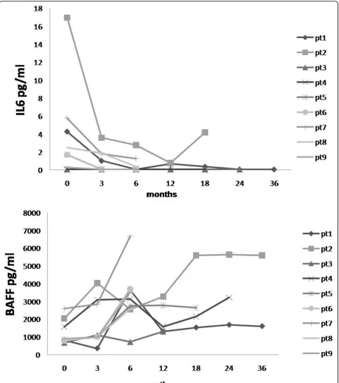
Biological markers
At baseline, patients presented high levels of IL-6 (3.7 ± 5.3 pg/ml), that permanently decreased after six months (0.6 ± 0.9 pg/ml, P = 0.02; Table 2 and Figure 2a). Three months after the rituximab infusion, circulating B cells evaluated by flow-cytometry were depleted (periph-eral blood CD19 <0.1%) in all but one patient, and between 6 and 12 months they begun to repopulate. Upon B-cell depletion, BAFF levels increased relative to baseline (baseline: 1233.5 ± 683.3 pg/mlvs six months: 3257.8 ± 1571.8 pg/ml), while in one patient the BAFF levels did not increase until the time of repopulation (Figure 2b).
The autoantibody titers and IgG and IgA levels did not vary over the study period, while IgM levels decreased from 133.7 ± 21.7 mg/dl to 90.9 ± 28.9 mg/dl at six months follow up (P= 0.008), and to 83.0 ± 18.7 after 12 months of follow up (P= 0.04; Table 2).
Before rituximab treatment, one patient presented myositis with high creatine kinase levels, which decreased significantly after anti-CD20 treatment (data not shown). Creatine kinase levels remained within the normal range during the 36 months of follow up. The only patient with a long disease duration who did pre-sent the less significant clinical improvement, showed an important amelioration of her arthritis, with a change of disease activity score (DAS) from 4.3 to 2.0.
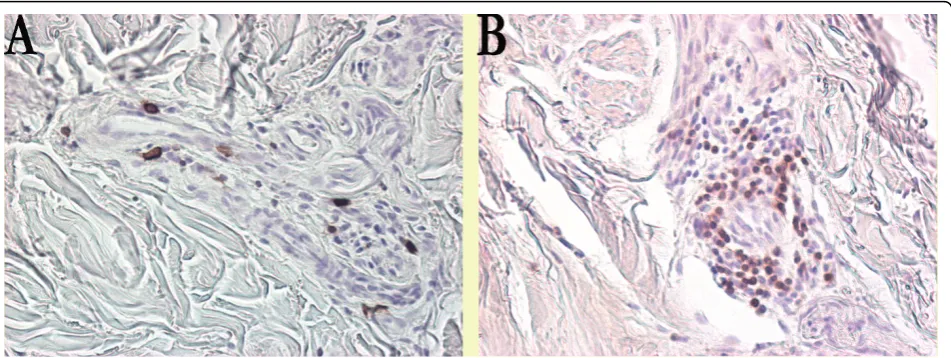
Three (42.9%) out of the seven patients, who under-went skin biopsies before treatment, presented CD20-positive cells on biopsies of the clinically involved skin and uninvolved skin; only one patient of these three repeated the biopsy after 12 months and it showed a depletion of dermal B cells. The other two patients were treated only for six months and they did not agree to a repeat biopsy.
Table 2 Efficacy of rituximab on clinical and biologic parameters of nine SSc patients treated with anti-CD20 during the follow up
Baseline After 3
Months
After 6 Months
After 12 months*
After 18 months**
After 24 months***
After 36 months****
Rodnan skin score
(mean (SD)) 21.1 (9.0) 15.2 (6.0) 12.0 (6.1) 7.0 (4.0) 7.0 (3.5) 5.0 (2.0) 4.0 (1.4)
(median (range)) 19.0 (7-36) 15.0 (5-27) 10.0 (4-26) 8.0 (3-14) 7.5 (3-10) 5.0 (3-7) 4.0 (3-5)
Disease activity index
(mean (SD)) 4.8 (1.3) 2.0 (1.2) 1.2 (1.2) 0.9 (1.0) 1.0 (1.3) 1.3 (1.4) 1.7 (1.8)
(median (range)) 4.5 (3.5-7.0) 2.0 (0.5-3.5) 0.5 (0.5-3.5) 0.5 (0.5-3.0) 0.5 (0.5-3.0) 0.5 (0.5-3.0) 1.7 (0.5-3.0)
Disease severity index
(mean (SD)) 10.5 (3.2) 8.3 (2.9) 7.2 (2.8) 6.2 (2.8) 6.7 (2.2) 7.7 (1.5) 8.5 (0.7)
(median (range)) 11.0 (6-15) 7.0 (4-13) 6.0 (4-11) 6.0 (3-9) 7.0 (4-9) 8.0 (6-9) 8.5 (8-9)
HAQ
(mean (SD)) 0.9 (0.7) 0.5 (0.5) 0.4 (0.5) 0.3 (0.7) 0.3 (0.6) 0.3 (0.6) 0.6 (0.6)
(median (range)) 0.8 (0.1-2.4) 0.4 (0-1.6) 0.2 (0-1.5) 0 (0-1.5) 0 (0-1.2) 0 (0-1.1) 0 (0-1.3)
GH(mean (SD)) 59.4 (20.9) 74.4 (16.5) 82.8 (16.6) 86.0 (10.8) 82.5 (21.8) 82.5 (17.7) 82.5 (17.7)
(median (range)) 60.0 (30-85) 80.0 (50-95) 90.0 (50-95) 90.0 (70-95) 92.5 (50-95) 82.5 (70-95) 82.5 (70-95)
Blood CD20%
(mean (SD)) 6.8 (3.9) 0.7 (1.5) 1.7 (2.4) 3.0 (2.7) 2.0 (1.6) 1.0 (1.5) 3.0 (3.3)
(median (range)) 6.0 (2.5-14.7) 0.1 (0.1-4.6) 1.0 (0.1-7.0) 4.0 (0.3-7.0) 2.0 (0.2-3.0) 1.0 (0.2-3.0) 3.0 (0.3-5.0)
IgG mg/ml
(mean (SD)) 1055 (233) 1001 (191) 1021 (158) 1028 (46.6) 946.2 (178) 944 (315) 951 (69)
(median (range)) 1140 (729-1340) 932 (884-1440) 1030 (802-1220) 1005 (1000-1110) 938 (738-1170) 896 (656-1280) 951 (902-1000)
IgA mg/ml
(mean (SD)) 184.0 (44.4) 174.5 (39.2) 177.8 (63.5) 179.4 (69.8) 152.5 (59.9) 139.0 (67.5) 100.0 (24.0)
(median (range)) 183.5 (119-249) 183.5 (119-262) 200.0 (75-262) 168.5 (93-281) 154.0 (79-222) 136.0 (73-208) 100.0 (83-117)
IgM mg/ml
(mean (SD)) 133.7 (21.7) 86.0 (12.8) 90.9 (28.9) 83.0 (18.7) 61.5 (6.7) 43.3 (10.6) 71.0 (1.4)
(median (range)) 132.5 (95-157) 91.0 (56-136) 96.0 (40-136) 73.0 (64-105) 61.5 (54-69) 45.0 (32-53) 71.0 (70-72)
BAFF pg/ml
(mean (SD)) 1233.5 (683.3) 1719.4 (1264.3) 3257.8 (1571.8) 2057.0 (912.5) 2988.0 (1804) 3520.0 (1999) 3608.0 (2824)
(median (range)) 875.6
(683-2601)
1008.6 (356-4038)
3141.8 (723-6682)
1580.6 (1321.6-3280)
2406.2 (1534-5605)
3224.8 (1684-5651)
3608.0 (1610-5605)
IL6 pg/ml
(mean (SD)) 3.7 (5.3) 1.0 (1.2) 0.6 (0.9) 0.4 (0.4) 1.2 (2.0) 0.1 (0.1) 0.1 (0.1)
(median (range)) 1.7 (0.1-16.9) 0.1 (0.1-3.6) 0.1 (0.1-2.8) 0.4 (0.1-0.8) 0.25 (0.1-4.2 0.1 (0.1-0.1) 0.1 (0.1-0.1)
Clinical and biological parameters of nine SSc patients treated with anti-CD20 at baseline, after 3, 6, 12, 18, 24 and 36 months. The values are indicated as the mean (SD) and median (range). All patients had trunk skin involvement.
*Five SSc patients reached 12 months of follow up. **Four SSc patients reached 18 months of follow up. ***Three SSc patients reached 24 months of follow up. *****Two patients reached 36 months of follow up.
number of CD3-positive cells was found in involved skin (44.3 ± 24.0) and in uninvolved skin (62.7 ± 23.4) of the five patients who underwent skin biopsies after 12 months.
Discussion
The results of our study suggest that B cell depletion in patients with early and progressive dSSc, leads to a clini-cally relevant decrease in skin involvement and to a sta-bilization of organ function. The only patient who showed a less clear-cut response either in terms of severity and activity indexes was the one with a long-standing disease.
In our study, the safety of anti-CD20 treatment in SSc patients was also confirmed in up to 36 months of fol-low up. The observed skin score improvement is more than expected as the spontaneous improvement in patients with similar disease, and comparable with the
study by Smith and colleagues [19]. Recently, two stu-dies assessed the safety of anti-CD20 treatment in scler-oderma patients. In the first open-label trial, eight SSc patients experienced a skin score improvement up to 43% after 24 weeks from the beginning of anti-CD20 treatment [19], while in the second, a cohort of 15 SSc patients, with a follow up of 12 months, showed no improvement in the skin score [20]. Only the first group used the corticosteroids premedication.
[image:6.595.58.539.88.442.2]could be a preclinical indicator of improvement of scler-oderma skin fibrosis. Furthermore, Lafyatis and collea-gues reported the presence of B cells in all but one skin specimen at baseline and a complete or nearly complete depletion of dermal B cells six months after administra-tion of rituximab [20]. This suggests a biological effect on the skin after drug administration that could with new courses of the drug lead to a clinical skin improve-ment. In fact, we treated patients with a progressive cutaneous disease after conventional cyclophosphamide therapy. Moreover, we decided to re-treat two of our patients, because they presented a slower improvement of the skin score in the first six months of follow up and an earlier repopulation of B cells, similar to the data reported by the Lafyatis and colleagues, in which the majority of patients presented a precocious recovery of B cells between 6 and 12 months [20].
Interestingly, none of the SSc patients in the current study treated with anti-CD20 showed a progression of major end-organ involvement in a population with early diffuse disease that had a relatively high risk of organ complication. Parameters of internal organ involvement remained stable, but a further follow up in a more con-sistent group of patients is needed before drawing any conclusions.
The clear fall in IL-6 levels observed in our study is in agreement with findings obtained in a mouse model after B cell depletion [8]. This fall could be related, at least for the first stages, to the high dose of methylpred-nisolone used for the premedication in ours and the cohort of patients in the study by Smith and colleagues [19], but considering the follow times of evaluation (3 to 6 to 12 months) it has to be related to the rituximab treatment. This may suggest that IL-6 might contribute to the active phase of the disease. The decrease in IL-6 at the systemic levels could be the biological premise of the improvement in skin fibrosis. In fact, it has been previously reported that chronic IL-6 administration induces an increased synthesis of collagen in dermal fibroblasts [31] and in the liver [32]. Furthermore, IL-6 has been demonstrated to enhance resistance of lung fibroblasts to apoptosis, contributing to the fibrotic effect [33].
Immunohistochemistry clearly demonstrated the pre-sence of T cells either in uninvolved or in involved skin, but B cells were seen only in some patients, as pre-viously reported [5,19]. These data suggest that the most relevant contribution of B cells comes from the systemic pool. In fact, it appears clear that the response of skin fibrosis to B cell depletion does not rely on the presence of B cells in the skin, because most of our treated patients had no B cells, but very likely depends upon the general contribution to the autoimmune derangement given by the B cell compartments in
[image:7.595.66.300.89.740.2]lymphoid organs. B cells, with their multiple mechan-isms as antibody-producing cells, antigen-presenting cells and profibrotic and proinflammatory cytokines pro-ducing cells (IL-6, IL-4, transforming growth factor-b), seem to be of great impact in the development of fibro-sis. Thus, their modulation could inhibit skin fibrosis, as reported in the scleroderma mouse model [8], but the data on BAFF levels need to be interpreted since, as observed in patients with Sjogren’s syndrome [34] or rheumatoid arthritis [35], the levels went up after B cell depletion.
Conclusions
Our data suggest that anti-CD20 treatment is well tol-erated and that dSSc patients experience an improve-ment of the skin score and of clinical symptoms. The clear fall in IL-6 levels may contribute to the skin fibrosis improvement, while the presence of B cells in the skin seems to be irrelevant with respect to the out-come after B cell depletion. Although we cannot draw any conclusion due to the limited number of cases, the response in the early disease patients was striking sug-gesting that a trial is warranted to confirm these preli-minary data.
Abbreviations
ANA: antinuclear antibodies; BAFF: B-cell activating factor; DAS: disease activity score; DLCO: diffusing capacity for carbon monoxide; dSSc: diffuse systemic sclerosis; ECG: electrocardiogram; ELISA: enzyme-linked immunosorbent assay; FVC: forced vital capacity; GH: Global Health Status; HAQ: Health Assessment Questionnaire; HRCT: high-resolution computed tomography; Ig: immunoglobulin; IL-6: interleukin-6; LVEF: left ventricular ejection fraction; mAbs: monoclonal antibodies; PASP: pulmonary artery systolic pressure; RT: room temperature; SD: standard deviation; SSc: systemic sclerosis; TBS: Tris-buffered saline.
Acknowledgements
Written consent for publication was obtained from all patients.
Author details
1Division of Rheumatology, Catholic University, Medical School, Via G.
Moscati, 31 - Rome, 00168, Italy.2Institute of Histology and Embryology, Catholic University, Medical School, L.go F.Vito, 1 - Rome, 00168, Italy.
Authors’contributions
BS conceived and designed the study, collected data, performed the statistical analysis, interpreted and analysed data, and drafted the manuscript. MDS conceived and designed the study, collected data, interpreted and analysed data, and drafted the manuscript. LG carried out the immunohistochemistry, interpreted and analysed data, and revised the manuscript. SC carried out the immunohistochemistry, collected data, interpreted and analysed data, and revised the manuscript. AC carried out the immunohistochemistry, and collected data. TB carried out immunoassay and collected data. SG participated in the design of the study, analysed data and revised the manuscript. GF conceived and designed the study, interpreted and analysed data and drafted the manuscript. All authors read and approved the final manuscript.
Competing interests
The authors declare that they have no competing interests.
Received: 5 May 2009 Revised: 29 November 2009 Accepted: 25 March 2010 Published: 25 March 2010
References
1. Okano Y:Antinuclear antibody in systemic sclerosis (scleroderma).Rheum Dis Clin North Am1996,22:709-735.
2. Famularo G, Giacomelli R, Alesse E, Cifone MG, Morrone S, Boirivant M, Danese C, Perego MA, Santoni A, Tonietti G:Polyclonal B lymphocyte activation in progressive systemic sclerosis.J Clin Lab Immunol1989, 29:59-63.
3. Sato S, Fujimoto M, Hasegawa M, Takehara K:Altered blood B lymphocyte homeostasis in systemic sclerosis: expanded naive B cells and diminished but activated memory B cells.Arthritis Rheum2004, 50:1918-1927.
4. Sato S, Hasegawa M, Fujimoto M, Tedder TF, Takehara K:Quantitative genetic variation in CD19 expression correlates with autoimmunity. J Immunol2000,165:6635-6643.
5. Whitfield ML, Finlay DR, Murray JI, Troyanskaya OG, Chi JT,
[image:9.595.62.538.89.268.2]Pergamenschikov A, McCalmont TH, Brown PO, Botstein D, Connolly MK:
Systemic and cell type-specific gene expression patterns in scleroderma skin.Proc Natl Acad Sci USA2003,100:12319-12324.
6. Asano N, Fujimoto M, Yazawa N, Shirasawa S, Hasegawa M, Okochi H, Tamaki K, Tedder TF, Sato S:B lymphocyte signaling established by the CD19/CD22 loop regulates autoimmunity in the tight-skin mouse.Am J Pathol2004,165:641-650.
7. Saito E, Fujimoto M, Hasegawa M, Komura K, Hamaguchi Y, Kaburagi Y, Nagaoka T, Takehara K, Tedder TF, Sato S:CD19-dependent B lymphocyte signaling thresholds influence skin fibrosis and autoimmunity in the tight-skin mouse.J Clin Invest2002,109:1453-1462.
8. Hasegawa M, Hamaguchi Y, Yanaba K, Bouaziz JD, Uchida J, Fujimoto M, Matsushita T, Matsushita Y, Horikawa M, Komura K, Takehara K, Sato S, Tedder TF:B-lymphocyte depletion reduces skin fibrosis and
autoimmunity in the tight-skin mouse model for systemic sclerosis.Am J Pathol2006,169:954-966.
9. Yoshizaki A, Iwata Y, Komura K, Ogawa F, Hara T, Muroi E, Takenaka M, Shimizu K, Hasegawa M, Fujimoto M, Tedder TF, Sato S:CD19 regulates skin and lung fibrosis via Toll-like receptor signaling in a model of bleomycin-induced scleroderma.Am J Pathol2008,172:1650-1663. 10. Novobrantseva I, Majeau GR, Amatucci A, Kogan S, Brenner I, Casola S,
Shlomchik MJ, Koteliansky V, Hochman PS, Ibraghimov A:Attenuated liver fibrosis in the absence of B cells.J Clin Invest2005,115:3072-3082. 11. Scala E, Pallotta S, Frezzolini A, Abeni D, Barbieri C, Sampogna F, De Pità O,
Puddu P, Paganelli R, Russo G:Cytokine and chemokine levels in systemic sclerosis: relationship with cutaneous and internal organ involvement. Clin Exp Immunol2004,138:540-546.
12. Hasegawa M, Sato S, Fujimoto M, Ihn H, Kikuchi K, Takehara K:Serum levels of Interleukin 6 (IL-6), oncostatin M, soluble IL-6 receptor, and soluble gp130 in patients with systemic sclerosis.J Rheumatol1998,25:308-316. 13. De Santis M, Bosello S, La Torre G, Capuano A, Tolusso B, Pagliari G,
Pistelli R, Danza FM, Zoli A, Ferraccioli F:Functional, radiological and biological markers of alveolitis and infections of the lower respiratory tract in patients with systemic sclerosis.Respir Res2005,6:96-106. 14. Koch AE, Kronfeld-Harrington LB, Szekanecz Z, Cho MM, Haines GK,
Harlow LA, Strieter RM, Kunkel SL, Massa MC, Barr WG, Jimenez SA:In situ expression of cytokines and cellular adhesion molecules in the skin of patients with systemic sclerosis. Their role in early and late disease. Pathobiology1993,61:239-246.
15. Kadono T, Kikuchi K, Ihn H, Takehara K, Tamaki K:Increased production of interleukin 6 and interleukin 8 in scleroderma fibroblasts.J Rheumatol 1998,25:296-301.
16. Kondo K, Okada T, Matsui T, Kato S, Date K, Yoshihara M, Nagata Y, Takagi H, Yoneda M, Sugie I:Establishment and characterization of a human B cell line from the lung tissue of a patient with scleroderma; extraordinary high level of IL-6 secretion by stimulated fibroblasts. Cytokine2001,13:220-226.
17. Matsushita T, Hasegawa M, Yanaba K, Kodera M, Takehara K, Sato S: Elevated serum BAFF levels in patients with systemic sclerosis: enhanced BAFF signaling in systemic sclerosis B lymphocytes.Arthritis Rheum2006,54:192-201.
18. Matsushita T, Fujimoto M, Hasegawa M, Matsushita Y, Komura K, Ogawa F, Watanabe R, Takehara K, Sato S:BAFF antagonist attenuates the development of skin fibrosis in tight-skin mice.J Invest Dermatol2007, 127:2772-2780.
19. Smith V, Van Praet JT, Vandooren B, Van der Cruyssen B, Naeyaert JM, Decuman S, Elewaut D, De Keyser F:Rituximab in diffuse cutaneous systemic sclerosis: an open-label clinical and histopathological study. Ann Rheum Dis2010,69:193-197.
20. Lafyatis R, Kissin E, York M, Farina G, Viger K, Fritzler MJ, Merkel PA, Simms RW:B cell depletion with Rituximab in patients with diffuse cutaneous systemic sclerosis.Arthritis Rheum2009,60:578-583. 21. McGonagle D, Tan AL, Madden J, Rawstron AC, Rehman A, Emery P,
Thomas S:Successful treatment of resistant scleroderma-associated interstitial lung disease with rituximab.Rheumatology2008,47:552-553. 22. Calguneri M, Apras S, Ozbalkan Z, Ertenli I, Kiraz S, Ozturk MA, Celik I:The efficacy of oral cyclophosphamide plus prednisolone in early diffuse systemic sclerosis.Clin Rheumatol2003,22:289-294.
23. Subcommittee for Scleroderma Criteria of the American Rheumatism Association Diagnostic and Therapeutic Criteria Committee:Preliminary criteria for the classification of systemic sclerosis (scleroderma).Arthritis Rheum1980,23:581-590.
24. LeRoy EC, Black C, Fleischmajer R, Jablonska S, Krieg T, Medsger TA Jr, Rowell N, Wollheim F:Scleroderma (systemic sclerosis): classification, subset and pathogenesis.J Rheumatol1988,15:202-205.
25. Valentini G, D’Angelo S, Della Rossa A, Bencivelli W, Bombardieri S: European Scleroderma Study Group to define disease activity criteria for systemic sclerosis. IV: Assessment of skin thickening by modified Rodnan skin score.Ann Rheum Dis2003,62:904-905.
26. Valentini G, Silman AJ, Veale D:Assessment of disease activity.Clin Exp Rheumatol2003,21(Suppl 29):S39-S41.
27. Medsger TA Jr, Bombardieri S, Czirjak L, Scorza R, Della Rossa A, Bencivelli W:Assessment of disease severity and prognosis.Clin Exp Rheumatol2003,21(Suppl 29):S42-S46.
28. Egan JJ, Martinez FJ, Wells AU, Williams T:Lung function estimates in idiopathic pulmonary fibrosis: the potential for a simple classification. Thorax2005,60:270-273.
29. Behr J, Furst DE:Pulmonary function tests.Rheumatol2008,47 Suppl 5: v65-v67.
30. Kissin EY, Merkel PA, Lafyatis R:Myofibroblasts and hyalinized collagen as markers of skin disease in systemic sclerosis.Arthritis Rheum2006, 54:3655-3660.
31. Duncan MR, Berman B:Stimulation of collagen and glycosaminoglycan production in cultured human adult dermal fibroblasts by recombinant human interleukin 6.J Invest Dermatol1991,97:686-692.
32. Choi I, Kang HS, Yang Y, Pyun KH:IL-6 induces hepatic inflammation and collagen synthesis in vivo.Clin Exp Immunol1994,95:530-535.
33. Moodley YP, Misso NL, Scaffidi AK, Fogel-Petrovic M, McAnulty RJ, Laurent GJ, Thompson PJ, Knight DA:Inverse effects of interleukin-6 on apoptosis of fibroblasts from pulmonary fibrosis and normal lungs.Am J Respir Cell Mol Biol2003,29:490-498.
34. Pers JO, Devauchelle V, Daridon C, Bendaoud B, Le Berre R, Bordron A, Hutin P, Renaudineau Y, Dueymes M, Loisel S, Berthou C, Saraux A, Youinou P:BAFF-modulated repopulation of B lymphocytes in the blood and salivary glands of rituximab-treated patients with Sjögren syndrome.Arthritis Rheum2007,56:1464-1477.
35. Lavie F, Miceli-Richard C, Ittah M, Sellam J, Gottenberg JE, Mariette X: Increase of B cell-activating factor of the TNF family (BAFF) after Rituximab treatment: insights into a new regulating system of BAFF production.Ann Rheum Dis2007,66:700-703.
doi:10.1186/ar2965
Cite this article as:Boselloet al.:B cell depletion in diffuse progressive systemic sclerosis: safety, skin score modification and IL-6 modulation in an up to thirty-six months follow-up open-label trial.Arthritis Research & Therapy201012:R54.
Submit your next manuscript to BioMed Central and take full advantage of:
• Convenient online submission
• Thorough peer review
• No space constraints or color figure charges
• Immediate publication on acceptance
• Inclusion in PubMed, CAS, Scopus and Google Scholar
• Research which is freely available for redistribution