© 2018, IRJET | Impact Factor value: 6.171 | ISO 9001:2008 Certified Journal | Page 1015
AN EFFECTIVE BRAIN Tumor SEGMENTATION USING K-means
clustering
1
K.Pravallika,
1G.Veena Lokeswari,
1B.Buelah,
1G.Venkata Sowmya,
2V.Purna Chandra Reddy.
1B.Tech Student, Dept of ECE, VVIT, Nambur, Guntur District, A.P., India. 2Assistant Professor, Dept of ECE, VVIT, Nambur, Guntur District, A.P., India.
---***---Abstract -
Image segmentation technology has alwaysbeen one of key technologies in image processing, in recent years, many algorithms are applied in the field of image segmentation. Image segmentation technology divides the image into several areas, and needed information is extracted. General segmentation technology are based on threshold segmentation method, based on region segmentation method, the segmentation method based on edge, and the segmentation method based on the specific theory. In this paper study the different clustering methods in image segmentation. In view of the traditional clustering image segmentation algorithm for image segmentation accuracy is low problem, put forward a kind of fuzzy control based on C-means clustering image segmentation method. Methods firstly in clustering image segmentation algorithm based on fast, using fuzzy C-means clustering algorithm for image segmentation. The experimental results show that the algorithm in clustering, to optimize the performance of the same premise, image segmentation edge clear, segmentation better than traditional clustering algorithm
for image segmentation..
Key Words: Image segmentation, fuzzy c-means clustering algorithm and optimization
1.INTRODUCTION
Stroke or cerebrovascular accident is a disease which affects the vessels that supply blood to the brain. The blockage of the blood vessel and the bursts of the blood vessels causes the brain stroke [1-2]. There are three main kinds of stroke: Ischemic strokes (severely reduced blood flow), Hemorrhagic strokes (leakage of blood) and Transient ischemic attacks (TIAs-also referred to as mini-strokes) [3, 4]. Ischemic brain stroke is one of the leading causes of death and disability in major industrialized countries [5]. To detect the Ischemic brain cells, the computed tomography (CT) and Magnetic Resonance Image (MRI) is the two important screening tests [6, 7]. The existing methods used the conventional threshold techniques, but this method only has the limited accuracy and repeatability [8, 9]. The clear image of the affected region of the brain image is not provided by the above techniques, but, for the early identification of the brain infection, the MRI is more efficient than the CT [10].
The brain stroke detection techniques consolidate many methods such as pre-processing, segmentation, feature
extraction and classification [11-13]. The researchers proposed many techniques for the segmentation and classification of images. A fuzzy clustering approach [14] to the segmentation followed by 3D connected components to build the tumor shape, Atlas-based medical image segmentation techniques [15] which convert the segmentation of an MR image into a non rigid registration problem between the MR image of the patient and the MR image used to create the brain atlas. Unfortunately, this requires using a large seed that mask atlas structures, potentially leading to erroneous results. The drawback of FCM (Fuzzy C-Mean) [16] is that the spatial neighborhood term is computed at each iteration step, which is very time-consuming. Fast generalized FCM (FGFCM) algorithm [17] which introduces a local similarity measure, but the computational cost is very high in this method.
After extracting the features of the segmented image, the researchers introduce many types of classification techniques to classify the stroke and non-stroke regions. The Artificial Neural Networks (ANN) [18] which is used to anticipate the fate of ischemic tissues on three different stroke groups. But it may suffer from an overfitting problem. The Random Decision Forests [19] is a popular classifier, but it can contain errors, noise and image artifacts, which may lead to uncertainties.
1.1 Fuzzy C-means (FCM)
© 2018, IRJET | Impact Factor value: 6.171 | ISO 9001:2008 Certified Journal | Page 1016 Where,
‘n’ is the number of data points ‘Vj’ represents the jth cluster centre ‘m’ is the fuzziness index m €[1,∞] ‘c’ represents the number of cluster centre
‘μij’ represents the membership of ith data to jth cluster centre.
‘dij’represents the Euclidean distance between ith data and jth cluster centre.
‘xi’ is the ith of d-dimensional measured data ‘cj’ is the d-dimension centre of the cluster
is any norm expressing the similarity between any measured data and the centre.
The main objective of fuzzy c-means algorithm is to minimize
Where, is the Euclidean distance between ith data
and jth cluster centre
1) Algorithmic steps for fuzzy C-means clustering: Let X = {x1, x2, x3, ....xn}be the set of data points and V = { v1, v2, v3, ....vc }be the set of cluster centres.
Step1: Randomly select ‘c’ cluster centres
Step2: Calculate the fuzzy membership ‘μij’using the equation
Step3: Compute the fuzzy centres ‘vj’using
Step4: Repeat step2 and step3 until the minimum ‘J’ value is achieved or ║U(k+1)-U(k)║< β
Where, ‘k’ is the iteration step
‘β’ is the termination criterion between [0,1] is the fuzzy membership matrix ‘J’ is the objective function
The first loop of the algorithm calculates membership values for the data points in clusters and the second loop recalculates the cluster centres using these membership values. When the cluster centre stabilizes the algorithm ends.
The FCM algorithm gives best result for overlapped data set and also gives better result than k-means algorithm. Here, the data point can belong to more than one cluster centre. The main drawback of FCM is 1) the sum of embership value of a data point xi in all the clusters must be one but the outlier points has more membership value. So, the algorithm has difficulty in handling outlier points. 2) Due to the influence of all the data members, the cluster centres tend to move towards the centre of all the data points [Cox (2005)].
It only considers image intensity thereby producing unsatisfactory results in noisy images [Hall et al. (1992)]. A bunch of algorithms are proposed to make FCM robust against noise and in homogeneity but it’s still not perfect [Hall et al. (1992), Lions et al. (1992), Acton et al. (2000), Tolias and Panas (2008), Dave (1991), Zhang and Chen (2004)].
2.Proposed k-means clustering
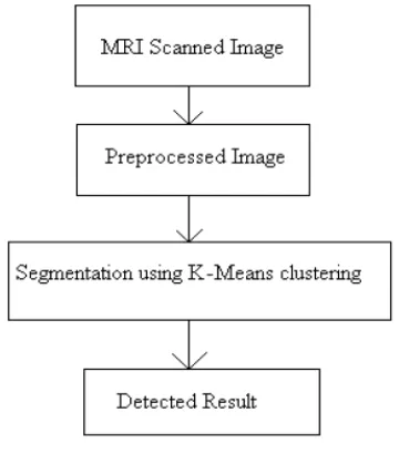
[image:2.595.340.520.459.664.2]We have proposed segmentation of the brain MRI images for detection of tumors using K-Means clustering technique. A cluster can be defined as a group of pixels where all the pixels in certain group defined by similar relationship. Clustering is also unsupervised classification because the algorithm automatically classifies objects based on user given criteria. Here K-Means clustering algorithm for segmentation of the image is used for tumor detection from the brain MRI images. The proposed block diagram is as shown.
Fig 1. Proposed Methodology
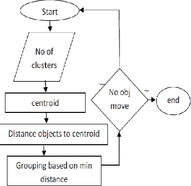
© 2018, IRJET | Impact Factor value: 6.171 | ISO 9001:2008 Certified Journal | Page 1017 The pre-processed image is given for image segmentation
[image:3.595.66.259.125.312.2]using K-Means clustering algorithm.
Fig.2. Proposed K-means Clustering Algorithm
2.1.Experiment and Analysis
In order to test the performance of the algorithm, this paper adopts brain MRI images of the Montreal neurological institute for simulation. Traditional clustering image segmentation result is shown in Fig.1, the noise elimination of image using the median filter is shown in fig 2. The tradition approach for the detection of tumor is implemented using K means Clustering is shown in fig 3. Final tumor detection using K-means Clustering is shown in fig 4. Traditional FCM image segmentation result is shown in Fig.5, and image segmentation result of proposed algorithm of section 2. The proposed fast image segmentation is superior to the traditional algorithm, the edge details is clear, this algorithm can ensure clustering optimization performance unchanged, reduce the cost of operation, and obviously improves the segmentation efficiency.
Fig 3: input image and histogram for K-means Clustering method
Fig4 noise elimination using Filtering Technique
Fig5: K-means Clustering processing for tumor detection
Fig6 Tumor Detection using K-means Clustering
3. CONCLUSIONS
© 2018, IRJET | Impact Factor value: 6.171 | ISO 9001:2008 Certified Journal | Page 1018 REFERENCES
[1] Y. Wang, A. Katsaggelos, X. Wang, and T. Parrish, "A deep symmetry convnet for stroke lesion segmentation", In: Proc. of the IEEE International Conference on Image Processing (ICIP), Phoenix, AZ, USA, pp.111-115, 2016.
[2] S. Bauer, P. Gratz, J. Gralla, M. Reyes, and R. Wiest, "Towards automatic MRI volumetry for treatment selection in acute ischemic stroke patients", In: Proc. of the 36th Annual International Conference of the IEEE Engineering in Medicine and Biology Society, Chicago, IL, USA, pp. 1521-1524, 2014.
[3] N.H. Rajini and R. Bhavani, "Computer aided detection of ischemic stroke using segmentation and texture features", Measurement, Vol. 46, No. 6, pp. 1865-1874, 2013.
[4] N. Rost, S. Sadaghiani, A. Biffi, K. Fitzpatrick, L. Cloonan, J. Rosand, D. Shibata, and T. Mosley, "Setting a gold standard for quantification of leukoaraiosis burden in patients with ischemic stroke: The Atherosclerosis Risk in Communities Study", Journal of Neuroscience Methods, Vol. 221, No. 1, pp. 196-201, 2014.
[5] N. Nabizadeh, N. John, and C. Wright, "Histogram-based gravitational optimization algorithm on single MR modality for automatic brain lesion detection and segmentation", Expert Systems with Applications, Vol. 41, No. 17, pp. 7820-7836, 2014.
[6] O. Maier, M. Wilms, J. von der Gablentz, U. Krämer, T. Münte, and H. Handels, "Extra Tree forests for sub-acute ischemic stroke lesion segmentation in MR sequences", Journal of Neuroscience Methods, Vol. 240, No. 1, pp. 89-100, 2015.
[7] M. Artzi, O. Aizenstein, T. Jonas-Kimchi, V. Myers, H. Hallevi, and D. Ben Bashat, "FLAIR lesion segmentation: Application in patients with brain tumors and acute ischemic stroke", European Journal of Radiology, Vol. 82, No. 9, pp. 1512-1518, 2013.
[8] M. Benders, N. van der Aa, M. Roks, H. van Straaten, I. Isgum, M. Viergever, F. Groenendaal, L. de Vries, and F. van Bel, "Feasibility and Safety of Erythropoietin for Neuroprotection after Perinatal Arterial Ischemic Stroke", The Journal of Pediatrics, Vol. 164, No. 3, pp. 481-486.e2, 2014.
[9] N. Ghosh, Y. Sun, B. Bhanu, S. Ashwal, and A. Obenaus, "Automated detection of brain abnormalities in neonatal hypoxia ischemic injury from MR images", Medical Image Analysis, Vol. 18, No. 7, pp. 1059-1069, 2014.
[10] A. Patel, B. van Ginneken, F. Meijer, E. van Dijk, M. Prokop, and R. Manniesing, "Robust cranial cavity segmentation in CT and CT perfusion images of trauma
and suspected stroke patients", Medical Image Analysis, Vol. 36, No. 2, pp. 216-228, 2017.
[11] J. Griffis, J. Allendorfer, and J. Szaflarski, "Voxel-based Gaussian naïve Bayes classification of ischemic stroke lesions in individual T1-weighted MRI scans", Journal of Neuroscience Methods, Vol. 257, No. 1, pp. 97-108, 2016.
[12] N. Yassi, B. Campbell, B. Moffat, C. Steward, L. Churilov, M. Parsons, P. Desmond, S. Davis, and A. Bivard, "Know your tools—concordance of different methods for measuring brain volume change after ischemic stroke", Neuroradiology, Vol. 57, No. 7, pp. 685-695, 2015.
[13] B. Menze, K. Van Leemput, D. Lashkari, T. Riklin-Raviv, E. Geremia, E. Alberts, P. Gruber, S. Wegener, M. Weber, G. Szekely, N. Ayache, and P. Golland, "A Generative Probabilistic Model and Discriminative Extensions for Brain Lesion Segmentation— With Application to Tumor and Stroke", IEEE Transactions on Medical Imaging, Vol. 35, No. 4, pp. 933-946, 2016.
[14] D. Chyzhyk, R. Dacosta-Aguayo, M. Mataró, and M. Graña, "An active learning approach for stroke lesion segmentation on multimodal MRI data", Neurocomputing, Vol. 150, No. 6, pp. 26-36, 2015.
[15] L. Chen, P. Bentley, and D. Rueckert, "Fully automatic acute ischemic lesion segmentation in DWI using convolutional neural networks", NeuroImage: Clinical, Vol. 15, No. 6, pp. 633-643, 2017.
[16] S.A. Kumar and B. Harish, "A Modified Intuitionistic Fuzzy Clustering Algorithm for Medical Image Segmentation", Journal of Intelligent Systems, Vol. 0, No. 0, pp. 1-15, 2017.
[17] K. Murphy, N. van der Aa, S. Negro, F. Groenendaal, L. de Vries, M. Viergever, G. Boylan, M. Benders, and I. Išgum, "Automatic quantification of ischemic injury on
diffusion-weighted MRI of neonatal hypoxic ischemic
encephalopathy", NeuroImage: Clinical, Vol. 14, No. 1, pp. 222-232, 2017.
[18] L. Mirtskhulava, J. Wong, S. Al-Majeed, and G. Pearce, "Artificial Neural Network Model in Stroke Diagnosis", In: Proc. of the 17th UKSim-AMSS International Conference on Modelling and Simulation (UKSim), Cambridge, UK, pp. 50-53, 2015.
[19] I. Ibrahim and T. Khatib, "A novel hybrid model for hourly global solar radiation prediction using random forests technique and firefly algorithm", Energy Conversion and Management, Vol. 138, No. 8, pp. 413-425, 2017.
© 2018, IRJET | Impact Factor value: 6.171 | ISO 9001:2008 Certified Journal | Page 1019 [21] O. Maier, B. Menze, J. von der Gablentz, L. Häni, M.
Heinrich, M. Liebrand, S. Winzeck, A. Basit, P. Bentley, L. Chen, D. Christiaens, and F. Dutil, "ISLES 2015 - A public evaluation benchmark for ischemic stroke lesion segmentation from multispectral MRI", Medical Image Analysis, Vol. 35, No. 1, pp. 250-269, 2017.
[22] N.H. Rajini and R. Bhavani, "Computer aided detection of ischemic stroke using segmentation and texture features", Measurement, Vol. 46, No. 6, pp. 1865-1874, 2013.
[23] M. Gong, Y. Liang, J. Shi, W. Ma, and J. Ma, "Fuzzy C-Means Clustering With Local Information and Kernel Metric for Image Segmentation", IEEE Transactions on Image Processing, Vol. 22, No. 2, pp. 573-584, 2013.
[24] P.R. Filho, R. Sarmento, G. Holanda, and D. de Alencar Lima, "New approach to detect and classify stroke in skull CT images via analysis of brain tissue densities", Computer Methods and Programs in Biomedicine, Vol. 148, No. 11, pp. 27-43, 2017.