to the comorbidity of pain and depression
Hyangin Kim, … , Yinghong Tian, Jianren Mao
J Clin Invest. 2012;
122(8)
:2940-2954.
https://doi.org/10.1172/JCI61884
.
Pain and depression are frequently comorbid disorders, but the mechanism underlying this
association is unknown. Here, we report that brain indoleamine 2,3-dioxygenase 1 (IDO1), a
rate-limiting enzyme in tryptophan metabolism, plays a key role in this comorbidity. We
found that chronic pain in rats induced depressive behavior and IDO1 upregulation in the
bilateral hippocampus. Upregulation of IDO1 resulted in the increased
kynurenine/tryptophan ratio and decreased serotonin/tryptophan ratio in the bilateral
hippocampus. We observed elevated plasma IDO activity in patients with both pain and
depression, as well as in rats with anhedonia induced by chronic social stress.
Intra-hippocampal administration of IL-6 in rats, in addition to in vitro experiments, demonstrated
that IL-6 induces IDO1 expression through the JAK/STAT pathway. Further, either Ido1
gene knockout or pharmacological inhibition of hippocampal IDO1 activity attenuated both
nociceptive and depressive behavior. These results reveal an IDO1-mediated regulatory
mechanism underlying the comorbidity of pain and depression and suggest a new strategy
for the concurrent treatment of both conditions via modulation of brain IDO1 activity.
Research Article
Neuroscience
Find the latest version:
Research article
Brain indoleamine 2,3-dioxygenase
contributes to the comorbidity
of pain and depression
Hyangin Kim, Lucy Chen, Grewo Lim, Backil Sung, Shuxing Wang, Michael F. McCabe, Gabriel Rusanescu, Liling Yang, Yinghong Tian, and Jianren Mao
MGH Center for Translational Pain Research, Department of Anesthesia, Critical Care and Pain Medicine, Massachusetts General Hospital, Harvard Medical School, Boston, Massachusetts, USA.
Pain and depression are frequently comorbid disorders, but the mechanism underlying this association is
unknown. Here, we report that brain indoleamine 2,3-dioxygenase 1 (IDO1), a rate-limiting enzyme in
tryp-tophan metabolism, plays a key role in this comorbidity. We found that chronic pain in rats induced
depres-sive behavior and IDO1 upregulation in the bilateral hippocampus. Upregulation of IDO1 resulted in the
increased kynurenine/tryptophan ratio and decreased serotonin/tryptophan ratio in the bilateral
hippocam-pus. We observed elevated plasma IDO activity in patients with both pain and depression, as well as in rats with
anhedonia induced by chronic social stress. Intra-hippocampal administration of IL-6 in rats, in addition to
in vitro experiments, demonstrated that IL-6 induces IDO1 expression through the JAK/STAT pathway.
Fur-ther, either Ido1 gene knockout or pharmacological inhibition of hippocampal IDO1 activity attenuated both
nociceptive and depressive behavior. These results reveal an IDO1-mediated regulatory mechanism underlying
the comorbidity of pain and depression and suggest a new strategy for the concurrent treatment of both
con-ditions via modulation of brain IDO1 activity.
Introduction
Pain and depression often coexist in the clinical setting, which complicates the treatment of both conditions. The prevalence rate of depression is several times higher in patients with chronic pain than in the general population (1), whereas depression signifi-cantly increases the risk of developing chronic pain (2). Currently, antidepressants and analgesics are often prescribed in combina-tion for symptomatic management, but this clinical approach has achieved only limited success (3). To date, the cellular mechanism underlying the comorbid relationship between pain and depres-sion remains unclear.
Tryptophan is an essential amino acid and the precursor of sero-tonin and kynurenine, two neuromodulators critically implicated in the regulation of neuronal excitation (4) and depression (5). Indoleamine 2,3-dioxygenase 1 (IDO1) is a rate-limiting enzyme in tryptophan metabolism. Relative to its basal expression in immune cells, IDO1 is significantly upregulated in response to inflammation (5, 6). Recent studies in the depression and immunology fields have shown that IDO1 activity is linked to (a) decreased serotonin content (1) and depression (1, 5)and (b) increased kynurenine content and neuroplastic changes through the effect of its derivatives such as quinolinic acid on glutamate receptors (7). Moreover, IDO1 expression has been shown to be induced by proinflammatory cytokines, leading to the increased kynurenine production (8–10).
Since proinflammatory cytokines including IL-6 have been implicated in the pathophysiology of both pain (11) and depres-sion (12), it is possible that regulation of brain IDO1 by proin-flammatory cytokines could serve as a critical mechanistic link in the comorbid relationship between pain and depression through
the regulation of tryptophan metabolism. We tested this hypoth-esis by utilizing a rat model of induced depressive behavior result-ing from persistent hind paw inflammatory pain (13).
Results
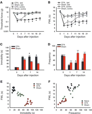
Persistent nociception induces depressive behavior. Inflammatory arthritis in Wistar rats induced by the injection of CFA into the right tibiotarsal joint produced mechanical allodynia (Figure 1A; ANOVA, F(3,104) = 3.11, P < 0.05) and thermal hyperalgesia (Fig-ure 1B; F(3,121) = 8.99, P < 0.05), which lasted for at least 21 days as compared with sham control rats injected with incomplete Fre-und’s adjuvant. This condition of persistent nociception induced depressive behavior in these same rats when examined on days 7 and 14, but not on day 1, in the forced swimming test (FST) (ref. 14 and Figure 1C; F(3,37) = 18.91; P < 0.01) and open field test (OFT) (refs. 15, 16, and Figure 1D; F(3,31) = 6.08, P < 0.05). A shorter hind paw withdrawal latency in arthritic rats correlated with a longer immobility time in FST (Figure 1E) and a lower frequency in OFT (Figure 1F), demonstrating a comorbid relationship between pain and depression in these rats. Of note, the increased immobility time in FST and reduced frequency in OFT were observed in both arthritic and sham control rats on day 1 but only in arthritic rats on day 7 and day 14. Testing of depressive behavior was not extended beyond day 14 in order to avoid habituation to the testing environ-ment, because there were no differences after day 14 in nociceptive behavior. The intensity of exploratory behavior (e.g., rearing and crossing in OFT) was similar in arthritic and sham control rats, although arthritic rats had a lower frequency of exploratory behav-iors. Moreover, there were no differences in a rotarod test between rats with or without hind paw arthritis on day 7 (Supplemental Figure 1A; supplemental material available online with this article; doi:10.1172/JCI61884DS1), suggesting that the observed depres-sive behavior was unlikely due to changes in motor function.
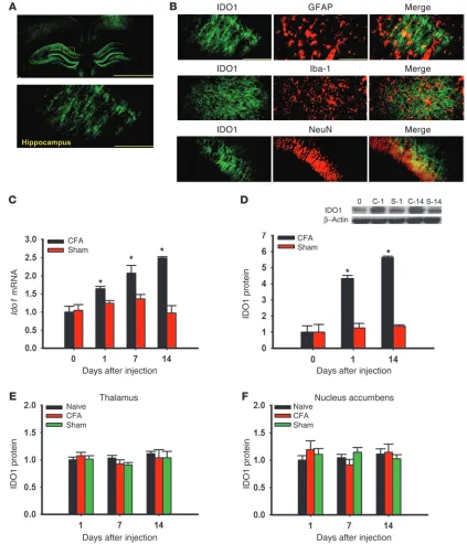
Hippocampal IDO1 expression is upregulated in rats with coexistent nociceptive and depressive behavior. We first examined whether brain IDO1 expression (hippocampus, thalamus, and nucleus accum-bens) would differ in rats with or without coexistent nociceptive and depressive behavior. IDO1 immunoreactivity in the hippo-campus (Figure 2A) was co-localized with glial fibrillary acidic protein (GFAP; astrocyte marker), Iba-1 (microglial marker), and NeuN (neuronal marker) (Figure 2B), consistent with both in vivo and in vitro expression of IDO1 in immune cells and neu-rons (17, 18). While the basal Ido1 mRNA level (real-time PCR) in the bilateral hippocampus was similar in arthritic and sham rats (naive, day 0; contralateral side only), the Ido1 mRNA level was progressively increased on days 1, 7, and 14 in arthritic but not sham rats (Figure 2C; P < 0.05). The IDO1 protein level (West-ern blot analysis) was also elevated in the hippocampus (Figure 2D; contralateral side only, P < 0.05), but not in the thalamus or nucleus accumbens (Figure 2, E and F), of arthritic rats. Moreover, there was a temporal relationship between IDO1 upregulation and nociceptive (Figure 1, A and B) and depressive behavior (Fig-ure 1, C and E) in these same rats.
Increased IDO1 enzyme activity alters ratios of hippocampal tryp-tophan metabolites. To examine the role of IDO1 enzyme activ-ity in tryptophan metabolism in both arthritic and sham rats, we first measured the content of tryptophan, serotonin, and kynurenine in the hippocampus using HPLC and then
deter-mined the ratio of serotonin or kynurenine to tryptophan. There were no baseline differences in the kynurenine/tryp-tophan or serotonin/trypkynurenine/tryp-tophan ratio between arthritic and sham control rats (Figure 3, A and B). However, the kynurenine/tryptophan ratio was significantly increased (Fig-ure 3A; P < 0.05), while the serotonin/tryptophan ratio was decreased (Figure 3B; P < 0.05), in arthritic rats as compared with sham control rats when measured on both day 1 and day 14. In contrast, IDO1 enzyme activity (both kynurenine/tryp-tophan and serotonin/trypkynurenine/tryp-tophan ratios) was not changed in the thalamus of arthritic rats (Figure 3, C and D). In addition, the plasma kynurenine/tryptophan ratio was also significantly increased (Figure 3E; P < 0.05) in arthritic rats as compared with sham control rats, although the plasma serotonin/tryp-tophan ratio remained unchanged (Figure 3F), when both were examined on day 14. Consistent with the IDO1 upregulation in arthritic rats, altered ratios of tryptophan metabolites in the hippocampus indicate increased IDO1 enzyme activity in rats with coexistent nociceptive and depressive behavior.
[image:3.585.43.338.82.446.2]Patients with both chronic back pain and depression also showed a significantly elevated plasma IDO1 level (Figure 3G) and increased IDO1 enzyme activity (Figure 3H; increased kynurenine/tryptophan ratio) as compared with healthy con-trol subjects without pain and depression (P < 0.05). In con-trast, the serotonin/tryptophan ratio was not different between
Figure 1
research article
healthy control subjects and patients with both pain and depression (Figure 3I). Although the data were obtained in a cross-sectional observational setting, these findings suggest that a relationship could also exist in human subjects between IDO1 activity and combined pain and depression.
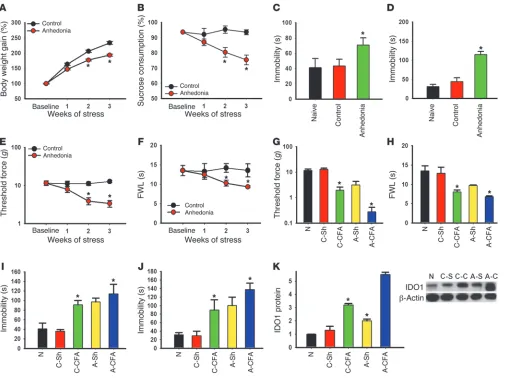
[image:4.585.73.496.84.577.2]Presence of anhedonic behavior exacerbates nociceptive behavior. In order to examine the generality of hippocampal IDO1 expression in relation to the interaction between nociception and depression, we used an established rat model of anhedonia induced by chronic social stress (see Methods). After 2 weeks of chronic social stress, rats
Figure 2
demonstrated a significant decrease in body weight gain and sucrose preference relative to control rats without social stress (Figure 4A,
F(1,23) = 438.20, P < 0.05; Figure 4B, F(1,23) = 172.12; P < 0.05), indi-cating the presence of anhedonic behavior. These rats also exhibited other depressive behaviors, manifesting as a longer immobility time in both FST (Figure 4C; F(2, 35) = 31.86, P < 0.05) and tail suspension test (TST) (Figure 4D; F(2,35) = 369.08, P < 0.05). Moreover, these same rats exhibited a progressively lower baseline nociceptive thresh-old in response to mechanical (Figure 4E; F(1,23) = 312,85, P < 0.05) and thermal stimulation (Figure 4F; F(1,23) = 100.276, P < 0.05) dur-ing 3 weeks of chronic social stress, indicatdur-ing that the presence of anhedonic behavior also influenced baseline nociceptive response.
To examine whether preexisting anhedonic behavior would exacerbate nociceptive behavior following hind paw arthritis, we exposed anhedonic and control rats, following 3 weeks of
[image:5.585.42.510.77.555.2]social stress and sham control respectively, to either CFA hind paw arthritis or sham control and examined behavioral changes 1 week later. Both mechanical allodynia (Figure 4G; F(4,35) = 164.98, P < 0.05) and thermal hyperalgesia (Figure 4H; F(4,35) = 63.72, P < 0.05) were exacerbated in anhedonic rats as compared with control rats, associated with a significantly longer immo-bility time in FST (Figure 4I; F(4,35) = 63.19, P < 0.05) and TST (Figure 4J; F(4,33) = 73.74, P < 0.05) in these same rats. Moreover, the IDO1 expression (Western blot) in the bilateral hippocam-pus was significantly increased in anhedonic rats as compared with control rats with or without hind paw arthritis (Figure 4K; contralateral side only, P < 0.05). These results indicate that noci-ceptive behavior was exacerbated in rats with preexisting anhedo-nic behavior, which was also associated with the upregulation of IDO1 expression in the hippocampus.
Figure 3
research article
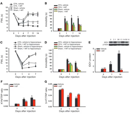
Inhibition of IDO1 activity concurrently attenuates nociceptive and depressive behavior. To examine whether inhibition of IDO1 activity would influence nociceptive and depressive behaviors in arthritic rats, we administered the IDO1 inhibitor l
-1-meth-yl-tryptophan (1-MT; 10 mg/d) (19) or vehicle intraperitoneally twice daily for 14 consecutive days. Treatment with 1-MT, but not vehicle, significantly attenuated both nociceptive (Figure 5A; F(3,119) = 11.33; P < 0.05) and depressive (Figure 5B: F(3,63) = 5.54); P < 0.05) behaviors in arthritic rats. Systemic 1-MT treat-ment alone did not alter behaviors in sham controls rats (Figure 5, A and B), nor did it change the appearance of arthritic hind paw (e.g., redness or swelling).
To examine the brain site of 1-MT action, we microinjected 1-MT (5 μg in 0.5 μl volume, once daily for 7 days) into the hippocampus contralateral to the arthritic hind paw.
Intra-hip-pocampal 1-MT treatment also attenuated both nociceptive (Figure 5C; F(3,71) = 5.54, P < 0.05) and depressive (Figure 5D;
F(3,31) = 14.70, P < 0.05) behaviors in arthritic rats without changing behaviors in sham control rats, indicating that the hippocampus is a critical brain locus of IDO1 activity. The pro-cedure of brain cannula implantation itself, used for intra-hip-pocampal microinjection, did not alter the baseline behavioral response when examined 5 days after the surgery (Supplemen-tal Figure 1, B–D).
[image:6.585.44.549.82.460.2]Intraperitoneal 1-MT treatment also (a) downregulated IDO1 expression (Figure 5E, P < 0.05), (b) lowered the kynurenine/ tryptophan ratio (Figure 5F, P < 0.05), and (c) elevated the sero-tonin/tryptophan ratio (Figure 5G, P < 0.05) in the hippocam-pus of arthritic rats. Together with the behavioral data, these results indicate that concurrent attenuation of nociceptive and
Figure 4
depressive behavior by the 1-MT treatment was mediated by the regulation of hippocampal IDO1 activity, thereby normalizing the content of tryptophan metabolites in the hippocampus.
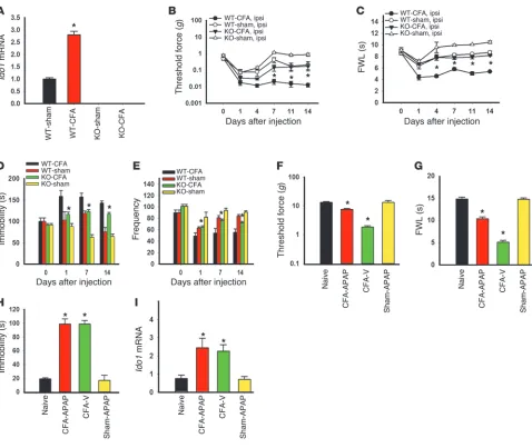
Ido1 gene knockout attenuates both nociceptive and depressive behavior. To further confirm the role of IDO1 in the behavioral manifesta-tion of pain and depression, we used IDO1-knockout and matched wild-type mice under the same experimental condition as that for Wistar rats. Both basal and arthritis-induced IDO1 expression in the hippocampus, as observed in age-matched wild-type mice, was absent in IDO1-knockout mice (Figure 6A). There were no base-line differences in behavioral tests for nociception (Figure 6, B and C; day 0) and depression (Figure 6, D and E; day 0) between IDO1-knockout and wild-type mice (each P > 0.05). In
IDO1-knockout mice, however, both mechanical allodynia (Figure 6B;
F(3,120) = 9.86, P < 0.01) and thermal hyperalgesia (Figure 6C;
F(3,122) = 5.73, P < 0.05) were significantly attenuated as compared with wild-type mice after the CFA injection into the right tibiotar-sal joint. In these same knockout mice, the immobility time (FST) was not increased, nor was there a decrease in the frequency in OFT, as compared with wild-type mice (Figure 6D; F(3,74) = 5.40; Figure 6E; F(3,49) = 32.175, each P < 0.05). These results indicate that Ido1
gene knockout concurrently attenuated nociceptive and depressive behavior induced by persistent hind paw nociception.
To examine whether selective reduction of nociceptive behav-ior would influence depressive behavbehav-ior and hippocampal IDO1 expression, was given acetaminophen (N
-acetyl-para-aminophe-Figure 5
[image:7.585.71.504.78.458.2]research article
nol, 100 mg/kg) (20, 21), an analgesic agent without the anti-in-flammatory effect, or vehicle once intraperitoneally on day 14 to arthritic or sham rats. When examined at 1 hour after the treat-ment, acetaminophen, but not vehicle, significantly reduced mechanical allodynia (Figure 6F; F(3,23) = 128.80, P < 0.05) and thermal hyperalgesia (Figure 6G; F(3,23) = 839.97, P < 0.05). The acetaminophen treatment did not acutely reverse depressive behavior (Figure 6H; FST), nor did it alter the Ido1 mRNA level in the same arthritic rats (Figure 6I). These results indicate that the correlation between nociception and depression demonstrated in these rats was not a simple coincidence but rather that the two were linked by the hippocampal IDO1 expression.
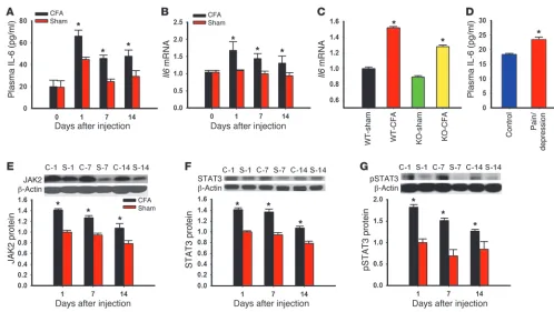
[image:8.585.50.528.79.482.2]IL-6 and JAK/STAT are increased in rats with nociceptive and depres-sive behavior. Proinflammatory cytokines including IL-6 have been shown to be involved in the cellular mechanisms of both pain and depression (11, 12, 22). To examine the hypothesis that proinflam-matory cytokines such as IL-6 and one of its downstream signaling pathways (JAK and STAT) (23, 24) would mediate hippocampal IDO1 upregulation, we first examined whether the IL-6 level and JAK/STAT expression would be increased in rats with coexistent nociceptive and depressive behavior. Both the plasma IL-6 level and hippocampal Il6 mRNA expression were significantly increased in rats with nociceptive and depressive behavior as compared with sham rats (Figure 7, A and B; P < 0.05). The hippocampal Il6 mRNA
Figure 6
level was also elevated in IDO1-knockout and wild-type mice after CFA injection into a tibiotarsal joint (Figure 7C; P < 0.05), indicat-ing that the IL-6 increase was upstream of IDO1 upregulation. In patients with both chronic pain and depression, the plasma IL-6 content was also elevated as compared with that in healthy con-trol subjects (Figure 7D; P < 0.05). Of note, plasma IL-6 content in human subjects was measured in a cross-sectional observational setting and could have been influenced by the subjects’ underlying pain condition and other variations such as body weight. More-over, the expression of IL-6 signaling elements, including JAK2, STAT3, and p-STAT3, was all elevated in the hippocampus of rats with nociceptive and depressive behavior as compared with sham controls (Figure 7, E–G; each P < 0.05).
IL-6 induces in vitro IDO1 upregulation. To examine a direct rela-tionship between IL-6 and IDO1 expression at the cellular level, we exposed cultured Neuro2a cells to exogenous IL-6 (0.5 ng/ml) or vehicle (PBS) for 24 hours. IDO1 immunoreactivity was detected in the perinuclear cytoplasm of Neuro2a cells and increased following exposure to IL-6 for 24 hours (Figure 8A). Exposure of cultured Neu-ro2a cells to exogenous IL-6, but not vehicle, significantly increased
Ido1 mRNA (real-time PCR) and protein (Western blot analysis) expression (Figure 8, B and C; P < 0.05), resulting in the increased kynurenine/tryptophan ratio and decreased serotonin/tryptophan ratio (HPLC) in these Neuro2a cells (Figure 8, D and E; P < 0.05).
Furthermore, we used a hippocampal organotypic slice culture taken from postnatal rats to examine the in vitro effect of IL-6 on hippocampal IDO1 expression and activity. After being cul-tured for 1 week, hippocampal slices were treated with IL-6 (100 ng/ml) or vehicle (PBS) for 24 hours. Exposure of exogenous IL-6, but not vehicle, increased IDO1 immunoreactivity (Figure 8F) and upregulated the expression of Ido1 mRNA (real-time PCR, Figure 8G; P < 0.05) and protein (Western blot analysis, Figure 8H;
P < 0.05) in cultured slices. Under the same experimental condi-tion, the kynurenine/tryptophan ratio was significantly increased, whereas the serotonin/tryptophan ratio was decreased in the cul-ture medium (HPLC, Figure 8I; P < 0.05). Collectively, the results indicate that IL-6 has a direct cellular effect on IDO1 expression in the hippocampus.
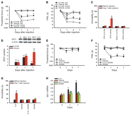
[image:9.585.39.537.79.360.2]IL-6–mediated hippocampal IDO1 expression concurrently regulates nociceptive and depressive behavior. To examine the functional role of IL-6 signaling in hippocampal IDO1 expression as well as its contribution to both nociceptive and depressive behavior, we microinjected an IL-6 antiserum into the hippocampus of arthritic or sham control rats. Microinjection of IL-6 antiserum (0.5 μg, once daily for 7 days) (22), but not control serum (vehi-cle), into the hippocampus contralateral to arthritic hind paw significantly attenuated mechanical allodynia (Figure 9A; F(1,39) = 9.28, P < 0.05), thermal hyperalgesia (Figure 9B; F(1,39) = 7.46,
Figure 7
research article
P < 0.05), and depressive behavior (Figure 9C; F(3,19) = 155.99,
P < 0.001). The same IL-6 antiserum treatment also prevented IDO1 upregulation in the hippocampus (Figure 9D; P < 0.05), consistent with the invitro results of IL-6–induced IDO1 expres-sion (Figure 8, B and C).
Conversely, microinjection of exogenous IL-6 (recombinant rat IL-6, 0.1 μg, once daily for 7 days) (25), but not vehicle, into the left hippocampus of naive rats (without arthritis) induced right hind paw mechanical allodynia (Figure 9E: F(3,79) = 2.54, P < 0.05) and thermal hyperalgesia (Figure 9F: F(3,103) = 11.24, P < 0.01), as well as depressive behavior (Figure 9G: F(3,19) = 65.20, P < 0.001)
and increased Ido1 mRNA expression in the hippocampus (Fig-ure 9H; P < 0.05). These IL-6 effects were prevented when IL-6 was co-administered with the JAK/STAT inhibitor AG490 (5 μg, once daily for 7 days) (26) into the hippocampus (Figure 9, E–H; each
P < 0.05). Intra-hippocampal microinjection of AG490 alone had no effect on the baseline behavioral response and Ido1 mRNA expression in naive rats (Figure 9, E–H; each P > 0.05).
[image:10.585.44.542.79.512.2]Taken together with the data obtained using the IDO1 inhibitor 1-MT, these findings indicate that the hippocampus is a central site of IL-6–regulated IDO1 expression critically contributory to the comorbid interaction between pain and depression.
Figure 8
Discussion
We have demonstrated that IDO1 expression was selectively upregulated in the hippocampus of Wistar rats with coexistent nociceptive and depressive behavior. The IDO1 level was also ele-vated in patients with both pain and depression. Either Ido1 gene knockout or inhibition of IDO1 activity, but not a transient rever-sal of nociceptive behavior alone by acetaminophen, concurrently attenuated nociceptive and depressive behavior. At the cellular level, the hippocampal IDO1 expression was mediated through IL-6 and its downstream JAK/STAT signaling pathway, which
in turn altered the kynurenine/tryptophan and serotonin/tryp-tophan ratios in the hippocampus. The results indicate that brain IDO activity played a critical role in regulating the comorbid inter-action between nociceptive and depressive behaviors.
[image:11.585.57.520.82.487.2]A comorbid relationship between pain and depression has long been recognized in the clinical setting (27, 28). Earlier stud-ies focused on a temporal relationship between pain and depres-sion (e.g., antecedent hypothesis versus consequence hypothesis) (29, 30), but the cellular mechanism underlying this relationship remains unknown. Recent neurobiological studies have suggested
Figure 9
research article
Studies in the immunology field have consistently shown a rela-tionship between inflammatory mediators and IDO expression in immune cells (48, 49). Studies using central administration of cytokines have indicated a role for cytokines in various behav-ioral manifestations. For example, intracerebroventricular (i.c.v.) administration of IL-6 or IL-1β elicited hyperalgesia (50), as well as fever, anorexia, and reduction of social exploratory behavior (51, 52). Three recent studies including ours have shown that periph-eral nerve injury induced depressive behavior in rats (53–55), which is associated with an increased IL-1β expression in the fron-tal cortex (54). The present data demonstrate a direct link between cytokine signaling and IDO expression in the hippocampus. Given that IDO changes were also demonstrated in a rat model of anhe-donia, regulation of brain IDO expression may have a broad impli-cation in the interaction between pain and depression. It will be of considerable interest in future studies to determine whether a similar cytokine and IDO link would be relevant to other pain con-ditions such as neuropathic pain.
The present study supports a central (hippocampal) effect of IL-6–mediated IDO activity on the behavioral interaction between pain and depression. First, intra-hippocampal microinjection of the IDO1 inhibitor 1-MT attenuated both nociceptive and depres-sive behavior similar to that after systemic 1-MT administration. Second, neither systemic 1-MT nor intra-hippocampal administra-tion of IL-6, IL-6 antiserum, or 1-MT changed signs of hind paw inflammation (e.g., redness, swelling), suggesting that the effect of 1-MT on nociceptive and depressive behavior is unlikely to be mediated through a peripheral mechanism at the site of hind paw arthritis. Third, the plasma IDO activity, reflected by an increased kynurenine/tryptophan ratio, was only transiently increased on day 1 but not day 7 and 14 after hind paw inflammation. Fourth, exogenous IL-6 directly upregulated IDO1 expression and enhanced IDO activity in Neuro2a cells and an organotypic hippocampal tissue culture. Fifth, intra-hippocampal microinjec-tion of IL-6 in naive rats induced hippocampal IDO upregulamicroinjec-tion as well as nociceptive and depressive behavior, which was blocked by AG490 (a JAK/STAT inhibitor). Thus, converging evidence from immunology literature and the current study suggests an impor-tant role of IDO activity in the central nervous system in addition to its critical role in immunoregulation (56).
Clinical studies have demonstrated that the plasma IL-6 level was increased in patients with painful neuropathy, cancer, inflam-mation (57, 58), and depression (59, 60). In this study, the plasma IL-6 and IDO level, as well as IDO enzyme activity, was increased in patients with chronic back pain and depression, consistent with the findings from animal studies. This raises the possibility that concurrent treatment of both pain and depression might be possible through regulation of brain IDO activity, in contrast to the current approach of symptomatic management using anti-depressants and analgesics (3). Although the neural and cellular mechanism underlying the interaction between pain and depres-sion is likely to be complex and involves other neurotransmitters and neuromodulators (25–33), the present findings may suggest a new strategy of clinical intervention. This new strategy focuses on both prevention and reversal of comorbid interactions between pain and depression by targeting its underlying mechanism involv-ing altered ratios of endogenous tryptophan metabolites resultinvolv-ing from upregulated IDO expression in certain brain regions. Since IDO inhibitors have been tried in clinical studies of depression and cancer treatment (61–63), it will be of considerable interest that both depression and chronic pain may involve the
monoamin-ergic system, the hypothalamic/pituitary/adrenal axis, as well as various other neurotransmitters/neuromodulators including acet-ylcholine, GABA, substance P, cholecystokinin, endogenous opioid, and brain-derived neurotrophic factor (31–35). Despite some prog-ress, clinical treatment of pain and depression has so far been lim-ited to symptomatic management (3, 36). A number of studies have also suggested that the effect of antidepressants on chronic pain is not necessarily related to their anti-depression property (36–38).
In the present study, the data from human subjects suggests an apparent relationship between IDO expression/enzyme activity and clinical symptoms of pain and depression, but this cross-sectional clinical observation does not explore the causal relationship between IDO and clinical conditions. On the other hand, the data from animal experiments suggest a novel mecha-nistic link between pain and depression via a critical role of IDO1 in the hippocampus. Regulation of hippocampal IDO1 is likely mediated through an IL-6 signal transduction pathway, because (a) upregulation of IL-6 as well as downstream JAK and STAT3 preceded the IDO expression in rats with combined nociceptive and depressive behavior; (b) the hippocampal Il6 mRNA level was elevated in Ido1 gene knockout mice in response to inflammatory arthritis; (c) inhibition of IDO1 activity by systemic 1-MT treat-ment did not prevent elevation of the plasma IL-6 level in rats with both nociceptive and depressive behavior (data not shown); and (d) IL-6 directly upregulated IDO1 expression in both Neuro2a cells and an organotypic hippocampal tissue culture. Our data also indicate that the hippocampus is a critical brain region of IDO regulation, because IDO1 was selectively upregulated in the hip-pocampus, but not in the thalamus or nucleus accumbens. These findings are consistent with the previous reports that (a) certain brain regions including the hippocampus play a critical role in the integration of mood changes and pain (31, 39, 40) and (b) the hip-pocampus is related to nociceptive perception and its exacerbation by mood disorders such as anxiety (41, 42).
Behavioral tests
Nociceptive test. Animals were habituated to a test setting (30-minute
ses-sion) for 3 consecutive days. Thermal withdrawal threshold was assessed using the Hargreaves apparatus and method (68). Mechanical withdrawal threshold was assessed using von Frey filaments (69).
FST. FST was performed according to the method of Porsolt et al. (14). The total duration of immobility (non-swimming) within a 5-minute session was recorded as immobility scores (in seconds) and compared among groups.
TST. TST was carried out as described previously (70). Briefly, a rat was hung on a hook, using adhesive tape placed 20 mm from the tip of its tail, on a 50-cm-height rod. The immobility time during a 6-minute period of tail suspension was recorded with a stopwatch.
OFT. OFT was carried out in a Plexiglas square box (57 × 57 cm) with walls 50 cm in height (71). Behaviors were observed for 10 minutes under a dim light. After the first 5 minutes (habituation), the number of squares crossed by a rat was recorded for the next 5 minutes.
Sucrose preference test. A sucrose preference test (SPT) was performed as
described in previous studies (72, 73). To perform this test, 1% sucrose solution was offered once weekly in a rat’s home cage. Each rat had a free choice between 1% sucrose solution bottle and a tap water bottle. Sucrose and tap water intakes were separately measured by weighing each bottle before and after the test (a 24-hour period). The sum of tap water and sucrose water intake (in grams) was calculated as the total water intake. The sucrose preference was expressed as the percentage of sucrose water intake relative to the total water intake.
Rotarod test. A rotarod test, using an accelerating rotarod apparatus
(Columbus Instruments), was performed in both arthritic and sham con-trol rats. Rats were given 3 training sessions and then placed on a 9-cm-di-ameter rod with speed increased from 0.5 to 30 rpm over a 60-second period. Each rat was tested in 3 consecutive trials with 15-minute intervals. The duration of time on the rotarod was determined automatically by a timer that recorded to the nearest second.
Cultures
Neuro2a cell culture. Neuro2a cells were cultured in Eagle’s minimal essential medium (Invitrogen) supplemented with 10% fetal bovine serum, 100 U/ml penicillin, and 100 μg/ml streptomycin (Invitrogen) in a humidified atmo-sphere of 5% CO2 and 95% air at 37°C. Cells were treated with 0.5 ng/ml IL-6 or vehicle and collected 24 hours later for assays (74).
Organotypic hippocampal tissue culture. According to a modified method
(75, 76), organotypic hippocampal cultures were prepared using hippo-campus slices from 5- to 8-day-old Wistar rats. Rat pups were euthanized by decapitation, brains were rapidly removed, and the hippocampus was separated from the brain. Hippocampal slices (350 μm in thickness) were made using a McIlwain tissue chopper (Campden Instruments Ltd.) and placed in Gey’s balanced salt solution (Sigma-Aldrich) with 2 mg/ml d
-glu-cose. Slices were then placed on 30-mm Millicell-CM (Millipore) porous (0.4 μm) membranes. These membranes were transferred into 6-well cul-ture plates filled with an incubation medium (1 ml) consisting of 50% vol/ vol minimal essential medium, 25% heat-inactivated horse serum, and 25% HBSS (Invitrogen), supplemented with 25 mM d-glucose. Plates were incu-bated in a humidified atmosphere of 5% CO2 and 95% air at 37°C. Culture medium was changed 3 times a week. Slices were treated with 100 ng/ml IL-6 or vehicle and collected 24 hours later for assays (77).
Immunohistochemistry
Rats were anesthetized with pentobarbital (50 mg/kg, intraperitoneally) and perfused transcardially with cold 0.9% saline (pH 7.4 ± 0.1), fol-lowed by cold 4% paraformaldehyde in a phosphate buffer (PB; 0.1 M,
to examine whether using IDO inhibitor, alone or in combination with other agents blocking IDO upregulation or regulating tryp-tophan metabolism, would be able to achieve concurrent allevia-tion of pain and depression in the clinical setting.
Methods
Human subjects
Male and female subjects between 18 and 65 years old were recruited. A total of 33 subjects were recruited from the greater Boston area. Inclusion criteria included (a) chronic lumbar or cervical radicular pain due to disk herniation or radiculitis for at least 3 months; (b) clinical depression with-out hospitalization within the past 12 months or medication changes; (c) no history of drug abuse confirmed by a negative urine drug screen; (d) no other psychiatric disorders, including schizophrenia, bipolar disorder, and eating disorders; and (e) for normal control subjects, no pain or depression for at least the past 6 months. Board-certified physicians on the research team reviewed and confirmed clinical diagnosis of pain and depression. Blood samples were taken once and used for HPLC and ELISA assays. Of note, the data from human subjects was obtained in a cross-section obser-vational setting, which could be confounded by potential clinical varia-tions such as body weight and a subject’s underlying pain condition as well as a small sample size.
Experimental animals
Male Wistar rats (250–300 g; Charles River) or 7- to 10-week-old male B6.129-Ido1tm1Alm/J(IDO–/–) and C57BL/6J wild-type mice (Jackson Labo-ratory) were used. Animals were housed individually (21 ± 2°C, relative humidity 50% ± 10%, 12-hour light/12-hour dark cycle), with water and food available ad libitum.
Surgical procedures
Hind paw monoarthritis. Hind paw monoarthritis was induced by the injection
of CFA (50 μl) into a unilateral tibiotarsal joint cavity under brief isoflurane anesthesia (13). Animals in sham groups were injected with 50 μl incomplete Freund’s adjuvant. Local redness and joint swelling were observed in CFA-in-jected rats but not control rats during the experimental period.
Brain cannula implantation and drug injection. Under brief isoflurane
anes-thesia, a guide cannula (26 gauge, Plastics One) was implanted just above the hippocampus (anteroposterior [AP]: –3.6 mm; left: +2.0 mm from bregma; depth: –4.0 mm from skin) (64). An injection needle (33 gauge, Plastics One) was inserted through the guide cannula, and drug solution or vehicle (0.5 μl) was slowly injected over 5 minutes using a Hamilton syringe. Locations of the cannula placement were confirmed at the time of tissue harvest (Supplemental Figure 3A).
Anhedonia induced by chronic social stress
research article
reader (Synergy HT, BioTEK). A standard curve, provided by the manufac-turer, was generated for each set of samples assayed, and the concentration of each sample was obtained according to the standard curve.
Real-time RT-PCR
Total RNA was isolated from hippocampal samples using TRIzol reagent (Invitrogen). Four micrograms of total RNA was used to synthesize the first-strand cDNA using a SuperScript III kit (Invitrogen). Four microli-ters of cDNA was put into a 20-μl vessel using 1 μl of ×20 TaqMan Gene Expression Assay. TaqMan Gene Expression Assays (Applied Biosystems) containing primers and a TaqMan probe were used to quantify each gene of interest. The genes examined were Il6 (Rn01410330 m1, M26744.1) and Ido1 (Rn00576778 m1, AF312699.1) for rats and Il6 (Mm00446190 m1, X54542.1) and Ido1 (Mm00492586 m1, M69109.1) for mice. The reaction was performed in duplicate with the following conditions: denaturing at 95°C for 30 seconds, annealing at 60°C for 2 minutes, extension at 68°C for 2 minutes for 40 cycles using a 7300 Real-Time PCR System (Applied Biosystems). The RNA expression of GAPDH was measured as control.
HPLC
Hippocampal tryptophan, kynurenine, and serotonin content was deter-mined by HPLC (78, 79). All samples were taken between 2 and 5 p.m. Kynurenine was measured by a UV detector (Shimadzu, SPD-10Avp; 360-nm wavelength). Tryptophan and serotonin were measured by a fluo-rescence detector (Shimadzu, RF-10Axl; 286-nm excitation and 366-nm emission wavelengths). Each day, external calibration was made by using freshly prepared control samples of the same concentrations (100 nmol/l tryptophan, 100 μmol/l kynurenine, and 100 nmol/l serotonin).
Statistics
Repeated measures 2-way ANOVA was used and followed by the Tukey test to detect interactions between test time points and groups. Correla-tions between nociceptive and depressive behavior were determined using Pearson correlation coefficient analysis. HPLC, Western blot, and ELISA data were analyzed using 1-way ANOVA followed by a Tukey or Dunn test. Statistical analyses were performed using SigmaStat (Systat, version 11), with a significance level of P < 0.05.
Study approval
Informed consent was obtained from study participants. Research proto-cols were approved by the Massachusetts General Hospital Institutional Animal Care and Use Committee. Experiments were conducted with the experimenter being unaware of the group assignment.
Acknowledgments
This study was supported by NIH grants R01DE18214, R01DE18538, and P20DA26002 (to J. Mao).
Received for publication November 11, 2011, and accepted in revised form May 16, 2012.
Address correspondence to: Jianren Mao, MGH Center for Transla-tional Pain Research, Department of Anesthesia, Critical Care and Pain Medicine, Massachusetts General Hospital, Harvard Medical School, Boston, Massachusetts 02114, USA. Phone: 617.726.2338; Fax: 617.724.2719; E-mail: [email protected].
pH 7.4 ± 0.1). Brains were removed and placed in the same fixative for post-fixation at 4°C for 6 hours. For cryoprotection, the samples were kept with cold 30% sucrose in PBS at 4°C until samples sank to the bot-tom. Coronal brain sections (30 μm) were cut using a cryostat and then floated in PBS. Sections were blocked with 5% normal donkey serum in 0.3% Triton X-100/PBS for 1 hour at room temperature. Free-float-ing sections were incubated overnight at 4°C on a rocker with one of the following primary antibodies: 1:100 IDO1 rabbit polyclonal (Santa Cruz Biotechnology Inc.); 1:1,000 NeuN mouse monoclonal (Chemi-con); 1:1,000 GFAP mouse monoclonal (BD Biosciences — Pharmin-gen); 1:1,000 Iba-1 mouse monoclonal (Abcam). Sections were then washed with PBS 3 times and incubated with 1:300 FITC- or cyanine 3–conjugated secondary antibody (Jackson ImmunoResearch Laborato-ries Inc.) for 1 hour at room temperature. Blue fluorescent DAPI (1:300; Invitrogen) was used to stain the nucleus of fixed, cultured Neuro2a cells for 15 minutes.
Colocalization was examined by adding a second primary antibody fol-lowing the same procedure as described above. Controls were performed by either omitting primary antibody or using antigen absorption (Supple-mentary Figure 3B). Images were examined with a fluorescence microscope (Olympus) and captured with a digital camera. The images were analyzed using Adobe Photoshop (version 7).
Western blot analysis
Rats were sacrificed under pentobarbital anesthesia. The hippocampus was separated from the brain and immediately stored on dry ice and kept at –80°C until use. Samples were homogenized in a lysis buffer with SDS containing a cocktail of proteinase inhibitors (Roche). Samples were separated on an SDS-PAGE gel (4%–15% gradient gel; Bio-Rad) and transferred to PVDF membranes (Millipore). Membranes were blocked with 5% nonfat dried milk for 1 hour at room temperature and incu-bated overnight at 4°C on a rocker with one of following primary anti-bodies: 1:200 IDO1 (Santa Cruz Biotechnology Inc.; rabbit polyclonal antibody), 1:200 IDO1 (Novus Biologicals; rabbit polyclonal antibody), 1:500 JAK2 (Santa Cruz Biotechnology Inc.; rabbit polyclonal antibody), 1:1,000 STAT3 (Millipore; rabbit polyclonal antibody), 1:5,000 p-STAT3 (Abcam; rabbit monoclonal antibody). After membranes were washed with PBS 4 times, membranes were incubated with 1:8,000 HRP-conju-gated secondary antibody (Amersham Biosciences, GE Healthcare) for 1 hour at room temperature on a rocker. The membranes were washed with PBS 4 times, and blots were visualized in enhanced chemilumines-cent (ECL) solution (Pierce) for 5 minutes and exposed to hyperfilms (Kodak) for 15 minutes. Blots were again incubated in a stripping buffer (Pierce) for 15 minutes at room temperature on a rocker and reprobed with 1:12,000 anti–β-actin antibody (Abcam Inc.) as a loading control. Western blots were made in triplicate. Band density was measured and normalized against a loading control band.
ELISA
Under pentobarbital anesthesia, whole blood was collected from the left ventricle of the heart and placed in a serum collection tube (BD Vacutainer). Blood clots were formed after 30–60 minutes at room tem-perature, and the tube was centrifuged for 10 minutes at 9,659.52 g. An ELISA Quantikine kit (R&D Systems) and IDO ELISA kit (TSZ ELISA) were used according to the manufacturers’ instructions. Optical density of samples was read at wavelengths of 450 and 570 nm using a microplate
1. Sullivan MJ, Reesor K, Mikail S, Fisher R. The treat-ment of depression in chronic low back pain: review and recommendations. Pain. 1992;50(1):5–13. 2. Von KM, Simon G. The relationship between
pain and depression. Br J Psychiatry Suppl. 1996; (30):101–108.
3. Mao J, Gold MS, Backonja MM. Combination drug therapy for chronic pain: a call for more clinical
studies. J Pain. 2011;12(2):157–166.
1986;236(1):293–299.
5. Dantzer R, O’Connor JC, Freund GG, Johnson RW, Kelley KW. From inflammation to sickness and depression: when the immune system subjugates the brain. Nat Rev Neurosci. 2008;9(1):46–56. 6. Popov A, et al. Indoleamine
2,3-dioxygenase-expressing dendritic cells form suppurative granu-lomas following Listeria monocytogenes infection.
J Clin Invest. 2006;116(12):3160–3170.
7. Heyes MP, et al. Quinolinic acid and kynurenine pathway metabolism in inflammatory and non-inflammatory neurological disease. Brain. 1992;115(pt 5):1249–1273.
8. Heyes MP, Lackner A. Increased cerebrospinal fluid quinolinic acid, kynurenic acid, and L-kynurenine in acute septicemia. J Neurochem. 1990;55(1):338–341. 9. Yoshida R, Urade Y, Tokuda M, Hayaishi O.
Induc-tion of indoleamine 2,3-dioxygenase in mouse lung during virus infection. Proc Natl Acad Sci U S A. 1979;76(8):4084–4086.
10. O’Connor JC, et al. Interferon-gamma and tumor necrosis factor-alpha mediate the upregulation of indoleamine 2,3-dioxygenase and the induc-tion of depressive-like behavior in mice in response to bacillus Calmette-Guerin. J Neurosci. 2009;29(13):4200–4209.
11. De Jongh RF, Vissers KC, Meert TF, Booij LH, De Deyne CS, Heylen RJ. The role of interleu-kin-6 in nociception and pain. Anesth Analg. 2003; 96(4):1096–1103.
12. Maes M, Verkerk R, Bonaccorso S, Ombelet W, Bos-mans E, Scharpe S. Depressive and anxiety symp-toms in the early puerperium are related to increased degradation of tryptophan into kynurenine, a phe-nomenon which is related to immune activation. Life Sci. 2002;71(16):1837–1848.
13. Butler SH, Godefroy F, Besson JM, Weil-Fugazza J. A limited arthritic model for chronic pain studies in the rat. Pain. 1992;48(1):73–81.
14. Porsolt RD, Le PM, Jalfre M. Depression: a new ani-mal model sensitive to antidepressant treatments.
Nature. 1977;266(5604):730–732.
15. Hall CS. Emotional behavior in rat. J Comp Physiol Psychol. 1934;18(3):385–403.
16. Pare WP. Open field, learned helplessness, condi-tioned defensive burying, and forced-swim tests in WKY rats. Physiol Behav. 1994;55(3):433–439. 17. Guillemin GJ, Smythe G, Takikawa O, Brew BJ.
Expression of indoleamine 2,3-dioxygenase and production of quinolinic acid by human microglia, astrocytes, and neurons. Glia. 2005;49(1):15–23. 18. Roy EJ, Takikawa O, Kranz DM, Brown AR, Thomas
DL. Neuronal localization of indoleamine 2,3-diox-ygenase in mice. Neurosci Lett. 2005;387(2):95–99. 19. O’Connor JC, et al.
Lipopolysaccharide-in-duced depressive-like behavior is mediated by indoleamine 2,3-dioxygenase activation in mice.
Mol Psychiatry. 2009;14(5):511–522.
20. Hama AT, Sagen J. Cannabinoid receptor-mediated antinociception with acetaminophen drug combi-nations in rats with neuropathic spinal cord injury pain. Neuropharmacology. 2010;58(4–5):758–766. 21. Mitchell D, Gelgor L, Weber J, Kamerman PR.
Antihypernociceptive synergy between ibuprofen, paracetamol and codeine in rats. Eur J Pharmacol. 2010;642(1–3):86–92.
22. Wang S, Lim G, Mao J, Sung B, Mao J. Regulation of the trigeminal NR1 subunit expression induced by inflammation of the temporomandibular joint region in rats. Pain. 2009;141(1–2):97–103. 23. Heinrich PC, Behrmann I, Muller-Newen G,
Schaper F, Graeve L. Interleukin-6-type cytokine signalling through the gp130/Jak/STAT pathway.
Biochem J. 1998;334(pt 2):297–314.
24. Legendre F, Dudhia J, Pujol JP, Bogdanowicz P. JAK/STAT but not ERK1/ERK2 pathway mediates interleukin (IL)-6/soluble IL-6R down-regulation of Type II collagen, aggrecan core, and link protein
transcription in articular chondrocytes. Associa-tion with a down-regulaAssocia-tion of SOX9 expression.
J Biol Chem. 2003;278(5):2903–2912.
25. Wallenius K, Wallenius V, Sunter D, Dickson SL, Jansson JO. Intracerebroventricular interleukin-6 treatment decreases body fat in rats. Biochem Bio-phys Res Commun. 2002;293(1):560–565. 26. Dominguez E, Rivat C, Pommier B, Mauborgne A,
Pohl M. JAK/STAT3 pathway is activated in spinal cord microglia after peripheral nerve injury and contributes to neuropathic pain development in rat. J Neurochem. 2008;107(1):50–60.
27. Dworkin RH, Gitlin MJ. Clinical aspects of depression in chronic pain patients. Clin J Pain. 1991;7(2):79–94.
28. Delgado PL. Common pathways of depression and pain. J Clin Psychiatry. 2004;65(suppl 12):16–19. 29. Dohrenwend BP, Raphael KG, Marbach JJ,
Gal-lagher RM. Why is depression comorbid with chronic myofascial face pain? A family study test of alternative hypotheses. Pain. 1999;83(2):183–192. 30. Blackburn-Munro G, Blackburn-Munro RE.
Chronic pain, chronic stress and depression: coincidence or consequence? J Neuroendocrinol. 2001;13(12):1009–1023.
31. Schatzberg AF. The relationship of chronic pain and depression. J Clin Psychiatry. 2004;65(suppl 12):3–4. 32. Jasmin L, Rabkin SD, Granato A, Boudah A, Ohara
PT. Analgesia and hyperalgesia from GABA-medi-ated modulation of the cerebral cortex. Nature. 2003;424(6946):316–320.
33. Andre J, Zeau B, Pohl M, Cesselin F, Benoliel JJ, Becker C. Involvement of cholecystokininergic systems in anxiety-induced hyperalgesia in male rats: behavioral and biochemical studies. J Neurosci. 2005;25(35):7896–7904.
34. Gameiro GH, da Silva AA, Nouer DF, Ferraz de Arruda Veiga MC. How may stressful experiences contribute to the development of temporomandib-ular disorders? Clin Oral Investig. 2006;10(4):261–268. 35. Tsigos C, Chrousos GP. Hypothalamic-pitu-itary-adrenal axis, neuroendocrine factors and stress. J Psychosom Res. 2002;53(4):865–871. 36. Max MB, et al. Amitriptyline relieves diabetic
neu-ropathy pain in patients with normal or depressed mood. Neurology. 1987;37(4):589–596.
37. Sharav Y, Singer E, Schmidt E, Dionne RA, Dubner R. The analgesic effect of amitriptyline on chronic facial pain. Pain. 1987;31(2):199–209.
38. Atkinson JH, et al. A placebo-controlled random-ized clinical trial of nortriptyline for chronic low back pain. Pain. 1998;76(3):287–296.
39. Zhang L, Zhang Y, Zhao ZQ. Anterior cingulate cortex contributes to the descending facilitatory modulation of pain via dorsal reticular nucleus.
Eur J Neurosci. 2005;22(5):1141–1148.
40. Vogt BA. Pain and emotion interactions in sub-regions of the cingulate gyrus. Nat Rev Neurosci. 2005;6(7):533–544.
41. Derbyshire SW, Jones AK, Gyulai F, Clark S, Townsend D, Firestone LL. Pain processing dur-ing three levels of noxious stimulation produces differential patterns of central activity. Pain. 1997; 73(3):431–445.
42. Ploghaus A, et al. Exacerbation of pain by anxiety is associated with activity in a hippocampal network.
J Neurosci. 2001;21(24):9896–9903.
43. Miura H, Ozaki N, Sawada M, Isobe K, Ohta T, Nagatsu T. A link between stress and depression: shifts in the balance between the kynurenine and serotonin pathways of tryptophan metabolism and the etiology and pathophysiology of depression.
Stress. 2008;11(3):198–209.
44. Myint AM, Kim YK. Cytokine-serotonin interaction through IDO: a neurodegeneration hypothesis of depression. Med Hypotheses. 2003;61(5-6):519–525. 45. Mico JA, Gibert-Rahola J, Casas J, Rojas O, Serrano
MI, Serrano JS. Implication of beta 1- and beta
2-adrenergic receptors in the antinociceptive effect of tricyclic antidepressants. Eur Neuropsychopharmacol. 1997;7(2):139–145.
46. Sawynok J, Esser MJ, Reid AR. Antidepressants as analgesics: an overview of central and periph-eral mechanisms of action. J Psychiatry Neurosci. 2001;26(1):21–29.
47. Stone TW, Perkins MN. Quinolinic acid: a potent endogenous excitant at amino acid receptors in CNS. Eur J Pharmacol. 1981;72(4):411–412. 48. Kwidzinski E, et al. Indolamine
2,3-dioxygen-ase is expressed in the CNS and down-regulates autoimmune inflammation. FASEB J. 2005; 19(10):1347–1349.
49. Yan Y, et al. IDO upregulates regulatory T cells via tryptophan catabolite and suppresses enceph-alitogenic T cell responses in experimental auto-immune encephalomyelitis. J Immunol. 2010; 185(10):5953–5961.
50. Hori T, Oka T, Hosoi M, Aou S. Pain modulatory actions of cytokines and prostaglandin E2 in the brain. Ann N Y Acad Sci. 1998;840:269–281. 51. Lenczowski MJ, et al. Central administration of
rat IL-6 induces HPA activation and fever but not sickness behavior in rats. Am J Physiol. 1999; 276(3 pt 2):R652–R658.
52. Huang Y, Henry CJ, Dantzer R, Johnson RW, God-bout JP. Exaggerated sickness behavior and brain proinflammatory cytokine expression in aged mice in response to intracerebroventricular lipopolysac-charide. Neurobiol Aging. 2008;29(11):1744–1753. 53. Zeng Q, et al. Exacerbated mechanical allodynia
in rats with depression-like behavior. Brain Res. 2008;1200:27–38.
54. Norman GJ, Karelina K, Zhang N, Walton JC, Mor-ris JS, Devries AC. Stress and IL-1beta contribute to the development of depressive-like behavior following peripheral nerve injury. Mol Psychiatry. 2010;15(4):404–414.
55. Yalcin I, et al. A time-dependent history of mood disorders in a murine model of neuropathic pain.
Biol Psychiatry. 2011;70(10):946–953.
56. Pallotta MT, et al. Indoleamine 2,3-dioxygenase is a signaling protein in long-term tolerance by den-dritic cells. Nat Immunol. 2011;12(9):870–878. 57. Kiefer R, Kieseier BC, Stoll G, Hartung HP. The
role of macrophages in immune-mediated damage to the peripheral nervous system. Prog Neurobiol. 2001;64(2):109–127.
58. Smith PC, Hobisch A, Lin DL, Culig Z, Keller ET. Interleukin-6 and prostate cancer progression.
Cytokine Growth Factor Rev. 2001;12(1):33–40. 59. Connor TJ, Leonard BE. Depression, stress and
immunological activation: the role of cytokines in depressive disorders. Life Sci. 1998;62(7):583–606. 60. Thomas AJ, Davis S, Morris C, Jackson E, Harrison
R, O’Brien JT. Increase in interleukin-1beta in late-life depression. Am J Psychiatry. 2005;162(1):175–177. 61. Hou DY, et al. Inhibition of indoleamine 2,3-diox-ygenase in dendritic cells by stereoisomers of 1-methyl-tryptophan correlates with antitumor responses. Cancer Res. 2007;67(2):792–801. 62. Okamoto A, et al. Indoleamine
2,3-dioxygen-ase serves as a marker of poor prognosis in gene expression profiles of serous ovarian cancer cells.
Clin Cancer Res. 2005;11(16):6030–6039. 63. Metz R, Duhadaway JB, Kamasani U,
Lau-ry-Kleintop L, Muller AJ, Prendergast GC. Novel tryptophan catabolic enzyme IDO2 is the preferred biochemical target of the antitumor indoleamine 2,3-dioxygenase inhibitory compound D-1-meth-yl-tryptophan. Cancer Res. 2007;67(15):7082–7087. 64. Paxinos G, Watson C. The Rat Brain in Stereotaxic Coordinates. New York, New York, USA: Academic Press; 1998.
research article
66. Strekalova T, Spanagel R, Bartsch D, Henn FA, Gass P. Stress-induced anhedonia in mice is associated with deficits in forced swimming and exploration.
Neuropsychopharmacology. 2004;29(11):2007–2017. 67. Rygula R, Abumaria N, Flugge G, Fuchs E, Ruther
E, Havemann-Reinecke U. Anhedonia and moti-vational deficits in rats: impact of chronic social stress. Behav Brain Res. 2005;162(1):127–134. 68. Hargreaves K, Dubner R, Brown F, Flores C, Joris
J. A new and sensitive method for measuring ther-mal nociception in cutaneous hyperalgesia. Pain. 1988;32(1):77–88.
69. Tal M, Bennett GJ. Extra-territorial pain in rats with a peripheral mononeuropathy: mechano-hyperal-gesia and mechano-allodynia in the territory of an uninjured nerve. Pain. 1994;57(3):375–382. 70. Steru L, Chermat R, Thierry B, Simon P. The tail
suspension test: a new method for screening anti-depressants in mice. Psychopharmacology (Berl).
1985;85(3):367–370.
71. Denenberg VH. Open-field beavior in the rat: what does it mean? Ann N Y Acad Sci. 1969;159(3):852–859. 72. Willner P, Towell A, Sampson D, Sophokleous
S, Muscat R. Reduction of sucrose preference by chronic unpredictable mild stress, and its restora-tion by a tricyclic antidepressant. Psychopharmacol-ogy (Berl). 1987;93(3):358–364.
73. Sigwalt AR, et al. Molecular aspects involved in swimming exercise training reducing anhedonia in a rat model of depression. Neuroscience. 2011; 192:661–674.
74. Zorina Y, Iyengar R, Bromberg KD. Cannabinoid 1 receptor and interleukin-6 receptor together induce integration of protein kinase and transcrip-tion factor signaling to trigger neurite outgrowth.
J Biol Chem. 2010;285(2):1358–1370.
75. Stoppini L, Buchs PA, Muller D. A simple method for organotypic cultures of nervous tissue. J
Neuro-sci Methods. 1991;37(2):173–182.
76. Fu X, Zunich SM, O’Connor JC, Kavelaars A, Dantzer R, Kelley KW. Central administration of lipopolysaccharide induces depressive-like behav-ior in vivo and activates brain indoleamine 2,3 dioxygenase in murine organotypic hippocampal slice cultures. J Neuroinflammation. 2010;7:43. 77. Hakkoum D, Stoppini L, Muller D. Interleukin-6
promotes sprouting and functional recovery in lesioned organotypic hippocampal slice cultures.
J Neurochem. 2007;100(3):747–757.
78. Widner B, Werner ER, Schennach H, Wachter H, Fuchs D. Simultaneous measurement of serum tryptophan and kynurenine by HPLC. Clin Chem. 1997;43(12):2424–2426.