Original Research Article
Lung Ultrasound in Diagnosis of Acute Respiratory Failure:
BLUE Protocol Based Evaluation
Niyas. K. Naseer
1, Muhammad Shafeek
2*, Rajani. M
3, Manoj. D. K
31Junior Resident, 2*Assistant Professor, 3Professor,
Department of Respiratory Medicine, Pariyaram Medical College, Kannur, Kerala, India.
ABSTRACT
Introduction: Acute respiratory failure is a distressing situation for the patient, demanding dynamic evaluation and interventions. Lung ultrasound is a bedside technique, very promising in this setting. Aims/Objectives: To evaluate the diagnostic accuracy of the lung ultrasound algorithm (BLUE protocol) in ICU patients admitted with acute respiratory failure.
Methodology: This study was conducted in the department of
respiratory medicine in consensus emergency medicine in Pariyaram Medical College, Kannur over a period of 1 year investigating 111 consecutive adult patients who presented with acute respiratory failure. A double blinded screening lung ultrasonography was performed according to the blue protocol, and was compared with final diagnosis. Uncertain diagnoses and rare causes were excluded. Three items were assessed: artefacts (horizontal A lines or vertical B lines indicating interstitial syndrome), lung sliding, and alveolar consolidation and/or pleural effusion.
Results: 111 patients were evaluated (63.2% male), mean age
was 62 years (SD ± 12.9). Anterior absent lung sliding plus A lines plus lung point indicated pneumothorax with 68% sensitivity, 100 % specificity and an accuracy of 94.1%. A normal anterior profile plus deep venous thrombosis indicated pulmonary embolism with 50% sensitivity, 96.6% specificity
and 97% accuracy. Multiple anterior diffuse B lines with lung sliding indicated pulmonary oedema with 90.5% sensitivity, 97.5% specificity and 96% accuracy. C profile and A/B profile showed > 80% accuracy in pneumonia.
Conclusion: Lung ultrasound, as proposed in the BLUE
protocol, has good accuracy, and seems reproducible and useful in acute respiratory failure.
Key Words: Lung Ultrasound, Acute Respiratory Failure,
BLUE Protocol.
*Correspondence to:
Dr. Muhammad Shafeek, Assistant Professor,
Department of Respiratory Medicine,
Pariyaram Medical College, Kannur, Kerala, India. Article History:
Received: 09-02-2017, Revised: 02-03-2017, Accepted: 14-03-2017
Access this article online
Website:
www.ijmrp.com
Quick Response code
DOI:
10.21276/ijmrp.2017.3.2.024
INTRODUCTION
Acute respiratory failure is a distressing situation, demanding dynamic evaluation and interventions. It is frequently encountered in intensive care units (ICUs), remains a major cause of morbidity and mortality.1,2 Furthermore, there are often situations of severe
respiratory distress in which an urgent diagnosis is required within minutes to direct potentially life-saving therapy.3,4
Management of such critically ill patients requires imaging techniques, which are essential for optimizing diagnostic and therapeutic procedures. To date, chest imaging has relied on bedside chest radiography and lung computed tomography (CT).5
To perform a lung CT scan, however, requires transportation to the department of radiology, a risky procedure necessitating the presence of trained physicians and sophisticated cardio-respiratory monitoring.6 In addition, helical multi-detector row CT
exposes the patient to a substantial radiation dose, which limits the repeatability of the procedure.7 General and cardiac
ultrasound can be easily performed at the bedside by physicians working in the intensive care unit (ICU) and may provide accurate
information with diagnostic and therapeutic relevance. It has become an attractive diagnostic tool in a growing number of situations, including evaluation of cardiovascular status, acute abdominal disease such as peritoneal collections, hepatobiliary tract obstruction, acalculous acute cholecystitis and diagnosis of deep venous thrombosis.8 Lung ultrasound is an imaging modality
which is easily available at bedside, is real time and free of radiation hazards in comparison to conventional imaging. Lichtenstein and colleagues proposed an algorithm approach with a diagnostic accuracy of 90.5%, named Bedside Lung Ultrasound in Emergency (BLUE protocol).9 Chest ultrasonography, enables a
quick bedside examination of the patient, the patient does not absorb any ionizing radiations, and the scan can be performed by the emergency physician (EP), who can immediately integrate the findings with the clinical data.10
MATERIALS AND METHODS
The present study was done in the intensive care units in the respiratory medicine department, Pariyaram Medical College, Kannur, Kerala. It was a hospital based observational study (double blinded). The period of study was one year. We prospectively recruited 111 adult patients admitted for acute respiratory failure (ARF) to the ICU whose final diagnosis was established in the hospitalization report using standardized tests by the pulmonologists (at discharge).
Acute respiratory failure was defined based on the classical clinical and biological criteria of admission to the ICU. Patients were diagnosed with acute respiratory failure clinically according to symptoms and signs of hypoxemia with or without hypercapnia, and biologically by arterial blood gases indicating very low oxygen tension (PO2 less than 60 mmHg) or hypoxic index less than 300 with or without hypercapnia (CO2 more than 45 mmHg) and low PH.11
All patients underwent standard medical care including History, clinical examination, routine lab investigations, Plain X-ray chest and CT chest or other imaging techniques (when needed) and other diagnostic tools when needed (as pleura aspirate examination and fibro-optic bronchoscopy).The diagnosis of the underlying etiology of acute respiratory failure was established by exhausting all investigations and Standardized tests by the pulmonologists and not including lung U/S. Thoracic ultrasound examination was done for all patients by trained emergency
medicine physician who did not participate in the patients’
management by using Micromaxx ultrasound sonosite machine. The ultrasound was performed to all patients on admission without interrupting their management. Ultrasound was performed with both deep (2.5 MHZ) and superficial (5 MHZ) probes in a semi-recumbent position, or supine if intubated.
Regarding ultrasound examination, signs with dual answers (Absent or present) were assessed, as follows: artifact analysis (A or B lines), lung sliding, abnormal ultrasonic shadow (alveolar consolidation or pleural effusion). A lateral sub-posterior search for postero lateral alveolar and/or pleural syndrome (PLAPS) was essential. Other ultrasonic signs such as lungpoint sign and sinusoid signs were reported when they were identified (Table
1).9,12,13 In patients suspected to have pulmonary embolism, deep
venous thrombosis was sought using the same probe.
Visualization of anatomic echoic intraluminal thrombosis or absence of compressibility was considered as a positive finding. The signs observed in each disease were methodically collected; then the ultrasound data were compared with the diagnosis established by the pulmonologist. Uncertain diagnoses and rare causes were excluded: patients with non-respiratory as or rare causes of acute respiratory failure and patients given multiple final diagnoses at the end of hospitalization.
Statistical Analysis
Data were collected, tabulated, and then analyzed using the SPSS software package version 16.
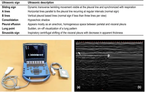
Table 1: Description of common chest ultrasonic signs
Ultrasonic sign Ultrasonic description
Sliding sign Dynamic transverse twinkling movement visible at the pleural line and synchronized with respiration
A lines Horizontal lines parallel to the pleural line recurring at regular intervals (normal sign)
B lines Vertical pleural based lines (normal sign if less than three lines per view)
Consolidation Hypoechoic shadow
Pleural effusion Appears mostly as an anechoic, homogeneous space between parietal and visceral pleura
Lung point Sudden, on–off visualization of a lung pattern
Sinusoids sign Inspiratory centrifugal shifting of the visceral pleura with decrease in apparent thickness
Figure1: Ultra Sound Machine Figure 2: Lung Point In M-Mode
RESULTS
This study included 111 patients with acute respiratory failure. 10 patients were excluded of the analysis because they had rare/uncertain diagnosis. Out of 101 patients, 64 (63.4%) were
Table 2: Distribution of the sample according to age
Age Count Percent
<=50 21 20.8
51 – 60 23 22.8
61 – 70 29 28.7
>70 28 27.7
Mean ± SD 62 ± 12.9
Table 3: Distribution of the sample according to sex
Sex Count Percent
Male 64 63.4
Female 37 36.6
According to history, clinical examination and investigations (not including ultrasound examination), the final etiological diagnosis of acute respiratory failure in the 111 studied patients was: pneumonia 22 cases (CAP 15 cases and HAP 5 cases, aspiration pneumonia 2 cases), acute exacerbation of chronic obstructive pulmonary disease (AECOPD) 21 cases, pulmonary edema 21 cases, pulmonary embolism (PE) 6 cases, massive effusion/empyema 5 cases, bronchial asthma (BA) 12 cases, pneumothorax 19 cases, idiopathic pulmonary fibrosis (IPF) 3 cases and no/multiple diagnosis in 2 cases. (Table 4)
US Examination
To facilitate the comparison of the ultrasound data to the final etiological diagnosis obtained by the conventional diagnostic tools in the ICU, the characteristic ultrasound signs were collected together and interpreted as different ultrasound profiles according to the previously designated nomenclatures.9,13 The A profile
designates anterior predominant bilateral A lines associated with
lung sliding (with possible focalized B lines).The A’ profile is an A
profile with abolished lung sliding and without lung point. The B profile designates anterior predominant bilateral B lines associated with lung sliding (with possible focalized A lines). The
B’ profile is a B profile with abolished lung sliding. The A/B profile designates anterior predominant B lines on one side and predominant A lines on the other. The C profile designates anterior alveolar consolidation(s). PLAPS represents posterior and/or lateral alveolar and/or pleural syndrome. All these definitions are based on the patient being supine or semi recumbent. The normal profile associates the A profile without PLAPS (regardless of posterior A or B lines).
The ultrasound profiles of different final underlying etiologies of ARF: (Table 5)
Pneumonia was presented by: A (normal) profile in 9% of patients, AB profile in 23% of patients, A+ PLAPS profile in 23% of patients
,B’ profile in 14% and C profile in 32% of patients.
Acute exacerbation of chronic obstructive pulmonary disease (AECOPD) was presented by: A (normal) profile in 66.6% of
patients, A’ profile in 19.1%, B profile + PLAPS in 9.5% and A+
PLAPS profile in 4.7% of patients. Pulmonary edema was presented by: A (normal) profile in 9.5% of patients, and B + PLAPS profile in 90.5% of patients.
Pulmonary embolism (PE) was presented by: A (normal) profile in 33.3%, C profile in 17% of patients and A+ PLAPS + DVT profile in 50% of patients.
Bronchial asthma (BA) was presented by: A (normal) profile in 91.7% of patients and A +PLAPS profile in 8.3% of patients.
Pneumothorax was presented by: A ’profile in 31.6% of patients
and lung point profile in 68.4% of patients.
Rare diagnosis, unknown diagnosis and multiple diagnosis cases were omitted.
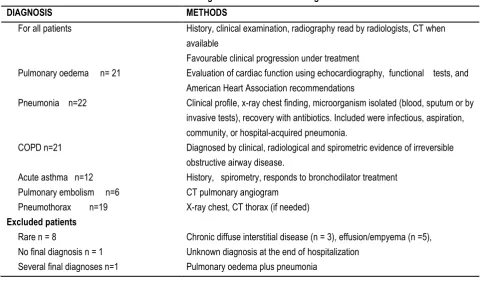
Table 4: Final Diagnoses and Methods of Diagnosis
DIAGNOSIS METHODS
For all patients History, clinical examination, radiography read by radiologists, CT when available Favourable clinical progression under treatment
Pulmonary oedema n= 21 Evaluation of cardiac function using echocardiography, functional tests, and American Heart Association recommendations
Pneumonia n=22 Clinical profile, x-ray chest finding, microorganism isolated (blood, sputum or by invasive tests), recovery with antibiotics. Included were infectious, aspiration, community, or hospital-acquired pneumonia.
COPD n=21 Diagnosed by clinical, radiological and spirometric evidence of irreversible obstructive airway disease.
Acute asthma n=12 History, spirometry, responds to bronchodilator treatment Pulmonary embolism n=6 CT pulmonary angiogram
Pneumothorax n=19 X-ray chest, CT thorax (if needed) Excluded patients
Rare n = 8 Chronic diffuse interstitial disease (n = 3), effusion/empyema (n =5), No final diagnosis n = 1 Unknown diagnosis at the end of hospitalization
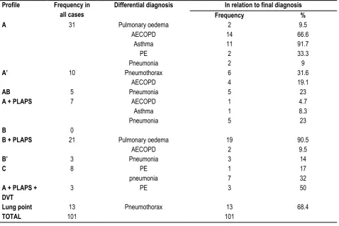
Table 5: Ultrasound profile in relation to final etiological diagnosis
Profile Frequency in
all cases
Differential diagnosis In relation to final diagnosis
Frequency %
A 31 Pulmonary oedema 2 9.5
AECOPD 14 66.6
Asthma 11 91.7
PE 2 33.3
Pneumonia 2 9
A’ 10 Pneumothorax 6 31.6
AECOPD 4 19.1
AB 5 Pneumonia 5 23
A + PLAPS 7 AECOPD 1 4.7
Asthma 1 8.3
Pneumonia 5 23
B 0
B + PLAPS 21 Pulmonary oedema 19 90.5
AECOPD 2 9.5
B’ 3 Pneumonia 3 14
C 8 PE 1 17
pneumonia 7 32
A + PLAPS + DVT
3 PE 3 50
Lung point 13 Pneumothorax 13 68.4
TOTAL 101 101
Table 6: Accuracy of USG profile
Final diagnosis USG Profile Sensitivity
(%)
Specificity (%)
Positive Predictive value (%)
Negative Predictive value (%)
Accuracy (%)
COPD + asthma A Profile 75.76 91.2 80.7 88.6 86.1
Pneumonia A/B Profile 22.7 100 100 82.3 83.2
C Profile 31.8 98.7 87.5 83.9 84.2
Pneumo thorax Lung point
Profile
.68.4 100 100 93.2 94.1
A’ profile 31.6 95.1 60 85.7 77.2
Pulmonary embolism
A Profile with Plaps +
DVT
50 96.9 50.0 96.9 97.0
Pulmonary edema
B Profile with Plaps
90.5 97.5 90.5 97.5 96.0
A profile 9.5 63.75 6.5 72.86 52.5
Accuracy of ultrasound diagnosis
As some profiles were presented in more than one aetiology, the sensitivity, specificity, positive predictive value and negative predictive value were calculated to determine the accuracy of each profile to diagnose each aetiology of acute respiratory failure.
Characteristic profiles that produced specificities >90% for each of the final etiologic diagnosis are presented in Table 6.
DISCUSSION
The key inference from our study was the significant diagnostic accuracy of applying chest ultrasound in comparison to the standard approach that included clinical, radiologic and biologic data. Moreover, the greater diagnostic ability of the ultrasound
approach was demonstrated when each main etiologic entity of acute respiratory failure was independently analysed.
The main diagnostic profiles were as follows:
The AB profile (asymmetric anterior interstitial syndrome) and the C profile (anterior consolidation) indicated pneumonia. Since pneumonia has numerous causes, several pathologic and radiologic presentations, and can be found in a wide variety of locations, it generates, several profiles, mainly asymmetry (from left to right, explaining the A/B profile, from posterior to anterior, explaining the A-profile plus PLAPS), and anterior consolidations (C-profile).9,14 The AB and the C profiles were the profiles
COPD and asthma are bronchial diseases assumed to yield a normal lung surface. They are diagnosed mainly by their clinical presentations and are not usually accompanied by significant radiologic presentations that can be echoed by ultrasound, explaining the predominance of the A profile (i.e., normal lung surface) in these patients.9 Our study showed less sensitivity and
specificity compared to the Lichtenstein et al’s study.
Alveolar interstitial syndrome (AIS) constitutes a group of diseases that is caused by an increase in lung fluid and/or a reduction in its air content. The major causes of AIS are pulmonary oedema, interstitial pneumonia and pulmonary fibrosis. The sonographic appearance of AIS is vertical artefacts called B lines. When compared with CT scan images, B-lines correspond to oedema of the interlobular septa. When diffuse B-lines are visualized, this gives the B profile and suggests a diagnosis of AIS. The causes of diffuse AIS presented to the ICUs in our study were pulmonary oedema and interstitial pulmonary fibrosis. The B profile (with or without PLAPS due to gravitational filling of dependent alveoli) characterizes pulmonary oedema with high accuracy of 96%.16,17
Lung ultrasound for the diagnosis of pulmonary embolism (PE) is a relatively new concept. There are several limitations of lung ultrasound to diagnose PE. Only two-thirds of the lung area is accessible to ultrasound examination because the remainder is covered by bony structures. However, almost 80% of lesions are located in the lower lobes, which are accessible to examination.18
Only thromboembolic lesions extending to the pleura can be detected. However, it has been demonstrated that central and peripheral lesions occur concurrently in 80% of cases.19
Furthermore, the criteria used to diagnose PE are not standard. However, Lichtenstein proposed simplified criteria to diagnose PE. According to his Bedside Lung Ultrasound in Emergency (BLUE)
protocol, in the severely dyspnoeic patient not in shock, a normal lung ultrasound (presence of A-lines throughout) plus signs of deep vein thrombosis (DVT) is 81% sensitive and 99% specific for PE.9 Similar results were found in our study where, in pulmonary
embolism where A-profile associated with deep vein thrombosis presented a sensitivity of 50% and specificity of 96%, suggesting that the search for venous thrombosis should be associated with the lung analysis.
In pneumothorax, we found that, absent lung sliding (A’ profile)
and the lung point, had a specificity of 95% and 100% with a sensitivity of 31% and 68%, respectively. The confirmation of lung sliding, which is the back-and forth movement of the bright echogenic parietal and visceral pleura occurring during respiration, has a 100% negative predictive value for the absence of pneumothorax.20 It is important to recognize that, although the
presence of lung sliding effectively rules out pneumothorax, its absence does not necessarily rule it in pneumothorax. The appearance of a single B-line confirms the opposition of both pleural surfaces and effectively rules out a pneumothorax. Therefore, the absence of a single B line can safely rule in a pneumothorax.20
The lung point refers to the point on the chest wall where the visceral pleura has been separated from the parietal pleura and, therefore, defines where the pneumothorax begins. Visualization of the lung point is 100% specific for the diagnosis of pneumothorax.21
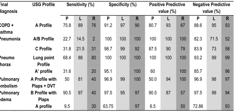
Our study has got 94% accuracy for pneumothorax, 97% accuracy for pulmonary embolism, 96.0% for pulmonary oedema, 86% for asthma, and COPD. Table 7 Demonstrates comparison of our study with the original study done by Lichtenstein et al. and Rasha Daabis et al.9,15
Table 7: Comparison with Lichtenstein et al. and Rasha Daabis et al. study Final
diagnosis
USG Profile Sensitivity (%) Specificity (%) Positive Predictive
value (%)
Negative Predictive value (%)
P L R P L R P L R P L R
COPD + asthma
A Profile 75.8 89 76 91.2 97 90 80.7 93 67 88.6 95 93
Pneumonia A/B Profile 22.7 14.5 2 100 100 100 100 100 100 82.3 71.5 52
C Profile 31.8 21.5 31 98.7 99 92 87.5 90 79 83.9 73 58
Pneumo thorax
Lung point Profile
68.4 88 80 100 100 100 100 100 100 93.2 99 99
A’ profile 31.6 20 95.1 100 60 100 85.7 96
Pulmonary embolism
A Profile with Plaps + DVT
50 81 40 96.9 99 100 50.0 94 100 96.9 98 97
Pulmonary edema
B Profile with Plaps
90.5 97 40 97.5 95 97 90.5 87 57 97.5 99 94
A profile 9.5 30 63.75 97 6.5 50 72.86 93
P= Present study, L= Lichtenstein et al., R= Rasha Daabis et al.
Acute respiratory failure is one of the most distressing situations for the patient. The ultrasound approach with the profiles described by Lichtenstein et al can be used in such patients, as it was corresponding to the underlying physiopathology in most of the cases, and resulted in a high diagnostic accuracy, which can be further increased when combined with simple clinical data.
CONCLUSION
most disorders. It can be repeated at will and generates standardized, reproducible patterns with high feasibility.
ACKNOWLEDGEMENT
Authors would like to thank Dr. Vimal Rohan, Emergency medicine physician, Pariyaram Medical College and Mr. Ommenn, statistician.
LIMITATIONS Single centered study.
Few final diagnoses were subsequently excluded from this study.
ETHICAL APPROVAL
The study was approved by the Institutional Ethics Committee.
REFERENCES
1. J.L. Vincent, S. Akc. a, A. De Mendonc¸ a, et al, SOFA Working Group. The epidemiology of acute respiratory failure in critically ill patients, Chest 2002; 121 (5); 1602–1609.
2. O.R. Luhr, K. Antonsen, M. Karlsson, et al, The ARF Study Group. Incidence and mortality after acute respiratory failure and acute respiratory distress syndrome in Sweden, Denmark, and Iceland, Am. J. Respir. Crit. Care Med.1999; 159 (6); 1849–61. 3. B. Afessa, I.J. Morales, P.D. Scanlon, S.G. Peters, Prognostic factors, clinical course, and hospital outcome of patients with chronic obstructive pulmonary disease admitted to an intensive care unit for acute respiratory failure, Crit. Care Med.2002; 30 (7); 1610-1615.
4. J.R. Zahar, E. Azoulay, E. Klement, et al, Delayed treatment contributes to mortality in ICU patients with severe active pulmonary tuberculosis and acute respiratory failure, Intens.Care Med.2001; 27 (3); 513–520
5. B. Bouhemad, M. Zhang, Lu.Q. Qin, J.J. Rouby, Clinical review: bedside lung ultrasound in critical care practice, Crit.Care. 2007; 11; 205–213.
6. Beckmann U, Gillies DM, Berenholtz SM, Wu AW, Pronovost P: Incidents relating to the intra-hospital transfer of critically ill patients. An analysis of the reports submitted to the Australian Incident Monitoring Study in Intensive Care. Intensive Care Med 2004, 30:1579-15856.
7. Mayo JR, Aldrich J, Muller NL. Radiation exposure at chest CT: a statement of the Fleischner Society. Radiology 2003, 228:15-21. 8. Lichtenstein D, Biderman P, Meziere G, Gepner A: The
“sinusogram”, a real-time ultrasound sign of maxillary sinusitis. Intensive Care Med; 1998, 24:1057-1061.]
9. Lichtenstein DA, Mezière GA. Relevance of lung ultrasound in the diagnosis of acute respiratory failure: The BLUE protocol. Chest 2008; 134:117 25
10. M. Zanobetti, C. Poggioni, R. Pini, Can chest ultrasonography
replace standard chest radiography for evaluation of acute dyspnea in the ed? Chest. 2011; 139 (5); 1140–1147.
11. M. Ata, S. Sat, Respiratory failure, Am. J. Respir. Crit. Care. 2009; 179 (3); 220–227.
12. Seth J. Koenig, Mangala Narasimhan, Paul H. Mayo, Thoracic ultrasonography for the pulmonary specialist, Chest. 2011; 140; 5. 13. D.A. Lichtenstein, Lung ultrasound in the critically ill, Neth. J.Crit. Care; 2012; 16 (2); 43–51.
14. J.P. Turner, J. Dankoff, Thoracic ultrasound, Emerg. Med.Clin. N. Am.2012; 30; 451–473.
15. Rasha Daabis, Laila Banawan, Abdelmoneim Rabea, Abdelaziz Elnakedy, Ayman Sadek. Relevance of chest sonography in the diagnosis of acute respiratory failure: Comparison with current diagnostic tools in intensive care units Egyptian Journal of Chest Diseases and Tuberculosis. 2014; 63, 979–985.
16. L. Cardinale, G. Volpicelli, F. Binello, et al, Clinical application of lung ultrasound in patients with acute dyspnoea: differential diagnosis between cardiogenic and pulmonary causes, Radiol.Med. 2009; 114 (7); 1053–1064.
17. A. Reissig, C. Kroegel, Transthoracic sonography of diffuse parenchymal lung disease: the role of comet tail artefacts, J. Ultrasound Med. 2003; 22; 173–180.
18. A. Reissig, Sonography of lung and pleura in pulmonary embolism: sonomorphologic characterization and comparison with spiral CT scanning, Chest; 2001; 120 (6); 1977–1983.
19. D.A. Lichtenstein, Lung sonography in pulmonary embolism, Chest. 2003; 123 (6); 2154–2155.
20. D. Lichtenstein, G. Mezie´ re, P. Biderman, et al, The comet-tail artifact: an ultrasound sign ruling out pneumothorax, Intens. Care Med. 1999; 25 (4); 383–388.
21. D. Lichtenstein, G. Mezie´ re, P. Biderman, et al, The ‘‘lung point’’: an ultrasound sign specific to pneumothorax, Intens. Care
Med. 2000; 26 (10); 1434–1440.
[
Source of Support: Nil. Conflict of Interest: None Declared.
Copyright: © the author(s) and publisher. IJMRP is an official publication of Ibn Sina Academy of Medieval Medicine & Sciences, registered in 2001 under Indian Trusts Act, 1882. This is an open access article distributed under the terms of the Creative Commons Attribution Non-commercial License, which permits unrestricted non-commercial use, distribution, and reproduction in any medium, provided the original work is properly cited.