- 14 -
Predicting the Impact of PHEX, FGF23 and DMP1 Gene Variants
Found in Malaysian Malay Patients with Hypophosphataemic Rickets
Through In Silico Analysis of Protein Function and mRNA Secondary
Structure
Nurul Nadirah Razali
1, Tzer Hwu Ting
2, King Hwa Ling
1,3, Marina L. Kennerson
4,5,6,
Karuppiah Thilakavathy
1,3*
1Department of Biomedical Science, Faculty of Medicine & Health Sciences, Universiti Putra Malaysia,
43400 UPM Serdang, Selangor, Malaysia.
2Department of Paediatrics, Faculty of Medicine & Health Sciences, Universiti Putra Malaysia,
43400 UPM Serdang, Selangor, Malaysia.
3Genetics and Regenerative Medicine Research Center, Faculty of Medicine & Health Sciences, Universiti Putra Malaysia,
43400 UPM Serdang, Selangor, Malaysia.
4Northcott Neuroscience Laboratory, ANZAC Research Institute,
University of Sydney Concord NSW 2139, Australia.
5Sydney Medical School, University of Sydney, Sydney, Australia. 6Molecular Medicine Laboratory, Concord Hospital, Concord, Australia.
*Corresponding author:
Associate Professor Dr. Thilakavathy Karuppiah, Department of Biomedical Sciences, Faculty of Medicine and Health Sciences,
Universiti Putra Malaysia, 43400 UPM Serdang,
Selangor, Malaysia.
Email: [email protected]
INTRODUCTION
Hypophosphataemic rickets (HR) is a subtype of rickets due to hypophosphataemia or low serum phosphate level. The low serum phosphate in the body is related to the low reabsorption and high
excretion of phosphate by renal tubules [1, 2]. Parathyroid hormone (PTH), fibroblast growth factor 23 (FGF23) and 1,25 dihydroxyvitamin D [1, 25(OH)2D] are the major regulators for phosphate homeostasis in the human body [3, 4].
HISTORY
Received: 22nd November 2019
Received in revised form: 19th December 2019 Accepted: 19th December 2019
ABSTRACT
Hypophosphataemic Rickets (HR) is a rare bone disorder characterised by chronic hypophosphataemia caused by defective phosphate reabsorption in the renal tubules. Variants in phosphate-regulating endopeptidase homolog, X-linked (PHEX), fibroblast growth factor-23 (FGF23) and dentin matrix protein-1 (DMP1) genes contribute to X-linked dominant, autosomal dominant and autosomal recessive forms of HR, respectively. In this study, four Malaysian patients’ DNA samples were subjected to polymerase chain reaction and Sanger sequencing to identify the types and locations of the variants. Then, in silico study was conducted based on the variants found to predict the effects of amino acid substitution on protein functions using SIFT and PolyPhen-2 software and RNAfold was used to construct the mRNA secondary structure. Mutational analyses had revealed two variants in PHEX; c.10G>C (E4Q), c.1970A>G (Y657C), one mutation in FGF23; c.716C>T (T239M) and three variants on DMP1; c.309A>T (S69C), c.1322C>T (S406S), c.1334G>A (E410E). The variants in these Malay patients were previously reported in different ethnic HR patients. Protein prediction programs suggested that the PHEX Y657C and DMP1 S69C variants may affect protein function. All variants were predicted to alter the secondary mRNA structure. These findings suggest that these missense and silent variants may lead to changes in protein function and mRNA secondary structure that are associated with the manifestation of HR phenotype.
KEYWORDS
Hypophosphataemic Rickets PHEX
FGF23 DMP1 in silico analysis
JOURNAL OF BIOCHEMISTRY, MICROBIOLOGY
AND BIOTECHNOLOGY
Website: https://journal.hibiscuspublisher.com/index.php/JOBIMB
JOBIMB VOL 7 NO 2 2019
- 15 - Familial HR is a rare disease with a low rate of occurrence; 1 in 20, 000 live births worldwide [5]. Numerous studies have been carried out on HR in North American, European, African American, Saudi Arabian and subcontinent Indian, however, in Malaysia, previously only one study has been reported on the pattern of inheritance of a family with two HR individuals [6].
Individuals with familial HR tend to have growth retardation, short stature, lower limb deformities, dental abscesses, early tooth loss, and bone pain [5,7,8]. X-linked hypophosphataemia (XLH, MIM307800), autosomal dominant HR (ADHR, MIM193100) and autosomal recessive HR 1 (ARHR, MIM241520) are the hereditary forms of HR that are mainly caused by mutations in PHEX, FGF23
and DMP1, respectively. PHEX accounts for the most in HR disorder, approximately 80% in all reported cases of familial HR, followed by FGF23 and DMP1 [9]. To date, there are more than 300 mutations reported on PHEX [10].
The phosphate regulating endopeptidase homolog, X-linked (PHEX), fibroblast growth factor 23 (FGF23) and dentin matrix protein-1 (DMP1) genes are known to be mutated in familial HR.
PHEX helps in bone formation as it encodes a protein expressed in osteoblasts, osteocytes, and odontoblasts [11, 12]. DMP1 shares the same functions as PHEX, where it encodes for a protein that is highly expressed in the osteoblasts and osteocytes [1]. It is responsible for bone and dentin mineralisation [13] and plays a role in downregulating FGF23 [2]. Whilst FGF23 has a role in bone formation and mineralisation [14], the overexpression of FGF23 causes down regulation of membrane expression of sodium-phosphate co-transporter in the kidney and suppresses 1,25 (OH)2D
production that subsequently interferes the process of phosphate reabsorption in the gut [3]. Gain or loss of function mutations in these genes can lead to a decrease in blood phosphate levels via elevation of FGF23, thereby contributing to the hypophosphataemia [15]. FGF23 levels will increase due to loss of function mutations in PHEX [16] and DMP1 [2] and gain of function mutations in FGF23 [14].
It is interesting to note that all the patients diagnosed with HR in several hospitals in Malaysia were of Malay ethnicity. Finding the gene mutations could give a better understanding of the disease pathogenesis in this population. We have screened the PHEX,
FGF23 and DMP1 genes in HR patients of Malay ethnicity and identified six known variants. In silico analyses predicted the effect of the particular PHEX, DMP1 and FGF23 variants on protein functions and mRNA secondary structures.
METHODS Patients
Children and adolescents aged less than 18 years diagnosed with hypophosphataemic rickets were recruited for this study. Records of clinical features and laboratory parameters of each patient since the diagnosis of HR were obtained from the clinic medical records. Criteria for inclusion in the study were manifestation of HR clinical features (short stature and bone deformities), reduced serum phosphate levels, raised alkaline phosphatase, normal serum calcium levels and reduced maximum rate of renal tubular reabsorption of phosphate normalised to the glomerular filtration rate (TmP/GFR) [17]. Patients with raised serum creatinine and urinary calcium levels, urine glycosuria and metabolic acidosis on venous blood gas were excluded to eliminate the possibility of renal failure, Fanconi syndrome or other renal tubulopathies.
Informed consent was obtained from the parents or guardians of the patients prior to sample collection in accordance with the approved Ethics Committee guidelines; Universiti Putra Malaysia..Ethics..Committee..(UPM/TNCPI/RMC/1.4.18.1(JKE
UPM)/F2) and Ministry of Health Medical Research and Ethics
Committee (NMRR-13-609-16031).
DNA analysis
Genomic DNA was extracted from the buffy coat of patient blood samples using the QIAamp® DNA Blood Mini Kit (Qiagen, USA) according to the protocol recommended by the manufacturer. Exons and exon-intron boundaries were amplified with sets of previously described primer pairs specific to PHEX, FGF23 and
DMP1 [18, 19].
PCR reactions were performed in 20 µL reactions, containing 1X Colorless GoTaq® Reaction Buffer, 0.8-1.5 mM MgCl2
solution, 0.2 mM dNTP Mix, 2.5 U GoTaq® DNA Polymerase (Promega, USA), nuclease free water (Vivantis, Inc. USA), 0.3 µM primers and 100ng of template DNA. Thermal cycling was initiated with 10 min at 95oC, followed by 40 cycles of 95oC (45 s),
annealing temperature (45 s) and extension at 72oC (45 s), with a
final extension step at 72oC for 10 min using a PCR Mastercycler
Gradient machine (Eppendorf, Germany). PCR amplicons were size fractionated on 2% (w/v) agarose gels in 1X TAE buffer. PCR product purification was performed using the MEGAquick-spinTM
Total – Fragment DNA purification kit (Intron Biotechnology, Inc., Korea). The purified PCR products were sent for direct sequencing service (First Base Laboratories, Singapore). Sequencing traces were viewed and analysed using DNA Baser software (v4.16.0).
In silico analysis
All missense variants were analysed by using in silico study; SIFT..(http://sift.jcvi.org/)..and..PolyPhen..(http://genetics.bwh.ha rvard.edu/pph2/) for prediction of the effects of amino acids substitution on the protein functions. RNAfold (http://rna.tbi.univie.ac.at/cgi-bin/RNAfold.cgi) software was used to predict the secondary structure of mRNA. The predicted function by SIFT and PolyPhen-2 were interpreted following the parameters of the program. The range of SIFT and PolyPhen-2 scores is from 0 to 1, to categorize tolerated or deleterious. For SIFT, score < 0.05 is considered as deleterious and score > 0.05 is tolerated. PolyPhen-2 scores benign from 0 to 0.5, possibly damaging from > 0.5 to 0.9 and probably damaging from > 0.9 to 1.0 [20, 21].
RESULTS
Clinical findings on hypophosphataemic rickets patients
- 16 -
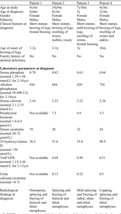
Table 1. Clinical data at diagnosis of hypophosphataemic rickets patients.
Patient 1 Patient 2 Patient 3 Patient 4 Age at study 9y3m 14y9m 7y10m 4y5m Age at diagnosis 6y6m 6y1m 3y 3y3m Gender Female Female Female Female Ethnicity Malay Malay Malay Malay Clinical features at
diagnosis Short stature, bowing of legs, frontal bossing
Short stature, bowing of legs, swelling of wrists, rachitic rosary
Short stature, mild bowing of legs, swelling of wrists, frontal bossing
Short stature, bowing of legs, swelling of wrists and ankles
Age of onset of
bowing of legs 1-2y 2-3y 2y 18m Family history of
skeletal deformity No No No No
Laboratory parameters at diagnosis
Serum phosphate (normal 1.29-1.94 mmol/L for 2-18yo)
0.78 0.82 0.63 0.64
Alkaline phosphatase (normal 58-400 U/L for 2-18yo)
436 644 620 726
Serum calcium (normal 2.1-2.55 mmol/L)
2.16 2.35 2.22 2.34
Parathyroid hormone (normal 1.6-6.9 pmol/L)
Not available 7.5 4.9 3.3
Serum creatinine (normal 20-72 µmol/L)
39 28 32 24
25-hydroxyvitamin D
(normal >50 nmol/L)
56.5 51.6 55.0 90.9
TmP/GFR (normal 1.15-2.44 mmol/L for 2-15yo)
Not available 0.68 0.49 0.51
Urine
calcium/creatinine (normal <0.7)
Not available 0.15 0.22 0.5
Radiological findings at diagnosis Osteopenia, splaying and fraying of femoral and tibial metaphyses Splaying and fraying of femoral and tibial metaphyses Mild splaying and fraying of radial, ulnar and tibial metaphyses Cupping, splaying and fraying of tibial metaphyses
Note: TmP/GFR - maximum rate of renal tubular reabsorption of phosphate normalised to the glomerular filtration rate
Variants screening on PHEX, FGF23 and DMP1 genes in
hypophosphataemic rickets patients
Based on the DNA sequencing screening, total six variants have been found in this study (Table 2). Two variants were identified in Patient 1; c.716C>T in FGF23 exon 3 and c.309A>T in DMP1 exon 6. Meanwhile, Patient 2 and Patient 3 have three variants in all three studied genes; P02 has variants of c. 10G>C in PHEX exon 1, c.716C>T in FGF23 exon 3 and c.309A>T in DMP1 exon 6, whereas P03 has variants of c.1970A>G in PHEX exon 20, c.716C>T in FGF23
exon 3 and c.309A>T in DMP1 exon 6. As for P04, variants were only found in exon 6 of DMP1; c.1322C>T and c.1334G>A.
Table 2. Description of variants on PHEX, FGF23 and DMP1 discovered in four Malaysian hypophosphataemic rickets patients.
Patient 1 Patient 2 Patient 3 Patient 4 Gene FGF23 DMP1 PHEX FGF23 DMP1 PHEX FGF23 DMP1 DMP1 DMP1 Exon 3 6 1 3 6 20 3 6 6 6 Location c.716C>T c.309A>T c.10G>C c.716C>T c.309A>T c.1970A>G c.716C>T c.309A>T c.1322C>T c.1334G>A
Amino acid change
T239M S69C E4Q T239M S69C Y657C T239M S69C S406S E410E
Mutation
type m m m m m m m m s s Note: m missense, s silent
Exon 1 of PHEX is 118 bp in size, and at location of c.10, the heterozygous substitution of G to C (rs147859619) lead to amino acid changes, from glutamic acid to glutamine at codon 4 (E4Q). Whereas exon 20 of PHEX has 105 bp, and a substitution of A to G at location c.1970 causing the tyrosine to be replaced by cysteine at codon 657 (Y657C). Exon 3 of FGF23 comprises of 441 bp and C>T at location c.716 (rs7955866) causes amino acid changes from threonine to methionine at codon 239 (T239M). Exon 6 is the longest exon of DMP1 with 1360 bp from A>T at location c.309 (rs10019009) alters the amino acid from serine to cysteine at codon 69 (S69C). Meanwhile from C>T at c.1322 (rs2615498) and G>A at c.1334 (rs2615497) led to no changes of amino acid serine at codon 406 and glutamic acid at codon 410, respectively.
In silico analysis on protein function based on amino acid substitutions by using SIFT and PolyPhen-2 software
Analyses in predicting the impact of amino acid substitutions on protein function were conducted via prediction tools; SIFT and PolyPhen-2 (Table 3). SIFT studies the type of amino acid changes and its position by aligning the related protein sequence with the query. Ng & Henikoff [22] explained the multistep procedure of SIFT which begins with searching for similar sequences, and then the closely related sequences that share similar function will be selected and aligned. From the alignment, the normalised probabilities of all possible substitutions occurring at each position of the alignment will be calculated. From the calculation, if the probability is less than a chosen cutoff, it is predicted as deleterious, while it will become tolerated if the value is greater or equal to the cutoff. PolyPhen-2 operates by using eight sequence-based and three structure-based predictive features which have been chosen automatically by an iterative greedy algorithm [23]. Position-Specific Independent Counts (PSIC) score is then calculated based on the difference between wild type (ancestral, normal) and mutant (derived, disease causing) amino acid, hence classifying the results into benign, possibly damaging, probably damaging [24].
Table 3. Prediction of SIFT and PolyPhen-2 on the impact of missense nucleotide substitutions on protein function.
Gene Amino acid changes SIFT SIFT score PolyPhen-2 PolyPhen-2 score
PHEX E4Q Y657C Tolerated 0.29 Damaging 0.01 Probably damaging 0.981 Probably damaging 1.000
FGF23 T239M Tolerated 0.29 Benign 0.001
DMP1 S69C Damaging 0.02 Probably damaging 1.000 Based on the computational analysis, both software predicted with high confidence that the PHEX Y657C and DMP1 S69C variants affect protein function or structure (SIFT score <0.05, PolyPhen-2 score 1.0), whilst FGF23 T239M variants was predicted as not affecting the protein function (SIFT score >0.05, PolyPhen-2 score 0.001). PHEX E4Q has mixed prediction of tolerated by SIFT (score >0.05) and probably damaging by PolyPhen-2 (score 0.981).
Determination of mRNA secondary structure by using RNAfold
- 17 -
- 18 - The c.10G>C variant caused increase in the loop structure size compared to the wild type structure (Fig.1D and E). Similarly, a change in structure was observed between wild type and the c.1970A>G variant. The substitution predicted the formation of different size of loop structures on different locations of the mRNA branch (Fig. 1F and 1G). Fig. 2 shows the mRNA secondary structure prediction of the wild type and FGF23 variant. Obvious changes in the structure of FGF23 were seen in Fig.2B, where it had increased number of branches compared to the wild type structure (Fig.2A). The MFE value for wild type FGF23 was -750.21 kcal/mol which was higher than MFE for variant -750.74 kcal/mol. Fig.2C and 2D show the location of the substitution of c.716C>T (U). c.716C was originally located near a loop structure, however, due to the nucleotide change, the mutated position was shifted onto a hairpin structure. Additional branches and loops existed in the variant mRNA structure too.
The mRNA secondary structure of wild type and variant DMP1 are shown in Fig.3A, 3B and 3C. At a glance, structure of variant c.309A>T (U) and wild type DMP1 appear to be similar, nevertheless, the MFE values for both structures were different; wild type had higher value (-464.30 kcal/mol) than variant (-467.40 kcal/mol). Meanwhile mRNA secondary structure of variant c.1322C>T (U) and c.1334A>G variant exhibited obvious changes on the right-side branch and recorded a higher MFE (-463.90 kcal/mol) when compared with the wild type structure. The dissimilarity between wild type and variant c.309A>T (U) structures was not noticeable. However, a close-up on the loop near the end of the mutated branch showed that the size of the loop in the altered structure was smaller compared to that in the wild type (Fig.3D and 3E).
- 19 -
- 20 - The presence of double variants c.1322C>T (U) and c.1334A>G in
DMP1 changed the secondary structure of the mRNA extensively (Fig. 3F and 3G). Separate analysis on the mRNA secondary structure for each variant of the dual variants was carried out and it showed that the DMP1 structure was greatly impacted by the presence of one of the variants, c.1322C>T (U) (Fig.4A), with MFE value of -463.90 kcal/mol.
DISCUSSION
According to Li et al., [27], the PHEX mutation database shows different frequencies of mutation types; 25% frameshifts, 23% alternative splicing, 22% missense, 18% nonsense, 8% deletion and 4% polymorphisms. Although mutations in PHEX are the most common in HR cases, in this study, not all patients had a PHEX
variant. Patient 1 has only variants on FGF23 and DMP1, while Patient 4 carried DMP1. On the other hand, Patient 2 and Patient 3 recorded variants in all three studied genes. Nevertheless, HR is a single gene disorder [28], hence a single altered gene is enough to cause the disease. The severity of the disease is not associated with the location of variant and gender as no correlation has been found between these two factors and severity of HR in previous studies [29, 30].
In this study, the missense substitution found on PHEX
(c.10G>C) has been reported in a previous study among Chinese patients [31]. This substitution causes E4Q amino acid change and was predicted to be non-pathogenic. However, the study by St-louis [32] revealed that E4Q was associated with bone mineralisation. This substitution is believed in obstructing the interaction of PHEX to COPII; which is responsible for training secretory vesicles. This interference subsequently affecting the incorporation of PHEX into calcification vesicles that would be important in mineralisation process. Thus, although E4Q was predicted with no change in protein function, the phenotype may result from transformed mRNA secondary structure and altered translation efficiency. The prediction tools indicated c.1970A>G substitution in PHEX leading to Y657C alteration as damaging. There were various additions of loops with different sizes and hairpin on the right-side branch that made the top structure of the variant appeared completely different from the wild type. Fig.4B
showed the variant mRNA secondary structure with the presence of c.1334A>G. This variant did not seem to change the mRNA structure. However, its MFE value differ from the wild type; 463.20 kcal/mol.
This variant was also reported to occur sporadically in European HR patients [33]. It was speculated that this substitution occurred in the conserved region, consequently giving an impact on protein functions. SIFT and PolyPhen-2 prediction on this variant concur with the speculation.
PHEX is a zinc metalloendopeptidase and the protein has high specificity of binding with matrix extracellular glycoprotein (MEPE), a protein involved in biomineralisation. PHEX mutations may fail to interrupt the process of intermediate cleavage of MEPE that will generate free protease-resistant acidic serine aspartate rich MEPE associated (ASARM) peptides. ASARM peptide is a mineralisation inhibitor in bone and teeth, and causes mineralisation defects and inhibits renal and intestinal phosphate uptake [34]. Thus, interruption in phosphate regulation results in higher phosphate excretion and reach hypophosphataemia state.
Variant at c.716C>T in FGF23 was also found in hypophosphataemic stone former due to renal phosphate leak [35]. Despite being predicted as benign and tolerable, previous in vitro
studies by Rendina et al., [35] indicated that T239M modification influenced FGF23 biological function. This was supported by Merlotti et al., [36] whose study showed that T239M enhanced FGF23 protein secretion. Mutated FGF23 causes prevention of proteolytic degradation and promotes FGF23production, increased FGF23 results in renal phosphate loss leading to hypophosphataemia [14].
The three different variants in exon 6 of DMP1 in our study were also reported by Ruppe et al., [8] in American HR patients. Missense substitution c.309A>T (S69C) was foretold by the prediction tools used in this study that it might be likely damaging the protein function. Turan et al., [37] in their study stated that
- 21 - S69C was associated with bone mineralisation and concluded the importance of DMP1 in the pathogenesis of autosomal recessive HR and in the normal regulation of phosphate homeostasis. DMP1
is a member of small integrin-binding ligand N-linked glycoprotein (SIBLING). ASARM is one of the features that are highly conserved in SIBLING protein. Defective DMP1 may enhance ASARM peptides in bone and teeth that lead to hypophosphataemia state [34].
RNAfold was used to predict the mRNA secondary structure and calculate the MFE values of wild type and altered PHEX, FGF23 and DMP1. As stated by de Smit & Van Duin [38] mRNA secondary structure is a key factor in determining the translation efficiency. Thus, studying the mRNA secondary structure widens the scope of analyzing the impact of all types of substitution variants. Stability is an important factor in mRNA secondary structure, as stable complexes improve translation efficiency [39]. MFE is provided on all structures constructed by RNAfold.
MFE indicates the stability of a structure, where it is hypothesised that the lower the MFE, more stable is the secondary structure, thus enhancing protein translation [39]. In this study, the mRNA secondary structures of all variant genes with missense substitutions showed lower MFE than the wild type, indicating higher mRNA structural stability. Opposing the MFE hypothesis, study by Dvir et al., [40] found a significant association between stable structure (lower MFE) and less protein translation. Hence, they suggested that stable mRNA secondary structures would not necessarily yield more translation of protein, instead the stable secondary structures would have probability and tendency of impediment on the translation initiation, thus producing less protein.
In contrast, the two synonymous DMP1 variants in exon 6 showed less stability as indicated by higher MFE and would hypothesis there is less protein translation. Komar [41] showed in studies of translation kinetics and protein folding that abnormal translation kinetics arose from a silent mutation. Abnormal translation kinetics describes the behaviour of ribosome speed that can be faster or slower through certain regions of mRNA, hence producing different protein conformation. Therefore, it is possible that defective protein translation could occur due to the presence of variants found in the three genes in this study regardless of their MFE value and mRNA structure stability.
Hairpin, loop and stem in mRNA secondary structures cause interference in protein translation [42]. Thus, comparing the mRNA secondary structure of wild type and mutant gene is necessary as small changes in the structure might give adverse effects. Paek et al., [43] reported that translational process is enhanced by the presence of loopings. Paulus et al., [44] explained the importance of the RNA stem-loop for mediating translation initiation and concluded that the presence of stem loop may enhance formation of a translation initiation domain. Any deletion or addition of a single loop may give impact on the translation and affects the protein yield eventually.
The mRNA secondary structures of the altered PHEX, FGF23
and DMP1 depicted different structures from the wild type, with the obvious addition of loopings and branchings. Although mRNA secondary structures of mutant PHEX c.10G>C and DMP1 c.309A>T (U) did not seem to appear different from the wild type, the presence of different sizes of loops could be spotted near the vriant sites. Apart from the presence of hairpin and loop, codon usage bias could be another promising factor in interfering the efficiency of protein translation [46, 47]. Codon usage is calculated based on the occurrence frequency of a synonymous codon in a
coding sequence, which is the actual region of DNA that is translated to form proteins. It has been proven by study conducted by Athey et al., [48], where they have shown that codon usage is affecting protein structure and function through interference of translation kinetics and co-translational protein folding.
Moreover, previous study by Huang et al., [49] analysed the relationship between the codon usage frequencies of open reading frame and their translation rate. Their findings confirmed the strong correlation of codon usage frequencies and translation efficiency, with additional identification of which codon usage may increase and decrease the translation efficiency. Higher frequency of AAC, TCT, ACC, TCC, GCC, GCT, CCA codons are believed to have tendency elevating the rate of protein synthesis, meanwhile translation could be reduced with the frequent presence of ATA, CGA, TGC, GTA, GGA, CTT, AGG, CGG, TAT codons [49]. Recent study on manipulation of codon usage by Frumkin et al., [50] had shown quite similar finding, where genes with higher occurrence of CGG codon has demonstrated reduce translation efficiency. In contrast, there was elevation of translation efficiency in the genes that are supplemented with CGU, CGC, and CGA codons. Nevertheless, in this study we did not assess the correlation of the variants with codon usage bias manipulation.
These discoveries perhaps support the reasons how protein translation was affected by the presence of synonymous and nonsynonymous variants, despite of none or slight mRNA structural changes, as shown by PHEX c.10G>C and DMP1
c.1334A>G variants.
In this study, the existence of dual silent variants on DMP1
especially by the presence of c.1322C>T (U), has changed the mRNA secondary structure greatly at the upper part; 1151-1647 bp. In fact, these are the only variants that give less structure stability due to high MFE values. Therefore, this result support the report by Supek et al., [45] that synonymous variants may no longer be silent and could impact the protein function through oncogenic potential or by altering transcript splicing.
CONCLUSION
This study provides new knowledge for the genetics of HR in an unstudied Malaysian population. This information provides genetic knowledge of ethnicity in relation to patients described from other countries. For further validation on HR variants, further functional studies are required to understand the impact of the variants on protein function.
ABBREVIATION
HR Hypophosphataemic Rickets
PHEX Phosphate-regulating endopeptidase homolog, X-linked FGF23 Fibroblast growth factor-23
DMP1 Dentin matrix protein-1 PTH Parathyroid hormone 1,25(OH)2D 1,25 dihydroxyvitamin D
TmP/GFR Maximum rate of renal tubular reabsorption of phosphate normalised to the glomerular filtration rate
MFE Minimum free energy
COPII Coatomer, a type of vesicle coat protein MEPE matrix extracellular glycoprotein ASARM acidic serine aspartate rich MEPE associated SIBLING small integrin-binding ligand N-linked glycoprotein
DECLARATION OF INTEREST
- 22 -
ACKNOWLEDGEMENTS
This work was supported by the Fundamental Research Grant Scheme (Grant No. 04-02-13-1327FR) funded by the Ministry of Education, Malaysia. We would like to thank Dr Lim Poi Giok, Department of Paediatrics, Hospital Kuala Lumpur; patients and parents for their support for the study.
AUTHOR CONTRIBUTIONS
All authors have made a significant contribution to the research described in this manuscript. NNR: Study concept and design, acquisition of data, analysis and interpretation of data, and manuscript writing. KT: Study concept and design, interpretation of data, and manuscript writing. THT: Acquisition of clinical data and manuscript writing. KHL: Interpretation of in silico data and revision of the manuscript. MLK: Revision of the manuscript. All authors approved the final manuscript as well as the authorship list.
REFERENCES
1. Pettifor JM. What’s new in hypophosphataemic rickets? Eur J Pediatr 2008; 167: 493–9.
2. Jagtap VS, Sarathi V, Lila AR, Bandgar T, Menon P, Shah NS. Hypophosphataemic rickets. Indian J Endocrinol Metab 2012; 16: 177– 82.
3. Bergwitz C, Jüppner H. Regulation of phosphate homeostasis by PTH, vitamin D, and FGF23. Annu Rev Med 2010; 61: 91–104.
4. Penido MGMG, Alon US. Phosphate homeostasis and its role in bone health. Pediatr Nephrol 2012; 27: 2039–48.
5. Douyere D, Joseph C, Gaucher C, Chaussain C, Courson F. Familial Hypophosphataemic vitamin D-resistant rickets--prevention of spontaneous dental abscesses on primary teeth: a case report. Oral Surg Oral Med Oral Pathol Oral Radiol Endod 2009; 107: 525–30. 6. Yong SM, Aik S. X-linked Hypophosphataemic Rickets - a report of 2
cases and review of literature. Med J Malaysia. 2000 Sep; 55 Suppl C: 101-4.
7. Bhadada SK, Bhansali A, Upreti V, Dutta P, Santosh R, Das S, et al. Hypophosphataemic rickets/osteomalacia: a descriptive analysis. Indian J Med Res 2010; 131: 399–404.
8. Ruppe MD, Brosnan PG, Au KS, Tran PX, Barbara W. Mutation Analysis of PHEX, FGF23 and DMP1 in a cohort of patients with Hypophosphataemic rickets. Clin Endocrinol (Oxf) 2012; 74: 312–318.
9. Medscape. Hypophosphataemic Rickets. Available at
emedicine.medscape.com/article/922305-overview#showall. Accessed: 22 Sept 2014.
10.Sabbagh Y, Jones AO, Tenenhouse HS. PHEXdb, a locus-specific database for mutations causing X-linked hypophosphataemia. Hum Mutat 2000;16:1-6.
11.Sabbagh Y, Boileau G, DesGroseillers L, Tenenhouse HS. Disease-causing missense mutations in the PHEX gene interfere with membrane targeting of the recombinant protein. Hum Mol Genet 2001; 10: 1539– 46.
12.Durmaz E, Zou M, Al-Rijjal RA, Baitei EY, Hammami S, Bircan I, et al. Novel and de novo PHEX mutations in patients with Hypophosphataemic rickets. Bone 2013; 52: 286–91.
13.Farrow EG, Davis SI, Ward LM, Summers LJ, Bubbear JS, Keen R, et al. Molecular analysis of DMP1 mutants causing autosomal recessive Hypophosphataemic rickets. Bone 2009; 44: 287–94.
14.Fukumoto S, Yamashita T. FGF23 is a hormone-regulating phosphate metabolism-unique biological characteristic of FGF23. Bone 2007; 40:1190–5.
15.Razali NN, Ting TH, Thilakavathy K. Phosphate homeostasis and genetic mutations of familial Hypophosphataemic rickets. J Pediatr Endocrinol Metab 2015; 28:1009-17.
16.Nelson AE, Mason RS, Robinson BG. The PEX gene: not a simple answer for X-linked hypophosphataemic rickets and oncogenic osteomalacia. Mol Cell Endocrinol 1997; 132: 1–5.
17.Baroncelli GI, Toschi B, Bertelloni S. Hypophosphataemic Rickets. Curr Opin Endocrinol Diabetes Obes 2012; 19:460–467.
18.
19.Goji K, Ozaki K, Sadewa AH, Nishio H, Matsuo M. Somatic and Germline Mosaicism for a Mutation of the PHEX Gene Can Lead to
Genetic Transmission of X-Linked Hypophosphataemic Rickets That Mimics an Autosomal Dominant Trait. J Clin Endocrinol Metab 2006; 91: 365–370.
20.Koshida R, Yamaguchi H, Yamasaki K, Tsuchimochi W, Yonekawa T, Nakazato M. A novel nonsense mutation in the DMP1 gene in a Japanese family with autosomal recessive Hypophosphataemic rickets. J Bone Miner Metab 2010; 28: 585–90.
21.Pauline CN, Henikoff S. SIFT: predicting amino acid changes that affect protein function. Nucleic Acids Res. 2003; 31:3812-4.
22.Adzhubei I, Jordan DM, Sunyaev SR. Predicting Functional Effect of Human Missense Mutations Using PolyPhen-2. Curr Protoc Hum Genet 2013; 7.
23.Ng PC, Henikoff S. Predicting Deleterious Amino Acid Substitutions. Genome Res 2001; 11: 863–874.
24.Adzhubei IA, Schmidt S, Peshkin L, Ramensky VE, Gerasimova A, Bork P, et al. A Method and Server for Predicting Damaging Missense Mutations. Nat Methods 2010; 7:248-9.
25.Tavtigian SV, Greenbelt MS, Lesueur F. Byrnes GB. In silico analysis of missense substitutions using sequence- alignment based methods. Hum Mutat 2012; 29: 1327–1336.
26.Gruber AR, Lorenz R, Bernhart SH, Neuböck R, Hofacker IL. The Vienna RNA websuite. Nucleic Acids Res 2008; 36: W70–4. 27.Sato K, Tajima T, Nakae J, Adachi M, Asakura Y, Tachibana K, et al.
Three novel PHEX gene mutations in Japanese patients with X-linked Hypophosphataemic rickets. Pediatr Res 2000; 48: 536–40.
28.Li, SS, Gu JM, Yu WJ, He JW, Fu WZ, Zhang ZL. Seven novel and six de novo PHEX gene mutations in patients with Hypophosphataemic rickets. Int J Mol Med 2016; 38: 1703-1714.
29.Farrow EG, Yu X, Summers LJ, Davis SI, Fleet JC, Allen MR, et al. Iron deficiency drives an autosomal dominant Hypophosphataemic rickets (ADHR) phenotype in fibroblast growth factor-23 (Fgf23) knock-in mice. Proc Natl Acad Sci U S A 2011; 108:E1146-55. 30.Brame LA, White KE, Econs MJ. Renal phosphate wasting disorders:
clinical features and pathogenesis. Semin Nephrol 2004; 24: 39–47. 31.Kim J, Yang KH, Nam JS, Choi JR, Song J, Chang M, et al. A novel
PHEX mutation in a Korean patient with sporadic Hypophosphataemic rickets. Ann Clin Lab Sci 2009; 39: 182–7.
32.Sun Y, Xia W, Li M, Jiang Y, Wang O, Nie M, et al. Genetic analysis of 128 Chinese patients with Hypophosphataemic rickets. Bone Abstract. 2010; 47; S385-458.
33.St-louis M. Contrôle de l ’ expression de la protéine PHEX et rôle de PHEX et FGF23 dans la minéralisation par les cellules. PhD thesis, Université de Montréal, 2009. Available from: https://papyrus.bib.umontreal.ca/xmlui/handle/1866/5136.
34.Gaucher C, Walrant-Debray O, Nguyen TM, Esterle L, Garabedian M, et al. PHEX analysis in 118 pedigrees reveals new genetic clues in Hypophosphataemic rickets. Hum Genet 2009; 125:401-11.
35.Rowe PSN. Regulation of Bone–Renal Mineral and Energy Metabolism: The PHEX, FGF23, DMP1, MEPE ASARM Pathway. Crit Rev Eukaryot Gene Expr. 2012; 22(1):61-86.
36.Rendina D, Esposito T, Mossetti G, De Filippo G, Gianfrancesco F, Perfetti A, et al. A functional allelic variant of the FGF23 gene is associated with renal phosphate leak in calcium nephrolithiasis. J Clin Endocrinol Metab 2012; 97:E840-4.
37.Merlotti D, Rendina D, Gennari L, Esposito T, Magliocca S, De Filippo G, et al. Interaction between FGF23 R176W mutation and C716T nonsynonymous change (T239M, rs7955866) in FGF23 on the clinical phenotype in a family with autosomal dominant Hypophosphataemic rickets. Bone Abstract 2013; 1: PP120.
38.Turan S, Aydin C, Bereket A, Akcay T, Güran T, Yaralioglu BA, et al. Identification of a novel dentin matrix protein-1 (DMP-1) mutation and dental anomalies in a kindred with autosomal recessive hypophosphataemia. Bone. 2010; 46: 402–9.
39.De Smit MH, Van Duin J. Secondary structure of the ribosome binding
site determines translational efficiency : A quantitative analysis. Proc
Natl Acad Sci U S A. 1990; 87:7668-72.
40.Gaspar P, Moura G, Santos MAS, Oliveira JL. mRNA secondary structure optimization using a correlated stem-loop prediction. Nucleic Acids Res 2013; 41:e73
41.Dvir S, Velten L, Sharon E, Zeevi D, Carey LB, Weinberger, A. et al. Deciphering the rules by which 5′-UTR sequences affect protein expression yeast. Proc Natl Acad Sci U S A 2013; 110:E2792-801. 42.Komar AA. SNPs, Silent But Not Invisible. Science 2007; 315, 466–
- 23 - 43.Hrzenjak A, Artl A, Knipping G, Kostner G, Sattler W, Malle E. Silent
mutations in secondary Shine – Dalgarno sequences in the cDNA of human serum amyloid A4 promotes expression of recombinant protein in Escherichia coli. Protein Eng Des Sel 2001; 14:949-52.
44.Paek KY, Hong KY, Ryu I, Park SM, Keum SJ, Kwon OS, et al. Translation initiation mediated by RNA looping. Proc Natl Acad Sci U S A 2015; 112:1041-1046.
45.Paulus M, Haslbeck M, Watzele M. RNA stem–loop enhanced expression of previously non-expressible genes. Nucleic Acids Res 2004; 32:e78.
46.Supek F, Miñana B, Valcárcel J, Gabaldón T, Lehner B. Synonymous mutations frequently act as driver mutations in human cancers. Cell 2014; 156:1324–35.
47.Mao Y, Liu H, Liu Y, Tao S. Deciphering the rules by which dynamics of mRNA secondary structure affect translation efficiency in Saccharomyces cerevisiae. Nucleic Acids Res 2014; 42:4813-22. 48.Pop C, Rouskin S, Ingolia NT, Han L, Phizicky EM, Weissman JS, et
al. Causal signals between codon bias, mRNA structure, and the efficiency of translation and elongation. Mol Syst Biol 2014; 10:770 49.Athey J, Alexaki A, Osipova E, Rostovtsev A, Santana-Quintero LV,
Katneni U, et al. A new and updated resource for codon usage tables. BMC Bioinformatics 2017; 18: 391.
50.Huang T, Wan S, Xu Z, Zheng Y, Feng K-Y, Li H-P, et al. Analysis and Prediction of Translation Rate Based on Sequence and Functional Features of the mRNA. PLoS ONE 2011; 6(1): e16036.