INTRODUCTION
Endothelial dysfunction is a hallmark of peripheral artery disease (PAD) (1). Central to the development of endothelial dysfunction, regardless of its cause, is a reduction in the bioavailability of nitric oxide (NO) derived from endothelial ni-tric oxide synthase (eNOS). Three funda-mental mechanisms can compromise NO bioavailability: loss of eNOS expression,
loss of eNOS-derived NO production (that is, functional inactivation of eNOS) and inactivation of NO by a superoxide anion (O2–) to form peroxynitrite (OONO–) (2,3). It is likely that all three mechanisms contribute to the endothelial dysfunction characteristic of PAD because increased oxidative stress is a common antecedent in the pathogenesis of this dis-ease and can reduce NO bioavailability.
At least two characteristics of eNOS render it susceptible to oxidative stress. First, eNOS transcription, posttransla-tional modification and trafficking to the caveolae are attenuated by the accumula-tion of reactive oxygen species (ROS) within the endothelial cell (4). Second, the eNOS cofactor tetrahydrobiopterin (BH4) is highly susceptible to oxidation (5). BH4 maintains eNOS in its functional dimeric form; in the absence of BH4, eNOS be-comes uncoupled so that the electron flux is diverted away from the L-arginine
bind-ing site and instead reduces molecular oxygen, generating O2–(6). This circum-stance initiates a vicious cycle, wherein eNOS catalytic activity produces O2–, not NO, worsening existent oxidative stress.
These molecular characteristics of eNOS predict that several therapeutic options might prove effective for the
Synergistically to Decrease Oxidative Stress, Increase Nitric
Oxide and Improve Blood Flow after Induction of Hindlimb
Ischemia in the Rat
Jinglian Yan, Guodong Tie, and Louis M Messina
Department of Surgery, University of Massachusetts Medical School, Worcester, Massachusetts, United States of America
Nitric oxide (NO) derived from endothelial nitric oxide synthase (eNOS) is a potent vasodilator and signaling molecule that plays an essential role in vascular remodeling of collateral arteries and perfusion recovery in response to hindlimb ischemia. In ischemic conditions, decreased NO bioavailability was observed because of increased oxidative stress, decreased L-arginine and
tetrahy-drobiopterin. This study tested the hypothesis that dietary cosupplementation with tetrahydrobiopterin (BH4), L-arginine, and
vita-min C acts synergistically to decrease oxidative stress, increase nitric oxide and improve blood flow in response to acute hindlimb ischemia. Rats were fed normal chow, chow supplemented with BH4 or L-arginine (alone or in combination) or chow
supple-mented with BH4 + L-arginine + vitamin C for 1 wk before induction of unilateral hindlimb ischemia. Cosupplementation with BH4 + L-arginine resulted in greater eNOS expression, Ca2+-dependent NOS activity and NO concentration in gastrocnemius from the
is-chemic hindlimb, as well as greater recovery of foot perfusion and more collateral artery enlargement than did rats receiving ei-ther agent separately. The addition of vitamin C to the BH4 + L-arginine regimen did further increase these dependent variables,
although only the increase in eNOS expression reached statistical significances. In addition, rats given all three supplements demonstrated significantly less Ca2+-independent activity, less nitrotyrosine accumulation, greater glutathione:glutathione
disul-fide (GSH:GSSG) ratio and less gastrocnemius muscle necrosis, on both macroscopic and microscopic levels. In conclusion, co-supplementation with BH4 + L-arginine + vitamin C significantly increased vascular perfusion after hindlimb ischemia by
increas-ing eNOS activity and reducincreas-ing oxidative stress and tissue necrosis. Oral cosupplementation of L-arginine, BH4 and vitamin C holds
promise as a biological therapy to induce collateral artery enlargement. Online address: http://www.molmed.org
doi: 10.2119/molmed.2011.00103
Address correspondence toLouis M Messina, Department of Surgery, University of Massa-chusetts Medical School, 55 Lake Avenue North, Worcester, MA 01655. Phone: 508-856-5599; Fax: 508-856-8329; E-mail: [email protected].
treatment of PAD, namely dietary sup-plementation with an antioxidant, or with the eNOS substrate L-arginine, or
with the eNOS cofactor BH4. Vitamin C, or L-ascorbic acid, is a potent antioxidant
and has been shown to preserve BH4 levels and enhance endothelial NO pro-duction in vitro(7). ONOO–reacts with BH4 6–10 times faster when in the pres-ence of ascorbate. The intermediate prod-uct of the reaction between ONOO–and BH4 is the trihydrobiopterin radical (BH3•), which is also reduced back to BH4 by ascorbate. Thus, ascorbate does not protect BH4 from oxidation but rather recycles the BH3 radical back to BH4 (8). Vitamin C levels are low in PAD patients (9), and acute (10) or short-term (11) vitamin C supplementation reduces PAD symptoms; however, cross-sectional epidemiological surveys have failed to find a clear link between long-term vita-min C intake and PAD symptoms or dis-ease progression (12,13). L-arginine
sup-plementation showed exciting promise in short-term studies of PAD (14,15), but this effect was not observed in a subse-quent long-term study by the same group (16). BH4 improves eNOS- dependent va-sodilation in long-term smokers and pa-tients with type 2 diabetes, conditions as-sociated with increased oxidative stress (17,18). To our knowledge, BH4 has not been specifically evaluated as a therapeu-tic modality in PAD.
An important gap in our understand-ing of BH4, L-arginine and L-ascorbic
acid in the prevention and treatment of PAD is the potential synergistic effect of combined therapy, and there is convinc-ing evidence to suggest that such an ap-proach would prove successful. For ex-ample, supplementation with L-arginine
alone might prove deleterious in the face of endothelial oxidative stress inasmuch as the resultant increase in eNOS cat-alytic activity might generate O2–, not NO, if BH4 levels were reduced by oxi-dation. Thus, cosupplementation of L
-arginine with L-ascorbic acid and BH4
might enhance the therapeutic outcome by reducing oxidant stress and preserv-ing eNOS in its functional dimeric form,
respectively. This action would enhance eNOS-derived NO production and, by quenching existent O2–, reduce NO inac-tivation by its reaction with O2–.
In our previous work (19), we observed decreased eNOS expression, decreased bioavailable NO and increased oxidative stress in hindlimb ischemic rats, and oral supplementation of BH4 increased the beneficial effect of eNOS gene transfer. The goal of this study was to test the hy-pothesis that combined dietary supple-mentation with BH4, L-arginine and
vita-min C will act synergistically to improve hindlimb perfusion recovery and preser-vation of tissue integrity in response to severe hindlimb ischemia. To this end, we generated severe hindlimb ischemia in the rat by means of femoral artery exci-sion. Measured dependent variables in-cluded gastrocnemius muscle eNOS ex-pression and activity, hindlimb laser Doppler perfusion and collateral artery enlargement, and gastrocnemius nitroty-rosine accumulation and tissue necrosis.
MATERIALS AND METHODS
Materials
Dietary supplements included the fol-lowing: BH4 (10 mg/kg/d; Schircks Lab-oratories, Jonas, Switzerland); L-arginine,
provided as L-arginine α-ketoglutarate
(hereafter L-arginine; 88.5 mg/kg/d;
Body Tech, North Bergen, NJ, USA); and
L-ascorbic acid (that is, vitamin C; 88.5
mg/kg/d, Sigma-Aldrich, St. Louis, MO, USA). The dose of tetrahydrobiopterin was selected on the basis of a published report of its use in rats (35). And the dose of L-arginine and vitamin C was on the
basis of their clinical dose in patients.
Animals
All protocols were approved by the Institutional Animal Care and Use Com-mittee at the University of California, San Francisco, and University of Massachu-setts Medical School. Adult male Sprague-Dawley rats weighing 265–285 g (Charles River Laboratories, Wilmington, MA, USA) were maintained in a clean housing facility on a 12-h light/dark cycle.
Preparation
Severe ischemia was induced in the left hindlimb. The femoral artery was lig-ated between the inguinal ligament and popliteal fossa, and the ligated section and its branches were excised. This pro-cedure was carried out under anesthesia with 2% isofluorane. The untreated right hindlimb served as an internal control for each rat. An additional group of sham-operated rats were also used for selected assays. These rats underwent isolation of the femoral artery in the left hindlimb under 2% isofluorane anesthe-sia, but the artery was left intact.
Study Design
All rats were fed standard chow in powder form and water ad libitum
(Deans Feeds, Redwood City, CA, USA). Animals were randomly selected to re-ceive normal chow (control), chow with a single added supplement (BH4 or
L-arginine), chow supplemented with
BH4 + L-arginine or chow supplemented
with BH4 + L-arginine + L-ascorbic acid.
Dietary supplementation was com-menced 7 d before the induction of hindlimb ischemia and was continued until sacrifice of the animal. Chow was replaced every 2 d. The time of sacrifice varied with the measured endpoint under consideration, as described below.
Western Blotting
concentration determined (Pierce Biotech-nology, Rockford, IL, USA). A total of 100 µg protein per sample was separated
on a 7.5% or 12% sodium dodecyl sul-fate–polyacrylamide gel for determination of eNOS or nitro tyrosine, respectively, and then electro blotted on nitrocellulose membranes (Bio-Rad, Hercules, CA, USA). Membranes were incubated overnight at 4°C with mouse monoclonal anti-nitrotyrosine (Cayman Chemical, Ann Arbor, MI, USA; 1:1,000), mouse monoclonal anti-eNOS (BD Transduction Laboratories, San Diego, CA, USA; 1:1,000) or rabbit anti-p-eNOS (cell signal-ing) and then incubated for 2 h with horseradish peroxidase– conjugated anti-mouse or rabbit IgG antibody (Pierce Biotechnology; 1:5,000). Immunoreactive bands were visualized using the en-hanced chemiluminescence system (Amersham, Arlington Heights, IL, USA). Band density was quantitated by stan-dard densitometry, and the intensity of the band of interest was expressed as a function of the α-tubulin band.
eNOS Activity
Rats were sacrificed 14 d after induc-tion of ischemia for determinainduc-tion of gas-trocnemius muscle NOS activity. The tim-ing of sacrifice was selected on the basis of previous work that demonstrated max-imal postischemic change in this variable at this time (19). Muscles were homoge-nized in ice-cold buffer (250 mmol/L Tris-HCl, pH 7.4, 10 mmol/L EDTA, 10 mmol/L EGTA [ethylene glycol-bis{β
-aminoethyl ether}-N,N,N´N´-tetraacetic acid]) and centrifuged, and the protein in the supernatant was adjusted to 5 µg/mL.
Samples were incubated in 10 mmol/L NADPH, 1 µCi/µL 14C-Arg, 6 mmol/L
CaCl2, 50 mmol/L Tris-HCl (pH 7.4), 6µmol/L BH4, 2 µmol/L flavin adenine
dinucleotide (FAD) and 2µmol/L flavin
mononucleotide (FMN) for 30 min at 37°C. The reaction was stopped with 400 µL of 50 mmol/L HEPES, pH 5.5, and
5 mmol/L EDTA. Identical samples were prepared without CaCl2, and all reactions were performed in duplicate. The radioac-tivity of the sample eluate was measured
and expressed as carboxypeptidase M (CPM)/µg protein. The Ca2+- dependent
NOS activity, which corresponds to the sum of the endothelial and neural NOS isoforms, was calculated by subtracting the NOS activity measured in the absence of CaCl2from the NOS activity measured in the presence of CaCl2. The Ca2+ -inde-pendent NOS activity corresponds to in-flammatory isoform of NOS, or iNOS.
NOx (Nitrite + Nitrate) Assay The left gastrocnemius muscles after ischemic d 14 were collected and homog-enized in phosphate-buffered saline (PBS). After centrifugation, ultrafilter tis-sue homogenates were passed through a 10-kDa molecular weight cutoff filter. Fil-trate was used for nitrite and niFil-trate
con-centration measurement according to the protocol of the Nitrate/Nitrite fluoro-metric assay kit (Cayman Chemical).
Assay of GSH:GSSG Ratio
The left gastrocnemius muscles were ho-mogenized and deproteinated. And then the supernatant was used for the measure-ment of glutathione disulfide (GSSG) and glutathione (GSH) according to the Glu-tathione assay kit (Cayman Chemical).
Hindlimb Perfusion
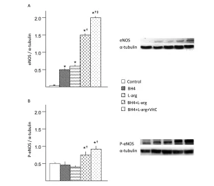
Hindlimb blood flow was determined by means of laser Doppler imaging (Moor Instruments, Devon, UK). Flow was mea-sured preoperatively, immediately after arterial excision, and then 3, 7, 14, 21, 28, 35 and 42 d after induction of ischemia. Figure 1.Effects of BH4, L-arginine (L-arg) and vitamin C (VitC) on eNOS and phosphorylated
eNOS expression in the ischemic gastrocnemius muscle. Muscle was harvested 14 d after the induction of hindlimb ischemia, and supernatants from muscle homogenates were used in these assays. (A) Expression of eNOS was increased by all dietary additives, although the greatest increase was noted in rats that received BH4 + L-arginine + vitamin C. (B) Expression
of p-eNOS was increased in rats fed BH4 + L-arginine or BH4 + L-arginine + vitamin C. Control
rats were fed a standard diet, whereas the four treatment groups consumed diets supple-mented with additives noted in the bar graph. Concentrations of these additives are noted in the text. Data are mean ± standard deviation (sd); n = 5–6. *p < 0.05 versus control; †p <
Scans were obtained during inhalation of 1% isofluorane while core body tempera-ture was maintained between 36.8 and 37.2°C. Scans were repeated three times, and the average for each rat was deter-mined. Data were expressed as the ratio of ischemic to non- ischemic hindlimb.
Angiograms
Angiograms were performed 42 d after induction of ischemia. Barium sulfate (2.5 mL; EZPaqe, Merry X-Ray, South San Francisco, CA, USA) was infused into the infrarenal aorta after ligation of the proxi-mal aorta and inferior vena cava during inhalation of 2% isofluorane. A grid was superimposed over the film between the greater trochanter of the femur to the
patella. The number of intersections be-tween contrast-filled vessels and gridlines was determined independently by three blinded observers. The angioscore was cal-culated as the average ratio of intersections to the total number of gridlines. Within the experimental setting of this study, the angioscore is a marker of collateral artery enlargement; thus, as collateral arteries di-late and remodel in response to femoral artery excision, their diameters increase, enhancing their visibility on the X-ray film, thus increasing the angioscore (19).
Nitroblue Tetrazolium Staining to Detect Muscle Necrosis
The left gastrocnemius muscle was re-moved 7 d after induction of ischemia.
The timing of sacrifice was selected on the basis of previous work that demon-strated maximal postischemic muscle necrosis at this time (19). The muscle was cut transversely into three 2-mm sections. Two sections were used for nitro blue tetrazolium (NBT) staining, while the third was frozen (–80°C) in optimal cut-ting temperature (OCT) embedding com-pound. Sections for NBT staining were incubated in PBS containing 0.033% NBT (Fisher Biotech, Austin, TX, USA) and 0.133% NADH (Roche Diagnostics, Indi-anapolis, IN, USA) at 21°C for 10 min. The samples were then fixed in 4% paraformaldehyde for 24 h. The areas of viable tissue, indicated by dark blue color, and nonviable tissue, indicated by white color, were measured by quantita-tive image analysis (ImagePro; Media Cy-bernetics, Bethesda, MD, USA). Data were expressed as the ratio of nonviable tissue to total tissue area. Measurements were made on the four exposed cut sur-faces, and the average was taken and used as a single data point for each ani-mal. The frozen section was used to pre-pare cryosections (10 µm) for
hema-toxylin and eosin (H&E) staining to evaluate histological integrity of the mus-cle, as well as to detect the presence of an inflammatory infiltrate.
STATISTICAL ANALYSIS
Analyses were carried out by means of analysis of variance (ANOVA). Post hoc
Student-Newman-Keuls tests were car-ried out if the ANOVA Fstatistic was sig-nificant to determine sites of difference within the ANOVA format. Probability values less <0.05 were accepted as signif-icant for all statistical calculations.
RESULTS
Effects of Oral BH4, L-Arginine, and Vitamin C on Gastrocnemius eNOS and p-eNOS Expression
Dietary supplementation of BH4,
L-arginine and vitamin C significantly
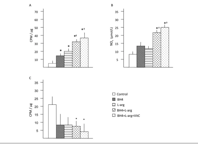
af-fected eNOS and p-eNOS expression in the ischemic gastrocnemius muscle (Fig-ure 1). Rats given single supplementation Figure 2.Effects of BH4, L-arginine (L-arg) and vitamin C (VitC) on NOS activity in the
is-chemic gastrocnemius muscle. Muscle was harvested 14 d after the induction of hindlimb ischemia, and supernatants from muscle homogenates were used in these assays. (A) Ca2+-dependent NOS activity was increased by all dietary additives, although the
greatest increase was noted in mice receiving BH4 + L-arginine or BH4 + L-arginine +
vita-min C. Data are mean ± sd; n = 5–6. *p < 0.05 versus control; †p < 0.05 versus BH4 or
L-arginine groups; ‡p < 0.05 versus BH4 + L-arginine group. (B) NOx (nitrite + nitrate) was
in-creased in mice receiving BH4 + L-arginine or BH4 + L-arginine + vitamin C. Data are mean ± sd; n = 5. *p < 0.05 versus control; †p < 0.05 versus BH4 or L-arginine groups.
(C) Ca2+-independent NOS activity was less in mice receiving BH4 + L-arginine or BH4 +
L-arginine + vitamin C than in control mice. Data are mean ± sd; n = 5–6. *p < 0.05 versus
with BH4 or L-arginine demonstrated
similar levels of eNOS expression, and these levels were significantly greater than that of rats fed normal chow. Rats given two (BH4 + L-arginine) or three
(BH4 + L-arginine + vitamin C)
supple-ments also had greater levels of eNOS expression than rats given single supple-ments. Hence, the combination of BH4 and L-arginine had an additive effect on
eNOS expression, and the addition of vi-tamin C provided an additional benefi-cial effect. However, increased p-eNOS expression was only observed in BH4 +
L-arginine– and BH4 + L-arginine +
vita-min C–fed rats.
Effects of Oral BH4, L-Arginine, and Vitamin C on Gastrocnemius Ca2+
-Dependent NOS Activity
Dietary supplements affected Ca2+ dependent NOS activity in the ischemic gastrocnemius muscle (Figure 2A). Rats
given a single dietary supplement (BH4 or L-arginine) demonstrated Ca2+
dependent NOS activities that were simi-lar to each other and greater than that of rats fed normal chow. Rats given two (BH4 + L-arginine) or three (BH4 + L-arginine + vitamin C) supplements
dis-played significantly greater Ca2+ -depen-dent NOS activity than rats given a single dietary supplement. The combina-tion of BH4 and L-arginine generated an
additive effect. The addition of vitamin C further increased Ca2+-dependent NOS activity, although it did not reach statisti-cal significance.
Effects of Oral BH4, L-Arginine and Vitamin C on Gastrocnemius NOx Levels
The final products of NO in vivoare ni-trite (NO2–) and nitrate (NO3–). NOx is the sum of NO2–and NO3–and it is the best index of total NO production. The
NOx concentration in ischemic gastroc-nemius was significantly increased from rats fed BH4 and L-arginine or BH4, L-arginine and vitamin C (Figure 2B).
There were no changes in rats fed with either BH4 and L-arginine alone. Effects of Oral BH4, L-Arginine and Vitamin C on Gastrocnemius Ca2+
-Independent NOS Activity
Ca2+-independent NOS activity was greater in the ischemic gastrocnemius from rats fed normal chow than in the ischemic gastrocnemius of dietary fed rats (Figure 2C). Rats fed a single sup-plement (BH4 or L-arginine), or the
com-bination of these two agents, demon-strated Ca2+-independent NOS activity statistically similar to the normal chow group (that is, there was no beneficial ef-fect noted in these dietary intervention groups). However, rats provided with three dietary supplements (BH4 + L
-argi-nine + vitamin C) demonstrated Ca2+ -in-dependent NOS activity levels in the is-chemic gastrocnemius muscle that were lower than the other dietary intervention groups.
Effects of Oral BH4, L-Arginine and Vitamin C on Gastrocnemius Oxidative Stress
Dietary supplementation affected ni-trotyrosine accumulation and the ratio of GSH versus GSSG in the ischemic gas-trocnemius muscle (Figure 3). Rats given a single dietary supplement (BH4 or
L-arginine), or the combination of these
dietary agents, displayed similar nitroty-rosine levels. These levels were signifi-cantly less than the level noted in rats fed normal chow. Rats provided with all three dietary supplements (BH4 +
L-arginine + vitamin C) exhibited a
ni-trotyrosine level significantly less than rats given a single supplement (BH4 or
L-arginine) or the combination of these
two agents. The ratio of reduced versus oxidized glutathione (GSH:GSSG) was also measured as another index of ox-idative stress in the ischemic gastrocne-mius muscle. GSH:GSSG ratio was in-creased in the ischemic gastrocnemius of Figure 3.Effects of BH4, L-arginine (L-arg) and vitamin C (VitC) on oxidative stress in the
is-chemic gastrocnemius muscle. Muscle was harvested 14 d after the induction of hindlimb ischemia, and supernatants from muscle homogenates were used in these assays. (A) Ni-trotyrosine expression was decreased by all dietary additives, although this decrease was greatest in rats receiving BH4 + L-arginine + vitamin C. (B) The GSH:GSSG ratio was
in-creased in rats receiving BH4 + L-arginine; the addition of vitamin C to this regimen
rats fed the two dietary combination (BH4 + L-arginine) or the three dietary
combination (BH4 + L-arginine +
vita-min C). Addition of vitavita-min C had a beneficial effect in increasing GSH:GSSG ratio. Taken together, three dietary sup-plementation (BH4 + L-arginine +
vita-min C) would be much more effective in decreasing ischemic muscles oxidative stress after induction of hindlimb ische-mia in rats.
Effects of BH4, L-Arginine and Vitamin C on Hindlimb Perfusion
Dietary supplementation significantly increased the recovery of hindlimb per-fusion after induction of severe ischemia, and this effect was regimen and time de-pendent (Figure 4A). Rats given a single supplement (BH4 or L-arginine)
demon-strated similar degree of perfusion recov-ery in the foot, and this level was also similar to that noted in rats fed normal
chow. Rats provided with two (BH4 +
L-arginine) or three (BH4 + L-arginine +
vitamin C) supplements showed signifi-cantly greater recovery of foot perfusion than rats fed normal chow or rats given a single dietary supplement. This differ-ence was evident at the later phase of recovery, on d 21, 28 or 42 after induc-tion of ischemia, which was also the time of maximal collateral artery wall remod-eling (20). A similar pattern was noted for collateral artery angioscores deter-mined on d 42 after induction of ische-mia. Hence, the angioscore was signifi-cantly greater in rats given two (BH4 +
L-arginine) or three (BH4 + L-arginine +
vitamin C) supplements than in rats fed normal chow or in rats given a single supplement (Figures 4B, C).
Effects of Oral BH4, L-Arginine and Vitamin C on Gastrocnemius Muscle Necrosis
The extent of gastrocnemius necrosis was affected by the provision of dietary supplements. Rats given a single supple-ment (BH4 or L-arginine) or the
combina-tion of these agents manifest a similar degree of gastrocnemius muscle necrosis. Moreover, the extent of necrosis noted in these dietary intervention groups was similar to that noted in rats fed normal chow (that is, these dietary regimens did not improve postischemic muscle in-tegrity). However, rats provided with all three dietary supplements (BH4 +
L-arginine + vitamin C) had significantly
less gastrocnemius necrosis than rats fed normal chow, rats provided with a single dietary supplement or rats given the combination of BH4 + L-arginine. This
difference was evident on macroscopic and microscopic levels. The percentage of the cut surface of the ischemic gastroc-nemius muscle that was necrotic, deter-mined by NBT staining, was significantly less in the BH4 + L-arginine + vitamin C
group than in all other groups (Fig-ures 5A, B). Groups fed normal chow, or supplemented with BH4, L-arginine, or
both agents, demonstrated similar histo-logical evidence of severe necrosis: mus-cle numus-clei were nearly absent, intra-Figure 4.Effects of BH4, L-arginine and vitamin C on perfusion recovery after induction of
hindlimb ischemia. (A) Laser Doppler perfusion imaging (LDPI) data, expressed as the ratio of blood flow from the ischemic to nonischemic hindlimbs, was determined before, imme-diately after and then serially over the ensuing 6 wks. Group identity is shown in the color key. Data are mean ± sd; n = 6. *p < 0.05 for BH4 + L-arginine or BH4 + L-arginine + vitamin C groups versus all other groups. (B) The angioscore, determined 42 d after the induction of hindlimb ischemia and calculated as described in the text, was greater in rats receiving BH4 + L-arginine or BH4 + L-arginine + vitamin C than in all other groups. Data are mean ± sd;
myofiber vacuolization was substantial and the distance between myofibers was large (Figure 5C). A pronounced inflam-matory infiltrate was also present in these groups. In contrast, the BH4 +
L-arginine + vitamin C group
demon-strated good preservation of muscle his-tology and only a limited inflammatory cell infiltrate.
DISCUSSION
The following study hypothesis was supported by our findings: dietary cosupplementation with tetrahydrobiop -terin (BH4), L-arginine and vitamin C
acts synergistically to decrease oxidative stress, increase nitric oxide and thereby improve perfusion and tissue recovery in response to acute hindlimb ischemia more than supplementation with a single supplement. Interestingly, two patterns of effect emerged. Cosupplementation with BH4 + L-arginine increased the
de-pendent variables eNOS and p-eNOS ex-pression, Ca2+-dependent NOS activity, foot perfusion and the collateral artery angioscore more than the addition of ei-ther component separately, whereas the addition of vitamin C provided a further beneficial effect on these variables, al-though only eNOS expression reached statistical significance. In addition, co -administration of all three dietary sup-plements had a significantly greater ef-fect than BH4 or L-arginine, given
individually or in combination, when the dependent variables of Ca2+-independent NOS activity, oxidative stress or muscle necrosis were measured.
eNOS and p-eNOS expression, Ca2+ -dependent NOS activity, foot perfusion and the collateral artery angioscore are linked by established cause-and-effect re-lationships. eNOS-derived NO is a po-tent vasodilator (2); hence, the increased eNOS expression and activity present in the BH4 + L-arginine group should result
in an NO-dependent increase in foot per-fusion, and this expectation was realized by our findings. Moreover, derived NO is a critical determinant in the response to hindlimb ischemia (21–23). This effect is direct, on the basis
of the vasodilator effect of NO (24), but also indirect, insofar as eNOS-derived NO is critical to collateral artery remod-eling. These effects include mobilization of endothelial progenitor cells from bone marrow and their subsequent homing to the ischemic hindlimb (25). Once there, endothelial progenitor cells participate in
postischemic arteriogenesis, the process wherein existing collateral arteries un-dergo remodeling designed to restore vascular conductance (26). This process was evidenced by the increased an-gioscore, a marker of collateral artery enlargement, in rats provided with L
-arginine + BH4 dietary supplements. Figure 5.The effects of BH4, L-arginine (L-arg) and vitamin C (VitC) on necrosis in the
ische-mic gastrocnemius muscle. (A) Gastrocnemius muscle was removed 7 d after induction of hindlimb ischemia. Transverse sections of this muscle were stained with nitroblue tetra-zolium to determine the ratio of necrotic versus viable surface area. This ratio was less in rats receiving BH4 + L-arginine + vitamin C than in all other groups. Data are mean ± sd;
n = 6. *p < 0.05 for BH4 + L-arginine + vitamin C groups versus all other groups. (B)
Represen-tative images from each study group. (C) Histological sections of gastrocnemius muscle taken from the ischemic hindlimb 7 d after the induction of hindlimb ischemia were stained with H&E. Representative sections are shown. Note the loss of myofibers and the in-tense cellular infiltrate in the control, BH4 and L-arginine groups. Best preservation of muscle
Vitamin C likely exerted its beneficial effects in this study through a variety of molecular mechanisms. In its capacity as an antioxidant, it enhances NO bioavail-ability by quenching O2–, thus limiting the inactivation of NO that occurs when O2–and NO combine to produce OONO– (3). Vitamin C also stabilizes existing BH4 (8) and increases endothelial BH4 synthesis (27), thus minimizing eNOS “uncoupling,” which in turn lessens gen-eration of O2–by eNOS and reduces vas-cular oxidative stress (7). However, BH4 is itself a potent antioxidant (7), and ad-ministration of exogenous BH4 has been established to increase endothelial BH4 levels (28). Moreover, L-arginine directly
stimulates eNOS expression (29), en-hances eNOS activity by a dependent G protein–linked process (30) and limits the inhibitory effect of asym-metric dimethylarginine on derived NO production (31). We propose that under the experimental conditions imposed by hindlimb ischemia, addition of vitamin C to BH4 + L-arginine
signifi-cantly decreased oxidative stress and ac-cordingly decreased tissue necrosis. It
may also restore BH4 or NO levels, since we observed an increase in eNOS activ-ity and p-eNOS expression, although it did not reach statistical difference be-cause of the number of samples ana-lyzed, dosage of vitamin C or time of data collection. Cosupplementation with BH4 + L-arginine may have sufficiently
restored endothelial BH4 levels and thereby intracellular redox balance. Hence, the addition of vitamin C did not further p-eNOS expression or activity, and hence NO bioavailability.
Ca2+-independent NOS activity and tis-sue nitrotyrosine accumulation were sig-nificantly lower in rats receiving all three dietary supplements than in rats receiving BH4 or L-arginine, or the combination of
the two. When measured by methods used herein, Ca2+-independent NOS activ-ity is an authentic reflection of iNOS ac-tivity, inasmuch as the assay was con-ducted in vitro, in the absence of shear stress, which can activate eNOS in the ab-sence of Ca2+via phosphorylation (32). The marked elevation of iNOS activity in rats fed normal chow indicates the pres-ence of postischemia inflammation, which is also evidenced by the cellular inflam-matory infiltrate in this group. Nitrotyro-sine accumulation is indicative of
OONO–-induced damage (33), and it is in-teresting that the group that exhibited the least amount of tissue damage (rats pro-vided with all three supplements) also had the least nitrotyrosine accumulation. Moreover, rats in the triple therapy group demonstrated a virtual absence of nitroty-rosine. We interpret the present findings to indicate that muscle injury after ische-mia is not entirely contingent upon loss of perfusion, inasmuch as cosupplementa-tion with the antioxidant vitamin C clearly reduced tissue injury and elimi-nated nitrotyrosine accumulation, but did not affect perfusion or collateral artery en-largement. Instead, we speculate that vita-min C provided an antioxidant effect that limited tissue injury generated by inflam-matory cells for which action depends in part on oxidant production (for example, neutrophils and macrophages). This effect could be direct, due to the antioxidant
ac-tivity of vitamin C, or indirect, due to the beneficial effect of vitamin C on BH4 lev-els (8,27), insofar as BH4 also exhibits po-tent antioxidant activity (7).
Although it is well established that en-dothelial dysfunction related to vascular oxidative stress is a critical factor in PAD pathogenesis, dietary supplementation with L-arginine or antioxidants, such as
vitamin C, have had equivocal effects on long-term outcome (12,13,16). Dietary supplementation with L-arginine alone
has a beneficial effect when given acutely (that is, via intravenous infusion [14] or for short duration [2 months] [15]), and these clinical results are consistent with the positive effects observed in rats pro-vided with dietary L-arginine. However,
long-term administration of L-arginine
(6 months) not only failed to demonstrate a beneficial effect, but resulted in a degree of eNOS-dependent vascular reactivity significantly less than that of the placebo group (16). The present findings demon-strated increased iNOS activity after in-duction of ischemia. If a similar circum-stance is present in PAD, then the singular dietary supplementation with L-arginine,
the substrate for all NOS isoforms, might serve to worsen vascular inflammation, a critical participant in the pathogenesis of PAD (1). Vitamin C reduces vascular in-flammation (34), improves redox balance (11) and eNOS- dependent vascular reac-tivity (10), but these effects have only been evaluated on a short-term basis, whereas retrospective cross-sectional studies have failed to confirm that dietary supplementation with antioxidants im-proves PAD outcome (12,13). We interpret the present findings to indicate that provi-sion of BH4 + L-arginine + vitamin C
act-ing synergystically might prove a useful therapeutic alternative in PAD treatment. To this end, the use of sapropterin dihy-drochloride, a synthetic form of (6R)-L
-erythro-5,6,7,8-tetrahydrobiopterin re-cently approved for the treatment of phenylalanine hydroxylase deficiency (35), might provide a practical means for the provision of BH4.
In conclusion, cosupplementation with BH4 + L-arginine + vitamin C resulted in
Figure 6.Schematic illustration of possible mechanism of three dietary combined regimen can increase NO bioavailability and decrease oxidative stress, accord-ingly increase blood flow recovery and re-duce tissue necrosis. A–•, ascorbate; AH–,
increased eNOS activity and NO concen-tration as well as greater foot blood flow recovery than rats receiving normal chow or either agent separately. The ad-dition of vitamin C to the BH4 +
L-arginine regimen further reduced
ox-idative stress and tissue injury in ische-mic muscles (Figure 6). The clearly supe-rior outcome of rats provided with BH4 +
L-arginine + vitamin C warrants
investi-gation of a cosupplementation strategy as a therapeutic alternative in PAD.
ACKNOWLEDGMENTS
This work was supported by National Institutes of Health, National Heart, Lung, and Blood Institute grant RO-1 HL-75353 (to LM Messina), as well as grants from the Pacific Vascular Research Foundation and the Wayne and Gladys Valley Foundation (to LM Messina).
DISCLOSURE
The authors declare that they have no competing interests as defined by Molecu-lar Medicine, or other interests that might be perceived to influence the results and discussion reported in this paper.
REFERENCES
1. Silvestro A, Oliva G, Brevetti G. (2002) Intermit-tent claudication and endothelial dysfunction.
Eur. Heart J. 4:B35–40.
2. Marletta M. (1993) Nitric oxide synthase structure and mechanism. J Biol. Chem.;268:12231–12234. 3. Pacher P, Beckman J, Liaudet L. (2007) Nitric
oxide and peroxynitrite in health and disease.
Physiol. Rev.87:315–424.
4. Peterson T, et al.(1999) Opposing effects of reac-tive oxygen species and cholesterol on endothe-lial nitric oxide synthase and endotheendothe-lial cell caveolae. Circ. Res.85:29–37.
5. Kuzkaya N, Weissman N, Harrison D, Dikalov S. (2003) Interactions of peroxynitrite, tetrahydro-biopterin, ascorbic acid, and thiols. J. Biol. Chem.
278:22546–54.
6. Bevers L, et al.(2006) Tetrahydrobiopterin, but not L-arginine, decreases NO synthase uncou-pling in cells expressing high levels of endothe-lial NO synthase. Hypertension.47:87–94. 7. Schmidt T, Alp N. (2007) Mechanisms for the role
of tetrahydrobiopterin in endothelial function and disease. Clin. Sci.113:47–63.
8. Heller R, et al.(2001). L-ascorbic acid potentiates endothelial nitric oxide synthesis via a chemical stabilization of tetrahydrobiopterin. J. Biol. Chem.
276:40–7.
9. Langlois M, Duprez D, Delanghe J, De Buyzere M, Clement D. (2001) Serum vitamin C concen-tration is low in peripheral arterial disease and is associated with inflammation and severity of atherosclerosis. Circulation.103:1863–8. 10. Silvestro A, et al.(2002) Vitamin C prevents
en-dothelial dysfunction induced by acute exercise in patients with intermittent claudication. Athero-sclerosis.165:277–83.
11. Wijnen M, et al.(2001) Antioxidants reduce oxida-tive stress in claudicants. J. Surg. Res. 96:183–7. 12. Donnan P, Thomson M, Fowkes F, Prescott R,
Housley E. (1993) Diet as a risk factor for periph-eral arterial disease in the genperiph-eral population: the Edinburgh Artery Study. Am. J. Clin. Nutr.
57:917–21.
13. Klipstein-Grobusch K, et al.(2001) Dietary an-tioxidants and peripheral arterial disease: the Rotterdam Study. Am. J. Epidemiol. 154:145–9. 14. Böger R, et al.(1998) Restoring vascular nitric oxide formation by L-arginine improves the symptoms of intermittent claudication in pa-tients with peripheral arterial occlusive disease.
J. Am. Coll. Card.32:1336–44.
15. Maxwell A, Anderson B, Cooke J. (2000) Nutri-tional therapy for peripheral arterial disease: a double-blind, placebo-controlled, randomized trial of HeartBar. Vasc. Med.5:11–9.
16. Wilson A, Harada R, Nair N, Balasubramanian N, Cooke J. (2007) L-arginine supplementation in peripheral arterial disease: no benefit and possi-ble harm. Circulation.116:188–95.
17. Ueda S, et al.(1999) Tetrahydrobiopterin restores endothelial function in long-term smokers. J. Am. Coll. Card.35:71–5.
18. Heitzer T, Krohn, K, Albers S, Meinertz T. (2000) Tetrahydrobiopterin improves endothelium-de-pendent vasodilation by increasing nitric oxide activity in patients with type II diabetes mellitus.
Diabetologia.43:1435–8.
19. Yan J, et al.(2010) Oral tetrahydrobiopterin im-proves the beneficial effect of mediated eNOS gene transfer after induction of hindlimb ischemia. Mol. Ther.18:1482–9. 20. Park B, et al.(2010) Endothelial nitric oxide
syn-thase affects both early and late collateral arterial adaptation and blood flow recovery after induc-tion of hindlimb ischemia in mice. J. Vasc. Surg.
51:165–73.
21. Yan J, Tang G, Wang R, Messina L. (2005) Opti-mization of adenovirus-mediated endothelial ni-tric oxide synthase delivery in rat hindlimb is-chemia. Gene Ther. 12:1640–50.
22. Yu J, et al.(2005) Endothelial nitric oxide syn-thase is critical for ischemic remodeling, mural cell recruitment, and blood flow reserve. Proc. Natl. Acad. Sci. U. S. A.102:10999–1004. 23. Lloyd P, Yang H, Terjung R. (2001) Arteriogenesis
and angiogenesis in rat ischemic hindlimb: role of nitric oxide. Am. J. Physiol.281:H2528–38. 24. Mees B, et al.(2007) Endothelial nitric oxide
syn-thase activity is essential for vasodilation during
blood flow recovery but not for arteriogenesis.
Arterioscler. Thromb. Vasc. Biol.27:1–8.
25. Aicher A, et al.(2003) Essential role of endothelial nitric oxide synthase for mobilization of stem and progenitor cells. Nat. Med.9:1370–6. 26. Helisch A, Schaper W. (2003) Arteriogenesis: the
development and growth of collateral arteries.
Microcirculation. 10:83–97.
27. Huang A, Vita J, Venema R, Keaney J Jr. (2000) Ascorbic acid enhances endothelial nitric-oxide synthase activity by increasing intracellular tetrahydrobiopterin. J. Biol. Chem.275:17399–406. 28. Sawabe K, Wakasugi K, Hasegawa H. (2004)
Tetrahydrobiopterin uptake in supplemental ad-ministration: elevation of tissue tetrahydro-biopterin in mice following uptake of the exoge-nously oxidized product 7,8-dihydrobiopterin and subsequent reduction by an sensitive process. J. Pharmacol. Sci.96:124–33. 29. Kohil R. (2004) Dietary L-arginine
supplementa-tion enhances endothelial nitric oxide synthesis in streptozotocin-induced diabetic rats. J. Nutr.
134:600–8.
30. Joshi M, et al.(2007) Receptor-mediated activa-tion of nitric oxide synthesis by arginine in en-dothelial cells. Proc. Natl. Acad. Sci. U. S. A.
104:9982–7.
31. Böger R, Ron E. (2005) L-arginine improves vas-cular function by overcoming the deleterious ef-fects of ADMA, a novel cardiovascular risk fac-tor. Alt. Med. Rev. 10:14–8.
32. Ayajiki K, Kindermann M, Hecker M, Fleming I, Busse R. (1996) Intracellular pH and tyrosine phosphorylation but not calcium induce shear stress-induced nitric oxide production in native endothelial cells. Circ. Res. 78:750–8.
33. Beckman J, Koppenol W. (1996) Nitric oxide, su-peroxide, and peroxynitrite: the good, the bad, and ugly. Am. J. Physiol.271:C1424–37.
34. Aguirre R, May J. (2008) Inflammation in the vas-cular bed: importance of vitamin C. Pharmacol. Ther.119:96–103.