Protocol
Practical Method for Isolation of Phage
Deletion Mutants
Diana Gutiérrez, Lucía Fernández, Ana Rodríguez and Pilar García *
Instituto de Productos Lácteos de Asturias (IPLA-CSIC), Paseo Río Linares s/n, 33300 Villaviciosa, Spain; [email protected] (D.G.); [email protected] (L.F.); [email protected] (A.R.)
* Correspondence: [email protected]; Tel.: +34-985-89-21-31 (ext. 41)
Received: 12 December 2017; Accepted: 15 January 2018; Published: 17 January 2018
Abstract:The growing concern about multi-drug resistant pathogenic bacteria has led to a renewed interest in the study of bacteriophages as antimicrobials and as therapeutic agents against infectious diseases (phage therapy). Phages to be used for this purpose have to be subjected to in-depth genomic characterization. It is essential to ascribe specific functions to phage genes, which will give information to unravel phage biology and to ensure the lack of undesirable genes, such as virulence and antibiotic resistance genes. Here, we describe a simple protocol for the selection of phage mutants carrying random deletions along the phage genome. Theoretically, any DNA region might be removed with the only requirement that the phage particle viability remains unaffected. This technique is based on the instability of phage particles in the presence of chelating compounds. A fraction of the phage population naturally lacking DNA segments will survive the treatment. Within the context of phages as antimicrobials, this protocol is useful to select lytic variants from temperate phages. In terms of phage efficiency, virulent phages are preferred over temperate ones to remove undesirable bacteria. This protocol has been used to obtain gene mutations that are involved in the lysogenic cycle of phages infecting Gram-positive bacteria (StaphylococcusandLactobacillus).
Keywords:phage deletion mutant; chelating agent; temperate phage; virulent phage
1. Introduction
The isolation and characterization of phage deletion mutants is a widely used tool for the analysis of gene function and regulation. In this regard, homologous recombination using a removable plasmid is a common technique to delete or exchange a target genome sequence in temperate phages. The recombination process is performed while the phage is inserted in the host bacterial genome as a prophage, and then, the induction of the lytic cycle by an external signal will release the mutated viral particles. However, methods to select mutant particles of virulent phages are scarce, and most of them involve the use of UV radiation or chemicals [1,2]. More recently, molecular biology techniques based on CRISPR-Cas (clustered, regularly interspaced, short palindromic repeats technology for generating RNA-guided nucleases with customizable specificities) systems have allowed accurately removing and swapping DNA fragments of some virulent phages [3–6]. One main disadvantage of these techniques is the need of previous knowledge regarding the phage genome sequence, making these methodologies time-consuming. In addition, the complete protocol is not available for all bacterial species and their phages.
In this article, we report an easy and rapid way to isolate phage deletion mutants based on the instability of phage particles in the presence of chelating agents, such as EDTA (Ethylenediaminetetraacetic acid), sodium citrate, or sodium pyrophosphate. Chelating agents can react with the cations attached to the phage structure, breaking the bonds that maintain particle conformation [7]. Moreover, this destabilizing effect increases with temperature [8]. However,
a small percentage of bacteriophages within a large population may have spontaneously lost genome fragments, which would lessen the pressure inside the head structure, and, consequently, make the mutant viral particles more stable in the presence of the chelating agent. Early work using chelating agents to isolate deletion mutants was performed in wild-type stocks of phageλ[9].
The mutations that can be selected with this method are determined by two main requirements. On the one hand, the size of the selected deletions is determined by the minimum and maximum genome size that can be packed into the phage head. Thus, very large deletions would not be selected due to the low stability of the head when the genome size is too small. On the other hand, deletions can only affect non-essential genes, as mutant selection is performed by plating, which requires the successful completion of the lytic cycle. Moreover, the phenotype of the selected phage can in some cases be screened directly after plating. For instance, mutations in the lysogeny module leading to the loss of lysogenic capability can be easily identified by the “clear lysis plaque” phenotype. Similarly, deletion of genes that regulate burst size can be recognized by the presence of smaller lysis plaques.
The procedure for selection of phage deletion mutants has been previously used to identify the repressor-encoding gene (cI) of theLactobacillus caseibacteriophage A2. In this case, several phage deletion mutants were selected and analyzed. Deletions ranged from 0.5 to 3.5 kb and were all located in one of two genomic regions, comprising up to 7.9 kb, which were dispensable for lytic development. Those phages carrying deletions in the lysogeny module showed a “clear lysis plaque” phenotype and were unable to lysogenize their host [10]. Similarly, this method was useful to select lytic derivative mutants from theStaphylococcus aureustemperate phages phiH5 and phiA72. Indeed, lytic phages phiIPLA88 and phiIPLA35 (derived from phiH5 and phiA72, respectively) could only follow a lytic life cycle and turned out to be more efficient at removingS. aureus than their respective parent phages [11]. Further analysis revealed that point mutations in genes that were involved in lysogeny control explained their strictly lytic behavior. Thus, the repressor protein of phiIPLA35 lacks 25 amino acids at the C-terminus, while the start codon was lost in the phiIPLA88 repressor-encoding gene [12].
The protocol described here allows for the selection of phages carrying random DNA deletions, and, in addition, the subsequent isolation of those mutations affecting the lysis-lysogeny functions. This protocol was tested on S. aureus phages phiH5 and phiA72, but it can be applied to other phages infecting Gram-positive bacteria. In a phage therapy context, the genomic characterization of bacteriophages as well as the use of lytic phages is encouraged. Therefore, this straightforward methodology allows for the selection of lytic variants from temperate phages in a very short period of time.
2. Experimental Design
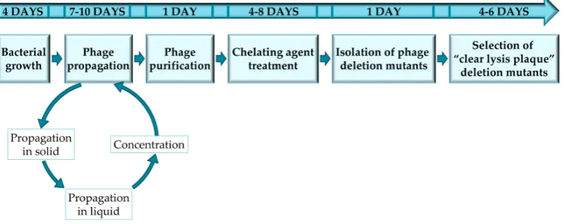
The protocol described here has been optimized for the selection of staphylococcal phage mutants with a “clear lysis plaque” phenotype, by using sodium pyrophosphate as a chelating agent. Thus, all the protocols, materials and methods are reliable for temperate phages infecting staphylococcal species; nevertheless, this protocol was also used in temperate bacteriophages infecting Lactobacillus[10]. However, it must be noted that the protocol may have to be adapted for each specific bacterium-phage pair in order to ensure successful results (Figure1). Furthermore, this protocol can also be used for the selection of mutant variants of virulent phages in which phage viability is not compromised. To note, all the steps performed in this protocol should be perform under sterile conditions using autoclaved material and a laminar airflow cabinet.
The following steps consist of propagation and purification of the phage on the selected host strain. Although these steps are not mandatory, it is advisable to purify the phage before obtaining the phage mutants, in order to ensure the success of the subsequent sodium pyrophosphate treatment. Thus, the presence of contaminants in the phage stock might entrap the sodium pyrophosphate molecules and decrease their activity, requiring a higher concentration of the compound to achieve the same effect. Phage propagation should be performed in two steps: first, in small volumes using the double-layer technique, followed by an up-scaled propagation in 1 liter of liquid medium. Prior to purification, the bacteriophage lysate should be filtered and concentrated by precipitation with NaCl and PEG 8000. Then, purification should be carried out by a conventional CsCl gradient using a ultracentrifuge. As mentioned previously, phage purification is recommended, but is not essential. However, skipping this step may lead to a lower efficiency of the protocol.
Figure 1.Experimental design to complete every stage of the protocol for obtaining and selecting “clear lysis plaque” phage deletion mutants.
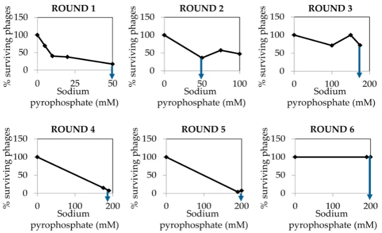
The chelating agent treatment is performed using a purified and concentrated phage stock and different concentrations of sodium pyrophosphate. Exposure of the phage to the chelating agent for 30 min at 37◦C will cause a dramatic decline in phage survival (ideally phage titers should decrease between 90 and 99% after treatment). From this treatment, phages were selected for the following rounds of treatment until obtaining a phage survival rate of 100%, even at high concentrations of the chelating agent. The concentrations of sodium pyrophosphate may vary depending on the specific phage.
After reaching a survival rate of 100%, the putative phage deletion mutants are isolated and propagated for further studies that are intended to characterize the mutations. Characterization is often carried out by DNA restriction analysis or PCR. For phages with a known genome sequence, PCR reactions can be designed to amplify regions along the genome and easily identify the deletion. For phages with an unknown genome sequence, DNA deletions should be identified by DNA extraction and further restriction analysis. This method will also provide an estimate of the deletion size.
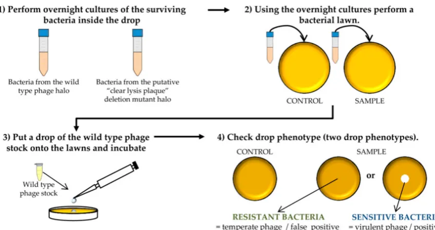
Moreover, the selection of “clear lysis plaque” phage deletion mutants is also performed by collecting the surviving phages, isolating single lysis plaques, and testing their ability to produce turbid or transparent halos by the drop assay. Surviving bacteria inside the halo will be finally tested for susceptibility to the phage.
2.1. Materials
•
S. aureusSa9 [11], isolated from a mastitic milk sample was used as the host strain of the phage.•
Phage phiH5, isolated from raw milk was used to select “clear lysis plaques” deletion mutants [11]•
Bacteriological agar (Cat. no.: A6686), Tris HCl (Cat. no.: 10812846001), MgSO4(Cat. no.: M2643), CaCl2(Cat. no.: C5670), NaCl (Cat. no.: S5886), KCl (Cat. no.: P9541), Na2HPO4 (Cat. no.: RES20908-A7), KH2PO4(Cat. no.: NIST200B), Sodium pyrophosphate (Cat. no.: P8135), PEG 8000 (Cat. no.: 89510), CsCl (Cat. no.: 203025) and RNAse (Cat. no.: R6513) where purchased at Sigma-Aldrich, now Merck, Darmstadt, Germany.2.2. Equipment
•
Incubator shaker model Excella E24 (New-Brunswick Scientific UK Ltd, Cambridge, UK).•
Sanyo MIR-153 refrigerated Incubator (Sanyo electric, Ora-Gun, Japan).•
Spectofotometer BioPhotometer Eppendorf (Eppendorf Ibérica S.L.U., Madrid, Spain).•
Laminar Airflow Cabinet FASTER TWO 30 Cabinet (Manson Technology, Dublin, Ireland).•
STUART Mini Gyro Rocker SSM3 (Cole-Parmer, Stone, UK).•
Microcentrifuge 5415 R Epperdorf (Eppendorf Ibérica S.L.U., Madrid, Spain).•
Beckman Optima MAX Ultracentrifuge (Beckman Coulter, Brea, CA, USA).•
Centrifuge Thermo Scientific™ Sorvall™ RC 6 Plus Centrifuge (Fisher Scientific, Loughborough, UK).•
Vortex ZX3 (VELP SCIENTIFICA, Usmate Velate, Italy).•
Vacuum pump Diaphragm pumps, VACUUBRAND (VACUUBRAND, GMBH, Wertheim, Germany). 0.45 µm cellulose acetate filter (Whatman™, GE Healthcare, Amersham, UK; Cat. no.: 462100).•
0.2µm polyethersulphone filter (VWR, Radnor, USA; Cat. no.: 28145-501).•
0.025µm VSWP MF-Millipore™ Membrane Filters (Merck, Millipore, Cork, Ireland; Cat. no.: VSWP02500).•
1 L, 0.45µm pore size, Corning®bottle-top vacuum filter system (Sigma-Aldrich, now Merck, Darmstadt, Germany; Cat. no.: CLS430516).•
1.5–3 mL VWR®Two-Sided Disposable Plastic Cuvette (VWR, Radnor, USA; Cat. no.: 97000-586).•
90 mm Polystyrene Petri dishes (Labbox, Labware S.L., Barcelona, Spain; Cat. no.: PDIP-09N-500).•
Tube, thin wall, Ultra-clear, 5 mL, 13×
51 mm ultracentrifuge tubes (Beckman Coulter, Brea, CA, USA; Cat. no.: 344057).•
PIPETMAN L Pipettes: P20L, P200L, and P1000L (Gilson, Dunstable, UK).•
Sterile aerosol filter pipet tips (VWR, Radnor, PA, USA).3. Procedure
3.1. Bacterial Growth Conditions and Phage Titration. 4 Days
•
Streak theS. aureushost strain onto a TSA (TSB supplemented with 2%w/vagar) plate using a sterile tip and incubate the plate at 37◦C for 18 h to obtain isolated colonies.Methods Protoc. 2018, 1, 6 4 of 14
• TSB, Tryptic Soy Broth (Scharlau, Barcelona, Spain; Cat. no.: 02-200-500).
• Bacteriological agar (Cat. no.: A6686), Tris HCl (Cat. no.: 10812846001), MgSO4 (Cat. no.: M2643),
CaCl2`(Cat. no.: C5670), NaCl (Cat. no.: S5886), KCl (Cat. no.: P9541), Na2HPO4 (Cat. no.:
RES20908-A7), KH2PO4 (Cat. no.: NIST200B), Sodium pyrophosphate (Cat. no.: P8135), PEG 8000
(Cat. no.: 89510), CsCl (Cat. no.: 203025) and RNAse (Cat. no.: R6513) where purchased at Sigma-Aldrich, now Merck, Darmstadt, Germany.
2.2. Equipment
• Incubator shaker model Excella E24 (New-Brunswick Scientific UK Ltd, Cambridge, UK). • Sanyo MIR-153 refrigerated Incubator (Sanyo electric, Ora-Gun, Japan).
• Spectofotometer BioPhotometer Eppendorf (Eppendorf Ibérica S.L.U., Madrid, Spain). • Laminar Airflow Cabinet FASTER TWO 30 Cabinet (Manson Technology, Dublin, Ireland). • STUART Mini Gyro Rocker SSM3 (Cole-Parmer, Stone, UK).
• Microcentrifuge 5415 R Epperdorf (Eppendorf Ibérica S.L.U., Madrid, Spain). • Beckman Optima MAX Ultracentrifuge (Beckman Coulter, Brea, CA, USA).
• Centrifuge Thermo Scientific™ Sorvall™ RC 6 Plus Centrifuge (Fisher Scientific, Loughborough, UK).
• Vortex ZX3 (VELP SCIENTIFICA, Usmate Velate, Italy).
• Vacuum pump Diaphragm pumps, VACUUBRAND (VACUUBRAND, GMBH, Wertheim, Germany).0.45 µm cellulose acetate filter (WhatmanTM, GE Healthcare, Amersham, UK; Cat. no.:
462100).
• 0.2 µm polyethersulphone filter (VWR, Radnor, USA; Cat. no.: 28145-501).
• 0.025 µm VSWP MF-Millipore™ Membrane Filters (Merck, Millipore, Cork, Ireland; Cat. no.: VSWP02500).
• 1 L, 0.45 µm pore size, Corning® bottle-top vacuum filter system (Sigma-Aldrich, now Merck, Darmstadt, Germany; Cat. no.: CLS430516).
• 1.5 ml - 3 ml VWR® Two-Sided Disposable Plastic Cuvette (VWR, Radnor, USA; Cat. no.: 97000-586).
• 90 mm Polystyrene Petri dishes (Labbox, Labware S.L., Barcelona, Spain; Cat. no.: PDIP-09N-500).
• Tube, thin wall, Ultra-clear, 5 ml, 13 × 51 mm ultracentrifuge tubes (Beckman Coulter, Brea, CA, USA; Cat. no.: 344057).
• PIPETMAN L Pipettes: P20L, P200L, and P1000L (Gilson, Dunstable, UK). • Sterile aerosol filter pipet tips (VWR, Radnor, PA, USA).
3. Procedure
3.1. Bacterial Growth Conditions and Phage Titration. 4 Days
• Streak the S. aureus host strain onto a TSA (TSB supplemented with 2% w/v agar) plate using a sterile tip and incubate the plate at 37 °C for 18 h to obtain isolated colonies.
PAUSE STEP After the colonies are grown, plates can be stored at 4 °C for up to 1 month. • Inoculate one S. aureus single colony from the TSA plate into a sterile 10 mL tube containing
5 mL of sterile TSB). Incubate at 37 °C with shaking (250 rpm) for 18 h to obtain an overnight (o/n) culture.
OPTIONAL STEP Take 1 mL of the o/n culture and add glycerol to a final concentration of 20% in order to prepare a frozen stock of the host strain. Store this stock at −80 °C.
• In order to perform a bacterial growth curve, inoculate 50 mL of TSB with 1% (v/v) of the S. aureus o/n culture into a 100 mL sterile bottle and incubate at 37 °C with shaking.
• Take 2 mL samples at time points during incubation (0, 1, 2, 3, 4, 5, 6, 7, 8 and 24 h). Transfer 1 mL sample to a plastic cuvette and measure the optical density at an absorbance wavelength of 600 nm (OD600). At the same time, perform 1:10 serial dilutions of the sample into PBS buffer.
PAUSE STEPAfter the colonies are grown, plates can be stored at 4◦C for up to 1 month.
•
Inoculate oneS. aureussingle colony from the TSA plate into a sterile 10 mL tube containing 5 mL of sterile TSB). Incubate at 37◦C with shaking (250 rpm) for 18 h to obtain an overnight (o/n) culture.OPTIONAL STEPTake 1 mL of the o/n culture and add glycerol to a final concentration of 20% in order to prepare a frozen stock of the host strain. Store this stock at
−
80◦C.•
In order to perform a bacterial growth curve, inoculate 50 mL of TSB with 1% (v/v) of theS. aureus o/n culture into a 100 mL sterile bottle and incubate at 37◦C with shaking.•
Count the number of colonies in the plate containing 30–300 colony forming units (CFU). Calculate the viable cell counts of the culture using the equation:CFU
mL
=
N×
1
DF
×
1 V ,
whereNis the number of colonies counted on the plate (expressed as CFU);DFis the dilution factor andVis the sample volume poured onto the plate (expressed in mL).
•
Repeat the experiment two extra times to obtain three biological replicates.•
Represent graphically the mean±
standard deviation of the CFU/mL in a logarithmic scale or the OD600in they-axis and the time of incubation of the culture in thex-axis.Methods Protoc. 2018, 1, 6 5 of 14
Add 100 µL of the desired dilution to a TSA plate and spread using a sterile L-shape spreader until liquid dries. Incubate the plate at 37 °C for 18 h.
• Count the number of colonies in the plate containing 30–300 colony forming units (CFU). Calculate the viable cell counts of the culture using the equation:
CFU
mL = 𝑁 × 1 𝐷𝐹 ×
1 𝑉 ,
where N is the number of colonies counted on the plate (expressed as CFU); DF is the dilution factor and V is the sample volume poured onto the plate (expressed in mL).
• Repeat the experiment two extra times to obtain three biological replicates.
• Represent graphically the mean ± standard deviation of the CFU/mL in a logarithmic scale or the OD600 in the y-axis and the time of incubation of the culture in the x-axis.
CRITICAL STEP The number of CFU/mL should be calculated for each host strain, but generally speaking, it is very similar among strains belonging to the same species. The calculation of the CFU/mL in the exponential culture (Cb) is necessary to allow for the accurate determination of the multiplicity of infection (MOI). Calculation of this parameter is decisive in order to achieve an optimum yield in the phage propagation procedure.
3.2. Phage Titration and Propagation in Small Volume. 2–5 Days
• Inoculate one S. aureus single colony to obtain an o/n culture (see above). • Make 1:10 serial dilutions of the initial phage stock.
• Determine the number of infective phage particles by using the double-layer plaque assay. Take 100 µL of a S. aureus o/n culture and mix with 100 µL of the desired phage dilution into a 15 mL tube. Add 3 mL of semisolid TSA (0.7% agar) and pour the mixture onto a TSA plate. Let the soft agar medium solidify, and then, incubate the plate at 37 °C for 18 h. This experiment must be performed in triplicate.
• After incubation, count the number of plaque forming units (PFU) in those plates where the number is between 30–300 PFU. Calculate the phage titer of the original phage sample by using the equation:
PFU
mL = 𝑁 × 1 𝐷𝐹 ×
1 𝑉 ,
where N is the number of plaques of lysis counted on the plate (expressed as PFU); DF is the dilution factor and V is the sample volume poured onto the plate (expressed in mL).
CRITICAL STEP If the titer of the phage suspension is ≥109 PFU/mL, then the phage
propagation and concentration protocol can be followed from Section 3.3. If not, propagation in solid medium should be performed before proceeding to the next step.
• In order to carry out the propagation in solid medium, take 100 µL of diluted phage stock mix with 100 µL of a S. aureus o/n culture into a 15 mL tube. Add 3 mL of TSA 0.7% and pour the mixture onto a TSA plate. Repeat the procedure to get 5 plates. Let the medium solidify and then, incubate the plates at 37 °C for 18 h.
CRITICAL STEP After incubation, the overlaid plate should be completely transparent reflecting the occurrence of confluent lysis. The minimum concentration of phages that should be plated to ensure confluent lysis varies between 104 and 106 PFU/plate. Add 1 mL of SM buffer
to each plate and incubate with gentle shaking (60 rpm) at 20 °C for 1 h. Recover the SM buffer from the five plates and transfer it to a 15 mL tube. Centrifuge 10 min at 13,600 × g. Transfer the supernatant containing the phages to a new sterile tube and perform phage titration again. If it is needed, solid propagation can be repeated again with this new phage stock in order to obtain
a concentration ≥109 PFU/mL.
PAUSE STEP Phage stock solutions can be kept at 4 °C for up to three months after filtration using a 0.45 µm cellulose acetate membrane filter. For longer periods of time, a phage stock can be kept at −80 °C with 20% of glycerol.
CRITICAL STEP The number of CFU/mL should be calculated for each host strain, but generally speaking, it is very similar among strains belonging to the same species. The calculation of the CFU/mL in the exponential culture (Cb) is necessary to allow for the accurate determination of the multiplicity of infection (MOI). Calculation of this parameter is decisive in order to achieve an optimum yield in the phage propagation procedure.
3.2. Phage Titration and Propagation in Small Volume. 2–5 Days
•
Inoculate oneS. aureussingle colony to obtain an o/n culture (see above).•
Make 1:10 serial dilutions of the initial phage stock.•
Determine the number of infective phage particles by using the double-layer plaque assay. Take 100µL of aS. aureuso/n culture and mix with 100µL of the desired phage dilution into a 15 mL tube. Add 3 mL of semisolid TSA (0.7% agar) and pour the mixture onto a TSA plate. Let the soft agar medium solidify, and then, incubate the plate at 37◦C for 18 h. This experiment must be performed in triplicate.•
After incubation, count the number of plaque forming units (PFU) in those plates where the number is between 30–300 PFU. Calculate the phage titer of the original phage sample by using the equation:PFU
mL
=
N×
1
DF
×
1 V ,
whereNis the number of plaques of lysis counted on the plate (expressed as PFU);DFis the dilution factor andVis the sample volume poured onto the plate (expressed in mL).
Methods Protoc. 2018, 1, 6 5 of 14
Add 100 µL of the desired dilution to a TSA plate and spread using a sterile L-shape spreader until liquid dries. Incubate the plate at 37 °C for 18 h.
• Count the number of colonies in the plate containing 30–300 colony forming units (CFU). Calculate the viable cell counts of the culture using the equation:
CFU
mL = 𝑁 × 1 𝐷𝐹 ×
1 𝑉 ,
where N is the number of colonies counted on the plate (expressed as CFU); DF is the dilution factor and V is the sample volume poured onto the plate (expressed in mL).
• Repeat the experiment two extra times to obtain three biological replicates.
• Represent graphically the mean ± standard deviation of the CFU/mL in a logarithmic scale or the OD600 in the y-axis and the time of incubation of the culture in the x-axis.
CRITICAL STEP The number of CFU/mL should be calculated for each host strain, but generally speaking, it is very similar among strains belonging to the same species. The calculation of the CFU/mL in the exponential culture (Cb) is necessary to allow for the accurate determination of the multiplicity of infection (MOI). Calculation of this parameter is decisive in order to achieve an optimum yield in the phage propagation procedure.
3.2. Phage Titration and Propagation in Small Volume. 2–5 Days
• Inoculate one S. aureus single colony to obtain an o/n culture (see above). • Make 1:10 serial dilutions of the initial phage stock.
• Determine the number of infective phage particles by using the double-layer plaque assay. Take 100 µL of a S. aureus o/n culture and mix with 100 µL of the desired phage dilution into a 15 mL tube. Add 3 mL of semisolid TSA (0.7% agar) and pour the mixture onto a TSA plate. Let the soft agar medium solidify, and then, incubate the plate at 37 °C for 18 h. This experiment must be performed in triplicate.
• After incubation, count the number of plaque forming units (PFU) in those plates where the number is between 30–300 PFU. Calculate the phage titer of the original phage sample by using the equation:
PFU
mL = 𝑁 × 1 𝐷𝐹 ×
1 𝑉 ,
where N is the number of plaques of lysis counted on the plate (expressed as PFU); DF is the dilution factor and V is the sample volume poured onto the plate (expressed in mL).
CRITICAL STEP If the titer of the phage suspension is ≥109 PFU/mL, then the phage
propagation and concentration protocol can be followed from Section 3.3. If not, propagation in solid medium should be performed before proceeding to the next step.
• In order to carry out the propagation in solid medium, take 100 µL of diluted phage stock mix with 100 µL of a S. aureus o/n culture into a 15 mL tube. Add 3 mL of TSA 0.7% and pour the mixture onto a TSA plate. Repeat the procedure to get 5 plates. Let the medium solidify and then, incubate the plates at 37 °C for 18 h.
CRITICAL STEP After incubation, the overlaid plate should be completely transparent reflecting the occurrence of confluent lysis. The minimum concentration of phages that should be plated to ensure confluent lysis varies between 104 and 106 PFU/plate. Add 1 mL of SM buffer
to each plate and incubate with gentle shaking (60 rpm) at 20 °C for 1 h. Recover the SM buffer from the five plates and transfer it to a 15 mL tube. Centrifuge 10 min at 13,600 × g. Transfer the supernatant containing the phages to a new sterile tube and perform phage titration again. If it is needed, solid propagation can be repeated again with this new phage stock in order to obtain
a concentration ≥109 PFU/mL.
PAUSE STEP Phage stock solutions can be kept at 4 °C for up to three months after filtration using a 0.45 µm cellulose acetate membrane filter. For longer periods of time, a phage stock can be kept at −80 °C with 20% of glycerol.
CRITICAL STEP If the titer of the phage suspension is
≥
109 PFU/mL, then the phage propagation and concentration protocol can be followed from Section3.3. If not, propagation in solid medium should be performed before proceeding to the next step.•
In order to carry out the propagation in solid medium, take 100µL of diluted phage stock mix with 100µL of aS. aureuso/n culture into a 15 mL tube. Add 3 mL of TSA 0.7% and pour the mixture onto a TSA plate. Repeat the procedure to get 5 plates. Let the medium solidify and then, incubate the plates at 37◦C for 18 h.Methods Protoc. 2018, 1, 6 5 of 14
Add 100 µL of the desired dilution to a TSA plate and spread using a sterile L-shape spreader until liquid dries. Incubate the plate at 37 °C for 18 h.
• Count the number of colonies in the plate containing 30–300 colony forming units (CFU). Calculate the viable cell counts of the culture using the equation:
CFU
mL = 𝑁 × 1 𝐷𝐹 ×
1 𝑉 ,
where N is the number of colonies counted on the plate (expressed as CFU); DF is the dilution factor and V is the sample volume poured onto the plate (expressed in mL).
• Repeat the experiment two extra times to obtain three biological replicates.
• Represent graphically the mean ± standard deviation of the CFU/mL in a logarithmic scale or the OD600 in the y-axis and the time of incubation of the culture in the x-axis.
CRITICAL STEP The number of CFU/mL should be calculated for each host strain, but generally speaking, it is very similar among strains belonging to the same species. The calculation of the CFU/mL in the exponential culture (Cb) is necessary to allow for the accurate determination of the multiplicity of infection (MOI). Calculation of this parameter is decisive in order to achieve an optimum yield in the phage propagation procedure.
3.2. Phage Titration and Propagation in Small Volume. 2–5 Days
• Inoculate one S. aureus single colony to obtain an o/n culture (see above). • Make 1:10 serial dilutions of the initial phage stock.
• Determine the number of infective phage particles by using the double-layer plaque assay. Take 100 µL of a S. aureus o/n culture and mix with 100 µL of the desired phage dilution into a 15 mL tube. Add 3 mL of semisolid TSA (0.7% agar) and pour the mixture onto a TSA plate. Let the soft agar medium solidify, and then, incubate the plate at 37 °C for 18 h. This experiment must be performed in triplicate.
• After incubation, count the number of plaque forming units (PFU) in those plates where the number is between 30–300 PFU. Calculate the phage titer of the original phage sample by using the equation:
PFU
mL = 𝑁 × 1 𝐷𝐹 ×
1 𝑉 ,
where N is the number of plaques of lysis counted on the plate (expressed as PFU); DF is the dilution factor and V is the sample volume poured onto the plate (expressed in mL).
CRITICAL STEP If the titer of the phage suspension is ≥109 PFU/mL, then the phage
propagation and concentration protocol can be followed from Section 3.3. If not, propagation in solid medium should be performed before proceeding to the next step.
• In order to carry out the propagation in solid medium, take 100 µL of diluted phage stock mix with 100 µL of a S. aureus o/n culture into a 15 mL tube. Add 3 mL of TSA 0.7% and pour the mixture onto a TSA plate. Repeat the procedure to get 5 plates. Let the medium solidify and then, incubate the plates at 37 °C for 18 h.
CRITICAL STEP After incubation, the overlaid plate should be completely transparent reflecting the occurrence of confluent lysis. The minimum concentration of phages that should be plated to ensure confluent lysis varies between 104 and 106 PFU/plate. Add 1 mL of SM buffer
to each plate and incubate with gentle shaking (60 rpm) at 20 °C for 1 h. Recover the SM buffer from the five plates and transfer it to a 15 mL tube. Centrifuge 10 min at 13,600 × g. Transfer the supernatant containing the phages to a new sterile tube and perform phage titration again. If it is needed, solid propagation can be repeated again with this new phage stock in order to obtain
a concentration ≥109 PFU/mL.
PAUSE STEP Phage stock solutions can be kept at 4 °C for up to three months after filtration using a 0.45 µm cellulose acetate membrane filter. For longer periods of time, a phage stock can be kept at −80 °C with 20% of glycerol.
CRITICAL STEP After incubation, the overlaid plate should be completely transparent reflecting the occurrence of confluent lysis. The minimum concentration of phages that should be plated to ensure confluent lysis varies between 104and 106PFU/plate. Add 1 mL of SM buffer to each plate and incubate with gentle shaking (60 rpm) at 20◦C for 1 h. Recover the SM buffer from the five plates and transfer it to a 15 mL tube. Centrifuge 10 min at 13,600
×
g. Transfer the supernatant containing the phages to a new sterile tube and perform phage titration again. If it is needed, solid propagation can be repeated again with this new phage stock in order to obtain a concentration≥
109PFU/mL.Methods Protoc. 2018, 1, 6 4 of 14
• TSB, Tryptic Soy Broth (Scharlau, Barcelona, Spain; Cat. no.: 02-200-500).
• Bacteriological agar (Cat. no.: A6686), Tris HCl (Cat. no.: 10812846001), MgSO4 (Cat. no.: M2643),
CaCl2`(Cat. no.: C5670), NaCl (Cat. no.: S5886), KCl (Cat. no.: P9541), Na2HPO4 (Cat. no.:
RES20908-A7), KH2PO4 (Cat. no.: NIST200B), Sodium pyrophosphate (Cat. no.: P8135), PEG 8000
(Cat. no.: 89510), CsCl (Cat. no.: 203025) and RNAse (Cat. no.: R6513) where purchased at Sigma-Aldrich, now Merck, Darmstadt, Germany.
2.2. Equipment
• Incubator shaker model Excella E24 (New-Brunswick Scientific UK Ltd, Cambridge, UK). • Sanyo MIR-153 refrigerated Incubator (Sanyo electric, Ora-Gun, Japan).
• Spectofotometer BioPhotometer Eppendorf (Eppendorf Ibérica S.L.U., Madrid, Spain). • Laminar Airflow Cabinet FASTER TWO 30 Cabinet (Manson Technology, Dublin, Ireland). • STUART Mini Gyro Rocker SSM3 (Cole-Parmer, Stone, UK).
• Microcentrifuge 5415 R Epperdorf (Eppendorf Ibérica S.L.U., Madrid, Spain). • Beckman Optima MAX Ultracentrifuge (Beckman Coulter, Brea, CA, USA).
• Centrifuge Thermo Scientific™ Sorvall™ RC 6 Plus Centrifuge (Fisher Scientific, Loughborough, UK).
• Vortex ZX3 (VELP SCIENTIFICA, Usmate Velate, Italy).
• Vacuum pump Diaphragm pumps, VACUUBRAND (VACUUBRAND, GMBH, Wertheim, Germany).0.45 µm cellulose acetate filter (WhatmanTM, GE Healthcare, Amersham, UK; Cat. no.:
462100).
• 0.2 µm polyethersulphone filter (VWR, Radnor, USA; Cat. no.: 28145-501).
• 0.025 µm VSWP MF-Millipore™ Membrane Filters (Merck, Millipore, Cork, Ireland; Cat. no.: VSWP02500).
• 1 L, 0.45 µm pore size, Corning® bottle-top vacuum filter system (Sigma-Aldrich, now Merck, Darmstadt, Germany; Cat. no.: CLS430516).
• 1.5 ml - 3 ml VWR® Two-Sided Disposable Plastic Cuvette (VWR, Radnor, USA; Cat. no.: 97000-586).
• 90 mm Polystyrene Petri dishes (Labbox, Labware S.L., Barcelona, Spain; Cat. no.: PDIP-09N-500).
• Tube, thin wall, Ultra-clear, 5 ml, 13 × 51 mm ultracentrifuge tubes (Beckman Coulter, Brea, CA, USA; Cat. no.: 344057).
• PIPETMAN L Pipettes: P20L, P200L, and P1000L (Gilson, Dunstable, UK). • Sterile aerosol filter pipet tips (VWR, Radnor, PA, USA).
3. Procedure
3.1. Bacterial Growth Conditions and Phage Titration. 4 Days
• Streak the S. aureus host strain onto a TSA (TSB supplemented with 2% w/v agar) plate using a sterile tip and incubate the plate at 37 °C for 18 h to obtain isolated colonies.
PAUSE STEP After the colonies are grown, plates can be stored at 4 °C for up to 1 month. • Inoculate one S. aureus single colony from the TSA plate into a sterile 10 mL tube containing
5 mL of sterile TSB). Incubate at 37 °C with shaking (250 rpm) for 18 h to obtain an overnight (o/n) culture.
OPTIONAL STEP Take 1 mL of the o/n culture and add glycerol to a final concentration of 20% in order to prepare a frozen stock of the host strain. Store this stock at −80 °C.
• In order to perform a bacterial growth curve, inoculate 50 mL of TSB with 1% (v/v) of the S. aureus o/n culture into a 100 mL sterile bottle and incubate at 37 °C with shaking.
• Take 2 mL samples at time points during incubation (0, 1, 2, 3, 4, 5, 6, 7, 8 and 24 h). Transfer 1 mL sample to a plastic cuvette and measure the optical density at an absorbance wavelength of 600 nm (OD600). At the same time, perform 1:10 serial dilutions of the sample into PBS buffer.
3.3. Phage Propagation in Large Volume and Concentration. 5 Days
•
Inoculate oneS. aureussingle colony from the TSA plate into a sterile 10 mL tube containing 5 mL of sterile TSB. Incubate at 37◦C with shaking (250 rpm) for 18 h to obtain an o/n culture.•
Inoculate one sterile 100 mL bottle containing 50 mL of sterile TSB with 1%v/vof theS. aureus o/n culture and incubate at 37◦C with shaking.OPTIONAL STEPSupplement the growth medium with 10 mM CaCl2 and 10 mM MgSO4. Addition of these cations is recommended for most phages, although it is not always strictly necessary, as some phages can be propagated efficiently without them.
•
Take 1 mL sample after 1 h incubation and measure the OD600. Continue incubating the culture at 37◦C with shaking until it reaches the exponential growth phase.Methods Protoc. 2018, 1, 6 5 of 14
Add 100 µL of the desired dilution to a TSA plate and spread using a sterile L-shape spreader until liquid dries. Incubate the plate at 37 °C for 18 h.
• Count the number of colonies in the plate containing 30–300 colony forming units (CFU). Calculate the viable cell counts of the culture using the equation:
CFU
mL = 𝑁 × 1 𝐷𝐹 ×
1 𝑉 ,
where N is the number of colonies counted on the plate (expressed as CFU); DF is the dilution factor and V is the sample volume poured onto the plate (expressed in mL).
• Repeat the experiment two extra times to obtain three biological replicates.
• Represent graphically the mean ± standard deviation of the CFU/mL in a logarithmic scale or the OD600 in the y-axis and the time of incubation of the culture in the x-axis.
CRITICAL STEP The number of CFU/mL should be calculated for each host strain, but generally speaking, it is very similar among strains belonging to the same species. The calculation of the CFU/mL in the exponential culture (Cb) is necessary to allow for the accurate determination of the multiplicity of infection (MOI). Calculation of this parameter is decisive in order to achieve an optimum yield in the phage propagation procedure.
3.2. Phage Titration and Propagation in Small Volume. 2–5 Days
• Inoculate one S. aureus single colony to obtain an o/n culture (see above). • Make 1:10 serial dilutions of the initial phage stock.
• Determine the number of infective phage particles by using the double-layer plaque assay. Take 100 µL of a S. aureus o/n culture and mix with 100 µL of the desired phage dilution into a 15 mL tube. Add 3 mL of semisolid TSA (0.7% agar) and pour the mixture onto a TSA plate. Let the soft agar medium solidify, and then, incubate the plate at 37 °C for 18 h. This experiment must be performed in triplicate.
• After incubation, count the number of plaque forming units (PFU) in those plates where the number is between 30–300 PFU. Calculate the phage titer of the original phage sample by using the equation:
PFU
mL = 𝑁 × 1 𝐷𝐹 ×
1 𝑉 ,
where N is the number of plaques of lysis counted on the plate (expressed as PFU); DF is the dilution factor and V is the sample volume poured onto the plate (expressed in mL).
CRITICAL STEP If the titer of the phage suspension is ≥109 PFU/mL, then the phage
propagation and concentration protocol can be followed from Section 3.3. If not, propagation in solid medium should be performed before proceeding to the next step.
• In order to carry out the propagation in solid medium, take 100 µL of diluted phage stock mix with 100 µL of a S. aureus o/n culture into a 15 mL tube. Add 3 mL of TSA 0.7% and pour the mixture onto a TSA plate. Repeat the procedure to get 5 plates. Let the medium solidify and then, incubate the plates at 37 °C for 18 h.
CRITICAL STEP After incubation, the overlaid plate should be completely transparent reflecting the occurrence of confluent lysis. The minimum concentration of phages that should be plated to ensure confluent lysis varies between 104 and 106 PFU/plate. Add 1 mL of SM buffer
to each plate and incubate with gentle shaking (60 rpm) at 20 °C for 1 h. Recover the SM buffer from the five plates and transfer it to a 15 mL tube. Centrifuge 10 min at 13,600 × g. Transfer the supernatant containing the phages to a new sterile tube and perform phage titration again. If it is needed, solid propagation can be repeated again with this new phage stock in order to obtain
a concentration ≥109 PFU/mL.
PAUSE STEP Phage stock solutions can be kept at 4 °C for up to three months after filtration using a 0.45 µm cellulose acetate membrane filter. For longer periods of time, a phage stock can be kept at −80 °C with 20% of glycerol.
CRITICAL STEPThe bacterial cells should be in the exponential growing state to ensure proper phage propagation.
•
Transfer the exponential growing bacteria to four sterile 50 mL tubes (10 mL in each tube). One tube will be the control and the other three tubes will be used for phage propagation with three different MOIs (0.1, 1 and 10). To calculate the volume (V0) of the phage that should be added to the different tubes follow the equation:V0
=
V1
×
C1C0 ,
whereV1is the final volume of the mixture (in this case 10 mL);C0is the concentration of the previously titrated phage stock (
≥
109PFU/mL), andC1is the desired final phage concentration. For calculatingC1follow the equation:C1
=
Cb×
MOI,whereCbis the concentration of the bacterial culture at an exponential growing phase (expressed
as CFU/mL) and MOI is the multiplicity of infection defined as PFU/CFU.
Methods Protoc. 2018, 1, 6 5 of 14
Add 100 µL of the desired dilution to a TSA plate and spread using a sterile L-shape spreader until liquid dries. Incubate the plate at 37 °C for 18 h.
• Count the number of colonies in the plate containing 30–300 colony forming units (CFU). Calculate the viable cell counts of the culture using the equation:
CFU
mL = 𝑁 × 1 𝐷𝐹 ×
1 𝑉 ,
where N is the number of colonies counted on the plate (expressed as CFU); DF is the dilution factor and V is the sample volume poured onto the plate (expressed in mL).
• Repeat the experiment two extra times to obtain three biological replicates.
• Represent graphically the mean ± standard deviation of the CFU/mL in a logarithmic scale or the OD600 in the y-axis and the time of incubation of the culture in the x-axis.
CRITICAL STEP The number of CFU/mL should be calculated for each host strain, but generally speaking, it is very similar among strains belonging to the same species. The calculation of the CFU/mL in the exponential culture (Cb) is necessary to allow for the accurate determination of the multiplicity of infection (MOI). Calculation of this parameter is decisive in order to achieve an optimum yield in the phage propagation procedure.
3.2. Phage Titration and Propagation in Small Volume. 2–5 Days
• Inoculate one S. aureus single colony to obtain an o/n culture (see above). • Make 1:10 serial dilutions of the initial phage stock.
• Determine the number of infective phage particles by using the double-layer plaque assay. Take 100 µL of a S. aureus o/n culture and mix with 100 µL of the desired phage dilution into a 15 mL tube. Add 3 mL of semisolid TSA (0.7% agar) and pour the mixture onto a TSA plate. Let the soft agar medium solidify, and then, incubate the plate at 37 °C for 18 h. This experiment must be performed in triplicate.
• After incubation, count the number of plaque forming units (PFU) in those plates where the number is between 30–300 PFU. Calculate the phage titer of the original phage sample by using the equation:
PFU
mL = 𝑁 × 1 𝐷𝐹 ×
1 𝑉 ,
where N is the number of plaques of lysis counted on the plate (expressed as PFU); DF is the dilution factor and V is the sample volume poured onto the plate (expressed in mL).
CRITICAL STEP If the titer of the phage suspension is ≥109 PFU/mL, then the phage
propagation and concentration protocol can be followed from Section 3.3. If not, propagation in solid medium should be performed before proceeding to the next step.
• In order to carry out the propagation in solid medium, take 100 µL of diluted phage stock mix with 100 µL of a S. aureus o/n culture into a 15 mL tube. Add 3 mL of TSA 0.7% and pour the mixture onto a TSA plate. Repeat the procedure to get 5 plates. Let the medium solidify and then, incubate the plates at 37 °C for 18 h.
CRITICAL STEP After incubation, the overlaid plate should be completely transparent reflecting the occurrence of confluent lysis. The minimum concentration of phages that should be plated to ensure confluent lysis varies between 104 and 106 PFU/plate. Add 1 mL of SM buffer
to each plate and incubate with gentle shaking (60 rpm) at 20 °C for 1 h. Recover the SM buffer from the five plates and transfer it to a 15 mL tube. Centrifuge 10 min at 13,600 × g. Transfer the supernatant containing the phages to a new sterile tube and perform phage titration again. If it is needed, solid propagation can be repeated again with this new phage stock in order to obtain
a concentration ≥109 PFU/mL.
PAUSE STEP Phage stock solutions can be kept at 4 °C for up to three months after filtration using a 0.45 µm cellulose acetate membrane filter. For longer periods of time, a phage stock can be kept at −80 °C with 20% of glycerol.
CRITICAL STEPThe optimal MOI for phage propagation should be determined by testing different MOIs (MOI = 0.1, 1 and 10) since it may differ between phages.
•
Incubate the tubes with shaking at 37◦C for at least 2 h. Check for lysis of the phage treated cultures by visual inspection.Methods Protoc. 2018, 1, 6 5 of 14
Add 100 µL of the desired dilution to a TSA plate and spread using a sterile L-shape spreader until liquid dries. Incubate the plate at 37 °C for 18 h.
• Count the number of colonies in the plate containing 30–300 colony forming units (CFU). Calculate the viable cell counts of the culture using the equation:
CFU
mL = 𝑁 × 1 𝐷𝐹 ×
1 𝑉 ,
where N is the number of colonies counted on the plate (expressed as CFU); DF is the dilution factor and V is the sample volume poured onto the plate (expressed in mL).
• Repeat the experiment two extra times to obtain three biological replicates.
• Represent graphically the mean ± standard deviation of the CFU/mL in a logarithmic scale or the OD600 in the y-axis and the time of incubation of the culture in the x-axis.
CRITICAL STEP The number of CFU/mL should be calculated for each host strain, but generally speaking, it is very similar among strains belonging to the same species. The calculation of the CFU/mL in the exponential culture (Cb) is necessary to allow for the accurate determination of the multiplicity of infection (MOI). Calculation of this parameter is decisive in order to achieve an optimum yield in the phage propagation procedure.
3.2. Phage Titration and Propagation in Small Volume. 2–5 Days
• Inoculate one S. aureus single colony to obtain an o/n culture (see above). • Make 1:10 serial dilutions of the initial phage stock.
• Determine the number of infective phage particles by using the double-layer plaque assay. Take 100 µL of a S. aureus o/n culture and mix with 100 µL of the desired phage dilution into a 15 mL tube. Add 3 mL of semisolid TSA (0.7% agar) and pour the mixture onto a TSA plate. Let the soft agar medium solidify, and then, incubate the plate at 37 °C for 18 h. This experiment must be performed in triplicate.
• After incubation, count the number of plaque forming units (PFU) in those plates where the number is between 30–300 PFU. Calculate the phage titer of the original phage sample by using the equation:
PFU
mL = 𝑁 × 1 𝐷𝐹 ×
1 𝑉 ,
where N is the number of plaques of lysis counted on the plate (expressed as PFU); DF is the dilution factor and V is the sample volume poured onto the plate (expressed in mL).
CRITICAL STEP If the titer of the phage suspension is ≥109 PFU/mL, then the phage
propagation and concentration protocol can be followed from Section 3.3. If not, propagation in solid medium should be performed before proceeding to the next step.
• In order to carry out the propagation in solid medium, take 100 µL of diluted phage stock mix with 100 µL of a S. aureus o/n culture into a 15 mL tube. Add 3 mL of TSA 0.7% and pour the mixture onto a TSA plate. Repeat the procedure to get 5 plates. Let the medium solidify and then, incubate the plates at 37 °C for 18 h.
CRITICAL STEP After incubation, the overlaid plate should be completely transparent reflecting the occurrence of confluent lysis. The minimum concentration of phages that should be plated to ensure confluent lysis varies between 104 and 106 PFU/plate. Add 1 mL of SM buffer
to each plate and incubate with gentle shaking (60 rpm) at 20 °C for 1 h. Recover the SM buffer from the five plates and transfer it to a 15 mL tube. Centrifuge 10 min at 13,600 × g. Transfer the supernatant containing the phages to a new sterile tube and perform phage titration again. If it is needed, solid propagation can be repeated again with this new phage stock in order to obtain
a concentration ≥109 PFU/mL.
PAUSE STEP Phage stock solutions can be kept at 4 °C for up to three months after filtration using a 0.45 µm cellulose acetate membrane filter. For longer periods of time, a phage stock can be kept at −80 °C with 20% of glycerol.
CRITICAL STEPIncubation should proceed until complete lysis of the culture is observed (transparent tube compared to the turbid control tube). This can take more or less time depending on the MOI, the phage and the host strain used.
•
Centrifuge the lysed cultures (13,600×
g, 15 min at 4◦C) and keep the supernatant. Titer the supernatants as indicated above.Methods Protoc. 2018, 1, 6 4 of 14
• TSB, Tryptic Soy Broth (Scharlau, Barcelona, Spain; Cat. no.: 02-200-500).
• Bacteriological agar (Cat. no.: A6686), Tris HCl (Cat. no.: 10812846001), MgSO4 (Cat. no.: M2643),
CaCl2`(Cat. no.: C5670), NaCl (Cat. no.: S5886), KCl (Cat. no.: P9541), Na2HPO4 (Cat. no.:
RES20908-A7), KH2PO4 (Cat. no.: NIST200B), Sodium pyrophosphate (Cat. no.: P8135), PEG 8000
(Cat. no.: 89510), CsCl (Cat. no.: 203025) and RNAse (Cat. no.: R6513) where purchased at Sigma-Aldrich, now Merck, Darmstadt, Germany.
2.2. Equipment
• Incubator shaker model Excella E24 (New-Brunswick Scientific UK Ltd, Cambridge, UK). • Sanyo MIR-153 refrigerated Incubator (Sanyo electric, Ora-Gun, Japan).
• Spectofotometer BioPhotometer Eppendorf (Eppendorf Ibérica S.L.U., Madrid, Spain). • Laminar Airflow Cabinet FASTER TWO 30 Cabinet (Manson Technology, Dublin, Ireland). • STUART Mini Gyro Rocker SSM3 (Cole-Parmer, Stone, UK).
• Microcentrifuge 5415 R Epperdorf (Eppendorf Ibérica S.L.U., Madrid, Spain). • Beckman Optima MAX Ultracentrifuge (Beckman Coulter, Brea, CA, USA).
• Centrifuge Thermo Scientific™ Sorvall™ RC 6 Plus Centrifuge (Fisher Scientific, Loughborough, UK).
• Vortex ZX3 (VELP SCIENTIFICA, Usmate Velate, Italy).
• Vacuum pump Diaphragm pumps, VACUUBRAND (VACUUBRAND, GMBH, Wertheim, Germany).0.45 µm cellulose acetate filter (WhatmanTM, GE Healthcare, Amersham, UK; Cat. no.:
462100).
• 0.2 µm polyethersulphone filter (VWR, Radnor, USA; Cat. no.: 28145-501).
• 0.025 µm VSWP MF-Millipore™ Membrane Filters (Merck, Millipore, Cork, Ireland; Cat. no.: VSWP02500).
• 1 L, 0.45 µm pore size, Corning® bottle-top vacuum filter system (Sigma-Aldrich, now Merck, Darmstadt, Germany; Cat. no.: CLS430516).
• 1.5 ml - 3 ml VWR® Two-Sided Disposable Plastic Cuvette (VWR, Radnor, USA; Cat. no.: 97000-586).
• 90 mm Polystyrene Petri dishes (Labbox, Labware S.L., Barcelona, Spain; Cat. no.: PDIP-09N-500).
• Tube, thin wall, Ultra-clear, 5 ml, 13 × 51 mm ultracentrifuge tubes (Beckman Coulter, Brea, CA, USA; Cat. no.: 344057).
• PIPETMAN L Pipettes: P20L, P200L, and P1000L (Gilson, Dunstable, UK). • Sterile aerosol filter pipet tips (VWR, Radnor, PA, USA).
3. Procedure
3.1. Bacterial Growth Conditions and Phage Titration. 4 Days
• Streak the S. aureus host strain onto a TSA (TSB supplemented with 2% w/v agar) plate using a sterile tip and incubate the plate at 37 °C for 18 h to obtain isolated colonies.
PAUSE STEP After the colonies are grown, plates can be stored at 4 °C for up to 1 month. • Inoculate one S. aureus single colony from the TSA plate into a sterile 10 mL tube containing
5 mL of sterile TSB). Incubate at 37 °C with shaking (250 rpm) for 18 h to obtain an overnight (o/n) culture.
OPTIONAL STEP Take 1 mL of the o/n culture and add glycerol to a final concentration of 20% in order to prepare a frozen stock of the host strain. Store this stock at −80 °C.
• In order to perform a bacterial growth curve, inoculate 50 mL of TSB with 1% (v/v) of the S. aureus o/n culture into a 100 mL sterile bottle and incubate at 37 °C with shaking.
• Take 2 mL samples at time points during incubation (0, 1, 2, 3, 4, 5, 6, 7, 8 and 24 h). Transfer 1 mL sample to a plastic cuvette and measure the optical density at an absorbance wavelength of 600 nm (OD600). At the same time, perform 1:10 serial dilutions of the sample into PBS buffer.
PAUSE STEPPhage suspensions can be kept at 4◦C for up to three months.
•
Repeat the propagation protocol twice by scaling up the volume, and using the MOI resulting in the highest concentrated phage titer. Use the 10 mL phage stock obtained previously to perform propagation in 100 mL. Then, use the 100 mL phage stock to perform propagation in 1 L.Methods Protoc. 2018, 1, 6 5 of 14
Add 100 µL of the desired dilution to a TSA plate and spread using a sterile L-shape spreader until liquid dries. Incubate the plate at 37 °C for 18 h.
• Count the number of colonies in the plate containing 30–300 colony forming units (CFU). Calculate the viable cell counts of the culture using the equation:
CFU
mL = 𝑁 × 1 𝐷𝐹 ×
1 𝑉 ,
where N is the number of colonies counted on the plate (expressed as CFU); DF is the dilution factor and V is the sample volume poured onto the plate (expressed in mL).
• Repeat the experiment two extra times to obtain three biological replicates.
• Represent graphically the mean ± standard deviation of the CFU/mL in a logarithmic scale or the OD600 in the y-axis and the time of incubation of the culture in the x-axis.
CRITICAL STEP The number of CFU/mL should be calculated for each host strain, but generally speaking, it is very similar among strains belonging to the same species. The calculation of the CFU/mL in the exponential culture (Cb) is necessary to allow for the accurate determination of the multiplicity of infection (MOI). Calculation of this parameter is decisive in order to achieve an optimum yield in the phage propagation procedure.
3.2. Phage Titration and Propagation in Small Volume. 2–5 Days
• Inoculate one S. aureus single colony to obtain an o/n culture (see above). • Make 1:10 serial dilutions of the initial phage stock.
• Determine the number of infective phage particles by using the double-layer plaque assay. Take 100 µL of a S. aureus o/n culture and mix with 100 µL of the desired phage dilution into a 15 mL tube. Add 3 mL of semisolid TSA (0.7% agar) and pour the mixture onto a TSA plate. Let the soft agar medium solidify, and then, incubate the plate at 37 °C for 18 h. This experiment must be performed in triplicate.
• After incubation, count the number of plaque forming units (PFU) in those plates where the number is between 30–300 PFU. Calculate the phage titer of the original phage sample by using the equation:
PFU
mL = 𝑁 × 1 𝐷𝐹 ×
1 𝑉 ,
where N is the number of plaques of lysis counted on the plate (expressed as PFU); DF is the dilution factor and V is the sample volume poured onto the plate (expressed in mL).
CRITICAL STEP If the titer of the phage suspension is ≥109 PFU/mL, then the phage
propagation and concentration protocol can be followed from Section 3.3. If not, propagation in solid medium should be performed before proceeding to the next step.
• In order to carry out the propagation in solid medium, take 100 µL of diluted phage stock mix with 100 µL of a S. aureus o/n culture into a 15 mL tube. Add 3 mL of TSA 0.7% and pour the mixture onto a TSA plate. Repeat the procedure to get 5 plates. Let the medium solidify and then, incubate the plates at 37 °C for 18 h.
CRITICAL STEP After incubation, the overlaid plate should be completely transparent reflecting the occurrence of confluent lysis. The minimum concentration of phages that should be plated to ensure confluent lysis varies between 104 and 106 PFU/plate. Add 1 mL of SM buffer
to each plate and incubate with gentle shaking (60 rpm) at 20 °C for 1 h. Recover the SM buffer from the five plates and transfer it to a 15 mL tube. Centrifuge 10 min at 13,600 × g. Transfer the supernatant containing the phages to a new sterile tube and perform phage titration again. If it is needed, solid propagation can be repeated again with this new phage stock in order to obtain
a concentration ≥109 PFU/mL.
PAUSE STEP Phage stock solutions can be kept at 4 °C for up to three months after filtration using a 0.45 µm cellulose acetate membrane filter. For longer periods of time, a phage stock can be kept at −80 °C with 20% of glycerol.
CRITICAL STEPThe need for scaling up and the number of extra propagation rounds may vary among phages, depending on both the phage concentration obtained and the optimal MOI used for propagation.