University of Pennsylvania
ScholarlyCommons
Publicly Accessible Penn Dissertations
Spring 5-17-2010
Evaluating the Use of Engineered Nervous Tissue
Constructs in the Repair of Peripheral Nerve
Lesions and Amputations
Niranjan KameswaranUniversity of Pennsylvania, [email protected]
Follow this and additional works at:http://repository.upenn.edu/edissertations Part of theBioelectrical and Neuroengineering Commons
This paper is posted at ScholarlyCommons.http://repository.upenn.edu/edissertations/171 Recommended Citation
Kameswaran, Niranjan, "Evaluating the Use of Engineered Nervous Tissue Constructs in the Repair of Peripheral Nerve Lesions and Amputations" (2010).Publicly Accessible Penn Dissertations. 171.
Evaluating the Use of Engineered Nervous Tissue Constructs in the Repair
of Peripheral Nerve Lesions and Amputations
Abstract
Severe trauma to the limbs can often result in the lesioning, or even amputation, of the underlying peripheral nerves. In these cases, endogenous neural repair mechanisms are compromised and a path to the end target may be lost, resulting in the need for surgical intervention. Current repair strategies are incapable of maintaining this regenerative pathway, or providing a bridge to a surrogate end target, often resulting in incomplete repair.
This thesis describes the development and evaluation of a novel method of addressing peripheral nerve lesions and amputations that utilizes living tissue-engineered neural grafts. These grafts are created by the controlled mechanical separation of axons spanning integrated neuron populations in vitro, resulting in axon tracts spanning several centimeters in length. Techniques were developed to encapsulate and transplant these tracts, with the goal of providing structural and nutrient support, while minimizing macrophage infiltration. The efficacy of these constructs in the treatment of lesions and amputations was then assessed using a rat sciatic nerve transection model.
In the first study, the ability of neural constructs to (a) encourage host regeneration from the proximal stump, while also (b) attenuating distal pathway degeneration, was evaluated. At the 4-week time point, the axonal constructs were observed to promote more robust host axonal and tissue regeneration across the graft when compared to unstretched grafts. A measurement of nerve conduction velocities also revealed a statistically significant improvement in the stretch-grown group, correlating with the observed increased fiber
regeneration. At the distal pathway, neural constructs were observed to prevent the atrophy of the support cells, and maintain the alignment of the Schwann cell columns for up to 4 months. These results suggest that the use of neural grafts may expand the time window within which successful nerve regeneration can occur.
The axon grafts were then shown to support and maintain regenerating host axon fibers for up to 4 weeks in the absence of a distal end target. Finally, axon grafts pre-attached to an implantable electrode substrate were shown to encourage host ingrowth to the vicinity of the substrate, showing promise for the development of a chronic brain-machine interface.
Degree Type Dissertation
Degree Name
Doctor of Philosophy (PhD)
Graduate Group Bioengineering
First Advisor
Keywords
living nerve grafts, axon stretch-growth, peripheral nerve repair, nerve regeneration, neural interface, neuroprosthesis
Subject Categories
EVALUATING THE USE OF ENGINEERED NERVOUS TISSUE
CONSTRUCTS IN THE REPAIR OF PERIPHERAL NERVE
LESIONS AND AMPUTATIONS
Niranjan Kameswaran
A Dissertation
in
Bioengineering
Presented to the Faculties of the University of Pennsylvania in Partial Fulfillment of the Requirements for the Degree of Doctor of Philosophy
2010
Supervisor of Dissertation
________________________________
Douglas H. Smith, M.D., Professor of Neurosurgery
Graduate Group Chairperson
________________________________
Susan S. Margulies, Ph.D., Professor of Bioengineering
Dissertation Committee
David F. Meaney, Ph.D., Professor of Bioengineering
Acknowledgements
I would like to thank the members of my thesis committee including Dr.
David Meaney, Dr. Dan Bogen, and Dr. Eric Zager for the many hours of
guidance, input and encouragement that they provided me during my thesis
work. Their feedback was invaluable in helping shape this thesis into its final
form.
I thank the various researchers and collaborators who provided assistance
for several of the studies in this thesis. Dr. Paul Janmey and Jessamine Winer
very graciously provided all the fibrin used in the various experiments. I thank
our collaborators at Integra LifeSciences Corp. for providing the NeuraGenTM
tubes that are an important component in the construct design. I would also like
to acknowledge Dr. Brian Salzberg and Dr. Ana Lia Obaid, who were early
collaborators in these experiments and have been sources of tremendous
support ever since.
I am immensely grateful for the support I have received from the
Engineering school. In particular, I would like to acknowledge Kate Venit and
Dawn Kelly for patiently guiding me through the various bureaucratic steps at
every stage of the process.
I am deeply appreciative of the support and friendship provided by the
training in cell culture and stretch-growth, and I owe him much. Kacy Cullen and
Kevin Browne were invaluable in helping me design the experiments, overcome
the numerous technical hurdles, and interpret the often-confusing results.
Nathan Ranalli was my collaborator on the distal nerve degeneration project, and
the work couldn’t have moved forward without his help. Andrew Eng and Lucy
Chong provided considerable assistance with tissue processing and histology,
and I am very grateful for their help. Finally, I thank Robin Armstrong and
Lyndsey Hauck for taking care of the various administrative headaches that I left
at their feet – I know it couldn’t have been easy.
I am deeply indebted to my advisor, Douglas H. Smith, for giving me the
time to find my feet and pushing me in the right direction when I strayed too far. I
doubt I could ever adequately express how truly grateful I am for his unwavering
mentorship.
Finally, I would like to thank my family for their unquestioning love and support. I
ABSTRACT
EVALUATING THE USE OF ENGINEERED NERVOUS TISSUE CONSTRUCTS
IN THE REPAIR OF PERIPHERAL NERVE LESIONS AND AMPUTATIONS
Niranjan Kameswaran
Douglas H Smith, MD
Severe trauma to the limbs can often result in the lesioning, or even
amputation, of the underlying peripheral nerves. In these cases, endogenous
neural repair mechanisms are compromised and a path to the end target may be
lost, resulting in the need for surgical intervention. Current repair strategies are
incapable of maintaining this regenerative pathway, or providing a bridge to a
surrogate end target, often resulting in incomplete repair.
This thesis describes the development and evaluation of a novel method
of addressing peripheral nerve lesions and amputations that utilizes living
tissue-engineered neural grafts. These grafts are created by the controlled mechanical
separation of axons spanning integrated neuron populations in vitro, resulting in
axon tracts spanning several centimeters in length. Techniques were developed
to encapsulate and transplant these tracts, with the goal of providing structural
and nutrient support, while minimizing macrophage infiltration. The efficacy of
these constructs in the treatment of lesions and amputations was then assessed
In the first study, the ability of neural constructs to (a) encourage host
regeneration from the proximal stump, while also (b) attenuating distal pathway
degeneration, was evaluated. At the 4-week time point, the axonal constructs
were observed to promote more robust host axonal and tissue regeneration
across the graft when compared to unstretched grafts. A measurement of nerve
conduction velocities also revealed a statistically significant improvement in the
stretch-grown group, correlating with the observed increased fiber regeneration.
At the distal pathway, neural constructs were observed to prevent the atrophy of
the support cells, and maintain the alignment of the Schwann cell columns for up
to 4 months. These results suggest that the use of neural grafts may expand the
time window within which successful nerve regeneration can occur.
The axon grafts were then shown to support and maintain regenerating
host axon fibers for up to 4 weeks in the absence of a distal end target. Finally,
axon grafts pre-attached to an implantable electrode substrate were shown to
encourage host ingrowth to the vicinity of the substrate, showing promise for the
CONTENTS
Page
Acknowledgements iii
Abstract v
Contents vii
List of Figures ix
Chapter 1: Introduction 1
1.1 Clinical relevance 2
1.2 Hypotheses and goals 3
Chapter 2: Overview of Stretch-induced Axonal 5
Growth
Chapter 3: Development of Nervous Tissue Encapsulation 17
Techniques
3.1 Materials and methods 20
3.2 Results 21
3.3 Conclusion 28
Chapter 4: Repair of Lesions in the Peripheral Nervous System 29
Using Stretch-Grown Axonal Constructs
4.1 Overview 30
4.2 Repair of a 1cm lesion PNS lesion 33
4.2.1 Relevant background 33
4.2.2 Materials and methods 34
4.3 Attenuation of distal degeneration 46
4.3.1 Relevant background 46
4.3.2 Materials and methods 48
4.3.3 Results 52
4.4 Discussion 54
4.5 Summary of Findings 59
Chapter 5: Development of a Neural Interface Platform with the 61
Peripheral Nervous System Using Stretch-Grown Axonal Constructs
5.1 Overview 61
5.2 Extension and maintenance of host neurites 68
5.2.1 Relevant background 68
5.2.2 Materials and methods 69
5.2.3 Results 73
5.3 Neural Interface 77
5.3.1 Relevant background 77
5.3.2 Materials and methods 79
5.3.3 Results 82
5.4 Discussion 85
5.5 Summary of Findings 89
Chapter 6: Synthesis and Future Work 91
REFERENCES 99
List of Figures
Page
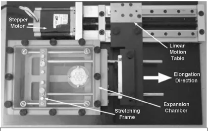
2.1 Axonal stretch-growth device 10
2.2 Stretch-growth of cortical axon tracts 11
2.3 Stretch-growth parameters and DRG axon tracts 12
2.4 Transplanted axonal constructs in the rat spinal cord 13
2.5 Transplanted axonal constructs in the peripheral nervous system 14
- preliminary results
2.6 Survival and stretch-growth of human DRG axons in vitro 16
3.1 Stretch-growth of DRG axons on an agarose hydrogel base layer 22
3.2 Axons spanning the interface of an electrode array and agarose 23
base layer
3.3 Preliminary evidence of axon ingrowth and vascularization within 25
the modified encapsulation design
3.4 Macrophage infiltration adjacent to transplanted DRG neurons 26
3.5 Schematic representation of final encapsulation design 27
static DRG neuron cultures
4.2 Schematic representation of the bridging of a 1cm lesion using 37
stretch-grown axonal constructs
4.3 Harvesting the neural constructs at the 4-week time point 40
4.4 Survival of transplanted DRG neurons within the graft region 42
4.5 Gross comparison of host regeneration within the stretch-grown 44
and static DRG groups
4.6 Identification of host and graft axon fibers and host Schwann cells 45
4.7 Comparison of normalized conduction velocities between the 46
stretch-grown and static culture groups
4.8 Example of columnar alignment of Schwann cells at 2 weeks post- 48
transection
4.9 Schematic representation of the protection of the distal segment 50
using stretch-grown axonal constructs
4.10 Comparison of treated and control distal nerve segments at 54
4-months post-transection
5.2 Examples of PNS-based neural interface systems 66
5.3 Schematic representation of a brain-machine interface using 67
targeted muscle reinnervation
5.4 Schematic representation of a PNS-based neural interface using 69
stretch-grown axonal constructs
5.5 Harvesting of the transplanted neural construct at 4 weeks 73
5.6 Assessment of tissue regeneration within the stretch-grown 76
construct group
5.7 Assessment of tissue regeneration within the static DRG 77
culture group
5.8 Schematic of proposed neural interface design 79
5.9 Gross assessment of the neural interface at transplantation 84
and harvesting
Chapter
1
Chapter 1: Introduction
1.1 Clinical Relevance
Peripheral nerve (PN) trauma is a serious condition affecting 2.8% of trauma
patients annually [Chiono V et al, 2009]. This form of injury is primarily caused
by traumatic accidents, tumor resection or iatrogenic effects of surgeries such as
orthopedic procedures [Kretschmer T et al, 2001], and can often lead to lifelong
pain and disability. Etiologies of PN trauma include penetrating injury, crush and
ischemia [Robinson LR, 2004]. Upper limb injuries occur in over 70% of reported
cases, with the ulnar nerve being most frequently damaged.
Table 1.1 – The Sunderland Classification of Nerve Injury
Sunderland Scale Description
First degree Neurapraxia – segmental demyelination
Second degree Axonotmesis with intact endoneurium
Third degree Axonotmesis with damaged endoneurium, intact perineurium
Fourth degree Axonotmesis with damaged perineurium, intact epineurium
Fifth degree Neurotmesis – disruption of entire nerve trunk
Campbell WW, 2008
The Sunderland classification is a widely used scheme for classifying
peripheral nerve injury [Table 1.1] (Sunderland S, 1990). According to this scale,
the mildest form of PN injury is neurapraxia, or segmental demyelination, which
results in an inability of the nerve to conduct action potentials across the affected
region. The loss of function persists until subsequent remyelination, and is
with crush injuries, refers to the loss of continuity of the axon wherein the
ensheathing structures remain at least partially undamaged. Repair typically
occurs naturally, and the extent of recovery is thought to be a function of the
location of the injury and the degree of disruption of the surrounding stroma. The
most severe form of PN lesion is neurotmesis. Here, the entire nerve trunk is
severed and the ability of the axons to functionally reconnect with their targets is
severely compromised. This type of injury typically requires surgical intervention,
and even minor functional recovery can take several months using current
treatments [Campbell WW, 2008].
The most debilitating form of PN damage occurs as a result of limb
amputation. Without presentation of a distal target, the proximal neurites
frequently undergo extensive collateral branching, resulting in either inaccurate
innervation of surrounding tissue or the formation of painful neuromas. Although
the nerve remains electrophysiologically functional, there currently exists no
chronic means of recording its activity.
1.2 Hypotheses and Goals
By exploiting a newly discovered method of creating axonal tracts in vitro
using mechanical tension, we have been able to create transplantable nerve
constructs that can be used to repair damaged axonal tracts and nerves. In the
lesions and amputations is assessed. The study design was motivated by the
following hypotheses: (1) neural constructs can provide regenerative support for
the proximal stump while attenuating degeneration of the distal pathway, and (2)
living axonal constructs can provide support for the extension and maintenance
of regenerating neurites in the absence of a distal target.
In chapter 2, a synopsis of the stretch-growth mechanism and its
adaptability for various therapeutic applications is provided. Chapter 3 describes
the development of techniques to encapsulate and transplant nervous tissue
constructs into a rat sciatic nerve lesion. Chapter 4 illustrates the beneficial
effects of using these constructs for (a) repairing a short nerve lesion, and (b)
maintaining the support architecture at the distal stump, potentially increasing the
window for successful target reinnervation. Finally, chapter 5 describes how
these axonal constructs can be used to (a) extend and maintain regenerating
proximal axons in the absence of a distal target, and (b) encourage host axonal
growth towards an electrode substrate, which may ultimately enable bi-directional
communication with the peripheral nervous system. A synthesis of these findings
Chapter
2
Overview of Stretch‐induced Axonal
Chapter 2: Overview of Stretch-induced Axonal Growth
The study of axon growth has traditionally comprised two distinct areas –
growth cone extension and guidance during embryogenesis, and nerve
regeneration following traumatic injury. In both these cases, the axon has a free
end that advances towards its final target using chemotaxic and/or haptotaxic
cues. However, we now know that there is a third form of axon growth that
occurs after synaptic integration with the target, and is triggered by the
application of mechanical tension. This form of growth, which we dub
“stretch-growth”, is a newly discovered phenomenon that challenges long-held notions on
axonal transport and protein assembly, and could potentially transform our
understanding of post-embryonic nerve development.
Traditional concepts on axon growth
Growth cone motility and axonal pathfinding during embryonic
development has been extensively studied over the years [Dickson, 2002; Yu
and Bargmann, 2001; Tessier-Lavigne and Goodman, 1996]. During this
process, pioneer axons forge the initial path to the target, following which all
of attractive and repulsive chemotaxic cues in the surrounding milieu, as well as
the internal mechanical forces generated at the tip of the growth cone. Trailing
axons, on the other hand, are believed to respond to a combination of these
chemotaxic cues and contact-influenced guidance. Protein assembly has been
shown to occur within the growing tip. Developing axons grow at a rate of
approximately 1mm/day, and this phase of neurogenesis is typically complete
within 8 weeks in humans.
The other form of axon growth that has been extensively researched is
post-trauma regeneration. Here, the proximal segment of a transected axon
attempts to rejoin its target by a process remarkably similar to that seen during
development. After injury, the neuron shifts from a transmitting mode to a growth
mode resulting in a growth cone lamelipodial formation at the tip (Dahlin, 2006).
This growth cone is guided across the lesion primarily via chemoattractive and
chemorepulsive cues until it reaches the distal support cells, after which the
guidance is primarily contact-mediated (Mueller, 1999; Goodman, 1996). The
response to injury can begin within hours of the insult and may persist for as long
as 12 months [Campbell WW, 2008].
While both of these forms of axonal outgrowth are essential for the
formation and maintenance of the nervous system, neither can account for the
rapid growth of already-integrated axonal tracts in the fetus and thereafter. Since
completely different mechanism, a process of stretch-growth that we are only
now beginning to understand.
Stretch-growth – a newly discovered form of axonal growth
The use of mechanical tension to extend the tip of an axon was first
demonstrated in 1984, where a movable substrate was used to tow chick
sensory ganglion growth cones [Bray, 1984]. This work has expanded upon
since, to induce transient lengthening of the growth cones of single axons in vitro
[Dennerll etal., 1989; Zheng et al., 1991; Heidemann et al., 1995; Chadaet al.,
1997]. The interpretation of these findings was that tension generated at the
growth cone (by internal cytoplasmic components) is a key factor for the growth
of axons. However, the idea that external mechanical forces may play a role in
the growth of synaptically-integrated axon tracts was suggested as far back as
1935 by RG Harrison and 1941 by Paul Weiss, who referred to it as “passive
stretching” and “towing” respectively. They noted that at the peak growth of the
human fetus, motor axons in the lower limb grow at rates approaching
1.2mm/day, which is well within the known rates of axonal transport. However,
evidence of far more rapid axonal growth can be seen elsewhere in nature. Axon
tracts in the giraffe neck have a peak growth rate of 2cm/day [Bannister et al,
1996; Dagg and Foster, 1982]. Axon tracts in the blue whale spine are estimated
to grow at even more astonishing rates, potentially exceeding 3cm/day [Smith
synthesis and transport. The rate of axonal growth has traditionally been thought
to be limited by slow transport – indeed, neurofilament proteins have been shown
to be shipped down the axon at the rate of a few millimeters per day [Brown,
2003; Nixon, 1998a,b; Roy et al, 2000], which is well below the rates described
here. Even at fast transport rates of 300mm/day, it would take over three months
for neurofilament proteins to be carried the 30m distance from the cell body down
the length of a blue whale axon [Smith DH, 2009]. A possible explanation for this
discrepancy could be local synthesis of protein within the axon itself [Alvarez J et
al 2000; Brittis PA et al, 2002], which would preclude the need for such
long-distance transport.
Experimental verification of stretch-growth using rat primary cortical neurons
The development of techniques to create transplantable stretch-grown
axon tracts evolved from in vitro experiments examining traumatic axonal injury
[Wolf et al, 2001] using rat primary cortical neurons. In these experiments,
tension was transiently applied to axons spanning two neural populations
spanning a short gap with the goal of producing a deleterious effect. However, it
was noticed that the axons demonstrated a resilience to stretch at less rapid
strain rates. Expanding upon these findings, an in vitro stretch-growth model
was developed wherein axons spanning two populations of either cortical or
N-tera 2 cell line neurons were allowed to integrate over several days and then
studies, the populations were separated at the rate of 3.5µm every 5 minutes,
resulting in 1cm axon tracts after 10 days of stretch-growth (Figure 2.2, left). The
axons were also seen to coalesce into highly organized fascicles (Figure 2.2,
right), potentially replicating the mechanism of organized axonal tract formation
during development [Smith et al, 2001].
2.1.4 Extreme stretch-growth of rat dorsal root ganglion axons
With a view towards development of a transplantable axonal construct for
eventual clinical usage, dorsal root ganglia (DRG) neurons were selected as our
primary cell source. Adult human DRG neurons can be harvested from patients
undergoing elective ganglionectomies, making them a viable autologous cell
source. Human and animal DRG neurons are also known to be robust in culture,
and can survive for extended periods of time while maintaining normal
electrophysiological function [Pfister et al, 2006a,b].
Subsequent experiments revealed strain rate and acclimation to be the
main determining factors for successful stretch-growth. Starting at the low rate of
1mm/day, the strain rate could be gradually increased, provided the axons were
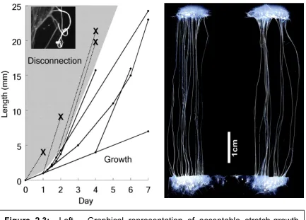
given a sufficient “rest period” in order to acclimate (Figure 2.3a). By employing
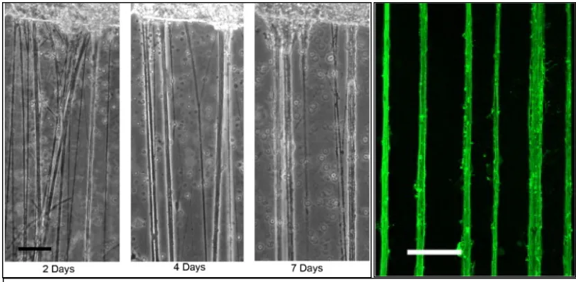
Figure 2.2: Left – The stretch growth of cortical axon tracts as seen at 2, 4 and 7 days. Adjacent fibers gradually coalesce into aligned axon bundles. Right – Confocal image of cortical axon bundles after 7 days of stretch-growth.
this strategy, growth rates of up to 1cm/day were reached within a few days of
commencing stretch-growth [Pfister et al 2004]. In addition, integrated axonal
tracts of up to 10cm length could be created in less than 2 weeks (5cm length
shown in Figure 2.3b).
Despite these high-growth rates, the stretch-grown axons were seen to
possess a normal morphology. However, the average diameter of these axons
was measured to be 30% greater than non-stretched DRG axons, possibly in
order to sustain increased stability and transport. Transmission electron
microscopy revealed a normal complement of microtubules and neurofilaments
per cross-section area. In addition, the axons were shown to be able to conduct
normal action potentials, although they did appear to possess an increased
density of sodium and potassium channels [Pfister et al, 2006a,b].
Using stretch-grown axon tracts to repair the nervous system
In addition to providing insight into the mechanisms of axongrowth during
development, stretch-grown axonal constructs may be exploited for nerve repair.
Living nerve tracts can be rapidly created to any desired length in vitro and
transplanted to bridge even extensive central and peripheral nervous system
lesions. In the case of spinal cord repair, transplanted axonal tracts were used to
bridge a 1cm defect spanning three vertebrae in the rat. The constructs were
seen to survive and remain structurally intact one month post-transplantation
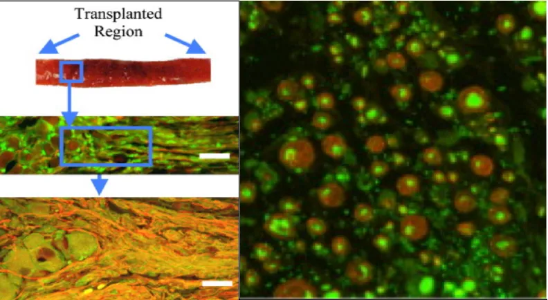
Figure 2.4: Survival of transplanted stretch-grown axons in the spinal cord at 1 month. The transplanted cells (indicated by the arrows) were found sandwiched between layers of collagen in the SCI cavity.
(Figure 2.4) [Iwata A et al, 2006]. Evidence of host axon penetration into the
graft area, as well as graft neurite outgrowth into the host tissue, was also
observed. It is still unclear whether the constructs form functional synaptic
connections with the host – however, these results demonstrate the feasibility of
this approach and merit further investigation.
Axonal constructs have also been used to bridge 1cm lesions in the rat
sciatic nerve (Figure 2.5). The graft tissue was seen to survive for at least 4
months without immunosuppression. Robust intermingling of host and graft
axons was seen in the repaired lesion area as well as in the proximal and distal
nerve segments, indicating graft neurite outgrowth into the host. Analysis of
cross-sections of these nerve segments revealed robust regeneration and
myelination of the axons.
Stretch-grown axons from Human dorsal root ganglia
Ultimately in order to be clinically viable, axonal constructs will need to be
generated from autologous tissue. As previously mentioned, human DRG
neurons can be isolated from patients undergoing elective ganglionectomy
procedures, and can be successfully maintained in vitro. We have been able to
obtain ganglia from live human patients, as well as from organ donors, and have
maintained the tissue in culture for at least 3 months, replicating the findings of
other studies. We have also been able to create stretch-grown axonal constructs
from these human DRGs, thus demonstrating the feasibility of this approach
Figure 2.6: Fluorescence micrographs of adult human DRG neurons and axons. The human DRG neurons were labeled using (A) CGRP or neurofilament proteins; (B) RMO-254; (C) SMI-31; and (D) neurofilament-200. Bottom – Aligned fascicles seen upon stretch-growth of human DRG axons.
Chapter
3
Development of Nervous Tissue
Chapter 3: Development of Nervous Tissue
Encapsulation Techniques
With the goal of creating transplantable axonal tracts that may be used to
bridge CNS and PNS lesions, we have developed techniques to encapsulate and
remove the constructs from the in vitro environment without disrupting their
structural integrity and alignment. The two major components required for
transplantation are an embedding hydrogel and an encapsulating nerve guidance
channel. The primary criteria behind hydrogel selection were structural support
offered, ability to incorporate nutrient supply and permissiveness to neurite
outgrowth. While our initial design solely consisted of a simple collagen
hydrogel, it has since evolved to include a variety of materials, each serving a
unique and critical purpose.
Initial design – collagen hydrogel
The first material employed for construct encapsulation and
removal was a simple collagen hydrogel (3.67mg/mL) in Dulbecco’s modified
Eagle’s medium supplemented with nerve growth factor (2.5S, 10ng/mL). The
collagen matrix was gently applied over the construct axons and allowed to
polymerize in the incubator at 37°C. Upon gelation (typically 10-15 minutes) the
immersed in the collagen. The embedded axons are then rolled along the axis of
the tracts, and the entire hydrogel-tissue combination is deposited into a nerve
guidance channel cut open lengthwise [Iwata et al, 2006].
This approach has several disadvantages. While the 80% collagen
does provide a permissive environment for neurite growth and is seeded with
growth factors, the structural support offered for the axons is minimal. Removal
from the culture environment using this technique can be precarious – axon
tracts are regularly damaged and discarded during the removal process, and
there is no certain way of determining if they are intact even upon successful
transference into the tube. With the goal of overcoming these limitations, and
facilitating easier removal from the culture environment, a variety of additional
hydrogel components were systematically evaluated.
Incorporation of agarose hydrogel
Agarose is a naturally occurring polysaccharide that has been extensively
studied in PNS repair. It can be easily formulated to a wide range of stiffnesses,
each of which has different effects on neurite outgrowth. Neither central nor
peripheral neurons have any known receptors that interact with agarose side
chains.
Our first goal was to evaluate the range of gel stiffnesses that could be
applicable to the construct encapsulation and transfer procedure. It has been
inversely proportional to the stiffness of the gel used, with minimal outgrowth at
formulations exceeding 2% (wt/vol). Our initial design assessment, therefore,
was limited to agarose formulations of 0.5-2.0% (wt/vol).
3.1 Materials and Methods:
Agarose is water-soluble at temperatures exceeding 65°C and typically
gels at a temperature range of 17-35°C. It is clear and stable once gelled, and
does not re-liquefy until heated above 65°C again. The agarose gels used in this
experiment were created in the range of 0.5% to 2.0% (0.25% increments, wt/vol)
by heating and stirring agarose powder (Ultrapure; Invitrogen, Carlsbad, CA) in
pH 7.4 1x Dulbecco’s phosphate buffered saline (PBS; Gibco, Grand Island, NY).
Dorsal root ganglion neurons were isolated from embryonic day 15 fetuses
from timed-pregrant Sprague-Dawley (Charles River, Wilmington, MA) rats. The
DRG explants were suspended at 5x10⁶ cells/mL in Neurobasal® medium
supplemented with 2% B-27, 0.4 mM L-glutamine, 1% penicillin/streptomycin, 2
mg/mL glucose (Sigma-Aldrich, St. Louis, Mo), 10 ng/mL 2.5S nerve growth
factor (NGF) and 1% fetal bovine serum (FBS) (HyClone, Logan, UT), and a
mitotic inhibitor formulation of 10 mM 5-fluoro-2’-deoxyuridine (FdU)
(Sigma-Aldrich), and 10 mM uridine (Sigma-Aldrich) to encourage non-neuronal cell
3.2 Results
Evaluation of agarose as an encapsulating medium
The isolated DRG explants were initially plated at the interface of two
overlapping membranes of Aclar pre-coated with rat-tail collagen (3.67mg/mL) in
order to approximate initial stretch-growth conditions. After allowing the
populations to integrate over 5 days, liquefied agarose formulations at 37°C were
added directly over the cultures and allowed to gel by cooling them to 4°C for 2
minutes, followed by placing them for 10 minutes in the incubator at 37°C. Using
a cell scraper, the agarose was then gently separated from the underlying Aclar
layers. At all formulations exceeding 1.5%, the cells remained preferentially
adhered to the underlying collagen substrate and did not separate from the Aclar.
Below 1%, however, the agarose was structurally fragile and transference into
the tube repeatedly resulted in fragmentation of the gel and disconnection of the
integrated axonal connections. Those instances of successful transfer also
necessitated the length-wise opening of the tubes for insertion and its
subsequent re-suturing. For these reasons the strategy of adding agarose to
already integrated tracts was abandoned, and its use as an underlying substrate
Evaluation of agarose as a base substrate
In the next design iteration, agarose was used as the base substrate on
which the cells were directly plated. Following from our previous findings, the
formulations were now limited to 1.0-1.5%. Liquefied agarose was added to the
bottom Aclar substrate in the elongator chamber, and allowed to gel by cooling it
to 4°C for 2 minutes followed by 10 minutes in the incubator at 37°C. The towing
membrane was then placed over the agarose a thin layer of collagen
(3.67mg/mL) was then coated over the interface and allowed to polymerize in the
incubator overnight. Isolated DRG explants were then added at the interface and
allowed to adhere and integrate over 5 days. In all 3 formulations (1%, 1.25%
and 1.5% wt/vol) axons were seen to span the two populations (Figure 3.1). In
order to assess ease of removal from the culture environment, matched
concentrations of agarose were added to the neural populations and the towing
membranes were gently separated using a cell scraper. The neurons were found
to remain encapsulated within the hydrogel, and axon tracts could be easily cut
out using a scalpel blade and directly inserted into the tube through the open
ends.
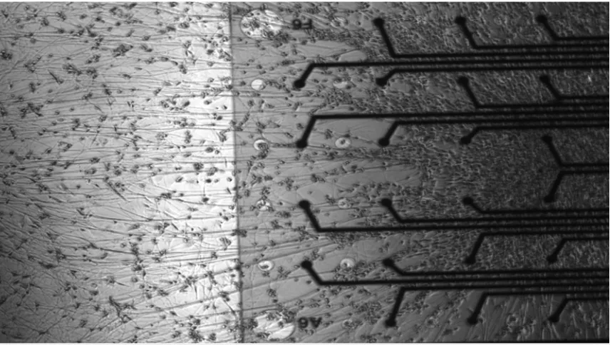
Adaptation of design to incorporate polyimide electrode array
A polyimide electrode array was then incorporated into the design, in
order to develop the optimal parameters for neural-interface encapsulation. After
formation of an initial agarose base layer and placement of the towing
membrane, a thin (1.5mm x 5mm) strip of polyimide was positioned length-wise
over the agarose at the hydrogel-towing membrane interface. A thin layer of
collagen (3.67mg/mL) was added at the towing membrane-polyimide junction,
and more agarose was added to the other end of the polyimide strip to anchor it
in place. As before, isolated DRG neurons were then plated at the interface, and
allowed to integrate over 5 days (Figure 3.2).
During encapsulation and removal from the elongation device, it was
found that the 1% agarose was unable to sufficiently secure the polyimide in
place, resulting in frequent defragmentation of the encapsulated construct during
removal and insertion into the tube. On the other hand, the 1.25% and 1.5%
agarose formulations did not suffer from this structural disintegration. As a result
of these findings, and with the goal of minimizing the agarose stiffness, a final
agarose concentration of 1.25% wt/vol was selected.
In vivo evaluation of encapsulation design
In order to assess the feasibility of this design in vivo, static neural
constructs were generated, encapsulated in 1.25% agarose, inserted into a 1cm
sciatic nerve. The constructs were harvested at 2 weeks and examined for signs
of nerve regeneration, vascularization and/or host immune response. The
agarose was still predominantly intact at this time. Axon infiltration into the tube
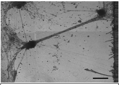
was observed, as were signs of vascular ingrowth (Figure 3.3). In addition
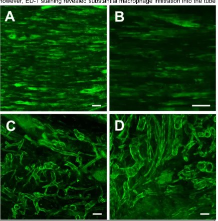
however, ED-1 staining revealed substantial macrophage infiltration into the tube
Figure 3.3: Confocal reconstructions of the regenerating sciatic nerve (GFP+)
(Figure 3.4), and in the general vicinity of the transplanted neurons. While
macrophages are a necessary element in the degradation of agarose, their
infiltration was perceived to be potentially disadvantageous considering the
presence of the allograft tissue.
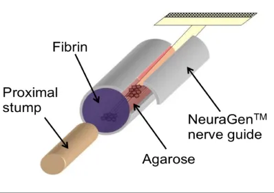
Final encapsulation design
With the goal of attenuating macrophage infiltration, salmon fibrin gel
(5mg/ml fibrinogen, 5 NIH units/ml thrombin in Neurobasal) was incorporated into
the final encapsulation design (Figure 3.5). Salmon fibrin has been shown to
degrade more slowly than mammalian fibrin, and is resistive to glial and
macrophagic infiltration [Ju Y et al, 2007; Georges PC et al, 2006]. Fibrin is also
extensively used as a surgical adhesive, thus making it a clinically appropriate
choice of end sealant.
Figure 3.5: Schematic representing final construct encapsulation design. The stretch-grown axons, pre-attached to a polyimide electrode array, are encapsulated in 1.25% agarose hydrogel and inserted into a NeuragenTM
3.3 Conclusion
By exploiting the mechanism of axonal growth, we have been able to
engineer living axonal tracts of several centimeter lengths in vitro. We have
demonstrated our ability to utilize these constructs to repair lesions in the central
and peripheral nervous system. By optimizing the encapsulation techniques, we
have been able to create a stable method of removing the axonal tracts from the
culture environment without disrupting their alignment and structural integrity.
This encapsulation design also permits the incorporation of a thin flexible
electrode substrate for the goal of creating a stable neural interface with the
Chapter
4
Repair of Lesions in the Peripheral
Nervous System Using Stretch‐Grown
Chapter 4: Repair of Lesions in the Peripheral Nervous
System Using Stretch-Grown Axonal Constructs
4.1 Overview
The current ‘gold standard’ for repair of PN lesions are autologous nerve
grafts. These grafts, typically of the sural nerve, provide a physical bridge across
the lesion and supply endogenous biological cues to guide regenerating axons
from the proximal nerve stump to the remaining distal nerve segment. However,
this approach is plagued by permanent loss of harvested nerve function and the
potential formation of painful neuromas [Lundborg, 2004; Sinis et al, 2007].
Furthermore, there are obvious limitations in the supply of donor nerves, making
autografts inadequate to repair extensive nerve damage. Alternative clinical
approaches to bridge PN lesions include synthetic tubes composed of
poly(glycolic acid) (PGA) or collagen; however, these conduits by themselves
have been only used for bridging relatively small peripheral nerve (PN) gaps
(Dellon, 2006; Trumble et al., 2006). Moreover, no current strategy
simultaneously addresses the steady degeneration of support cells in the nerve
segment distal to the injury site. This degeneration ultimately severely limits
recovery of function due to the eventual loss of cues to guide regenerating axons
beyond the lesion to their final targets. This need is particularly important in
several centimeters, or complex lesions to multiple nerves where conventional
approaches are overwhelmed or unsuitable. In these instances, the rate of
regeneration, ranging between 1-4 mm/day, may result in repair taking months or
even greater than one year, depending on the location of the injury, over which
time the distal support cells no longer remain. In addition, the end targets
themselves will undergo degeneration and atrophy after a period of denervation,
further impeding functional recovery [Lien SC et al, 2008]. Thus, there is clearly a
need to address the issue of distal pathway degeneration in conjunction with
regeneration of the proximal stump in order to enable complete functional
recovery.
More recently, tissue-engineered solutions have been sought to overcome
the limitations associated with autografts and nerve guidance channels. These
approaches include creating combinations of permissive scaffolds (such as
decellularized grafts or hydrogels), extracellular matrix (ECM), trophic factors,
and glial or stem cells (Evans et al., 2002; Frerichs et al., 2002; Lee et al., 2003;
Yu and Bellamkonda, 2003; Fansa and Keilhoff, 2004; Hu et al., 2005; Stang et
al., 2005; Chalfoun et al., 2006; Keilhoff et al., 2006; Nie et al., 2007). The
importance of anisotropy has been well recognized as an important spatial cue to
direct axon growth, and is typically achieved via gradients (e.g., neurotrophic or
ECM), longitudinally-aligned fibers, or tailored porosity (Matsumoto et al., 2000;
Typically, the optimization of these tissue-engineered constructs in vitro focuses
on developing permissive environments for axonal growth cone extension.
Unfortunately, these approaches typically ignore the fact that axonal
outgrowth in vivo occurs along Schwann cells and the basal lamina; thus,
strategies that were optimized based on directly promoting axonal outgrowth in
vitro may not translate mechanistically. In addition, all of these approaches are
geared towards promoting regeneration of the proximal stump; none of these
strategies can delay the eventual degeneration of the distal pathway.
We have recently begun investigating axon-mediated axon outgrowth as
an alterative mechanism of regeneration. By exploiting the newfound
mechanism of “stretch-growth”, whereby large axon tracts can be created by the
controlled and continuous separation of integrated neural populations, we have
created transplantable neural constructs that have been shown to effectively
bridge a 1cm sciatic nerve lesion in the rat 4 months post-transplantation.
However, the advantage of utilizing stretch-grown axonal constructs vis-à-vis a
simple (or “static”) neural culture was unknown. We also observed evidence of
graft axon outgrowth into the host nerve; this suggests the possibility of
maintaining the distal pathway support architecture by providing a temporary
axonal surrogate, a phenomenon often referred to as nerve “babysitting”.
Therefore, the objectives of this study were to (1) determine if stretch-grown
regeneration, and (2) assess whether axons emerging from the graft can
attenuate the degeneration of the distal nerve segment.
4.2 Repair of a 1cm PNS lesion using stretch-grown axons
4.2.1 Relevant Background
During development of the peripheral nervous system, axons known as
“pioneer axons” prescribe the initial path for subsequent axons to follow in a
process known as “selective fasciculation” [Dickson, 2002; Yu and Bargmann,
2001; Tessier-Lavigne and Goodman, 1996]. The pioneer axons are believed to
enable this targeted axonal outgrowth by a combination of neurotrophic support
and contact guidance. This phenomenon has inspired a variety of strategies in
the area of peripheral nerve repair. Several groups are investigating the use of
growth factors and/or Schwann and glial cell combinations to enhance nerve
growth within nerve guidance channels [Evans et al., 2002; Frerichs et al., 2002;
Lee et al., 2003; Yu and Bellamkonda, 2003; Fansa and Keilhoff, 2004; Hu et al.,
2005; Stang et al., 2005; Chalfoun et al., 2006; Keilhoff et al., 2006; Nie et al.,
2007]. Yet others have shown that axons appear to prefer longitudinally-aligned
fibers [Kim et al, 2008; Bellamkonda, 2006] as a regenerative substrate, and
attempts are being made to create trophic factor-eluting versions of these [Cao et
Building upon these findings, we hypothesize that the axonal portions of
the tissue-engineered neural constructs contribute more to host PN regeneration
than static cultures, as they may provide both trophic support, as well as a
longitudinally aligned pre-established pathway that spans the entire lesion. In
this study, we investigate the repair of a 1cm sciatic nerve lesion using
transplants of stretch-grown axonal constructs as well as static neural (dorsal
root ganglion) cultures.
4.2.2 Materials and Methods
Overview of Study Design
Building upon our previous findings on host nerve regeneration at 6 weeks
and 4 months [Huang J et al, 2009], the initial study design required harvesting
the nerve at the 2-week and 4-week time points in order to assess the
progression of the nerve through the graft. However, an assessment of host
regeneration at 2 weeks revealed similar results to the 4-week findings. This can
be attributed to the early time point – within days of repair, the proximal nerve
stump will undergo rapid regeneration and abundant collateral sprouting, both of
which will be maintained for several weeks to months. As a result of these
At 4-weeks post-transplantation, the nerves were re-exposed for
electrophysiological evaluation and then immediately harvested for
immunohistochemistry. This time-point was selected since the host nerve would
have regenerated through the length of the transplant, allowing us to compare
the extent (number of axons) and quality (nerve conduction velocity and latency)
of the repair.
Dorsal root ganglion neuron isolation
Dorsal root ganglion (DRG) neurons were isolated from embryonic day 15
fetuses from timed-pregrant Sprague-Dawley (Charles River, Wilmington, MA)
rats. The DRG explants were suspended at 5x10⁶ cells/mL in Neurobasal®
medium supplemented with 2% B-27, 0.4 mM L-glutamine, 1%
penicillin/streptomycin, 2 mg/mL glucose (Sigma-Aldrich, St. Louis, Mo), 10
ng/mL 2.5S nerve growth factor (NGF) and 1% fetal bovine serum (FBS)
(HyClone, Logan, UT), and a mitotic inhibitor formulation of 10 mM
5-fluoro-2’-deoxyuridine (FdU) (Sigma-Aldrich), and 10 mM uridine (Sigma-Aldrich) to
Static Nervous Tissue Constructs
Figure 4.1: Schematic representation of the bridging of a 1cm sciatic nerve lesion
using static DRG neuron cultures
The explants were plated into a collagen-filled (rat tail type 1, 3.67mg/mL)
agarose (1.25% in 1X PBS; Ultrapure, Invitrogen, Carlsbad, CA) trough
measuring 2mm x 1cm. The cell cultures were allowed to form axonal
connections over 12 days in vitro. The neural growth medium described above
Stretch-grown Nervous Tissue Constructs
Figure 4.2: Schematic representation of the bridging of a 1cm sciatic nerve lesion
using stretch‐grown axons
The explants were plated into mechanical elongation chambers
custom-fabricated for the stretch-growth procedure. The neurons were plated in two
aclar “towing” membrane and a base layer of agarose (1.25% in 1X PBS;
Ultrapure, Invitrogen, Carlsbad, CA), resulting in a separation of 50-100µm. Over
5 days in vitro, axonal connections were formed between these two populations.
The populations were then gradually separated over the course of 7 days using a
stepper motor system until the axons spanning them reach a length of 1cm.
Neural Construct Encapsulation
On post-isolation day 12, the neural cultures (both static and
stretch-grown) were removed from the incubation chambers and embedded in a
collagen-based matrix (3.0 mg/mL) in Dulbecco’s modified Eagle’s medium
supplemented with NGF (2.5S, 10 ng/mL). After gelation at 37°C, embedded
cultures were gently removed and placed within a 1cm absorbable collagen
nerve guidance channel (3mm inner diameter, NeuraGenTM, Integra LifeSciences
Corp, Plainsboro, NJ).
Peripheral Nerve Surgery and Tissue Construct Implantation
Stretch-grown (n=10) as well as static (n=8) constructs were implanted
into GFP+ rats (adult male, 400-450mg, strain TgN(act-EGFP)OsbCZ-004). The
prior to surgery. A longitudinal skin incision was made in the hind leg from the
sciatic notch proximally to the region of the popliteal fossa distally. The gluteal
muscle was freed up to expose the sciatic nerve and its posterior tibial branch. A
1cm segment of sciatic nerve was transected immediately proximal to the
trifurcation. The proximal and distal stumps were carefully inserted into the ends
of the NeuraGenTM tube with an overlap of 1mm, and the epineurium was
secured to the tube using 8-0 absorbable sutures. The ends of the tube were
sealed with salmon fibrin (5mg/ml fibrinogen, 5 NIH units/ml thrombin) to provide
additional stability, and to attenuate macrophage infiltration into the tube. Finally,
the muscle and skin layers were sutured over the graft with absorbable chromic
gut 4-0 sutures.
Nerve Electrophysiology
The sciatic nerve was re-exposed and 4 weeks (n=6) using the
aforementioned procedure. The animal was placed on a heating pad to maintain
body temperature at 37°C. A stimulating electrode was placed 2mm proximal to
the transection site, and the recording cuff was placed 2mm distal to the graft.
All recordings were obtained at a sampling frequency of 4000 samples/s for 20s
(MP-150; Biopac Systems Inc, Goleta, CA). Stimulation was provided using a
50µA, 100µs, constant current pulse train (SIU-91A; Cygnus Technology,
Delaware Water Gap, PA); the current pulse train was controlled by a gating
velocity. The procedure was repeated on the contralateral side for each animal,
and the ratio of conduction velocity of the operated side to the non-operated
nerve was determined. All related data analyses were performed using standard
software (Acknowledge 3.9.1; Biopac Systems Inc, Goleta, CA).
Nerve Harvest and Histological Analyses
Immediately following electrophysiological evaluation, the constructs were
harvested by transecting the nerve 2mm proximal and 2mm distal to the graft
(Figure 4.3), and were placed in 4% paraformaldehyde. The animals were
euthanized by an overdose of sodium pentobarbital.
Following overnight fixation in paraformaldehyde, the excised tissue was
removed and immersed in 30% sucrose solution. The tissue was then
cryosectioned longitudinally (15-25µm), and immunostained for (i)
neurofilament-200kDa (NF-200; N0142, 1:200; Sigma-Aldrich), (ii) myelin basic protein (MBP;
SMI-94R, 1:500, Covance Research Products, Princeton, NJ), (iii) S-100 (1:500;
Dako, Carpinteria, CA), or (iv) calcitonin gene-related peptide (CGRP; C8198,
1:1000 Sigma-Aldrich). The appropriate secondary fluorophore-conjugated
antibodies (TRITC- or AMCA-conjugated IgG; Jackson ImmunoResearch, West
Grove, PA) were used. The sections were examined under an epifluorescent
microscope (Eclipse E600; Nikon, Melville, NY) and the images were digitally
4.2.3 Results
Stretch-grown and static neural construct survival
Stretch-grown constructs were harvested at the 4-week (n=6) and 8-week
(n=2) time points. All static constructs (n=6) were harvested 4-weeks
post-transplantation. At both time-points the transplanted area could be clearly
identified as the collagen tube was still un-resorbed and maintained its structural
integrity. Within the tubes the transplanted ganglia and their axonal connections
were identified using the neuronal/neuritic cytoskeletal protein NF-200; these
areas did not co-localize with green fluorescent protein, indicating non-native
tissue (Figure 4.4). Examination of the grafts also revealed robust
vascularization along their entire lengths, which is a necessary requirement for
the survival and maintenance of host and graft tissue. Also, as observed in our
previous studies, there was no overt sign of immunologic response as denoted
by infiltrating macrophages and neutrophils despite the lack of immune
suppressant usage.
Host regeneration across stretch-grown constructs
Host nerve fibers were seen to extend across the length of the 1cm lesion
in the stretch-grown as well as the static neural transplants (Figure 4.5).
However, far most extensive host infiltration (neural and vascular) could be seen
in the stretch-grown group, whereas the static group had preferential neurite
growth along the walls of tube. The host fibers were identified as those
expressing NF-200 co-localized with GFP (Figure 4.6 A-C). Remnants of the
agarose were still present at 4 weeks; the axons were seen to grow between the
agarose and the walls of the collagen tube in both cases, presumably due to the
relative impermeability of the still-disintegrating agarose fragments. After 8
weeks in vivo, however, the nerve and accompanying vasculature was
At 4 weeks, all constructs were also seen to contain GFP+/S-100+
Schwann cell bodies in the vicinity of the host axon fibers (Figure 4.6D). These
same cell bodies did not positively express myelin basic protein (SMI-94),
suggesting that they had not yet converted into a myelinating phenotype.
As previously mentioned, the constructs were also evaluated at 2 weeks
(n=2 per group). However, as no measurable differences were observed when
compared with the 4-week time point, evaluation at 2 weeks was discontinued.
Figure 4.6: (A-C) Host regeneration within the construct. NF-200 staining (red) was expressed by both host and graft axons. Areas of co-localization (white arrow) indicate host axon fibers. (D) Schwann cells expressing S-100
Electrophysiological assessment
Evaluation of the electrophysiology of the repaired nerves at 4 weeks
revealed clear differences between the stretch-grown and static groups. The
ratio of the conduction velocities of the treatment and contralateral control sides
were calculated, and nerves with stretch-grown axons demonstrated a
statistically significant improvement over static cultures. This correlates with
observed differences in regenerated axonal fiber density. It should be noted that
the recorded conduction velocities are less than twenty times that of normal
0 0.5 1 1.5 2 2.5 3 3.5 4 4.5
Stretch‐grown axons Static Cultures
Percentage of contralateral side
Comparison of Normalized Conduction
Velocities at 4 weeks
nerves. This could be attributed in part to the still-incomplete regeneration as well
as the previously noted absence of myelination.
4.3 Attenuation of distal degeneration using a neural construct
4.3.1 Relevant Background
Following the transection of a nerve, distal axons – which have been
physically cut off from their neural cell bodies – undergo a gradual degenerative
process known as Wallerian degeneration. Within a few hours of transection, the
axolemma fuses and seals the end; the axons themselves begin to disintegrate
within a few days. Myelin and other cellular debris are phagocytosed by
macrophages within 6 weeks. Concurrent with the loss of the axons, Schwann
cells begin to proliferate at the site of the injury and organize themselves into
aligned columns known as bands of Bungner (Figure 4.8). These Schwann cells
upregulate their synthesis of neurotrophic factors including NGF, BDNF and NT-3
[Heumann, 1987; Thoenen et al, 1988; Funakoshi et al, 1993; Fu and Gordon,
1995; Raivich and Makwana, 2007] in order to attract regenerating axons of the
proximal stump into the aligned columns. If regeneration is successful, axonal
sprouts will advance through the bands of Bungner and ultimately innervate their
appropriate distal targets within a few (3-6) months of the injury. If reinnervation
does not occur within this timeframe, however, the recovery is likely to be
increase in the collagen content of the endoneurium and perineurium can lead to
an inhospitable environment for regenerating axons [Salonen et al, 1985].
However, the predominant factor contributing to this poor recovery is the gradual
reduction of neurotrophic support and eventual disintegration of the Schwann cell
basal lamina [Sunderland, 1950], which is almost entirely complete within 4-6
months.
A variety of strategies have been investigated with the goal of delaying
this distal atrophy and improving functional outcome. In the case of proximal
nerve injuries or long nerve gaps, clinicians regularly perform distal motor nerve
transfers in order to hasten muscle reinnervation [Mackinnon, Roque and Tung,
2007]; however, this results in impaired function at the donor site. Moreover, in
many situations a suitable motor nerve candidate is simply not available.
Figure 4.8: Example of columnar alignment of Schwann cells (S-100, green) in the sciatic nerve 2 weeks post-transection. Scale bar = 50µm.
Sensory nerve innervation of the distal stump has been shown to maintain a
favorable neurotrophic milieu for at least 6 months post-transection [Michalski et
al, 2008; Bain et al, 2001]. Here too, however, the sacrifice of an otherwise
healthy nerve is required for improvement of outcome at the graft site.
Building upon these approaches, and our observation of graft
axons extending into the host nerve stumps [Huang J et al, 2009], we
hypothesize that axonal processes extending from a transplanted neural
construct may maintain the support architecture of the distal segment, potentially
leading to improved functional recovery even after delayed reinnervation. In this
study, we compare the extent of degeneration of the distal stump over 16 weeks
after treatment with a transplanted static neural construct vis-à-vis no repair.
4.3.2 Materials and Methods
Dorsal root ganglion neuron isolation
Dorsal root ganglion (DRG) neurons were isolated from embryonic day 15
fetuses from timed-pregrant Sprague-Dawley (Charles River, Wilmington, MA)
rats. The DRG explants were suspended at 5x10⁶ cells/mL in Neurobasal®
medium supplemented with 2% B-27, 0.4 mM L-glutamine, 1%
penicillin/streptomycin, 2 mg/mL glucose (Sigma-Aldrich, St. Louis, Mo), 10
ng/mL 2.5S nerve growth factor (NGF) and 1% fetal bovine serum (FBS)
5-fluoro-2’-deoxyuridine (FdU) (Sigma-Aldrich), and 10 mM uridine (Sigma-Aldrich) to
encourage non-neuronal cell elimination.
Static Nervous Tissue Constructs
The explants were plated into a collagen-filled (rat tail type 1, 3.67mg/mL)
agarose (1.25% in 1X PBS; Ultrapure, Invitrogen, Carlsbad, CA) trough
measuring 2mm x 1cm. The cell cultures were allowed to form axonal
connections over 12 days in vitro. The neural growth medium described above
was replaced on post-isolation days 1, 3, 5 and 12.
Figure 4.9: Schematic representation depicting the use of neural constructs to
Neural Construct Encapsulation
On post-isolation day 12, the neural cultures were removed from the
incubation chambers and embedded in a collagen-based matrix (3.0 mg/mL) in
Dulbecco’s modified Eagle’s medium supplemented with NGF (2.5S, 10 ng/mL).
After gelation at 37°C, embedded cultures were gently removed and placed
within a 1cm absorbable collagen nerve guidance channel (3mm inner diameter,
NeuraGenTM, Integra LifeSciences Corp, Plainsboro, NJ).
Peripheral Nerve Surgery and Tissue Construct Implantation
All procedures were performed using GFP+ rats (adult male, 400-450g,
strain TgN(act-EGFP)OsbCZ-004). The animals were anesthetized with sodium
pentobarbital (Nembutal, 50 mg/kg, i.p.) prior to surgery. A longitudinal skin
incision was made in the hind leg from the sciatic notch proximally to the region
of the popliteal fossa distally. The gluteal muscle was freed up to expose the
sciatic nerve and its posterior tibial branch, and the nerve was transected 1cm
proximal to the trifurcation. For construct transplantations (n=6), the distal stump
was carefully inserted into one end of the NeuraGenTM tube with an overlap of
1mm, and the epineurium was secured to the tube using 8-0 absorbable sutures.
The ends of the tube were sealed with salmon fibrin (5mg/mL fibrinogen, 5 NIH
units/ml thrombin) to provide additional stability, and to attenuate macrophage
infiltration into the tube. For both neural construct transplantations and controls
(n=6), the proximal stump was capped and the distal stump/tube was covered in
inadvertent regeneration of the proximal stump into the transection site. Finally,
the muscle and skin layers were closed using absorbable chromic gut 4-0
sutures.
Nerve Harvest at 16 weeks and Histological Analyses
The surgical site was re-exposed at the 16-week time point (with the
exception of one neural construct graft and one control nerve that was left in vivo
for 8 months), and the distal stumps were harvested by transecting the nerve as
far as could be exposed distal to the graft (typically 1cm). The nerves and were
immediately placed in 4% paraformaldehyde and the animals were euthanized by
an overdose of sodium pentobarbital.
Following overnight fixation in paraformaldehyde, the excised tissue was
removed and immersed in 30% sucrose solution. The tissue was then
cryosectioned longitudinally (15-25µm), and immunostained for (i)
neurofilament-200kDa (NF-200; N0142, 1:200; Sigma-Aldrich), (ii) S-100 (1:250; Dako,
Carpinteria, CA), or (iii) calcitonin gene-related peptide (CGRP; C8198, 1:1000
Sigma-Aldrich). The appropriate secondary fluorophore-conjugated antibodies
(TRITC- or AMCA-conjugated IgG; Jackson ImmunoResearch, West Grove, PA)
were used. The sections were examined under an epifluorescent microscope
(Eclipse E600; Nikon, Melville, NY) and the images were digitally captured (Spot