Conserved bone microstructure in the shells of long-necked
and short-necked chelid turtles (Testudinata, Pleurodira)
Torsten M. Scheyer
Palontologisches Institut und Museum, Universitt Zrich, Karl Schmid-Strasse 4, 8006 Zrich, Switzerland. E-mail: [email protected]
Introduction
Pleurodira or side-necked turtles contain the three
ma-jor crown clades Chelidae, Pelomedusidae and
Podoc-nemidae, as well as the fossil clades Dortokidae,
Platy-chelyidae,
Euraxemydidae,
Bothremydidae
and
Araripemydidae (Lapparent de Broin 2001; Danilov
2005; Gaffney et al. 2006). Whether
Proterochersis
ro-busta
Fraas, 1913 lies within Pleurodira as sister taxon
to all other pleurodirans (e.g. Gaffney et al. 2006), or
outside of Testudines on the turtle stem (e.g. originally
proposed by Rougier et al. 1995; Sukhanov 2006; Joyce
2007; Sterli 2008), is contested.
Today, Chelidae represents about 55/60 living species
in 14/18 genera (lower numbers from Fritz & Havasˇ
2007; higher numbers from Bickham et al. 2007), but the
fossil record of Chelidae must still be considered to be
poor (Gaffney et al. 2006). Both modern and fossil
Che-lidae are restricted to the South American continent and
Australasia (Australia and New Guinea; Georges et al.
1998). Fossil chelids are unambiguously known from late
Early and Late Cretaceous as well as the Palaeogene and
Neogene strata of South America (Broin 1987; de la
Fuente et al. 2001; Lapparent de Broin & de la Fuente
2001; de la Fuente 2003) and from the Miocene and the
Eocene/?Palaeocene of Australia (Gaffney et al. 1989;
Lapparent de Broin & Molnar 2001). According to Joyce
et al. (2004), Early Cretaceous turtle remains from South
America may represent stem-group taxa of the Chelidae.
There is still an ongoing discussion about the
interre-lationships of chelid turtles (Fig. 1). In the classical and
more recent morphological approaches (e.g. Boulenger
1888a; Burbidge et al. 1974; Gaffney 1977; Gaffney &
Meylan 1988; de la Fuente 2003; Bona & de la Fuente
2005), two clades are recognised: (1) the long-necked
genera of South America (i.e.
Chelus
,
Hydromedusa
)
and Australasia (i.e.
Chelodina
) and (2) the South
American and Australasian short-necked chelid genera
(e.g.
Phrynops
,
Platemys
,
Emydura
). Gaffney et al.
(1989, p. 7) pointed out that the “
Emydura
group is
characterised almost entirely by features that are
plesio-morphic for Chelidae”. In contrast, phylogenetic
ana-lyses that are based on molecular and serological data
support the hypothesis (even though the support is only
weak in most cases) that the South American chelids
on the one hand, and the Australasian chelid taxa on
the other hand are more closely related to each other
respectively, regardless of the neck lengths (Seddon
et al. 1997; Shaffer et al. 1997; Georges et al. 1998;
Fu-jita et al. 2004; Near et al. 2005; Krenz et al. 2005).
Received 23 May 2008
Accepted 7 August 2008
Published 20 February 2009
Key Words
Testudines
bone histology
long-necked Chelidae
short-necked Chelidae
Platychelys oberndorferi
Abstract
Extant and fossil chelids are restricted to South America and Australasia. Based on
morphological data, long-necked and short-necked chelids are hypothesised to form
natural groups respectively, whereas molecular and serological data indicate South
American and Australasian chelids are monophyletic, regardless of neck-length. Here I
provide shell bone histological and microanatomical data and character mapping of
seven chelid taxa and the Late Jurassic stem-pleurodiran
Platychelys oberndorferi
The dimensions and shape of chelid shells are quite
diverse ranging from small to large sizes and from
smooth, unsculptured shells to strongly sculptured
morphs carrying longitudinal ridges or knobs (e.g. the
matamata). Similarly, the bone composition of the shell
of chelids is highly variable, as exemplified by the high
variation in the neural series from completely present
to complete reduced (Pritchard 1988, 2008; Thomson
& Georges 1996). The development of the pleurodiran
shell was recently studied, including a series of the
che-lid
Emydura subglobosa
(Krefft, 1876). Results were
found to be in overall accordance with data from
Cryp-todira and an alternative structural hypothesis for neural
reduction is presented, based on heterochronic shift
ac-companied by restricted influence of epaxial
muscula-ture and dermal-epidermal interaction in bone
forma-tion (Scheyer et al. 2008).
Recently, the presence of phylogenetic signals in
bone histological and microanatomical data was
veri-fied (e.g. Laurin et al. 2004; Cubo et al. 2005). The
mi-crostructure of turtle shell bones was found to be
simi-larly influenced to various degrees by phylogenetic and
functional influences and constraints (e.g. Scheyer &
Sander 2007; Scheyer & Anquetin 2008). In the current
study the question is thus addressed, whether the bone
microstructures of the shell of chelid turtles can be
used to test the competing phylogenies described above,
or whether other, i.e., functional, aspects that work on
the shell bone obscure any systematic value.
Accord-ingly, the microstructures of the turtle shell bones of
seven chelid taxa, including three extant long-necked
species, three extant short-necked species and a fossil
short-necked taxon are described. The palaeobiology of
each taxon is briefly reviewed. Furthermore, to analyse
how a stem-group pleurodiran taxon relates to the fossil
and modern chelid genera, the Late Jurassic European
taxon
Platychelys oberndorferi
Wagner, 1853 was
sampled. With the addition of the two stem taxa
Proga-nochelys quenstedti
Baur, 1887 and
Kayentachelys
sp.
(Scheyer 2007; Scheyer & Sander 2007), the bone
his-tological characters were then mapped on the two
com-peting hypotheses of chelid relationships (Fig. 1).
Material and methods
scriptions are mainly based on Francillon-Vieillot et al. (1990) and Scheyer & Snchez-Villagra (2007). If not specifically expressed, the plane of sectioning in costals is either anteroposteriorly (perpendicular to the progression of the ribs) or proximodistally oriented (parallel to the progression of the ribs), generally proximodistally oriented for peripherals and either anteroposteriorly or proximodistally oriented for plastral elements. Data on the samples used in this study were compiled into Table 1. For additional data on specimens refer to Scheyer (2007).
Due to the small sampling size, individual variation, ontogenetic variation, or variation based on sexual dimorphism cannot be ruled out; however, because significant changes in the bone histology seem to occur only on higher taxonomic levels, i.e. generic or family levels,
they are not expected either (Scheyer & Snchez-Villagra 2007; Scheyer & Sander 2007).
In the case of extant liquid-preserved (e.g. alcohol, formaldehyde) turtle specimens that are not macerated, the sampling was realised by core-drilling (e.g. Scheyer 2007; Scheyer & Snchez-Villagra 2007) and where applicable, the keratinous shield and dermal cover of the bones were left in place. Especially in larger specimens, it would be more sensible to sample the whole bone element to get a complete picture instead of a drilled core. However, the restricted sampling area of the drilling method parallels the situation in fragmentary fossils where the whole bone is not available for study.
Character mapping of the bone histological data was done using the software package Mesquite (Maddison & Maddison 2004).
Char-Table 1.
Material sectioned for the study including taxa names, accession numbers, element descriptions and general
re-marks.
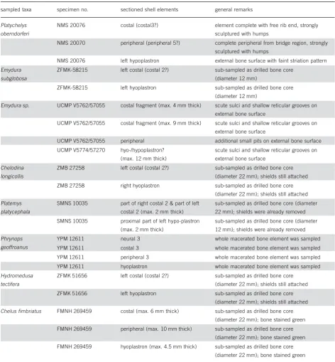
sampled taxa specimen no. sectioned shell elements general remarks
Platychelys
oberndorferi
NMS 20076 costal (costal3?) element complete with free rib end, strongly
sculptured with humps
NMS 20070 peripheral (peripheral 5?) complete peripheral from bridge region, strongly
sculptured with humps
NMS 20076 left hypoplastron external bone surface with faint striation pattern
Emydura
subglobosa
ZFMK-58215 left costal (costal 2?) sub-sampled as drilled bone core
(diameter 12 mm)
ZFMK-58215 left hyoplastron sub-sampled as drilled bone core
(diameter 12 mm)
Emydura sp. UCMP V5762/57055 costal fragment (max. 4 mm thick) scute sulci and shallow reticular grooves on
external bone surface
UCMP V5762/57055 costal fragment (max. 9 mm thick) scute sulci and shallow reticular grooves on
external bone surface
UCMP V5762/57055 peripheral additional small pits on external bone surface
UCMP V5774/57270 hyo-/hypoplastron?
(max. 12 mm thick)
scute sulci and shallow reticular grooves on
external bone surface
Chelodina
longicollis
ZMB 27258 left costal (costal 2?) sub-sampled as drilled bone core
(diameter 22 mm); shields still attached
ZMB 27258 right hyoplastron sub-sampled as drilled bone core
(diameter 22 mm); shields still attached
Platemys
platycephala
SMNS 10035 part of right costal 2 & part of left
costal 2 (max. 2 mm thick)
sub-sampled as drilled bone core (diameter
22 mm); shields were already removed
SMNS 10035 proximal part of left hypo-plastron
(max. 2 mm thick)
sub-sampled as drilled bone core (diameter
12 mm); shields were already removed
Phrynops
geoffroanus
YPM 12611 neural 3 whole macerated bone element was sampled
YPM 12611 costal 3 whole macerated bone element was sampled
YPM 12611 peripheral 3 whole macerated bone element was sampled
YPM 12611 hyoplastron whole macerated bone element was sampled
Hydromedusa
tectifera
ZFMK 51656 left costal (costal 2?) sub-sampled as drilled bone core
(diameter 22 mm); shields still attached
ZFMK 51656 left hyoplastron sub-sampled as drilled bone core
(diameter 22 mm); shields still attached
Chelus fimbriatus FMNH 269459 costal (max. 6 mm thick) sub-sampled as drilled bone core
(diameter 22 mm); bone stained green
FMNH 269459 peripheral (max. 10 mm thick) sub-sampled as drilled bone core
(diameter 22 mm); bone stained green
FMNH 269459 hyoplastron (max. 4.5 mm thick) sub-sampled as drilled bone core
acters were treated as ‘unordered’ and traced over the two composite hypotheses using ‘parsimony ancestral state reconstruction’. The tree length was recorded for both phylogenies using the ‘tree value using character matrix’ option, and additional tree descriptions and apomor-phy lists using both Acctran and Deltran optimisation were performed using PAUP 4.0beta10 (Swofford 2002).
Where appropriate, length and width measurements and maximum shell bone thicknesses are given for the sampled turtle shells, in which CCL stands for curved carapace length, CCW for curved cara-pace width, SPL for straight plastron length, and CPL for curved plastron length (see also Wyneken 2001).
Institutional Abbreviations. FMNH, The Field Museum, Chicago, Illi-nois, USA; IPB, Goldfuss-Museum, Institut fr Palontologie, Univer-sitt Bonn, Germany; SMNS, Staatliches Museum fr Naturkunde, Stuttgart, Germany; NMS, Naturmuseum Solothurn, Switzerland; UCMP, Museum of Paleontology, University of California at Berkeley, California, USA; YPM, Peabody Museum of Natural History at Yale University, New Haven, Connecticut; USA; ZFMK, Zoologisches For-schungsinstitut und Museum Alexander Koenig, Bonn, Germany; ZMB, Zoologische Sammlung, Museum fr Naturkunde, Humboldt-Universitt zu Berlin, Germany.
Platychelys oberndorferi Wagner, 1853. According to Joyce (2007), P. oberndorferiis one of only three taxa that are currently hypothe-sised to unambiguously represent the stem-group of Pleurodira. P. oberndorferiwas first described from lithographic limestone (Late Jurassic) near Kehlheim, southern Germany, but the taxon became much better known from Late Jurassic shallow marine limestones that were quarried near Solothurn, Switzerland (Brm 1965). Fossil re-mains of P. oberndorferiare relatively scarce though. It is hypothe-sised to be a freshwater turtle that inhabited fluvial systems, swamps, and lakes of near-shore environments (Brm 1965). Compared to the other turtle taxa from the Solothurn limestone (also known as the So-lothurn ‘turtle-limestone’ or ‘Schildkrtenkalk’) that most probably lived in the near-shore marine environments, P. oberndorferi repre-sents an allochthonous faunal element that was only occasionally brought into the marine limestone series. Typical for Platychelys oberndorferiare the humps and ridges of the shell that are also very clearly recognised in the thin-sections. As was recently presented by Scheyer & Anquetin (2008), the material from the Guimarota coal mine (Brm 1973), Portugal, originally assigned to aff.Platychelyssp. is a pleurosternid and not a platychelyid.
Short-necked Australasian Chelidae. Emydura subglobosa (Krefft, 1876) and fossil material of Emydurasp. were sampled. Today, all species ofEmydura Bonaparte, 1836 are semi-aquatic and restricted to New Guinea and Australia (Iverson 1992; Ernst & Barbour 1989) and there is no evidence for a more global distribution in the past. The carapace of extantEmyduraspp. (CL lengths of up to 300 mm) lack neurals so the costals meet medially above the vertebral column. A single neural was reported by Warren (1969) in a fossil specimen ofEmydurasp. aff.E. macquarifrom Oligocene or Miocene deposits of Tasmania.
The extant specimen (ZFMK-58215) ofE. subglobosa, preserved in liquid, had a CCL of 155 mm, a CCW of 125 mm, and a SPL of 130 mm. The specimen was labelledE. albertisiiBoulenger, 1888 in the collection (herein referred to as Boulenger 1888b), which is now considered to be a junior synonym of Emydura subglobosa (Krefft, 1876) by Iverson (1992) and Ernst & Barbour (1989). The specimen is from New Guinea, but no further data concerning a locality is available. Generally,E. subglobosadwells in rivers, lakes, and lagoons (e.g. Ernst & Barbour 1989).
Four fossil shell elements ofEmydurasp., collected in the Miocene Etadunna Formation of South Australia (Gaffney 1979, 1981), were sampled. As in other turtle groups, the taxonomy of chelids largely rests on cranial morphology and only to a lesser degree on shell mor-phology. Therefore, an assignment to one taxon or another will always be difficult without associated skull material. Although the sampled
specimens are more or less fragmentary, comparison to best preserved fossil chelid shells UCMP 77348 and UCMP 72492 (Gaffney 1979) from the Miocene Wipajiri Formation (which is younger than the Eta-dunna Formation), lead to the tentative assignment toEmydurasp. in this case.
The internal surface of the bones is smooth, whereas the external bone surfaces carried sulci and a reticular vascularisation pattern. At least five small shallow pits of different depths are present on the ex-ternal bone surface of the peripheral. It is apparent that these pits are not part of the sculpturing pattern. However, it is unclear if those pits appeared pre-mortem in the living animal, e.g. as the result of some kind of shell disease, or if they are diagenetic or decay structures.
Long-necked Australasian Chelidae. One liquid-preserved specimen ofChelodina longicollis (Shaw, 1794) was sampled. The species is known from eastern Australia (McCord & Thomson 2002) where it inhabits low currency streams, swamps and lagoons (e.g. Ernst & Bar-bour 1989). C. longicollis rarely basks outside the water, but it is known to travel overland to seek new water bodies in times of drought, or to even seek shelter on land for longer time periods (Leg-ler & Georges 1993; Greer 2006 and references therein). The thin neck can grow up to 60% of the total carapace length (Ernst & Bar-bour 1989) and the species is known to adapt its skin-colour to the surrounding background by melanophore cell contraction and expan-sion (Woolley 1957). The sampled specimen (ZMB 27258) of C. longicollishas a CCL of 210 mm, a CCW of 173 mm, and a CPL of 178 mm.
Short-necked South American Chelidae. Two short-necked species from South America, Platemys platycephala (Schneider, 1791) and Phrynops geoffroanus(Schweigger, 1812), were sampled. P. platyce-phalaor extant twist-necked turtle (CL up to 180 mm) lacks a neural series, and it is known to forage for longer time periods in the woods (e.g. Ernst & Barbour 1989). In the sampled specimen ofP. platyce-phala, the keratinous shields were already detached from the underly-ing dermis both from the carapacial and plastral side prior to sam-pling. The species does not occur as far south and southeast in South America as P. geoffroanus, a carnivorous species (CL up to about 350 mm) that basks frequently and lives in a wide range of freshwater habitats with slow currents (e.g. Ernst & Barbour 1989). The taxo-nomic status ofPhrynops-group species is still under debate (McCord et al. 2001; Joyce et al. 2004; Bickham et al. 2007).
Long-necked South American Chelidae. Two long-necked species from South America,Hydromedusa tectiferaCope, 1870 andChelus fimbriatus(Schneider, 1783), were sampled.H. tectiferais a rather in-conspicuous turtle that prefers a snail diet (e.g. Pritchard 1979).
The specimen (ZFMK 51656) ofH. tectifera comes from an area near Montevideo, Uruguay, but no further data is available. The li-quid-preserved specimen had a CCL of 223 mm, a CCW of 163 mm, and a SPL of 155 mm.
Results
The chelid turtle shells are described in a single
sec-tion, because they all have similar histologies.
Ob-served variation in the bone microstructures was
gener-ally
closely
related
to
differences
in
the
outer
morphology of the studied bone elements and is
pointed out where applicable. Additional histological
data on the taxa that exceed the scope of the present
paper are found in Scheyer (2007) and Scheyer &
San-der (2007).
Shell bone histology of Chelidae
The sampled shell bones all show a diploe structure
with external cortex (ECO) and internal cortex (ICO)
framing an interior area of cancellous bone (CB). The
ICO is reduced in thickness compared to the ECO in
the samples of
Emydura
,
Chelodina longicollis
, and
Chelus fimbriatus
, whereas cortices of the shell bones
of
Platemys platycephala
,
Phrynops geoffroanus
and
Hydromedusa tectifera
were of similar thickness. In
anteroposteriorly sectioned costals, the ICO is usually
thickest in the mid-part of the section (where the rib is
incorporated) and slightly thinner towards the sutured
margins of the bones. In the sampled costal of
P.
geof-froanus
, a slight decrease in thickness of both ECO and
ICO is observed from the proximal to the distal end of
the bone.
C. fimbriatus
is the taxon with the strongest
histological variation among the carapacial and plastral
samples. But, again, this variation is strongly connected
to the outer morphology of the elements. In all
sam-ples, bone cell lacunae lack canaliculi in the cortical
bone layers.
External cortex.
The ECO (Fig. 2A, B) consists of a
compact bone tissue of interwoven structural fibre
bundles (ISF). Fibre bundles of the ISF are of similar
length and thickness and usually extend perpendicular,
subparallel and diagonal to the surface of the bone,
thus lending the bone tissue an even, regular
appear-ance. In
Emydura
and
P. platycephala
, fibre bundles
that insert perpendicular to the bone surface into the
ECO are well observable, whereas these fibre bundles
are more inconspicuous in other taxa. Generally,
Shar-pey’s fibres are not well visible in the ECO. In
C. fimbriatus
, for example, Sharpey’s fibres are
pre-sent only in few of the humps and ridges of the
ECO.
The bone tissue is vascularised by a mixture of
scat-tered primary osteons, secondary osteons and primary
vascular canals. The primary canals anastomose
fre-quently
and
reticular
patterns
can
be
developed
(Fig. 2A), usually with no apparent dominant
orienta-tion of the canals. In
P. geoffroanus
, however,
horizon-tally arranged primary vascular canals appear to be
more numerous compared to vertically arranged canals.
The surface of the ECO is slightly rough, with primary
osteons and canals opening up as small foramina on
the surface of the bone. In
P. platycephala
, the ECO is
mainly vascularised by primary osteons. In
Emydura
,
only the external-most part of the ECO shows cyclical
incremental Growth marks (GM). In other species, GM
do occur but they are mostly too obscured to be
coun-table.
Cancellous bone.
The bone trabeculae are generally
secondarily remodelled, but interstitial areas within
the trabeculae are still composed of primary bone.
The bone trabeculae are thus lined with secondary
la-mellar bone towards the bone cavities (Fig. 2C, D).
Flattened and elongated bone cell lacunae follow the
centripetally
deposited
lamellar
bone
linings,
but
rounder cell lacunae are clustered in the remnants of
primary bone. In
C. fimbriatus
, the transition between
the external and internal cortical layer and the interior
CB is rather distinct instead of showing intermediate
zones. In this species, the trabecular bone of the CB
was largely primary with little secondary
reconstruc-tion.
The trabeculae in the CB are mostly short and thick
in diameter and irregularly arranged. In
P. platycephala
,
though, the trabeculae with dorsoventral orientation
slightly dominate the CB, thus leading to dorsoventrally
elongated vascular cavities. In
P. geoffroanus,
vascular
spaces and bone trabeculae are proximodistally
elon-gated in the longitudinal section of the costal. In the
thicker shell elements the larger vascular cavities are
found in the more ventrally situated part of the CB. In
fossil
Emydura
sp., the interior parts of the sampled
peripheral express larger cavities through fusion of
smaller vascular spaces. Here, the trabeculae may be
longer and are thinner in diameter.
At the axillary/inguinal buttress region of the
plas-tron sample of
Emydura
sp., the bone is further
influ-enced by directed growth and remnants of former
com-pact bone layers indicate earlier growth stages of this
shell element. In the extant (core-drilled) samples,
clumped and dried yellow-brown adipose tissue is
pre-sent in the centres of the bone cavities.
Internal cortex.
The ICO consists of parallel-fibred
bone (Fig. 2E, F). In
Emydura
sp. and
Emydura
subglo-bosa
, the parallel-fibred bone can locally grade into
la-mellar bone. Growth marks occur but they may not be
as distinct as true lines of arrested growth. The bone
tissue is avascular in
C. longicollis
and
H. tectifera
(Fig. 2E). In
P. platycephala
, scattered primary vascular
canals pervade the ICO. In
E. subglobosa
, the ICO can
be vascularised also by regularly scattered primary
os-teons, whereas erosion cavities lined with lamellar bone
sometimes reach well into the ICO in the
Emydura
spp.
(Fig. 2F).
ICO is well vascularised due to high amounts of
pri-mary osteons and canals, however the amounts of each
were different between the carapace and plastron
sam-ples (evenly spaced primary osteons are much more
common in the hyoplastron). Additionally, the
parallel-fibred bone of the ICO of
C. fimbriatus
locally shows
more irregular fibrous arrangement that is similar in
appearance to the ISF of the ECO.
Figure 2.
Bone histology of chelid turtles. Images
A
,
C
and
D
are in normal transmitted light, images
B
,
E
and
F
in polarised light.
A.
Close-up view of the external cortex and keratinous shield of costal of
Chelodina longicollis
. The cortical bone is vascularised by
reticular primary vascular canals.
B.
Detail of the external cortex of costal of fossil
Emydura
sp. The bone tissue, which consists of
interwoven structural fibre bundles, is vascularised by primary osteons and canals as well as scattered secondary osteons.
C
–
D.
Can-cellous bone of hyoplastron of extant
Emydura subglobosa
(
C
) and of costal of fossil
Emydura
sp. (
D
). In both specimens, bone
trabeculae are short and lined with secondary lamellar bone.
E
–
F.
Close-up views of the internal cortices and lower-most part of
cancellous bone in the costals of
Hydromedusa tectifera
(
E
) and fossil
Emydura
sp. (
F
). In both cases, the weakly vascularised cortical
bone consists of parallel-fibred bone tissue. Secondary osteons and large erosion cavities are lined with secondary lamellar bone.
Abbreviations:
ISF
–
interwoven structural fibre bundles;
KS
–
keratinous shield;
LB
–
lamellar bone;
PC
–
primary vascular canals;
Sutures
. The sutured margins (i.e. sampled in
P.
geof-froanus
and
P. platycephala
) are composed of bone
tis-sue similar to that found in the ECO. However, the
bone tissue is dominated by long, horizontal fibre
bun-dles, i.e., Sharpey’s fibres, thus trending perpendicular
to the lateral bone surface of the shell elements. The
Sharpey’s fibres gap the sutural spaces between the
bones.
Shell bone histology of
Platychelys oberndorferi
The carapacial fragments generally show thick external
cortices and thinner internal cortices framing an interior
part of cancellous bone, but the sampled hypoplastron
had cortices of similar thickness.
External cortex.
The ECO is composed of ISF, with the
fibre bundles being fine and evenly distributed.
Vascu-larisation of the external cortex is moderate with
scat-tered primary osteons and primary vascular canals
(Fig. 3A). Especially the primary osteons can occur as
orderly aligned rows through the successive layers of
the external cortex.
Cancellous bone
. The CB is almost completely
remo-delled with only few areas where primary bone tissue is
still preserved (Fig. 3A, B). The trabeculae are
gener-ally evenly distributed to form regular, similar sized,
of-ten rectangular shaped, bone cavities (Fig. 3B).
Scat-tered secondary osteons may be present at the transition
where the external cortex grades into the cancellous
bone.
Internal cortex.
The ICO consists of parallel-fibred
bone (Fig. 3B), where coarser fibre bundles
interdigi-tate with areas of finer fibre bundles. The direction of
the fibre bundles is largely subparallel to the interior
bone surface.
Variation
. In the samples of
P. oberndorferi
, light
varia-tion in the bone microstructure is present but restricted
to variation in thickness to the external cortex. In the
costals and peripherals, the thickness of the external
cortex is largely influenced by the occurrence of the
characteristic humps of the shell bones. The interior
cancellous bone can also be locally thickened in the
re-gion of the humps.
The free rib end of the sampled costal is sculptured
into bony protrusions and emarginations instead of
hav-ing a smooth bone surface (Fig. 3C).
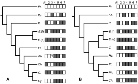
Character mapping
The character mapping (Tab. 2; Fig. 4) indicates that
most characters are distributed equally parsimoniously
on both the morphological and the
molecular/serologi-cal hypothesis. Only character #1, the reduction of
in-ternal cortex in carapacial bones, shows three steps on
the molecular tree instead of four steps on the
morpho-logical tree. In contrast, characters #6 (parallel-fibred
Figure 3.
Bone histology of
Platychelys oberndorferi
. All
images are in polarised light.
A.
Overview of the external
cor-tex and large parts of the interior cancellous bone of the costal.
bone grading into lamellar bone) and #7 (vascularisation
of internal cortex) needed only three instead of four and
two instead of three steps on the morphological
hypoth-esis, respectively. #7 supported the sister-group
relation-ship of
Chelodina
and
Hydromedusa
in the
morphologi-cal tree under both Acctran and Deltran optimisation.
With the present taxon sampling, #5 was recovered as a
synapomorphy for Chelidae. The morphology-based tree
had a tree length of 16, a consistency index (CI) of
0.4375 and a rescaled consistency index (RC) of 0.2188,
whereas the molecular-based tree had a tree length of 17
steps, a CI of 0.4118 and a RC of 0.1830.
Table 2.
Matrix of seven bone histological characters (#1
–
#
7
), which were mapped on the conflicting morphological and
molecular/serological hypotheses of Chelidae (Fig. 1). The basal taxa
Proganochelys quenstedti and Kayentachelys sp. have
been added as outgroups. The character values are given in parentheses. In the case of
Phrynops geoffroanus, character #3
was not clearly determinable. Character #1 internal cortex in carapacial bones reduced in thickness compared to external
cortex; #2 internal cortex in plastral bones reduced in thickness compared to external cortex; #3 fibre bundles that extend
perpendicular to external bone surface prominent in external cortex; #4 vascularisation of external cortex; #5 reticular
larisation patterns present in external cortex; #6 parallel-fibred bone grading into lamellar bone in internal cortex; #7
vascu-larisation of internal cortex.
#1 #2 #3 #4 #5 #6 #7
Proganochelys no (0) no (0) no (0) low to absent (0) no (0) yes (0) low (0)
Kayentachelys no (0) no (0) yes (1) low to absent (0) no (0) no (1) low (0)
Platychelys yes (1) no (0) no (0) moderate to strong (1) no (0) no (1) moderate to strong (1)
Emydura (fossil) yes (1) no (0) yes (1) moderate to strong (1) yes (1) yes (0) moderate to strong (1)
Emydura (extant) yes (1) yes (1) yes (1) moderate to strong (1) yes (1) yes (0) moderate to strong (1)
Platemys no (0) no (0) yes (1) moderate to strong (1) yes (1) yes (0) moderate to strong (1)
Phrynops no (0) no (0) ? (?) moderate to strong (1) yes (1) yes (0) moderate to strong (1)
Chelus yes (1) no (0) no (0) moderate to strong (1) yes (1) no (1) moderate to strong (1)
Chelodina yes (1) yes (1) no (0) moderate to strong (1) yes (1) no (1) low (0)
Hydromedusa no (0) no (0) no (0) moderate to strong (1) yes (1) no (1) low (0)
Figure 4.
Distribution of character states (see Tab. 2) mapped on the competing morphology-based (
A
) and
molecular/serological-based (
B
) phylogenies. See Figure 1 for information on phylogenies. Character state = 0 is depicted as a white square; character
state = 1 as a gray square. In
A
, character #3 is scored with a question mark in
Phrynops geoffroanus
. Abbreviations:
C.
–
Chelo-dina longicollis
;
Ch.
–
Chelus fimbriatus;
E.
(e)
–
Emydura subglobosa
(extant);
E
.
(f)
–
Emydura
sp. (fossil);
Hy
.
–
Hydromedusa
tectifera
;
Ka.
–
Kayentachelys
sp.;
P.
–
Platychelys oberndorferi
;
Ph.
–
Phrynops geoffroanus
;
Pl
.
–
Platemys platycephala
;
Pr.
–
Discussion
As expressed by the results of the character mapping,
neither the competing morphological nor the
molecu-lar/serological hypothesis is unambiguously well
sup-ported, arguing for a conserved condition of chelid
shell bone structures. Only one step less is counted on
the morphological than on the molecular hypothesis
when histological characters are mapped.
Based on comparison with pelomedusoides taxa
(Scheyer & Snchez-Villagra 2007),
P. oberndorferi
and
with stem turtles (Scheyer & Sander 2007), the diploe
structure of the shell with external and internal compact
bone framing interior cancellous bone, the metaplastic
incorporation of dermal interwoven structural fibre
bundles in the external cortex, the parallel-fibred bone
of the internal cortex and the presence of Sharpey’s
fi-bres in chelid shells are plesiomorphic for all turtle
shell bones (Scheyer 2007).
Whether the suture-like sculpturing of the free rib
end in the costal of
P. oberndorferi
fulfils some
func-tional aspect, i.e., to increase the bond between the
costal with the rib socket in the peripheral is debatable.
Aside from the weak phylogenetic aspect revealed
through the character mapping, observed variations
among the chelid turtle shell bones could be partly
re-lated to other influences. Characters that were
hypothe-sised to be functionally influenced (Scheyer & Sander
2007), the reduction of bone tissue, the overall
vascu-larisation of the shell elements, the increase in the
or-ganisation and ordered orientation of the bone
trabecu-lae and vascular cavities in the cancellous bone, were
also found within the chelid shell bones. Although a
quantitative analysis of the bone microstructures has
not been attempted yet, both the South American and
the Australasian chelids thus fit into the category of
semi-aquatic turtles which express low to moderate
adaptation to aquatic freshwater habitats as proposed by
Scheyer & Sander (2007). None of the chelid taxa, on
the other hand, show strong cortical bone reduction as
occurs, for example, in marine turtles.
The fossil
Emydura
sp. is regarded as being
moder-ately adapted to the aquatic environment, similar to its
extant relative
E. subglobosa
. Connections to a more
semi-aquatic versus a fully aquatic live style remain
speculative for the fossil
Emydura
, based only on the
bone histological evidence of its shell. Few chelid
tur-tles thus seem to reach the grade of order in trabecular
arrangement and overall bone vascularisation as the
stem pleurodiran
P. oberndorferi
, which shows moderate
adaptation to the aquatic environment (see also Scheyer
& Sander 2007).
Conclusions
Although the fossil record of chelid turtles reaches
back into the Mesozoic, presumably providing enough
time for potential phylogeny-induced variation to
accu-mulate in the shell bones, the shell bone microstructure
only slightly supports the morphology-based hypothesis
in which the short necked and the long necked chelids
form natural groups respectively. On the other hand, if
functional aspects are working on the bone
microstruc-tures in chelids, this can be used to assess the
palaeoe-cology of fossil taxa. Future work may further elucidate
whether and how the biology, habitat preference, and
behaviour of the turtle species respectively influence
shell bone microstructures.
Acknowledgements
The author likes to thank J. H. Hutchison and P. A. Holroyd, UCMP, W. G. Joyce and G. Watkins-Colwell, YPM, R. Gnther and collea-gues, MfN Berlin, A. Resetar and colleacollea-gues, FMNH, E. Mller, NMS, W. Bhme, ZFMK, and A. Schlter and colleagues, SMNS, for providing samples for histological sectioning. I thank M. Sander, IPB, for his supervision and support. W. Joyce, YPM, and N. Klein, IPB, are thanked for all discussions concerning turtles; O. Dlfer, R. Re-delstorff, and D. Wolff, IPB, for their various helps in preparing thin-sections. D. Kranz and G. Oleschinski, IPB are acknowledged for their various technical supports. This research was supported by DFG funding, grant #SA 469/15–1 and the Doris and Samuel P. Welles Fund (UCMP). W. G. Joyce, J. Sterli and one anonymous reviewer are thanked for their constructive comments on an earlier version of the manuscript.
References
Baur, G. 1887. Ueber den Ursprung der Extremitten der Ichthyopte-rygia. – Berichte ber die Versammlungen des Oberrheinischen Geologischen Vereines 20: 17–20.
Bickham, J. W., Iverson, J. B., Parham, J. F., Philippen, H.-D., Rhodin, A. G. J., Shaffer, B. S., Spinks, P. Q. & Van Dijk, P. P. 2007. An annotated list of modern turtle terminal taxa with comments on areas of taxonomic instability and recent change. – Chelonian Conservation and Biology Research Monographs 4: 173–199. Bona, P. & de la Fuente, M. S. 2005. Phylogenetic and
paleobiogeo-graphic implications of Yaminuechelys maior (Staesche, 1929) new comb., a large long-necked chelid turtle from the early Paleo-cene of Patagonia, Argentina.– Journal of Vertebrate Paleontol-ogy 25 (3): 569–582.
Bonaparte, C. L. 1836. Cheloniorum Tabula Analytica. Romae. Boulenger, G. A. 1888a. On the characters of the chelonian families
Pelomedusidae and Chelydidae. – Annual Magazine of Natural History, Series 61: 346–347.
Boulenger, G. A. 1888b. On the chelydoid chelonians of New Guinea.
– Annali del Museo Civico di Storia Naturale di Genova (2. Ser.) 6: 449–452.
Brm, H. 1965. Die Schildkrten aus dem oberen Jura (Malm) der Gegend von Solothurn. – Schweizerische Palontologische Ab-handlungen 83: 1–190.
Brm, H. 1973. Chelonia from the Upper Jurassic of Guimarota mine (Portugal). Contribuio para o conhecimento da fauna do Kimer-idgiano da Mina de Lignito Guimarota (Leiria, Portugal) III Par-te, VII. – Memorias dos Servios geolgicos de Portugal, (nova S r.) 22: 135–141.
Burbidge, A. A., Kirsch, J. A. W. & Main, A. R. 1974. Relationships within the Chelidae (Testudines: Pleurodira) of Australia and New Guinea.–Copeia 1974 (2): 392–409.
Cope, E. D. 1870. Seventh contribution to the herpetology of tropical America. – Proceedings of the American Philosophical So-ciety 11 (81): 147–169.
Cubo J., Ponton, F., Laurin, M., Margerie, E. de & Castanet, J. 2005. Phylogenetic signal in bone microstructure of sauropsids. – Sys-tematic Biology 54 (4): 562–574.
Danilov, I. G. 2005. Die fossilen Schildkrten Europas. In Fritz, U. (ed.). Handbuch der Reptilien und Amphibien Europas. Band 3/ IIIB: Schildkrten (Testudines) II. Aula-Verlag, Wiebelsheim: pp. 329–441.
Ernst, C. H. & Barbour, R. W. 1989. Turtles of the World. Smithonian Institution Press, Washington D.C.
Fraas, E. 1913. Proterochersis, eine pleurodire Schildkrte aus dem Keuper. – Jahreshefte des Vereins fr vaterlndische Naturkunde in Wrttemberg 80: 1–30.
Francillon-Vieillot, H., Buffr nil, V. de, Castanet, J., G raudie, J., Meunier, F. J., Sire, J. Y., Zylberberg, L. & Ricqls, A. de. 1990. Microstructure and mineralization of vertebrate skeletal tissues.In Carter, J. G. (ed.). Skeletal Biomineralization: Patterns, Processes and Evolutionary Trends. Van Nostrand Reinhold, New York: pp. 471–530.
Fritz, U. & Havasˇ, P. 2007. Checklist of chelonians of the world. –
Vertebrate Zoology 57 (2): 149–368.
de la Fuente, M. S. 2003. Two new pleurodiran turtles from the Porte-zuelo Formation (Upper Cretaceous) of Northern Patagonia, Ar-gentina. –Journal of Paleontology 77 (3): 559–575.
de la Fuente, M. S., Lapparent de Broin, F. de & Manera de Bianco, T. 2001. The oldest and first nearly complete skeleton of a chelid, of the Hydromedusa sub-group (Chelidae, Pleurodira), from the Upper Cretaceous of Patagonia.– Bulletin de la Soci t G ologi-que de France 2: 237–244.
Fujita, M. K., Engstrom, T. N., Starkey, D. E. & Shaffer, B. S. 2004. Turtle phylogeny: insights from a novel nuclear intron.– Molecu-lar Phylogenetics and Evolution 31: 1031–1040.
Gaffney, E. S. 1977. The side-necked turtle family Chelidae: a theory of relationships using shared derived characters.– American Mu-seum Novitates 2620: 1–28.
Gaffney, E. S. 1979. Fossil chelid turtles of Australia. – American Museum Novitates 2681: 1–23.
Gaffney, E. S. 1981. A review of the fossil turtles of Australia. –
American Museum Novitates 2720: 1–38.
Gaffney, E. S. & Meylan, P. A. 1988. A phylogeny of turtles.In Ben-ton, M. J. (ed.). The Phylogeny and Classification of the Tetra-pods. Volume 1: Amphibians, Reptiles, Birds. Clarendon Press, Oxford: pp. 157–219.
Gaffney, E. S., Archer, M. & White, A. 1989. Chelid turtles from the Miocene freshwater limestones of Riversleigh Station, Northwestern Queensland, Australia.–American Museum Novitates 2959: 1–10. Gaffney, E. S., Tong, H. & Meylan, P. A. 2006. Evolution of the
side-necked turtles: the families Bothremydidae, Euraxemydidae, and Araripemydidae. – Bulletin of the American Museum of Natural History 300: 1–698.
Georges, A., Birrell, J., Saint, K. M., McCord, W. & Donnellan, S. C. 1998. A phylogeny for side-necked turtles (Chelonia: Pleurodira) based on mitochondrial and nuclear gene sequence variation. –
Biological Journal of the Linnean Society 67: 213–246.
Greer, A. E. 2006. Chelidae. Encyclopedia of Australian Reptiles. –
Australian Museum Online. http://www.amonline.net.au/herpetol-ogy/research/encyclopedia.pdf; Version date: 7 August 2006. Iverson, J. B. 1992. A Revised Checklist with Distribution Maps of
the Turtles of the World. Privately published, Richmond, IN. Joyce, W. G. 2007. Phylogenetic relationships of Mesozoic turtles. –
Bulletin of the Peabody Museum of Natural History 48 (1): 3–
102.
Joyce, W. G., Parham, J. F. & Gauthier, J. A. 2004. Developing a pro-tocol for the conversion of rank-based taxon names to phylogen-etically defined clade names, as exemplified by turtles.– Journal of Paleontology 78 (5): 989–1013.
Krefft, G. 1876. Notes on Australian animals in New Guinea with description of a new species of fresh water tortoise belonging to the genusEuchelymys(Gray). –Annali del Museo Civico di Sto-ria Naturale di Genova 8: 390–394.
Krenz, J. G., Naylor, G. J. P., Shaffer, B. S. & Janzen, F. J. 2005. Mo-lecular phylogenetics and evolution of turtles.–Molecular Phylo-genetics and Evolution 37: 178–191.
Lapparent de Broin, F. de. 2001. The European turtle fauna from the Triassic to the Present.–Dumerilia 4 (3): 155–217.
Lapparent de Broin, F. de & Molnar, R. E. 2001. Eocene chelid turtles from Redbank Plains, Southeast Queensland, Australia. – Geodi-versitas 23 (1): 41–79.
Lapparent de Broin, F. de & Fuente, M. S. de la. 2001. Oldest world Chelidae (Chelonii, Pleurodira), from the Cretaceous of Patago-nia, Argentina. – Comptes Rendues de l’Acade´mie des Sciences Paris, Sciences de la Terre et des Plantes / Earth and Planetary Sciences 333: 463–470.
Laurin, M., Girondot, M. & Loth, M.-M. 2004. The evolution of long bone microstructure and lifestyle in lissamphibians. – Paleobiol-ogy 30 (4): 589–613.
Legler, J. M. & Georges, A. 1993. Family Chelidae.InGlasby, C. G., Ross, G. J. B. & Beesley, P. L. (eds). Fauna of Australia 2A– Am-phibia and Reptilia. Australian Government Publishing Service. http://www.environment.gov.au/biodiversity/abrs/publications/fau-na-of-australia/fauna-2a.html.
Maddison, W. P. & Maddison, D. R. 2004. Mesquite: a modular sys-tem for evolutionary analysis. Version 1.05. http://mesquitepro-ject.org.
McCord, W. P., Joseph-Ouni, M. & Lamar, W. W. 2001. Taxonomic reevaluation ofPhrynops(Testudines: Chelidae) with the descrip-tion of two new genera and a new species ofBatrachemys. – Re-vista de Biologa Tropical 49 (2): 1–57.
McCord, W. P. & Thomson, S. A. 2002. A new species ofChelodina (Testudines: Pleurodira: Chelidae) from Northern Australia. –
Journal of Herpetology 36 (2): 255–267.
Near, T. J., Meylan, P. A. & Shaffer, B. S. 2005. Assessing concor-dance of fossil calibration points in molecular clock studies: an example using turtles.–American Naturalist 165 (2): 137–146. Pritchard, P. C. H. 1979. Encyclopedia of Turtles. T. F. H. Publications,
Neptune, New Jersey.
Pritchard, P. C. H. 1988. A survey of neural bone variation among re-cent chelonian species, with functional interpretations.–Acta Zo-ologica Cracoviensia 31 (26): 625–686.
Pritchard, P. C. H. 2008. Evolution and structure of the turtle shell.In Wyneken, J., Godfrey, M. H. & Bels, V. (eds). Biology of Turtles. CRC Press, Boca Raton: pp. 45–83.
Rougier, G. W., de la Fuente, M. S. & Arcucci, A. B. 1995. Late Triassic turtles from South America. –Science 268 (5212): 855–
858.
Scheyer, T. M. 2007. Comparative bone histology of the turtle shell (carapace and plastron): implications for turtle systematics, func-tional morphology, and turtle origins. PhD Thesis, Mathematisch-Naturwissenschaftliche Fakultt, University of Bonn. pp. 343 [URN: http://nbn-resolving.de/urn:nbn:de:hbz:5N-12299; URL: http://hss.ulb.uni-bonn.de/diss_online/math_nat_fak/2007/schey-er_torsten].
Scheyer, T. M. & Anquetin, J. 2008. Bone histology of the Middle Jurassic turtle shell remains from Kirtlington, Oxfordshire, Eng-land.–Lethaia 41 (1): 85–96.
Scheyer, T. M. & Sander, P. M. 2007. Terrestrial palaeoecology for ba-sal turtles indicated by shell bone histology.– Proceedings of the Royal Society of London B 274 (1620): 1885–1893.
Scheyer, T. M., Brllmann, B. & Snchez-Villagra, M. R. 2008. The ontogeny of the shell in side-necked turtles, with emphasis on the homologies of costal and neural bones. –Journal of Morphology 269 (8): 1008–1021. [DOI: 10.1002/jmor.10637].
Schneider, J. G. 1783. Allgemeine Naturgeschichte der Schildkrten, nebst einem systematischen Verzeichnisse der einzelnen Arten und zwey Kupfern. Johann Gotfried Mllersche Buchhandlung, Leipzig.
Schneider, J. G. 1791. Beschreibung und Abbildung einer neuen Art von Wasserschildkrte nebst Bestimmungen einiger bisher wenig bekannten fremden Arten. – Schriften der Gesellschaft natur-forschender Freunde zu Berlin 10 (3): 259–284.
Schweigger, A. F. 1812. Prodromus monographiae Cheloniorum, Pt. 1. – Knigsberger Archiv fr Naturwissenschaft und Mathema-tik 1812: 271–458.
Seddon, J. M., Georges, A., Baverstock, P. R. & McCord, W. 1997. Phylogenetic relationships of chelid turtles (Pleurodira: Chelidae) based on mitochondrial 12S rRNA gene sequence variation. –
Molecular Phylogenetics and Evolution 7 (1): 55–61.
Shaffer, H. B., Meylan, P. & McKnight, M. L. 1997. Tests of turtle phylogeny: molecular, morphological, and paleontological ap-proaches.–Systematic Biology 46: 234–268.
Shaw, G. 1794. Zoology of New Holland. Sowerby, London.
Sterli, J. 2008. A new, nearly complete stem turtle from the Jurassic of South America with implications for turtle evolution. – Biol-ogy Letters 4 (3): 286–289.
Sukhanov, V. B. 2006. An archaic turtle,Heckerochelys romanigen. et sp. nov., from the Middle Jurassic of Moscow region, Russia. –
Fossil Turtle Research, Vol. 1, Russian Journal of Herpetolo-gy 13 (Supplement): 112–118.
Swofford, D. L. 2002. PAUP*. Phylogenetic Analysis Using Parsimony (*and Other Methods). Version 4. Sinauer Associates, Sunderland, Massachusetts. http://paup.csit.fsu.edu.
Thomson, S. & Georges, A. 1996. Neural bones in Australian chelid turtles.–Chelonian Conservation and Biology 2 (1): 82–86. Wagner, A. 1853. Beschreibung einer fossilen Schildkrte und etlicher
anderer Reptilien-berreste aus den lithographischen Schiefern und dem grnen Sandstein von Kehlheim. – Abhandlungen der mathematisch-physischen Classe der Kniglich-Bayerischen Aka-demie der Wissenschaften 7 (1): 239–264.
Warren, J. W. 1969. Chelid turtles from the mid-Tertiary of Tasmania.
–Journal of Paleontology 43 (1): 179–182.
Woolley, P. 1957. Colour change in a chelonian.–Nature 179 (4572): 1255–1256.
Wyneken, J. 2001. The anatomy of sea turtles.–National Oceanic and Atmospheric Administration (NOAA) Technical Memorandum NMFS-Southeast Fisheries Science Center (SEFSC) 470: 1–172. Zangerl, R. 1969. The turtle shell. InGans, C., Bellairs, A. d’A. &