Roles for Septins in Selection of Sites of Germ Tube Formation and
Hyphal Morphogenesis
Lifang Li,a,bChengda Zhang,aand James B. Konopkaa
Department of Molecular Genetics and Microbiologya
and Graduate Program in Genetics,b
Stony Brook University, Stony Brook, New York, USA
Septins were identified for their role in septation inSaccharomyces cerevisiaeand were subsequently implicated in other mor-phogenic processes. To study septins inCandida albicanshyphal morphogenesis, a temperature-sensitive mutation was created that altered the C terminus of the essential Cdc12 septin. Thecdc12-6cells grew well at room temperature, but at 37°C they dis-played expected defects in septation, nuclear localization, and bud morphogenesis. Although serum stimulated thecdc12-6cells at 37°C to form germ tube outgrowths, the mutant could not maintain polarized hyphal growth and instead formed chains of elongated cell compartments. Serum also stimulated thecdc12-6mutant to induce a hyphal reporter gene (HWP1-GFP) and a characteristic zone of filipin staining at the leading edge of growth. Interestingly,cdc12-6cells shifted to 37°C in the absence of serum gradually displayed enriched filipin staining at the tip, which may be due to the altered cell cycle regulation. A striking difference from the wild type was that thecdc12-6cells frequently formed a second germ tube in close proximity to the first. The mutant cells also failed to form the diffuse band of septins at the base of germ tubes and hyphae, indicating that this septin band plays a role in preventing proximal formation of germ tubes in a manner analogous to bud site selection. These studies demon-strate that not only are septins important for cytokinesis, but they also promote polarized morphogenesis and selection of germ tube sites that may help disseminate an infection in host tissues.
T
he human fungal pathogen Candida albicans is capable of causing severe systemic infections. Although immunocom-promised patients are particularly at risk, immunocompetent in-dividuals are susceptible to infection when the inoculum is high, which can occur under circumstances such as biofilm formation on medical devices. The major pathogenic effects ofC. albicansare due to invasive growth into tissues, which is facilitated in part by the ability ofC. albicansto switch between different morphologies(29,36).C. albicanscan grow as budding cells, chains of elongated
cells termed pseudohyphae, or as long filamentous cells with par-allel walls called hyphae (36). The filamentous morphology pro-motes invasive growth into agarin vitroand has been linked to invasive growth into tissuesin vivo(20,32). Previous studies also indicated thatC. albicansmust be able to switch between different morphologies to be fully pathogenic. Mutants that are locked in either the hyphal or budding form have been shown to be less virulent in models of hematogenously disseminated systemic can-didiasis (23,32,44).
The septin family of cytoskeletal filament-forming proteins has been shown to contribute to morphogenesis inC. albicans(16,28, 34,40). The septins were first identified in the yeastSaccharomyces
cerevisiaeas proteins that are needed for septum formation and
cytokinesis (15,25). The septins localize to the bud neck, where they form a scaffold to recruit other proteins that promote septum formation. The septins also act as a boundary domain to restrict proteins involved in septum formation to the proper position in the bud neck and also restrict certain proteins to the daughter cells (9,30). Deletion analysis of the seven different septin genes inC.
albicansrevealed that their relative importance is similar to the
orthologous genes inS. cerevisiae.CDC3andCDC12are essential, whereasCDC10,CDC11, andSEP7are not essential but contrib-ute to proper septation and morphogenesis (16,40). Mutation of
theC. albicansorthologs of theSPR3andSPR28septin genes that
are expressed during sporulation inS. cerevisiaedid not detectably affectC. albicansseptation or morphogenesis (40).
Septins have also been implicated in other morphogenic events. For example,S. cerevisiaeseptins promote proper phero-mone-induced morphogenesis, spore formation, and selection of the site of future bud formation (12). The possibility that the sep-tins may also play special roles duringC. albicanshyphal growth was suggested by studies that detected septins at sites distal to septum formation, including the base of the initial hyphal out-growths (know as germ tubes) and at the growing hyphal tips (35, 40). Consistent with this,cdc10⌬andcdc11⌬mutants exhibited partial defects in hyphal morphogenesis, including a greater fre-quency of curved hyphae, and slight inconsistencies in cell wall deposition (40). Significantly, thecdc10⌬andcdc11⌬mutants dis-played defects in morphogenesis and invasive growth in a mouse model of systemicC. albicansinfection (39). These mutants also had a related defect in the morphogenesis of the filamentous cells that produce chlamydospores (26).
In order to obtain a fuller understanding of the role of theC.
albicansseptins in hyphal growth, a new approach was necessary
to study the essential septin genesCDC3andCDC12. Therefore, a temperature-sensitiveCDC12mutant strain was constructed that was patterned after the well-studiedcdc12-6 S. cerevisiaemutant.
TheC. albicans cdc12-6strain grew well at room temperature but
not at 37°C, the temperature at which hyphal growth is induced. It
Received31 July 2012 Accepted3 August 2012
Published ahead of print10 August 2012
Address correspondence to James B. Konopka, [email protected].
Copyright © 2012, American Society for Microbiology. All Rights Reserved.
doi:10.1128/EC.00216-12
on September 8, 2020 by guest
http://ec.asm.org/
also showed temperature-sensitive defects in septation and bud morphogenesis, similar to those reported for the analogous mu-tation ofS. cerevisiae(1,17). Interestingly, theC. albicans cdc12-6
mutant also revealed important new roles for septins in maintain-ing highly polarized hyphal growth and for selection of the second site of germ tube formation.
MATERIALS AND METHODS
Strains and media.TheC. albicansstrains used in the present study are described inTable 1. The cells were propagated in rich YPD medium (2% glucose, 1% peptone, 2% yeast extract) or SD (yeast nitrogen base syn-thetic medium with dextrose) as described previously (33). Uridine at 80 mg/liter was added to cultures ofura3⌬cells.
AC. albicans cdc12⌬/CDC12heterozygous mutant was created in strain BWP17 by homologous recombination as described previously (40,42). In brief, a deletion cassette was constructed by PCR using primers that contain sequence homology to the 5=and 3=ends of the
CDC12open reading frame to amplify a cassette containing theARG4
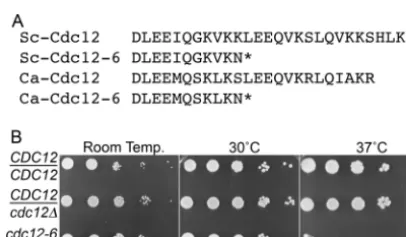
gene for use as a selectable marker to delete one copy ofCDC12. The C-terminal coding sequences of the remainingCDC12allele were then replaced by an altered sequence that was patterned after the changes found in theS. cerevisiae cdc12-6mutant allele (Brian Haarer, unpub-lished data). TheS. cerevisiae cdc12-6allele contains mutations that result in a Lys-to-Asn change at position 391, followed immediately by a TAG stop codon, truncating the mutant protein by 16 amino acids. Amino acid sequence alignments indicated that the Lys-391 of Sc-Cdc12 corresponds to Ser-384 of Ca-Sc-Cdc12. Therefore, the analogous mutation was constructed by mutating one allele ofCDC12to substi-tute Ser at position 384 with Asn and converting Leu at position 385 to the stop codon TAG (Fig. 1A). Oligonucleotide primers carrying the indicated changes were used to amplify theURA3gene using pGEM-URA3 as a template (42), and then the resulting cassette was used to select for integration of thecdc12-6allele intoC. albicans. The primer sequences were 5=-GACTAATATTATTAATGAAAGAAATAGATTGAA TCAAGACTTGGAAGAAATGCAATCGAAGTTGAAGAATTAGTTCTT GAGTTTGTAACAGCTGC-3=and 5=-GTAACCCAAACAACAGAAATT AAGTGGAAGTAATATATTGCTAGCAAAAAAAAACTAAAAAAATGT TAATCTCTCCCCCTTCACATTTATAAT-3=. Similar temperature-sen-sitive phenotypes for budding and hyphal morphogenesis were observed for independent transformants that werecdc12::ARG4/cdc12-6::URA3, so one strain (CZ10) was then used for all subsequent studies. Strain CZ10 was transformed with aHIS1gene to complement all of the auxotrophies and create strain LLF016. TheHIS1DNA fragment was isolated by PCR amplification using genomic DNA fromC. albicansstrain SC5314 as a template.
CDC10-GFPandNOP1-GFPfusion genes were created using PCR
primers that contained 80 bp of sequence homologous to the region im-mediately upstream and downstream of the stop codon to amplify a
GFP␥::HIS1cassette (43). This cassette contains the CaGFP␥version of green fluorescent protein (GFP) that is more photostable (43). Purified PCR product was transformed into the indicated strains, and then the resulting colonies were screened to identify strains that produced the ap-propriate GFP fusion protein. Similar methods were also used to create a strain carrying a hyphal reporter geneHWP1-GFP, except that GFP␥ se-quences were used to replace the entire open reading frame of the hypha-induced geneHWP1. The successful transformants were confirmed by microscopic examination of cells to demonstrate that that GFP expression was elevated in hyphal inducing condition.
Phenotypic analysis of thecdc12-6mutant.Temperature-sensitive growth properties of thecdc12-6mutant were demonstrated by spotting 3 l of a series of 10-fold dilutions of cells onto solid agar YPD medium, starting at a concentration of⬃107cells/ml. The plates were then
incu-bated at the indicated temperature for 2 days. Cells were also analyzed in liquid culture after shorter times of incubation at 37°C. Liquid cultures were grown overnight to log phase at room temperature in rich YPD medium. The cultures were split, and growth was then continued for the indicated times at room temperature or at 37°C. Cell morphology was examined using differential interference contrast (DIC) optics. Viability was determined by mixing the cells with an equal volume of 0.4% trypan blue (Sigma-Aldrich Corp.) and incubating them for 5 min, and then the cells were examined microscopically with visible light to detect
intracel-TABLE 1Strains used in this study
C. albicans
strain
Parent
strain Genotype
BWP17 Sc5314 ura3⌬::imm434/ura3⌬::imm434 his1::hisG/his1::hisG arg4::hisG/arg4::hisG
DIC185 BWP17 ura3⌬::imm434/URA3 his1::hisG/HIS1 arg4::hisG/ARG4
YLS685 BWP17 ura3⌬::imm434/URA3 his1::hisG/his1::hisG arg4::hisG/arg4::hisG
YAW12 BWP17 cdc12::ARG4/CDC12 ura3::imm434/ura3::imm434 his1::hisG/his1::hisG arg4::hisG/arg4::hisG
CZ10 YAW12 cdc12-6::URA3/cdc12⌬::ARG4 ura3::imm434/ura3::imm434 his1::hisG/his1::hisG arg4::hisG/arg4::hisG
CZ11 CZ10 cdc12-6strain CZ10, exceptCDC10-GFP␥::HIS1
CZ14 CZ10 cdc12-6strain CZ10, exceptNOP1-GFP␥::HIS1
LLF003 BWP17 BWP17, exceptCDC10-GFP␥::HIS1
LLF006 BWP17 BWP17, exceptNOP1-GFP␥::URA3
LLF009 LLF003 CDC10-GFP␥strain LLF003, exceptURA3/ura3::imm434
LLF010 CZ10 cdc12-6strain CZ10, exceptHWP1-GFP␥::HIS1
LLF012 YLS685 YLS685, exceptHWP1-GFP␥::HIS1
LLF016 CZ10 cdc12-6 strain CZ10,HIS1/his1::hisG
FIG 1TheC. albicans cdc12-6septin mutant is temperature sensitive for growth. (A) Alignment of the C-terminal sequences of theS. cerevisiaeandC. albicans Cdc12 proteins. (B) Tenfold dilutions of the wild-type control
CDC12/CDC12 (DIC185), cdc12-6/cdc12⌬ (LLF016), and heterozygote
CDC12/cdc12⌬(YAW12) strains were spotted onto rich-medium YPD plates. The plates were incubated for 48 h at the indicated temperature and then photographed.
on September 8, 2020 by guest
http://ec.asm.org/
lular staining indicate of plasma membrane lysis. GFP was analyzed di-rectly in live cells without further processing by using a fluorescence mi-croscope equipped with a fluorescein isothiocyanate (FITC) filter set. This filter set was used to detect GFP, since it was easier to recognize the auto-fluorescence of dead cells as a different color from the true GFP signal. Chitin staining was performed by incubating cells with 10g of Calco-fluor White/ml in phosphate-buffered saline (PBS) for 5 min, followed by two washes with PBS, and then viewing the cells without fixation using a UV filter set. Filipin staining was carried out essentially as described pre-viously (2,27). The cells were induced with or without serum for 2 h at 37°C, stained with 10g of filipin/ml, and then analyzed immediately by fluorescence microscopy using a UV filter set. The cells were viewed on an Olympus BH-2 microscope, and images were captured with an AxioCam digital camera (Carl Zeiss, Thornwood, NY) operated with Axiovision software. The fluorescence signal intensity for cells expressing HWP1-GFPwas quantified using Axiovision software. The results represent the average of three independent experiments in which at least 50 cells were analyzed each time.
Confocal microscopy was used to analyze septin ring structure in cells producing Cdc10-GFP. The cells were cultured overnight in log phase at room temperature, and then aliquots were incubated for 2 h at 23 or 37°C. The aliquot incubated at 37°C was further divided into two tubes. One tube was incubated in rich YPD medium to promote growth of budding cells. Bovine calf serum was added to the other tube to a final concentra-tion of 20% to induce hyphal growth. The cells were then analyzed by fluorescence microscopy using a Zeiss LSM510 META NLO two-photon laser scanning confocal microscope at the Stony Brook University Central Microscopy Imaging Center.
RESULTS
Construction of a C. albicans temperature-sensitivecdc12-6
mutant.The essential septin geneCDC12was examined by creat-ing a mutant allele based on the changes found in the well-studied temperature-sensitiveS. cerevisiae cdc12-6mutant (Haarer, un-published). The mutation alters the C-terminal sequences of Cdc12 as depicted inFig. 1A. These alterations may alter septin function because the C-terminal region of Cdc12 contributes to the stability of septin structures by promoting interaction between two septin filaments (4, 5,37). The other copy of CDC12was deleted so that only the mutantcdc12-6allele remained. TheC.
albicans cdc12-6strain displayed a strong temperature-sensitive
phenotype (Fig. 1B). Thecdc12-6strain grew as well as the wild-type control strain or acdc12⌬/CDC12heterozygote when spotted onto solid medium agar plates at temperatures up through 30°C, but it was not viable at 37°C. Thus, although the C termini of Cdc12 fromS. cerevisiaeandC. albicansare not identical, intro-duction of the analogous alterations of the Sc-cdc12-6allele into
Ca-CDC12still resulted in a strong temperature-sensitive
pheno-type.
cdc12-6mutant phenotypes during budding.The effects of
thecdc12-6mutation onC. albicansmorphogenesis were
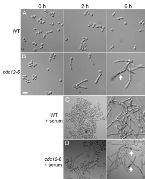
exam-ined at room temperature (0 h) and at different times after shifting cells to 37°C (Fig. 2). When grown at room temperature, the
cdc12-6cells formed buds, albeit some with a slightly abnormal
shape. Many cells were also present in clusters, indicating a partial defect in septation and cytokinesis. After 2 h at 37°C,cdc12-6cells formed elongated buds (Fig. 2B). After 6 h, continued growth of the mutant cells resulted in highly elongated filamentous cells. At around 6 h thecdc12-6cells began to lyse near the tips, as evi-denced by intracellular staining with trypan blue (Fig. 2D). The highly elongated buds formed by thecdc12-6mutant are similar to the morphology of septin mutants in S. cerevisiae, which are
thought to form due to activation of a cell cycle checkpoint path-way that prolongs apical growth of cells (30).
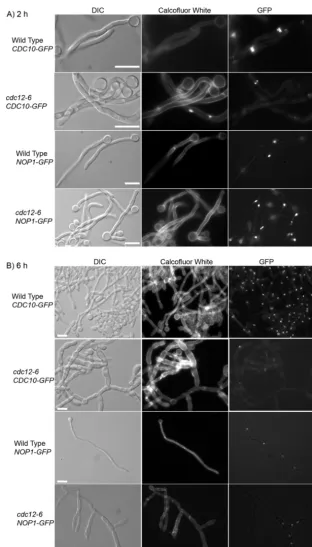
A hallmark ofS. cerevisiae septin mutants is their defect in septation. To assay this in theC. albicans cdc12-6mutant, log-phase cells shifted to 37°C were stained with Calcofluor White to detect the ring of cell wall chitin that forms at the septum (31). At room temperature, both the wild type and thecdc12-6mutant showed typical Calcofluor White staining at bud necks (Fig. 3A). Similar results were observed for the wild-type strain at 37°C. In contrast, sites of septation were rarely detected in thecdc12-6 mu-tant shifted to 37°C (Fig. 3B). This was most evident after 6 h of incubation at 37°C; thecdc12-6cells formed chains of elongated pseudohyphal-type cells with a few obvious septae (Fig. 3C). There were, however, patches of Calcofluor White staining that could represent aberrant attempts to form septae.
Septins also contribute to proper nuclear segregation in S.
cerevisiaeby interacting with microtubules to orient nuclear
mi-gration into the bud (21). The distribution of nuclei inC. albicans
was therefore examined by monitoring the nuclear-localized pro-tein Nop1-GFP. Wild-type cells typically contained one nucleus per cell, as expected. In contrast, thecdc12-6cell compartments frequently contained multiple nuclei or they lacked a nucleus, especially after the longer 6 h of incubation at 37°C, indicating a defect in nuclear segregation (Fig. 3C).
FIG 2Altered morphology and viability ofcdc12-6cells after a shift to 37°C. Wild-type (DIC185) andcdc12-6(LLF016) cells were cultured overnight in log phase in YPD medium at room temperature. The cells were then shifted to 37°C in the absence (A and B) or presence (C and D) of 20% serum and then incubated for the indicated times. Cells were stained with trypan blue and then examined by light microscopy with DIC optics. Examples of lysed cells that stain dark with trypan blue are indicated by an arrow. Bar, 10m. WT, wild type.
on September 8, 2020 by guest
http://ec.asm.org/
Septin localization in the cdc12-6 mutant was analyzed by studying the Cdc10 septin fused to GFP. Wild-type cells grown at either room temperature or 37°C exhibited the expected localiza-tion of Cdc10-GFP to rings at the bud neck (Fig. 3A). Thecdc12-6
cells grown at room temperature also showed bud neck localiza-tion of Cdc10-GFP (Fig. 3A). However, after a shift to 37°C for 2 h, approximately half of thecdc12-6cells lacked detectable Cdc10-GFP, and the others primarily contained faint patches or rings of Cdc10-GFP toward the growing end of the elongated cell cluster (Fig. 3B). Thus, the septins are still capable of forming a complex at 37°C, but it primarily appears near the leading edge of growth and does not stabilize at the pinched zones that correspond to bud necks. Cdc10-GFP was still showed a similar distribution in the
cdc12-6mutant after 6 h at 37°C (Fig. 3C). Although a majority of
cells appeared to lack Cdc10-GFP, a patch or ring of septins was frequently detected at the leading edge of growth or at sites of budding off the main filamentous cell clusters.
The three-dimensional structure of the Cdc10-GFP septin rings that formed in thecdc12-6mutants was analyzed by confocal microscopy (Fig. 4). Wild-type cells grown at room temperature or 37°C showed the expected Cdc10-GFP ring at the bud neck. In contrast, the Cdc10-GFP structures in thecdc12-6mutant were abnormal. At room temperature, Cdc10-GFP localized in a spec-trum of patterns ranging from typical ring structures, to partial rings with a break in the continuity, and very faint rings (Fig. 4). Shifting thecdc12-6cells to 37°C for 2 h resulted in much more severe defects in Cdc10-GFP localization. Cdc10-GFP most fre-quently appeared as a series of bars and did not form a contiguous
ring. The Cdc10-GFP rings incdc12-6cells showed similar defects for cells grown in the presence or absence of serum. Some of these structures appeared to be similar to the types of septin rings seen
inS. cerevisiaecells induced with mating pheromone or carrying a
mutation inGIN4(6,24) and inC. albicansmutants with hyper-active Cdc42 (11).
Hyphal morphogenesis defects incdc12-6mutant cells.The ability to undergo hyphal morphogenesis was examined by treat-ing cells with 20% bovine calf serum at 37°C. As expected, wild-type cells efficiently formed the initial polarized outgrowths termed germ tubes that continued to elongate in a highly
polar-FIG 3Altered localization of septins and nuclei incdc12-6cells at 37°C. Wild-type control andcdc12-6cells engineered to produce either Cdc10-GFP or Nop1-GFP were analyzed as indicated at the top of each column of panels. Cells were grown overnight to log phase at room temperature (A) or were shifted to 37°C for 2 h (B) or 6 h (C). As indicated on the left, cells were analyzed (i) using DIC optics to detect morphology and (ii) fluorescence microscopy to detect Calcofluor White (CW) staining of the cell wall chitin and the localization of GFP-tagged proteins (GFP). The strains used were wild-type cells carrying
CDC10-GFP(LLF009) orNOP1-GFP(LLF006) andcdc12-6cells carryingCDC10-GFP(CZ11) orNOP1-GFP(CZ14). Photos were taken at 0 and 2 h using a ⫻100 objective lens, while those at 6 h were taken with a⫻40 objective lens because of the larger cell size. Bars, 10m.
FIG 4Defects in septin ring formation by thecdc12-6mutant. Wild-type (LLF009) andcdc12-6(CZ11) cells engineered to produce Cdc10-GFP were grown at room temperature or shifted to 37°C for 2 h in YPD medium in the presence or absence of 20% serum as indicated. Cdc10-GFP was analyzed by confocal microscopy to determine the three-dimensional shape of the rings.
on September 8, 2020 by guest
http://ec.asm.org/
ized manner to form filamentous cells with multiple cell compart-ments termed hyphae (Fig. 2and5). Serum also inducedcdc12-6
cells to form germ tubes at 2 h that were generally similar to the wild-type cells (Fig. 2and5). Serum clearly induced a distinct
morphogenesis pathway in thecdc12-6mutant; most cell walls grew parallel and did not display the curvature that was seen in the absence of serum (Fig. 2 and 3). However, Cdc10-GFP localization was abnormal in thecdc12-6 cells induced with
FIG 5Cell morphology, septin ring, and nuclear localization are altered incdc12-6cells under hypha-inducing conditions. Cells were grown to log phase at room temperature, serum was added to 20% final concentration, and then the cultures were shifted to 37°C for 2 h (A) or 6 h (B). As labeled at the top of each column of photos, the cells were analyzed by light microscopy to detect morphology (DIC) or fluorescence microscopy to detect Calcofluor White staining of chitin (CW) or the GFP fusion protein (GFP) as indicated on the left. The strains used were wild-type cells carryingCDC10-GFP(LLF009) orNOP1-GFP(LLF006) and
cdc12-6cells carryingCDC10-GFP(CZ11) orNOP1-GFP(CZ14). Bars, 10m.
on September 8, 2020 by guest
http://ec.asm.org/
serum. About half of the cell clusters lacked detectable Cdc10-GFP, and the Cdc10-GFP structures that were present were typically fainter (Fig. 5A). In addition, the septin rings that formed incdc12-6cells had a wider diameter than those de-tected in the germ tubes and hyphae of wild-type cells (Fig. 4). This is likely due in part to the continued expansion of the width of thecdc12-6germ tubes (see below). In addition, the Cdc10-GFP rings incdc12-6cells typically had breaks in their continuity and some appeared as a series of bars, as was seen for cells grown in the absence of serum (Fig. 4).
After 6 h of incubation at 37°C with 20% serum, the morphol-ogy of thecdc12-6cells was very distinct from the wild type (Fig. 5B). The cdc12-6 cells formed filaments that were wider and curved, indicating the original germ tubes continued to grow in width, whereas new growth in wild-type cells is restricted to the apical tip. The hyphal inducing conditions did not appear to alter the viability of cells at 37°C. Trypan blue staining revealed that dead cells still began to accumulate by 6 h of incubation (Fig. 2).
Thecdc12-6cells also showed frequent branching of new offshoots
of filamentous outgrowth that was not seen for the wild type. Analysis of Nop1-GFP localization showed that many of these branched regions did not contain nuclei, whereas other regions contained multiple nuclei (Fig. 5). This indicates that nuclear di-vision continued in the absence of septation but that the nuclei were not segregating into the different cell compartments. The
cdc12-6cells rarely formed septae that could be detected by
Cal-cofluor White staining, even at the sites where Cdc10-GFP was localized. Instead, patches of Calcofluor White staining were com-monly detected in the new filamentous growth that may represent aberrant attempts to initiate septum formation. After 6 h there were patches or rings of Cdc10-GFP detected in ca. 30% of the cells. Interestingly, the Cdc10-GFP structures in thecdc12-6 mu-tant were frequently detected in the middle of the elongating germ tube, as seen for wild-type cells, and not at the tip of the filamen-tous cell as was seen forcdc12-6cells grown at 37°C in the absence of serum. This suggests that septin localization is affected by the altered cell morphogenesis or by distinct signaling pathways acti-vated in hyphae.
Hypha-induced responses.The ability of the mutant cells to induce hyphal genes was assayed by quantifying the expression of
aHWP1-GFPgene fusion. This reporter gene was constructed by
placingGFPexpression under the control of the hypha-induced
HWP1promoter. Cells carrying this reporter gene were grown in the presence or absence of the hyphal inducers serum or GlcNAc, and the relative induction was assessed by quantifying the signal intensity of GFP using fluorescence microscopy. Although the wild-type and mutant cells strongly induced HWP1-GFP (Fig. 6A), thecdc12-6cells were slightly less efficient than the wild type (P⬍0.003). Thus, septin function is not essential for induction hyphal genes.
Another hallmark of hyphal cells is that the apical region stains more readily with the ergosterol-binding agent filipin (27). As expected, essentially 100% of the wild-type cells induced with se-rum showed increased staining with filipin at hyphal tips (Fig. 6B). Similar results were observed forcdc12-6cells induced with se-rum. Surprisingly, control studies showed that ca. 31.4% (n⫽
191) of thecdc12-6cells shifted to 37°C for 1.5 h in the absence of serum also showed stronger filipin staining at the tips. This in-creased over time to 41.8% at 2 h (n⫽110) and 63.3% at 3 h (n⫽
128). These results for filipin staining contrasted with the
expres-sion ofHWP1-GFP, which required serum to be induced. Thus, this characteristic of hyphae could be induced incdc12-6cells in the absence of serum.
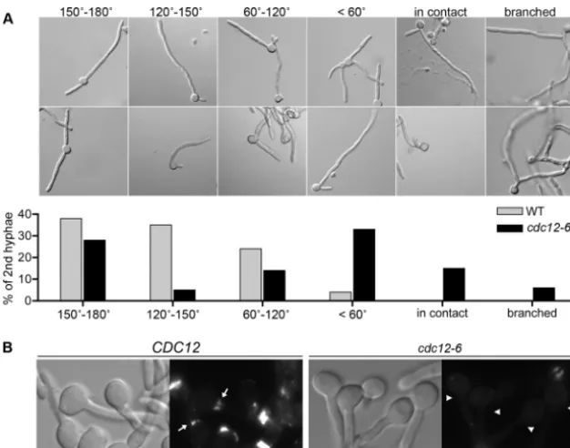
Altered position of second germ tubes incdc12-6mutant.
Thecdc12-6cells induced to form hyphae for an extended time
frequently formed a second germ tube very close to the first one, which was rarely observed in the wild type (Fig. 7) (10). To quan-tify the difference, the relative positions of the sites where the first and second germ tubes initiated were scored as one of six patterns: 150° to 180° apart, 120° to 150° apart, 60 to 120° apart,⬍60° apart, two germ tubes in contact, or a second germ tube that emerged from the first germ tube rather than the mother cell. Interestingly,
cdc12-6 cells showed significantly increased frequency of cells
forming a second germ tube proximal to the first (Fig. 7A). The majority ofcdc12-6cells formed a second germ tube within a 60° angle of the first, whereas essentially all of the wild-type cells formed a second germ tube that was more than 60° from the first.
Thecdc10⌬andcdc11⌬mutants occasionally formed proximal
germ tubes (40), but this defect was more extreme in thecdc12-6
cells.
The altered site selection for germ tube outgrowth in the
cdc12-6mutant indicates that septins influence this process. An
interesting possibility is that the basal band of septins may play a role in determining the site of the second germ tube (Fig. 7B). The basal band is a more diffuse type of septin ring that is located at the junction between the mother cell and the germ tube (35,40). The function of this basal band of septins is not clear, since
cyto-FIG 6Serum inducescdc12-6cells to expressHWP1-GFPand to display in-creased filipin staining at hyphal tips. (A) Expression of hyphal reporter gene
HWP1-GFPin budding and hyphal conditions for wild-type (LLF012) or
cdc12-6cells (LLF010). Cells were grown in the presence or absence of the hyphal inducer serum for 2 h or GlcNAc for 3.5 h. The level of GFP was quantified by fluorescence microscopy. Cells incubated in the absence of se-rum or GlcNAc did not show GFP levels above the background. (B) Wild-type (LLF009) andcdc12-6(CZ11) cells were incubated with or without serum for 2 h at 37°C. The cells were then stained with 10g of filipin/ml and analyzed by fluorescence microscopy. WT, wild type.
on September 8, 2020 by guest
http://ec.asm.org/
kinesis does not occur at this site. However, its location suggests that the basal septin band may function in germ tube site selection analogous to the role of the septin ring in bud site section (24). Consistent with this, the basal band of septins was not detected in
cdc12-6cells (Fig. 7B). Thus, proper Cdc12 function is required to
form both the basal septin band and the septin rings that form at sites of cytokinesis.
A relationship between bud site and germ tube site selection is also supported by previous studies which showed that the bud sites of wild-typeC. albicanswere clustered at an axial site at room temperature but were primarily not adjacent at 30 and 37°C (10). The budding mode ofcdc12-6cells could not be assessed at an elevated temperature, which is not permissive for this mutant. Therefore, we analyzed the nonessential cdc10⌬, cdc11⌬, and
sep7⌬septin mutants. These mutants also showed defects at 37°C that prevented accurate assessment of bud site selection but could be examined at 30°C. Interestingly, all three nonessential septin mutants budded primarily at a cluster of axial sites at room tem-perature (⬎90%;n⫽ ⬎200), similar to the wild type. In contrast, at 30°C only 33% of wild-type cells budded in an axial manner (n⫽202), whereas thecdc10⌬,cdc11⌬, andsep7⌬mutants all still budded primarily in an axial manner (⬎60%;n⫽ ⬎144). The effect was most obvious for thecdc11⌬mutant (76% axial bud-ding at 30°C), a finbud-ding consistent with thecdc11⌬mutant having the morphogenesis phenotype of the three. This suggests that the temperature-related switch in bud-site selection underlies the mechanisms that promote germ tube formation at distal rather than axial sites.
DISCUSSION
Temperature-sensitive septin mutants have played a valuable role
inS. cerevisiaefor identifying the function of septins in septation
and other morphogenic events, including mating and sporulation (12,25). Although hyphal morphogenesis could not be examined
inS. cerevisiae, studies of the nonessential septin genesCDC10,
CDC11, andSEP7indicated that they are important for normal
hyphal morphogenesis inC. albicans(16,34,40) and in filamen-tous fungi (7,14,22). Therefore, in the present study a tempera-ture-sensitivecdc12-6mutant was created to carry out the first analysis of an essential septin gene inC. albicans. Shifting theC.
albicans cdc12-6strain to 37°C caused a rapid defect in
morpho-genesis and septation, similar to those seen inS. cerevisiae. The
cdc12-6mutation likely causes a temperature-sensitive phenotype
because it alters the C-terminal region of Cdc12 that is important for stabilizing connections between two septin filaments (4,5,37). Temperature-sensitive mutants are well suited for the study ofC.
albicans hyphal morphogenesis because cells can be grown at
room temperature and then shifted to a nonpermissive tempera-ture of 37°C, the optimal temperatempera-ture for hyphal morphogenesis. The rapid inactivation of Cdc12 function at a high temperature is therefore expected to reveal better insight into septin function than the use of a regulated promoter, which would require a lon-ger incubation period to deplete the stable septin proteins. Thus, the newcdc12-6mutant represents an important new tool forC.
albicansresearch.
Induction and maintenance of hyphal morphogenesis.The
cdc12-6cells formed buds at room temperature and could be
stim-FIG 7Position of the second germ tube is altered incdc12-6cells. (A) Wild-type control (DIC185) andcdc12-6(LLF016) were shifted to 37°C and induced with 20% bovine calf serum for 3 h. Representative images of cells are shown with different positions of second germ tubes. The degree of separation between the first and second germ tubes was quantified, as shown in the table below. A total of 200 cells were counted from three independent hyphal induction experiments. (B) Wild-type control (LLF009) orcdc12-6(CZ11) cells engineered to produce Cdc10-GFP were induced with 20% serum for 1 h at 37°C and then analyzed by fluorescence microscopy to detect the basal septin band at the junction between the mother cell and germ tube. Arrows point to the presence of the basal band in the wild type. Arrowheads point to the absence of the basal band in thecdc12-6mutant. Note that basal septin band is more diffuse than the tight septin ring seen at sites of septation. WT, wild type.
on September 8, 2020 by guest
http://ec.asm.org/
ulated with serum to form germ tubes at 37°C (Fig. 2and5). The initial germ tube outgrowths in thecdc12-6mutant cells at 2 h did not appear to be significantly more defective than the wild type, whereas bud morphogenesis was clearly affected by 2 h (Fig. 2). This suggests that septin function is not required to initiate germ tubes. Previous studies showed that theC. albicans cdc10⌬and
cdc11⌬mutants frequently formed curved germ tube necks (26,
40), but this phenotype was not exacerbated in thecdc12-6 mu-tant.
Although thecdc12-6 mutant formed germ tubes, highly polarized hyphal morphogenesis was not maintained. The fil-amentous outgrowths became wider over time and took on the characteristics of pseudohyphal cells. Thecdc12-6mutant phe-notype was more extreme than the mutants lacking the nones-sential septin genesCDC10andCDC11, which showed subtler defects in maintaining polarized morphogenesis (40). This in-dicates that the septins have a special function in maintaining highly polarized growth. Altered septin localization is therefore likely to contribute to the abnormal hyphal morphogenesis of mutants that display defects in targeting septins to appropriate sites (11,18,41). However, some phenotypes ofcdc12-6cells may be due to the activation of stress pathways. The defects of
cdc12-6cells in septation and cell wall biogenesis (Fig. 2and5)
should activate cell wall stress pathways that could indirectly affect actin localization and morphogenesis. Activation of stress pathways could also account from some of the altered patches of Calcofluor White staining incdc12-6 cells at 37°C (Fig. 3and5). Unusual patches of Calcofluor White staining were also detected after treatment of cells with caspofungin, an inhibitor of cell wall-glucan synthesis (3).
Regulation of the Cdc11 septin by phosphorylation has also been implicated in properC. albicanshyphal morphogenesis. Mu-tation of a site in Cdc11 to prevent phosphorylation by Cdc28 (S394A) caused a defect in maintaining highly polarized hyphal growth (34). This defect in maintaining polarized growth was more extreme than the defects seen forcdc12-6cells. Mutation of a different site in Cdc11 to prevent phosphorylation by Gin4 (S395A) caused cells to sequentially initiate multiple short germ tubes, suggesting a role for septins in stabilizing the active site of polarized morphogenesis (34). However, thecdc10⌬,cdc11⌬, and
cdc12-6mutants rarely produce the multiple germ tube
protuber-ances seen in the wild type or that were seen so frequently in the
cdc11-S395Amutant (34). Thus, some phenotypes caused by
mu-tating Cdc11 phosphorylation sites are likely due to the dominant activity of a misregulated septin rather than the absence of septin function.
The cdc12-6 mutant at 37°C also showed more frequent
branching of the filamentous outgrowths. This phenotype likely relates to altered cell cycle regulation due to the failure of the germ tubes to undergo septation to form hyphae with different cell compartments. In wild-type cells, the mother cell vacuole swells to force most of the cytoplasmic constituents to the daughter cell compartment at the leading edge of growth (38). Consequently, the mother cells and subsequent subapical cells are delayed in initiating second germ tubes or branches until they can restore the cytoplasmic components. Consistent with this, septin rings persist in the hyphal cells at sites of septation and do not disassemble quickly after septation as they do in budding cells (35,40). More frequent branching was also seen for septin in mutants of the filamentous fungusAspergillus nidulans(19,22).
Hypha-induced gene expression and filipin staining. The ability of thecdc12-6mutant to induce hyphal responses was con-firmed by showing that serum and GlcNAc also induced expres-sion of theHWP1-GFPreporter gene (Fig. 6A). In addition, serum also induced a domain at the tips of the germ tubes in both the wild type and thecdc12-6mutant that stained more readily with the ergosterol binding agent filipin (Fig. 6B). The increased filipin staining is indicative of an altered lipid content in the plasma membrane at hyphal tips (27). Surprisingly, a high proportion (63%) of thecdc12-6cells shifted to 37°C in the absence of serum for 3 h also showed increased tip staining. This was unexpected because previous studies indicated that this filipin-staining do-main was only detected in hyphal and not pseudohyphal cells (27). These results could suggest thatcdc12-6cells induce this response in the absence of hyphal inducers. However, enriched filipin stain-ing is also transiently observed at sites of cytokinesis (2,27). Thus, a likely alternative possibility is that the filipin staining relates to altered cell cycle regulation incdc12-6cells. In support of this latter possibility, enriched filipin staining of the tips ofcdc12-6
cells increased over time rather than coinciding with the induction of the initial germ tube outgrowth as seen with serum induction (13,27).
Site of second germ tube formation.A striking defect of the
cdc12-6mutant is that the second germ tube usually formed
prox-imal to the first germ tube (Fig. 7). This contrasts with wild-type cells in which the second germ tube emerges at a distal position. The basal band of septins that forms at the junction between the mother cell and the germ tube is therefore implicated in this pro-cess. The role of the basal band of septins is not well understood; septation does not occur at this site, and the septins are detected in a more diffuse pattern than the septin ring seen at sites of septation (18,35,40). This raises the possibility that the function of the basal band is to prevent the initiation of second germ tubes proximal to the first. Consistent with this, the basal septin band is stably main-tained after the germ tube has undergone cytokinesis and turned into a hyphal cell. Thus, it is stably maintained until later stages when the second gem tube initiates. The formation of the second germ tube at a distal site is significant, since it would help dissem-inate an infection by promoting growth in a new direction.
This role for septins in germ tube site selection is likely related to their role in bud site selection. InC. albicans, wild-type cells form buds in an axial manner at room temperature but switch to forming buds at nonadjacent bipolar sites at 37°C (10). In con-trast, theC. albicansseptin mutants primarily budded in an axial manner at 30°C, where wild-type cells had mostly switched to budding at bipolar sites. Thus, septins play roles in preventing the formation of a new site morphogenesis adjacent to an existing bud or germ tube inC. albicans. In contrast, theS. cerevisiaeseptins are needed for proper axial budding of haploid cells (8), which means they act in a distinct manner to recruit the morphogenesis ma-chinery to an adjacent site.
Altogether, these studies demonstrate that, in addition to their essential role in septum formation, the septins are needed for maintenance of the highly polarized morphology and proper se-lection of sites of germ tube formation. Both of these are impor-tant for dissemination of an infection in host tissues. These con-clusions are supported by the defect of cdc10⌬ and cdc11⌬
mutants in invasive growth into tissues and virulence in a mouse model of candidiasis (39).
on September 8, 2020 by guest
http://ec.asm.org/
ACKNOWLEDGMENTS
This research was supported by a grant awarded to J.B.K. from the Na-tional Institutes of Health (RO1 AI47837).
We gratefully acknowledge Brian Haarer for providing unpublished data regarding the specific DNA sequence changes present in theS. cerevi-siae cdc12-6mutation. We also thank the members of our labs for their helpful advice and comments on the manuscript.
REFERENCES
1.Adams AEM, Pringle JR.1984. Relationship of actin and tubulin distri-bution to bud growth in wild-type and morphogenetic-mutant Saccharo-myces cerevisiae. J. Cell Biol.98:934 –945.
2.Alvarez FJ, Douglas LM, Konopka JB.2007. Sterol-rich plasma mem-brane domains in fungi. Eukaryot. Cell6:755–763.
3.Badrane H, et al. 2012. Rapid redistribution of phosphatidylinositol-(4,5)-bisphosphate and septins during theCandida albicansresponse to caspofungin. Antimicrob. Agents Chemother.56:4614 – 4624.
4.Bertin A, et al.2008.Saccharomyces cerevisiaeseptins: supramolecular organization of hetero-oligomers and the mechanism of filament assem-bly. Proc. Natl. Acad. Sci. U. S. A.105:8274 – 8279.
5.Bertin A, et al. 2012. Three-dimensional ultrastructure of the septin filament network inSaccharomyces cerevisiae. Mol. Biol. Cell23:423– 432. 6.Bharucha JP, Larson JR, Konopka JB, Tatchell K.2008.Saccharomyces cerevisiaeAfr1 protein is a protein phosphatase 1/Glc7-targeting subunit that regulates the septin cytoskeleton during mating. Eukaryot. Cell
7:1246 –1255.
7.Boyce KJ, Chang H, D’Souza CA, Kronstad JW. 2005. An Ustilago maydisseptin is required for filamentous growth in culture and for full symptom development on maize. Eukaryot. Cell4:2044 –2056. 8.Casamayor A, Snyder M.2002. Bud-site selection and cell polarity in
budding yeast. Curr. Opin. Microbiol.5:179 –186.
9.Caudron F, Barral Y.2009. Septins and the lateral compartmentalization of eukaryotic membranes. Dev. Cell16:493–506.
10. Chaffin WL.1984. Site selection for bud and germ tube emergence in
Candida albicans. J. Gen. Microbiol.130:431– 440.
11. Court H, Sudbery P.2007. Regulation of Cdc42 GTPase activity in the formation of hyphae inCandida albicans. Mol. Biol. Cell18:265–281. 12. Douglas LM, Alvarez FJ, McCreary C, Konopka JB.2005. Septin
func-tion in yeast model systems and pathogenic fungi. Eukaryot. Cell4:1503– 1512.
13. Douglas LM, Martin SW, Konopka JB. 2009. BAR domain proteins Rvs161 and Rvs167 contribute toCandida albicansendocytosis, morpho-genesis, and virulence. Infect. Immun.77:4150 – 4160.
14. Gladfelter AS.2010. Guides to the final frontier of the cytoskeleton: sep-tins in filamentous fungi. Curr. Opin. Microbiol.13:720 –726.
15. Gladfelter AS, Pringle JR, Lew DJ.2001. The septin cortex at the yeast mother-bud neck. Curr. Opin. Microbiol.4:681– 689.
16. Gonzalez-Novo A, et al.2008. Sep7 is essential to modify septin ring dynamics and inhibit cell separation duringCandida albicanshyphal growth. Mol. Biol. Cell19:1509 –1518.
17. Haarer BK, Pringle JR.1987. Immunofluorescence localization of the
Saccharomyces cerevisiae CDC12gene product to the vicinity of the 10-nm filaments in the mother-bud neck. Mol. Cell. Biol.7:3678 –3687. 18. Hausauer DL, Gerami-Nejad M, Kistler-Anderson C, Gale CA.2005.
Hyphal guidance and invasive growth inCandida albicansrequire the Ras-like GTPase Rsr1p and its GTPase-activating protein Bud2p. Eu-karyot. Cell4:1273–1286.
19. Hernandez-Rodriguez Y, Hastings S, Momany M.2012. The septin AspB inAspergillus nidulansforms bars and filaments and plays roles in growth emergence and conidiation. Eukaryot. Cell11:311–323.
20. Kumamoto CA.2008. Molecular mechanisms of mechanosensing and their roles in fungal contact sensing. Nat. Rev. Microbiol.6:667– 673.
21. Kusch J, Meyer A, Snyder MP, Barral Y.2002. Microtubule capture by the cleavage apparatus is required for proper spindle positioning in yeast. Genes Dev.16:1627–1639.
22. Lindsey R, Cowden S, Hernandez-Rodriguez Y, Momany M. 2010. Septins AspA and AspC are important for normal development and limit the emergence of new growth foci in the multicellular fungusAspergillus nidulans. Eukaryot. Cell9:155–163.
23. Lo HJ, et al.1997. NonfilamentousC. albicansmutants are avirulent. Cell
90:939 –949.
24. Longtine MS, Bi E.2003. Regulation of septin organization and function in yeast. Trends Cell Biol.13:403– 409.
25. Longtine MS, et al.1996. The septins: roles in cytokinesis and other processes. Curr. Opin. Cell Biol.8:106 –119.
26. Martin SW, Douglas LM, Konopka JB.2005. Cell cycle dynamics and quorum sensing inCandida albicanschlamydospores are distinct from budding and hyphal cells. Eukaryot. Cell4:1191–1202.
27. Martin SW, Konopka JB.2004. Lipid raft polarization contributes to hyphal growth inCandida albicans. Eukaryot. Cell3:675– 684.
28. Martin SW, Konopka JB. 2004. SUMO modification of septin-interacting proteins inCandida albicans. J. Biol. Chem.279:40861– 40867. 29. Odds FC.1988.Candidaand candidosis. Bailliere Tindall,
Philadel-phia, PA.
30. Oh Y, Bi E.2011. Septin structure and function in yeast and beyond. Trends Cell. Biol.21:141–148.
31. Pringle JR.1991. Staining of bud scars and other cell wall chitin with calcofluor. Methods Enzymol.194:732–735.
32. Saville SP, Lazzell AL, Monteagudo C, Lopez-Ribot JL.2003. Engineered control of cell morphology in vivo reveals distinct roles for yeast and filamentous forms ofCandida albicansduring infection. Eukaryot. Cell
2:1053–1060.
33. Sherman F.1991. Getting started with yeast. Methods Enzymol.194:3–21. 34. Sinha I, et al.2007. Cyclin-dependent kinases control septin phosphor-ylation inCandida albicanshyphal development. Dev. Cell13:421– 432. 35. Sudbery PE.2001. The germ tubes ofCandida albicanshyphae and
pseu-dohyphae show different patterns of septin ring localization. Mol. Micro-biol.41:19 –31.
36. Sudbery PE.2011. Growth ofCandida albicanshyphae. Nat. Rev. Micro-biol.9:737–748.
37. Versele M, et al.2004. Protein-protein interactions governing septin heteropentamer assembly and septin filament organization in Saccharo-myces cerevisiae. Mol. Biol. Cell5:4568 – 4583.
38. Veses V, Richards A, Gow NA.2009. Vacuole inheritance regulates cell size and branching frequency ofCandida albicanshyphae. Mol. Microbiol.
71:505–519.
39. Warenda AJ, Kauffman S, Sherrill TP, Becker JM, Konopka JB.2003.
Candida albicansseptin mutants are defective for invasive growth and virulence. Infect. Immun.71:4045– 4051.
40. Warenda AJ, Konopka JB.2002. Septin function inCandida albicans
morphogenesis. Mol. Biol. Cell13:2732–2746.
41. Wightman R, Bates S, Amornrrattanapan P, Sudbery P.2004. In Can-dida albicans, the Nim1 kinases Gin4 and Hsl1 negatively regulate pseu-dohypha formation and Gin4 also controls septin organization. J. Cell Biol.164:581–591.
42. Wilson RB, Davis D, Mitchell AP.1999. Rapid hypothesis testing with
Candida albicansthrough gene disruption with short homology regions. J. Bacteriol.181:1868 –1874.
43. Zhang C, Konopka JB.2010. A photostable green fluorescent protein variant for analysis of protein localization inCandida albicans. Eukaryot. Cell9:224 –226.
44. Zheng X, Wang Y.2004. Hgc1, a novel hypha-specific G1 cyclin-related protein regulatesCandida albicanshyphal morphogenesis. EMBO J.23: 1845–1856.