Abstract
Th e aim of this research was to assess the reactive changes of rat proximal tubules caused by gentamicin and the eff ect of relatively low doses of melatonin. adult male Wistar rats were distributed into six groups of equal size which all received one of the following daily intraperitoneal injections: vehicle ( ethanol in Ringer solution) during days (C); gentamicin ( mg/kg) during days (G), two groups which concomi-tantly received gentamicin ( mg/kg) during days and melatonin in two diff erent test doses ( or mg/kg) during days (GM, GM) and two groups treated only with melatonin in two diff erent doses ( or mg/kg) during days (M, M). Histological analysis included qualitative and semi-quantitative light microscopy analysis of proximal tubules. Exogenous melatonin had no signifi cant eff ect on the micro-structure, independently of dosis. Th e changes of proximal tubules microstructure induced by gentamicin were expressed in the form of gran-ulovacuolar degeneration, necrosis and desquamation. Th e grade of proximal tubular changes was smaller in animals who besides gentamicin received melatonin. Melatonin has a dose dependent protective eff ect on the structural alterations of proximal tubules of the kidney induced by gentamicin. © Association of Basic Medical Sciences of FB&H. All rights reserved
KEY WORDS: gentamicin, melatonin, proximal tubules, rats, histological analysis
melatonin on gentamicin induced structural
alterations of proximal tubules in rats
Dina Kapić*, Zakira Mornjaković, Esad Ćosović, Maida Šahinović
Institute of Histology and Embryology, Faculty of Medicine, University of Sarajevo, Čekaluša 90, 71000 Sarajevo, Bosnia and Herzegovina
INTRODUCTION
Gentamicin is an aminogyloside antibiotic widely used in the treatment of infections caused by Gram negative bacterias [, , ]. Th e use of gentamicin is limited by its nephrotoxicity []. In fact, the treatment with this antibiotic, even in therapeutic doses, causes the damage of proximal tubules of the kidney []. Despite the introduction of newer antibiotics of lesser toxicity, gentamicin is still often used, because it’s relatively low cost, and rapid bactericidal action [, ]. Th e mechanism of the genesis of gentamicin nephrotoxicity is still not com-pletely clear, but it is assumed that the reactive oxygen spe-cies (ROS) are one of the main causative agents [, ]. Th is is the reason why it is considered that the negative action of gentamicin could be reduced with the concurrent use of an antioxidant agent. N-acetyl--metoxytriptamine
(mela-tonin), as the main product of the pineal gland, was shown to be a good protector in diff erent models of kidney damage, e.g. ischemia-reperfusion injury [] or changes induced by cisplatin []. Beside its most famous function – the regula-tion of the circadian rhythm [], this molecule posseses other numerous functions including the immunomodula-tory [], the antiinfl ammaimmunomodula-tory [], and the antioxidative role []. Th e facts that melatonin functions as an antioxidant and that it reduces oxidative damage on the physiological and pharmacological level [] open the area of interest for use of melatonin in blocking the side eff ects of gentamicin. Therefore, the aim of this study was to test two dif-ferent doses of melatonin regarding the gentami-cin induced structural alterations of proximal tubules.
MATERIAL AND METHODS
Animals
Fourty eight adult male Wistar rats, weighing -g, were maintained in standardized laboratory conditions with a temperature of ± °C, and a -hour light-dark cycle. Both standard rat chow and water were provided ad libitum.
* Corresponding author: Dina Kapić,
Institute of Histology and Embryology, Faculty of Medicine, University of Sarajevo, Čekaluša 90, 71000 Sarajevo, Bosnia and Herzegovina Phone/Fax: +387 33 203 669
e-mail: [email protected], [email protected]
Submitted: 28 March 2013 / Accepted: 23 January 2014
Procedures
The rats were randomly assigned into six groups of equal size which all received one of the following daily intraperi-toneal injections: group (Control), the animals received vehicle ( ethanol in Ringer solution) during days, group (G) rats that received gentamicin ( mg/kg) dur-ing days, group (GM) rats which received gentamicin ( mg/kg) during days and melatonin ( mg/kg) days before and days concomitantly with gentamicin, group (GM) rats which received gentamicin ( mg/kg) dur-ing days and melatonin ( mg/kg) days before and days concomitantly with gentamicin, group (M) rats that received melatonin ( mg/kg) during days and group (M) rats that received melatonin ( mg/kg) during days. Th e animals were sacrifi ced under ether anesthesia hours after the last injection. Left kidneys were removed, fi xed in buff ered formalin and then prepared for the qualitative and semiquantitative histological analysis using hematox-ylin-eosin (H&E) and periodic acid - Schiff (PAS) staining. Th e study was carried out at the Institute of Histology and Embryology of the Faculty of Medicine of the University of Sarajevo, with the approval of the Local Ethics Committee. Th e histological analysis included qualitative and semiquan-titative analysis of the proximal tubules of the kidney at the level of light microscopy. Th e semiquantitative analysis of the kidney sections was performed using the technique of Houghton et al. []. Th e changes seen were graded as fol-lows: = normal; = areas of focal granulovacuolar epithelial cell degeneration and granular debris in tubular lumens with or without evidence of tubular epithelial cell desquamation in small foci (< of total tubule population involved by des-quamation); = tubular epithelial necrosis and desquamation easily seen but involving less than half of cortical tubules; = more than half of proximal tubules showing desquama-tion and necrosis but uninvolved tubules easily found; = complete or almost complete proximal tubular necrosis.
Statistical analysis
The chi-square test of independence was used to exam-ine the dependence of the applied dose of melatonin on the grade of damage of proximal tubules. Probability val-ues (p) less than . were considered to be statistically signifi cant.
RESULTS
Control group
The qualitative histological analysis revealed regu-lar renal parenchymal structure. The proximal tu-bules appeared intact, having uniform tubular epithe-lial cells with a preserved brush border, the cytoplasm
having homogenous tinctorial properties, and the nuclei hav-ing regular size, position and chromasia (Figure A and B).
G group
Th ere were visible wide fi elds of heavy and extensive necro-sis with numerous completely destroyed proximal tubules leaving only a bare basement membrane in the kidney cor-tex. Numerous proximal nephrocytes showed changed tinc-torial properties – some cells showed enhanced acidophily while others were of pale appearance. The brush border was damaged on several places, and completely missing on others. In numerous nephrocytes it was possible to observe vacuolisation and/or the accumulation of PAS positive ma-terial in the form of diff erently sized and shaped granules, from barely visible round-oval granules to large, irregular aglomerations, which exceeded the size of the nucleus. It was also revealed that the nuclei were of diff erent size, po-sition and chromasia, and that several cells had lost their nuclei. One part of the tubules showed dilatation; in some tubules was evident the obliteration of the lumina by the presence of a necrotic cellular mass (Figure C and D).
GM group
In the cortical parenchyma, we could observe relatively narrow areas of necrotically changed proximal tubules in comparison to areas of preserved structure. Histological changes of proximal tubular epithelial cells were more se-vere in the cortical then in the juxtamedullar tubules. Th e proximal nephrocytes showed signs of granulovacuolar de-generation and necrosis. Th e cytoplasm of the nephrocytes, showing necrosis, had changed tinctorial properties and the nuclei were pyknotic, and some nuclei showed signs of kariorexis. Necrotic cellular debris was observed in the lu-mina of some tubules. In a few nephrocytes it was possible to notice the presence of PAS positive granules of differ-ent size and shape, and it was possible to observe the vacu-olisation of the cytoplasm. Th e brush border of some cells was completely missing and in some cells its structure was only partially damaged. We could also notice a few cells with mitotic figures. A mildly expressed oedema and an inflammatory infiltrate composed predominantly of lym-phocytes were visible in the interstitium (Figure E and F).
GM group
Th e architecture of the cortical parenchyma was mainly rela-tively preserved, and the fi elds with granulovacuolar degener-ation, necrosis and/or desquamation were clearly spatially de-marcated and extremely narrow. Th e proximal tubules were lined by simple columnar epithelium composed of acido-philic cells with no discrete cell margins between them. Th eir round uniform nuclei were positioned in the basal portion
of the cells. A well preserved brush border was visible on the apical pole. Th e basement membrane was continuous, had a regular structure and uniform thickness. Some nephrocytes were showing signs of granulovacuolar degeneration and ne-crosis with typical changes of the cytoplasm and nucleus. Th e cortical interstitial connective tissue had mainly a regular cel-lularity and only somewhere it was possible to fi nd a sparse mononuclear inflammatory infiltrate (Figure G and H).
M and M group
Histological analysis of the cortex of the kidney in this two groups showed a regular architecture of the parechyma. The acidophilic cells of the proximal tubules were having a uniform appearance, and their round nuclei were having tipical basal position, tinctorial properties and size. Th e
in-tersitium had a preserved histological structure (Figure I). The result of the chi squared test of independence showed that the grade of proximal tubular damage (Table ) induced by gentamicin was dependent on the applied dose of melatonin (χ = ,; p<.).
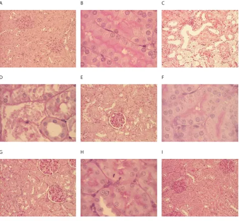
FIGURE 1. Histological fi ndings in kidneys of diff erent groups. A control group (PAS, x100), B control group (PAS, x400), C gentamicin group (H&E, x100), D gentamicin group (PAS, x400), E gentamicin + melatonin (5 mg/kg) group (PAS, x100), F gentamicin + melatonin (5 mg/kg) group (PAS, x400), G gentamicin + melatonin (20 mg/kg) group (PAS, x100), H gentamicin + melatonin (20 mg/kg) group (PAS, x400), I melatonin (20 mg/kg) group (PAS x100).
A
D
G
B
E
H
C
F
I
GROUP grade 0 grade I grade II grade III grade IV
Control 8 (100%) - - -
-G - - 1 (12,5%) 7 (87,5%)
-GM1 - - 6 (75%) 2 (25%)
-GM2 - 4 (50%) 4 (50%%) -
-M1 8 (100%) - - -
-M2 8 (100%) - - -
-TABLE 1. Histological degree of renal damage obtained by semi-quantitative analysis
DISCUSSION
Th e use of gentamicin is still very frequent in clinical practice despite its nephrotoxicity in - of therapeutic courses [, ]. Gentamicin induced nephrotoxicity is used in numerous
studies as the model for the research of aminoglycoside toxic-ity. Taking into account that the ROS are regarded as one of the main causative factors of nephrotoxicity of gentamicin [, ], research has been done lately in order to develop thera-pies to reduce oxidative damage. In our research, the treat-ment with gentamicin at the dose of mg/kg resulted with signifi cant lesions of the epithelium of the proximal tubules which were changed in form of necrosis and granulovacuolar degeneration whereat grade was recorded in . of ani-mals. Th e interstitium was reactively changed - oedematous and infi ltrated with mononuclear cells. Th e kidneys from the group of rats which were treated concomitantly with genta-micin and melatonin at the dose of mg/kg showed clearly that melatonin had a protective effect on the gentamicin induced structural changes. Namely, the areas of the kidney cortex enclosing tubular necrosis and degeneration were nar-rower; the changes of the tubules were of lesser extent, while the interstitial mononuclear infi ltrate and oedema were less prominent. Grade of changes was observed in of ani-mals, and grade only in . Histological analysis of kidneys of the rats which were beside gentamicin treated with mela-tonin at the dose of mg/kg showed that the changes in this group were the mildest. Th e protective eff ect of melatonin was even more pronounced comparing to the previous group, grade of changes was recorded in one half of the animals and grade in the other half. Th e kidneys of the rats treated with melatonin at either a dose of mg/kg or mg/kg did not diff er histologically signifi cantly and all the kidney sec-tions were graded with . We noticed a diff erence in the score obtained by the semiquantitative analysis between the group of rats treated only with gentamicin and the control group, as well as between the group treated with gentamicin and the groups treated concomitantly with gentamicin and melato-nin, while the degree of melatonin protection was dose de-pendent and highly signifi cant (p<.). Th e study of Sener and al. [], that pursued the protective action of melatonin on morpho-functional changes of the kidney caused by the administration of gentamicin, showed that the rats treated only with gentamicin had elevated values of urea and creati-nine, which was followed by morphological changes in the sense of severe degeneration of proximal tubular epithelial cells and their microvilli. Th e pretreatment with melatonin reduced the development of the mentioned changes, which is in accordance to our results. Shifow and al. [] were ex-amining the eff ect of melatonin on the development of bio-chemical and histological changes caused by gentamicin. Th e
values of urea and creatinin were increased in comparison to the control group, and the histological analysis showed loss of tubular epithelial cells with an intense granular degenera-tion, with the changes comprising more then of the renal cortex that is the total tubular necrosis was observed (grade ). The animals treated with melatonin had both lower se-rum values of urea and creatinine and a lower grade of struc-tural changes. Th e research conducted by Özbek and al. [] showed that melatonin attenuates the genatmicin induced morpho-functional changes. While the rats treated with gen-tamicin were graded between and , of the rats treated with genatmicin and melatonin had the grade of changes . Th e serum levels of γ-glutaminil transferase and the level of proteins in the urine were increased, while the values of superoxide dismutase, glutathion peroxidase and catalase in tissues were decreased in the gruop treated with gentamicin. In the group treated with gentamicin and melatonin a protec-tive eff ect was observed regarding all these parameters. Also the results of Lee and al. [] are in agreement with our re-sults and show that melatonin has a renoprotective eff ect on the oxidative damage induced by gentamicin. Lately, research has been done in order to examine the protective eff ect of melatonin on gentamicin induced structural alterations, and thereby heterogenous doses were used. Nevertheless, none of these studies had as its main interest several doses of mela-tonin with the aim of comparing the eff ects and locating the optimal protective dose, what we have tried to accomplish.
CONCLUSION
Our results show that the gentamicin induced structural alterations of the proximal tubules can be abated with pre-treatment and the concomitant application of mela-tonin, and that the level of protection is dose-dependent.
DECLARATION OF INTEREST
Th e authors state that there is no confl ict of interest.
REFERENCES
[] Ali BH, Al Za'abi M, Blunden G, Nemmar A. Experimental genta-micin nephrotoxicity and agents that modify it: a mini-review of re-cent research. Basic Clin Pharmacol Toxicol ; ():-. [] Kahlmeter G, Dahlager JI. Aminoglycoside toxicity - a review of
clinical studies published between and . J Antimicrob Chemother. ; Suppl A:-.
[] Mingeot-Leclercq MP, Tulkens PM. Aminoglycosides: nephrotox-icity. Antimicrob Agents Chemother. ;():-. [] Lopez-Novoa JM, Quiros Y, Vicente L, Morales AI,
Lopez-Hernan-dez FJ. New insights into the mechanism of aminoglycoside neph-rotoxicity: an integrative point of view. Kidney Int ; ():-. [] Martínez-Salgado C, Eleno N, Tavares P, Rodríguez-Barbero A,
García-Criado J, Bolaños JP, et al. Involvement of reactive oxygen
species on gentamicin-induced mesangial cell activation. Kidney Int ; ():-.
[] Kacew S, Bergeron MG. Pathogenic factors in aminoglycoside in-duced nephrotoxicity. Toxicol Lett ; ():-.
[] Leehey DJ, Braun BI, Th oll DA, Chung LS, Gross CA, Roback JA, et al. Can pharmacokinetic dosing decrease nephrotoxicity associ-ated with aminoglycoside therapy. J Am Soc Nephrol ; ():-.
[] Cuzzocrea S, Mazzon E, Dugo L, Serraino I, Di Paola R, Britti D, De Sarro A, Pierpaoli S, Caputi A, Masini E, Salvemini D. A role for superoxide in gentamicin-mediated nephropathy in rats. Eur J Pharmacol. ;():-.
[] Banday AA, Farooq N, Priyamvada S, Yusufi AN, Khan F. Time dependent eff ects of gentamicin on the enzymes of carbohydrate metabolism, brush border membrane and oxidative stress in rat kidney tissues. Life Sci ; (-):-.
[] Kurcer Z, Oguz E, Ozbilge H, Baba F, Aksoy N, Celik H, et al. Mela-tonin protects from ischemia/reperfusion-induced renal injury in rats: this eff ect is not mediated by proinfl ammatory cytokines. J Pi-neal Res ; ():-.
[] Parlakpinar H, Sahna E, Ozer MK, Ozugurlu F, Vardi N, Acet A. Physiological and pharmacological concentrations of melatonin protect against cisplatin-induced acute renal injury. J Pineal Res ; ():-.
[] Nowak R, McMillen IC, Redman J, Short RV. Th e correlation be-tween serum and salivary melatonin concentrations and urinary -hydroxymelatonin sulphate excretion rates: two non-invasive techniques for monitoring human circadian rhythmicity. Clin En-docrinol (Oxf) ; ():-.
[] Sutherland ER, Martin RJ, Ellison MC, Kraft M. Immunomodula-tory eff ects of mela¬tonin in asthma. Am J Respir Crit Care Med ; ():-.
[] Mayo JC, Sainz RM, Tan DX, Hardeland R, Leon J, Rodriguez C, et.
al. Anti-infl ammatory actions of melatonin and its metabolites, N-formyl--methoxykynuramine (AFMK) and N-acetyl--methoxykynuramine (AMK), in macrophages. J Neuroimmunol ; (-):-.
[] Hardeland R. Antioxidative protection by melatonin: multiplicity of mechanisms from radical detoxifi cation to radical avoidance. Endocrine ; :-.
[] Reiter RJ, Tan DX, Sainz RM, Mayo JC, Lopez-Burillo S. Melatonin: reducing the toxicity and increasing the effi cacy of drugs. J Pharm Pharmacol ; ():-.
[] Houghton DC, Plamp CE rd, DeFehr JM, Bennett WM, Porter G, Gilbert D. Gentamicin and tobramycin nephrotoxicity: a morpho-logic and functional comparison in the rat. Am J Pathol ; : -.
[] Martínez-Salgado C, López-Hernández FJ, López-Novoa JM. Glo-merular nephrotoxicity of aminoglycosides. Toxicol Appl Pharma-col ; ():-.
[] Yazar E, Elmas M, Altunok V, Sivrikaya A, Oztekin E, Birdane YO. Eff ects of aminoglycoside antibiotics on renal antioxidants, malo-ndialdehyde levels, and some serum biochemical parameters. Can J Vet Res ; ():-.
[] Sener G, Sehirli AO, Altunbas HZ, Ersoy Y, Paskaloglu K, Arbak S, et al. Melatonin protects against gentamicin-induced nephrotoxic-ity in rats. J Pineal Res ; ():-.
[] Shifow AA, Kumar KV, Naidu MU, Ratnakar KS. Melatonin, a pi-neal hormone with antioxidant property, protects against gentami-cin-induced nephrotoxicity in rats. Nephron ; ():-. [] Ozbek E, Turkoz Y, Sahna E, Ozugurlu F, Mizrak B, Ozbek M.
Melatonin administration prevents the nephrotoxicity induced by gentamicin. BJU Int ; ():-.
[] Lee IC, Kim SH, Lee SM, Baek HS, Moon C, Kim SH, et al. Mela-tonin attenuates gentamicin-induced nephrotoxicity and oxidative stress in rats. Arch Toxicol ; ():-.