1
The missing link in early emotional processing
Luis Carretié1, *, Raghunandan K. Yadav2, Constantino Méndez-Bértolo1
1 Facultad de Psicología, Universidad Autónoma de Madrid, 28049 Madrid, Spain
2 School of Life Sciences, Jawaharlal Nehru University, New Delhi 110067, India
* Corresponding author. E-mail: [email protected]
Abstract
Initial evaluation structures (IES) currently proposed as the earliest detectors of affective stimuli (e.g., amygdala, orbitofrontal cortex, or insula) are high order structures i) whose response latency cannot account for the first visual cortex emotion-related response (80ms), and ii) lack the necessary infrastructure to locally analyze the visual features that define emotional stimuli. Several thalamic structures accomplish both criteria. The lateral geniculate nucleus (LGN), a first-order thalamic nucleus that actively processes visual information, with the complement of the thalamic reticular nucleus (TRN), are proposed as core IESs. This LGN-TRN tandem could be supported by the pulvinar, a second order thalamic structure, and by other extra-thalamic nuclei. The visual thalamus, scarcely explored in Affective Neurosciences, seems crucial in early emotional evaluation.
1. Introduction
In certain and relatively frequent situations, the capability to rapidly evaluate and respond to salient stimuli, such as the abrupt onset or rapid approach of a dangerous animal or hazardous object, is crucial for survival. These and other salient stimuli cause, by definition, emotional reactions that manifest at the physiological, subjective and/or behavioral level. The term “emotional” will be employed hereafter to refer to salient stimuli as well as their inherent multi-level consequences (Carretié, 2014; but see different and more specific definitions of “emotion” and “emotional”, from diverse theoretical frames, in Brosch et al., 2010). Regarding the
behavioral level, each tenth of a second may be critical. Indeed, motor reactions clearly revealing a previous discrimination of emotional from non-emotional stimuli may occur between four and five tenths of a second in those tasks especially designed to favor rapid responses, such as Go/Nogo tasks (e.g., Zhang & Lu, 2012). Evidently, this emotional motor reaction is preceded by emotional neural processing, which should be also rapid.
Event-related potentials (ERPs) show that emotional modulation of the visual cortex occurs in humans before 100 milliseconds (ms) from stimulus onset. For example, C1, the first visual ERP component originated at the cortical level, presents increased amplitude in response to emotional stimuli (Acunzo et al., 2019; Elder et al., 2010; Pourtois et al., 2004). The onset of this component occurs around 50 ms (e.g., Di Russo et al., 2003), its peak being produced as early as 63 ms in response to certain spatial locations (e.g., Capilla et al., 2016), and in any case before 100 ms (Rauss et al., 2011). This is compatible with intracranial recordings in human patients, in which the V1/V2 response to visual stimuli is recorded at 60 ms (Krolak-Salmon et al., 2003). Emotional modulation of C1 is produced between 60 and 100 ms, peaking at 80 ms
(Acunzo et al., 2019; Eldar et al., 2010; Pourtois et al., 2004; see also Pizzagalli et al., 1999). This component originates in V1 or striate cortex (both terms will be employed indistinctly)
receiving preferential processing. Several proposals exist for these initial evaluation structures (IES), the most recurrent and noteworthy being the amygdala (e.g., see reviews or
meta-analyses in Adolphs, 2008; Costafreda et al., 2008; Ohman, 2002; Zald, 2003), due to the direct inputs it receives from the thalamus, an issue that will be discussed later. Mainly in human and non-human primates, ventral prefrontal -or orbitofrontal- cortex (vPFC) and insula have also been proposed as emotional evaluation structures (e.g., see reviews by Rudebeck & Murray, 2014 and Norman et al., 2011, respectively). All of them have in common their mutual direct
interconnections, as well as receiving early visual inputs and sending direct efferences to the visual cortex so they may modulate its activity through attentional changes (see a review on the connectivity of these structures in Carretié et al., 2009).
Two critical issues regarding the current proposals on initial emotional processing
motivate this review. The first one is related to time. If visual cortices show emotional modulation at 80 ms approximately (or maybe ~10 or 15 ms earlier, since 80 ms is the peak differentiation), these evaluative structures should previously present a distinctive activity for emotional stimuli (with respect to neutral). Moreover, this activity should occur with time enough to transmit their information to visual cortices prior to 80 ms. Human intracranial recordings of evaluation structures accumulated up to the present are not compatible with this prerequisite. Thus, the shortest reported responses sensitive to the emotional content of visual stimulation have been reported at 74 ms in the case of human amygdala, and only for faces; responses to other
emotional visual stimuli were produced beyond 150 ms (Méndez-Bértolo et al., 2016). In the case of the other two structures mentioned above, the earliest reported saliency activations are
produced over 80 ms: 120 ms in vPFC (for both faces and scenes: Adolphs et al., 2006), and 140 ms in the case of insula (only faces have been explored, to the best of our knowledge:
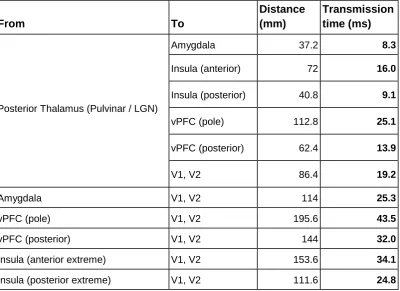
may act as IES and cause the emotional V1 peak response observed at 80 ms is even more remote (Table 1). In sum, and taking into account the available data, evaluative structures proposed up to present cannot be responsible for the emotion effect observed in V1 at 80 ms.
*** Table 1 ***
The second issue motivating this review is related to the architecture of IESs. These structures should be able to constantly monitor, evaluate and label (if pertinent) the ascending visual information. This requires an in situ processor of the visual input capable of recognizing certain visual features, such as shapes, colors or motion patterns. Ultimately, this visual
processing would require modules that constantly analyze the visual information transmitted by the retinal ganglion cells. Each module would process a visual region or receptive field -RF-, which is the information unit transmitted by ganglion cells. These modules could be
conceptualized as a sort of focal processors in terms of the total extension of the visual field. As explained in forthcoming sections, these focal processing modules, which indeed exist in different parts of the brain, consist of retinotopically organized visual processing cells. Parallely, IESs should also be capable of global processing. Global modules would transversally manage several focal modules to extract general information of the visual input, which often involves several focal RFs. For example, the shape of a spider close to us -its legs, its head, its abdomen- would involve several RFs (several focal processing modules), but the “spider shape” would require an in situ global processor that puts together the information of those individual RFs: Figure 1. These in situ processors should mark the ascending visual information if a relevant stimulus is detected, a “decision” that requires the capability of storing visual features that have been associated with emotional situations in the past. This storage should be also local, so initial evaluation may keep its speed.
*** Figure 1 ***
receive direct inputs from visual cortices, but this is not equivalent to be visual processors. Many other brain areas also receive visual inputs and respond to them, such as the hippocampus, the anterior cingulate cortex, the motor cortices, etcetera, but they are not considered visual
processors. Additionally, the infrastructure we are mentioning (retinotopy, focal and global feature processing) has not been described for them up to the present. A tentative explanation is that they are global processors (in the terms explained above), but this seems improbable, since global processors need to interconnect many focal modules to extract global features. This ramified interconnectivity with retinotopic structures of the brain has not been described for amygdala and neither for vPFC nor insula. However, the fact that these structures do not
accomplish the necessary conditions to be IESs does not mean they are not crucial in emotional processing. The key issue is that they receive pre-processed (pre-evaluated) information (also the amygdala, the fastest responding among the three: Pessoa & Adolphs, 2010). The most probable roles of these structures, and the stage within emotional processing in which they may intervene, will be discussed in the final section. Therefore, we are before a “missing link” in emotional processing. Concretely, the IESs are yet to be defined.
2. The thalamus hypothesis
Sensory thalamus, and particularly the visual thalamus, is often conceptualized as a relay to which sensory information arrives from the retina and from which this information is transmitted to the visual cortex and other brain areas. This role as a relay is understood as passive, the
capability of processing this information being assigned to other cerebral structures. In fact, as pointed out by Ghodrati and collaborators (2017), the models and representations of the visual processing route start at V1, never before. However, the capability of visual thalamus for
signals to visual cortices in response to exactly the same stimulus depending on whether it is attended or not. The proposal here is that this capability of the thalamus to process the information it receives may be extended to the emotional domain.
It is relevant to note that the idea of the thalamus as a structure crucial in “emotional labeling” of sensory inputs is about one century old. Cannon (1927), for example, postulated that “the peculiar quality of the emotion is added to simple sensation when the thalamic processes are roused” (p. 120). A similar idea was previously defended by Head (1920). These sound proposals were based, due to the scientific limitations of the epoch, on very scarce data mainly obtained from behavioral observations, and lacked concretion on the mechanisms by which the thalamus evaluated sensory inputs as emotional, or on the nuclei involved. However, Lashley, explicitly referring to this proposal, indicated a decade later that “the supposed evidence that the thalamus adds the affective or emotional character to sensations breaks down completely when subjected to critical analysis” (Lashley, 1938, p. 60). Parallely, the first influential model of the emotional brain was published by Papez (1937). In it, the key structure in charge of evaluating and labeling sensory inputs as emotional was not the thalamus, but the hypothalamus and other brain structures: “the sensory excitations (...) receive their emotional coloring from the concurrent processes of hypothalamic origin which irradiate them from the gyrus cinguli” (p. 729). Later, MacLean (1949), whose model of the emotional brain was strongly settled on Papez’s, successfully recovered the term “limbic” (coined by Broca in 1878), to refer to this “system” or circuit whose structures are in the limbo or intermediate area between brainstem and cortex. The limbic system, as the Papez’s circuit, included the thalamus, but its function in both models was connective and secondary. The central element of the MacLean model was, instead, the
hippocampus.
models, to other structures. For example, LeDoux (1992) signaled the amygdala as a core element of the emotional brain, proposing at the same time some strong and sound reasons to overcome the limbic system model, and Damasio (1994) stressed the crucial role of vPFC (whereas the amygdala was present in the MacLean’s model -not in Papez’s, who explicitly indicated that “its function is unknown”, 1937, p. 742-, and also the vPFC, their role was not as central as in contemporary Affective Neuroscience). This is currently the dominant vision since, as indicated, these structures have repeatedly been proposed to be involved in the emotional evaluation and labeling of visual inputs. In any case, this current vision inherits from the Papez-limbic age the idea of thalamus as a mere connecting hub from sensory organs to other brain structures, or between different brain structures. Indeed, thalamic nuclei have been largely ignored as recording placement during the 60-year history of human intracranial exploration of emotion, despite they have been repeatedly accessed for stimulation scopes (see a review in Guillory & Bujarsky, 2014). Similarly, non-invasive studies employing hemodynamic techniques have focused their regions of interest -ROIs- on the thalamus only in exceptional cases, as we are about to see.
characteristics, such as the gender of emotional expressions-). However, it is important to note that these studies employ haemodynamic techniques (fMRI or PET), and due to the spatial resolution limitations of the majority of scanners, this thalamic sensitivity to emotional stimulation is not located in specific nuclei except in a few studies (28.6%), the pulvinar (the biggest thalamic nucleus in humans and other mammals) being the most recurrent. This limitation, along with usual analytic strategies (e.g., defining relatively big cluster sizes), hinder the activity of small thalamic nuclei, such as lateral geniculate nucleus, to be detected (but see Van den Stock et al., 2011). More importantly, these recording techniques also present low temporal resolution, so brief responses are difficult to detect, and in any case, it is not possible to determine whether the recorded thalamic response is produced in the initial, ascending course of visual information or in later, recurrent phases (or in both). Scarce studies using electromagnetic brain signals,
characterized by their high temporal resolution, point to the former possibility, whereas results should be taken with certain caution due to their limitations in spatial resolution. Thus,
magnetoencephalographic (MEG) data show gamma band enhancement –an index of increased activity- in the thalamic area from 20 ms in response to fear facial expressions (Luo et al., 2007; this latency is not a discrete onset, but the beginning of the time-frequency analysis window), and another MEG study describes increased amplitudes of the thalamic response to emotional faces at 35 ms (Liu et al., 2015).
*** Table 2 ***
intracranial recordings in humans, but data from non-human primates, relatively numerous mostly in macaques and marmosets, provide information widely generalizable to our species. Crucially, as we are about to see, the three structures are capable of modulating the information they receive so it may be amplified or filtered prior to its submission to other brain areas, and their structure and function make them solid candidates to be IESs.
*** Figure 2 ***
3. First-order visual thalamus: LGN
As schematized in Figure 2, LGN is an “U” shaped structure, its neurons presenting a retinotopic distribution. LGN receives direct inputs from the retina through ganglion cells, and the majority of geniculate neurons receiving them directly project to the visual cortex, with no intermediate neurons (Weyand, 2016). These geniculo-cortical neurons, also named relay neurons, are of three different types, parvocellular (P), magnocellular (M) and koniocellular (K), and are
distributed in different layers. Each of these relay cells receive dominant inputs from one specific ganglion retinal cell, but also secondary inputs from other two or three ganglion cells (Ghodrati et al., 2017), even of different type (up to date, at least 17 types of ganglion cells have been
described in the primate visual system: Grünert & Martin, in press). Relay neurons would act mainly as focal processors (Figure 1), but there are several differences among the three types. Thus, P cells are more sensitive than M to color, higher spatial frequencies, lower temporal frequencies, and have lower sensitivity to luminosity contrast, whereas M cells are more
and MT receive geniculate inputs, mainly from K cells (Ghodrati et al., 2017). This direct transmission of information from the retina to visual cortices through a two-neuron design facilitates rapid transmission of information.
As indicated, this relay role classically stressed for the LGN in the literature, and
considered relatively passive, is actually modulated by several factors. First, only about 10% of synaptic inputs to LGN neurons proceed from the retina (Ghodrati et al., 2017). On one hand, the visual cortex sends feedback neurons to LGN and is able to modulate its activity (e.g., Marrocco et al., 1996). On the other hand, LGN neurons receive subcortical inputs, which account for approximately 30% of LGN synapses (Ghodrati et al., 2017). Interestingly, all subcortical structures innervating LGN reach K cells, which receive glutamate afferences from superior colliculus, gabaergic from the nucleus of the optic tract (NOT), cholinergic from both the
parabigeminal nucleus and the pedunculopontine tegmentum (PPT), and histaminergic from the tuberomammillary nucleus (TMN) (Casagrande et al., 2005; Zeater et al., 2019). P and M cells also receive inputs from NOT, TMN and PPT (Casagrande et al., 2005; Zeater et al., 2019).
IESs requirement of short latency. However, as discussed later, current data point to another thalamic nucleus, TRN (alone, or coordinately with interneurons) as the probable main global processor linked to LGN activity.
This interconnectivity of LGN (ascending routes being summarized in Figure 3) enables its role as an active processor capable of modulating the information conveyed by retinal ganglion cells prior to its re-transmission to visual cortices. Thus, data from fMRI reveal that, similar to visual cortices, the activity of human LGN is enhanced towards attended stimuli and attenuated towards unattended stimuli (O’Connor et al., 2002). fMRI data show also that human LGN increases its activity in response to complex shapes and figure vs ground processing, regardless if these stimuli are attended or not (Poltoratski et al., 2019). Whereas fMRI data may reflect a recurrent cortico-geniculate activation (Poltoratski et al., 2019), electrophysiological LGN recordings in macaques point to the possibility of local, pre-cortical modulation in some
circumstances. For example, the spike rate of M and P cells rapidly increases in response to attended stimuli -a bar with specific orientation- appearing in their corresponding RF, as compared to non-attended stimuli -a bar with another orientation- (McAlonan et al., 2008;
macaque). The latency of the attended vs. non-attended response differences is rapid enough to discard the involvement of the visual cortex, at least in early stages, in this attentional effect: 26 and 37 ms average from stimulus onset in M and P cells, respectively. Together, these fMRI and intracranial data point to a key LGN role in sensory gain or sensory filtering as a function of extrinsic variables, such as the attentional demands of the ongoing task, or intrinsic to the stimulus, such as its configurational characteristics.
al., 2015). This spike activity, linearly related to the intensity of the stimulus, is called tonic firing. Importantly, as indicated above, this tonic firing may be modulated by extrinsic or intrinsic characteristics of current stimulation.
Tonic firing may be also modulated by relatively steady neural states. For example, data exist showing shorter response latency in LGN neurons when the brain presents a
desynchronized state (characterized by a high-frequency / low amplitude electrical activity, reflecting enhanced vigilance) than in the synchronized state (low frequency / high amplitude, characteristic of lower vigilance situations), probably due to a global increase of membrane conductance in the former state (Wang, Chen et al., 2014). This shorter latency is also produced in different layers of V1, indicating an accumulative response speed in the visual pathway in vigilance situations. In relation to this, synchrony between LGN and V1 (this is a different concept from the global synchronized state of the brain, previously mentioned), particularly in the beta frequency, may enhance the transmission of visual information between both structures (see a review in Saalmann & Kastner, 2011). Another relatively steady, diffuse mechanism is the influence of certain neuromodulators, such as histamine (but also others such as serotonin), which reaches relay cells through neurons proceeding from the tuberomammillary nucleus, as already indicated, causing an increase in the LGN transfer ratio of information from the retina. According to Casagrande and collaborators (2005), “histamine release is often associated with negative stimuli. Thus, one might speculate that this pathway to the LGN functions to increase the transfer ratio of retinal signals in situations where potential danger exists” (p. 204).
2011; Alitto et al., 2019). As in tonic firing, the temporal characteristics of burst spikes, namely the amount of burst spikes and the interspike interval, could inform on specific visual
characteristics of the stimulus, such as the phase and the amplitude of spatial frequency (Ishii & Hosoya, 2020, regarding retinal bursts in salamanders, but probably generalizable to other visual levels and species).
As indicated, LGN is involved in complex feature processing. For example, it is able to categorize and prioritize stimuli -in terms of the firing rate towards visual cortices- according to their shape. Thus, P cells (M and K cells have not been explored at this respect) increase their spike rate, especially burst spikes, in response to a particular geometrical form but not to others 40 ms after their onset (Ortuño et al., 2014; macaque). This prioritization may be endogenous (i.e., primate subjects are conditioned -through juice or water administration- towards a particular shape) or exogenous (i.e., they are not instructed or conditioned towards a particular shape). In this latter case, an increase in burst occurrence rates is observed around 40 ms after the onset of novel shapes within a sequence of “standard” -more frequent- shapes (Ortuño et al., 2014). Also in the exogenous domain, burst firing increases also around 20 ms after luminosity or motion changes in naturalistic shapes -frames of forests or people- as compared to similar changes in artificial shapes -frames composed of black and white pixels randomly distributed- (Lesica & Stanley, 2004; cat). Despite only parvocellular cells (or X cells, in cats) have been explored up to the moment as regards shape recognition, similar results should be observed in other LGN relay cell types, as indicated by Ortuño and colleagues (2014). Whether emotional shapes are capable of activating LGN neurons to a greater extent than non-emotional has not been explored through intracranial recordings, but haemodynamic data in humans point to this possibility (despite this results could also reflect recurrent cortico-geniculate LGN activations rather than initial
evaluation: Van den Stock et al., 2011). In sum, LGN appears to accomplish the main criteria to be an IES, adequate latency of response and visual processing architecture, but is likely
4. Second order visual thalamus
4.1. Pulvinar
thorough review of Pessoa & Adolphs, 2010, at this respect), and may explain the relatively long response latencies in this structure.
Both PL and PI, devoted to visual processing as indicated, contain one or more
retinotopic maps (Halassa & Kastner, 2017). The latter may be further subdivided into four or five regions, depending on the primate species (Baldwin et al., 2017). Although the nomenclature is variable from one author to another, the most common is, from the lateral to the midline PI: the lateral shell (PILS), lateral (PIL), central (PIC), medial (PIM) and posterior (PIP) parts (Cola et al.,
1999; Gattass et al., 2018), with certain variations in humans (Figure 2; e.g., PIP is absent in our
species: Cola et al., 1999). Importantly, PIM (present also in humans) is the only pulvinar
subdivision receiving direct retinal (weak) inputs, along with superior colliculus (SC) inputs, and exclusively projects to the dorsal visual cortical pathway, concretely to area MT (Warner et al., 2010; marmoset): Figure 3. This retino/colliculo-pulvino-cortical route shows its peak functionality in newborns, and after a few months the LGN becomes the dominant thalamo-cortical visual output, axonal afferences from PlM to MT experiencing then a swift anatomical regression
(Warner et al., 2012). This route is significantly involved in visual processing in newborns, particularly in motion detection, and seems essential in the early acquisition of the idiosyncratic and sophisticated visomanual control in primates (Mundinano et al., 2018; marmoset). In adult primates, in which the superior colliculus (SC) is the main visual input, a residual of this pulvinar-MT path may remain, and is characterized by its fast transmission speed: total latency in the SC-Pulvinar-MT route is 5 ms average (Berman & Wurtz, 2010; macaque). In human adults, this route has been proposed to contribute to blindsight, a sort of unconscious vision caused by lesions in the striate cortex, although this contribution is controversial (see Kinoshita et al., 2019; but see Ajina & Bridge, 2018 or Schmid, 2010).
Some other PI areas also receive afferences from SC (PIL is not among them, as later
to the same stimuli in pulvinar and LGN; macaque). However, main visual inputs of the visual pulvinar are visual cortices. Thus, V1 to V4, or early ventral pathway, innervate PL and PIL (the
only PI subdivision directly connected to these visual cortices: Bridge et al., 2016), whereas MT and other early dorsal pathway areas send efferences to PIM and PIP (Bridge et al., 2016; Gattass
et al., 2018). Innervations are mutual: these PI subdivisions send projections to the same visual areas from which they receive inputs. Thus, the pulvinar is mainly considered as a cortico-cortical intercommunication and coordination hub (Eradath et al., 2020; Jaramillo et al., 2019).
Particularly, this role as a cortical hub could consist in regulating “cortico-cortical information flow by modifying synaptic efficacy within and across visual cortical regions, rather than by relaying visual features from one area to another” (Halassa & Kastner, 2017, p. 1673).
Electrophysiological data provide another clue on the cortical preeminence over pulvinar, since part of the visual cortex (concretely V4) leads pulvinar in gamma synchrony, a sign of attention enhancement, during stimulus processing (Zhou et al., 2016; macaque).
Data from different lines of research point to a significant role of pulvinar in emotional processing. A recent review by Soares, Maior and colleagues (2017) reports enhanced firing rates of primate pulvinar neurons in response to emotional stimuli such as snakes or emotional faces. Forming a sort of tandem with pulvinar, SC seems also sensitive to the saliency of stimulation, as will be discussed in section 5. Despite an important part of pulvinar activity is “post-cortical”, as indicated above, some data suggest a pre-cortical capability of pulvinar to discriminate salient stimuli, including emotional. Indeed, differences between faces and other visual stimuli are observed as early as ~50ms (Nguyen et al., 2013; macaque) or between snakes and other visual stimuli at ~55ms (Van Le et al., 2013; macaque), these latencies being incompatible with a cortically-mediated phasic intervention. Whereas the involvement of pulvinar in rapid emotional evaluation seems solidly backed by experimental data, the question on whether these latencies explain the initial V1 emotional discrimination in humans requires additional research. However, there are two complementary reasons that, together, and
Adolphs, 2010). Second, as previously explained and schematized in Figure 3, pulvinar nuclei innervating V1 and subsequent ventral visual cortex areas (PL and PIL) seem not to receive
ascending visual inputs (although this issue is controversial: Bridge et al., 2016), so something similar to what we explained above regarding SC-pulvino-amygdalar route occurs in this case. Thus, PI, the pulvinar area which receives visual ascending inputs (except PIL, which does not
receive them, as also indicated), can only indirectly trigger any activity at V1, V2 or V3, probably through TRN or cortico-cortical connections. In sum, the visual pulvinar could be a
complementary IES acting with some delay with respect to LGN, and would send additional emotion-labeled information to both common and different structures to those reached by the LGN.
4.2. Thalamic reticular nucleus (TRN)
The TRN is a laminae of GABAergic neurons that surrounds the thalamus laterally (Figure 2). This sort of shell, close to, but not within the body of the thalamus, is indeed an intricate net or reticula that interconnect thalamic nuclei, and connects them with non-thalamic structures (Guillery & Harting, 2003; Kimura, 2014; 2017). Thus, each portion of the TRN is neurally and functionally linked to the thalamic nuclei it covers, often presenting a topographical organization (Halassa & Kastner, 2017). The visual portion, organized in retinotopic maps (Bragg et al., 2017), corresponds to its posterior part. It is bidirectionally connected with pulvinar and LGN,
interconnecting them as already indicated (Saalman & Kastner, 2011). Additionally, this visual portion receives visual cortical information through collateral inputs from cortico-thalamic neurons (Bragg et al., 2017; Guillery & Harting, 2003). Globally, the TRN also receives direct afferences from brainstem nuclei, basal ganglia, ventral and dorsolateral prefrontal cortices, and amygdala (Ghodrati et al., 2017).
inhibitory-, they may also produce a disinhibitory effect on relay cells (Ghodrati et al., 2017). Each TRN neuron reaches multiple LGN cell types and layers regardless of their typology or ocularity, and this transversality and nonspecificity would enable interactions between different visual pathways (M, P, K), again pointing to the involvement of visual thalamus in relatively complex processing (Bragg et al., 2017). As regards its interconnections with pulvinar, which involve different TRN portions to those interconnected to LGN, data are scant. Recently, the pulvinar-TRN network has been involved, along with their cortical interconnections, in cognitive
computations associated with decision making and, more concretely, with decision confidence (Jaramillo et al., 2019).
This highly interactive structure, both at the thalamic (and visual-thalamic) and at the extrathalamic level, makes TRN a relevant global processor and modulatory element (Bragg et al., 2017). Indeed, it has been defined as an “attentional gate” that regulates the visual output of thalamus before reaching the cortex and other structures (McAlonan & Brown, 2002). In the case of visual processing, it could contribute to guide the attentional effects previously described in LGN relay neurons. Thus, while the visual input reaches M cells prior to TRN cells (P cells are reached after; K have not been studied), the effect of presenting an attended stimulus as compared to a non-attended one in the corresponding RF is observed 4 ms earlier in TRN cells than in M cells (22 ms and 26 ms average, respectively; 37 ms in P cells: McAlonan et al., 2008; macaque). The mechanism of action would start with a decrease in the disinhibitory effect of TRN. Thus, the arrival of the attended input triggers a reduction in TRN activity - as compared to the activity elicited by non-attended stimuli- and, since this nucleus is inhibitory in its direct
synapses with LGN relay cells, the immediate consequence of this reduction is that they increase their activity (McAlonan et al., 2008).
However, this is highly probable taking into account the capability of emotional stimuli to capture attention (Carretié, 2014). The fact that, as indicated above, TRN receives inputs from emotional processing structures, such as amygdala and vPFC, point to this same hypothesis. Importantly, TRN shows long-term potentiation, which seems to be regulated by burst firings of
thalamocortical cells (Sieber et al., 2013; rat), although this is a scarcely explored issue. This could imply a role of the TRN in mnemonic processes, which is a necessary element that allows the online comparison of the visual input with stored emotional features. Therefore, its role regarding initial emotional evaluation seems crucial and would complement the role of LGN and pulvinar.
5. First-order non-thalamic structures
Just as the thalamus has often been out of the focus of main models of cognitive and affective processing, and even out of models of visual processing, as indicated by Ghodrati and
colleagues (2017), a possible limitation in any proposal on early emotional processing of visual stimuli could be to ignore other non-thalamic structures that may be involved. It is probable that each node within the ascending visual routes in which a synapsis is produced develops a certain level of processing or classification of stimulation as regards its potential risk or other saliency indicators, as well as a certain modulation of the information it receives. Therefore, the “missing link” in early emotional processing could be potentially extended to some first order visual structures.
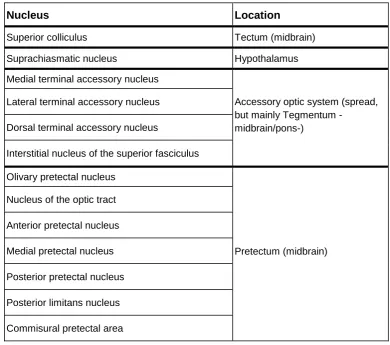
Whereas the majority of retinal outputs travel through the optic nerve to LGN, there are a dozen brain nuclei that also receive direct ganglion fibers (Table 3). In most of them, the main role is associated with oculomotor function and/or motion processing, but some studies propose their involvement, to a greater or lesser extent, in cognitive functions such as attention or
memory. Their involvement in affective processes has been scarcely studied (or not studied, in many cases), but the fact that dynamic stimuli are often emotional (predators, mates,
*** Table 3 *** 5.1. Superior colliculus (SC)
This structure is located in the top (tectum) part of the midbrain, and is organized in six layers in mammals, the two external or superficial (SCs) receiving visual inputs from retina and other visual structures such as the visual thalamus, visual striate and extrastriate cortices,
parabigeminal nucleus (a sort of SC satellite contributing to saccade control), pretectum nuclei (discussed in the next section) as well as from the locus coeruleus (involved in the regulation of arousal): Basso & May (2017). The SCs is organized retinotopically (Cerkevich et al., 2014) and sends projections to K layers and to pulvinar. It is involved in basic visual processing, mainly related to orienting and motion, and aimed at organizing orienting movements of the eyes and head (Krauzlis et al., 2013). Interestingly, SCs, despite not being connected to attention networks (such as fronto-parietal networks), responds differently as a function of physical stimulus
saliency, what suggests an autonomous capability to detect it (White et al., 2017). Intermediate and internal layers (SCi), also organized topographically (Wurtz, 2009), are multimodal, since they receive visual inputs from SCs and non-visual sensory inputs as well. The SCi layers
interact with many levels of the central nervous system including frontal and parietal cortices (but not with the visual thalamus), and would contribute to orienting behavior, and to cognitive
processes such as attention or decision making (Basso & May, 2017, Krauzlis et al., 2013). Several data suggest that SC is able to discriminate complex features. Through fMRI recordings in humans, SC responses to aversive stimuli are greater than to other visual stimuli (Almeida et al., 2015; Wang et al., 2020). Indeed, SC is proposed to form a sort of tandem with pulvinar as an early emotional detection mechanism, as indicated (Soares et al., 2017).
Intracranial electrophysiological data also point to the SC capability to discriminate complex shapes. For example, it responds more intensely to certain fractal forms -varying in colors, forms and sizes- as a function of their hedonic value previously conditioned with juice administration (Griggs et al., 2018; macaque), a discrimination which is patent at 95 ms from stimulus onset. This relatively long latency may be explained by proposals from other lines exploring the
maps of the visual context rather than stimulus features, and therefore would need to intercommunicate with other cerebral structures that process and evaluate these features (Krauzlis et al., 2013). According to this, the SC would act as an indexing system that pools together the signals of those structures to determine the content of perception. This idea would imply that the feature evaluation carried out by the SC is fruit of recurrent information. In any case, electrophysiological data are very scarce up to the present, and additional research is needed to characterize, both in temporal and functional terms, the involvement of SC in emotional evaluation.
5.2. Other structures
As indicated, whereas the main targets of ganglion cells are LGN and, to a lesser extent, SC, several brain nuclei also receive direct retinal inputs (Table 3). Some of them seem less involved in rapid processing of visual stimulation, such as the suprachiasmatic nucleus of the
hypothalamus, a key element in the control of circadian rhythms, or the pretectal olivary nucleus (PON), which controls the pupillary light reflex or triggers rapid eye movements during sleep. However, the majority of first-order nuclei, particularly those linked to oculomotor control, are indeed fast detectors of visual stimulation.
On one hand, the human accessory optic system (AOS) is a net of four nuclei also innervated by retinal ganglion cells which present large receptive fields and are direction
selective (Fredericks et al., 1988). The AOS is involved in motion processing and in the control of oculomotor mechanisms, but it is also involved in cognitive processes such as spatial memory and attention (Giolli et al., 2006). On the other hand, the six nuclei that form the pretectum -PON, previously mentioned, is among them- also receive direct retinal inputs (Gamlin, 2006). One nucleus of the pretectum, the nucleus of the optic tract (NOT), is especially relevant since it receives inputs from visual cortex (along with retina) and projects to LGN, pulvinar and TRN, as well as to AOS nuclei and SC, among other structures (Büttner‐Ennever et al., 1996).
adapts oculomotor activity to that motion (Gramlin, 2006). Indeed, and as AOS, the NOT participates in cognitive processes such as attention (Büttner‐Ennever et al., 1996).
6. Discussion
Figure 3 summarizes the information provided in previous sections on the main ascending routes of visual information, and the approximate timings –from stimulus onset- in which each
processing level (each synapsis) comes into play. The present review points to the visual
thalamus, and likely other associated non-thalamic nuclei, as crucially involved in early emotional processing of visual stimulation. Concretely, the LGN seems to be a key IES, rapidly labeling the sensory inputs as emotional thanks to its contrasted capability to locally process relative complex stimuli, with the fast concourse of TRN as a global evaluator of visual features. This information, labeled as emotional if pertinent, would reach the visual cortex around 60-70 ms after stimulus onet. In parallel, but some milliseconds later, the SC-pulvinar tandem could also evaluate the ascending information. Whereas the SC-pulvinar tandem would probably not contribute to the initial V1 (or V2/V3) response to emotional stimuli, peaking at 80 ms, its action would reinforce and complement the LGN-TRN evaluation in ascending phases. Later, the visual input already marked as emotional would follow the routes shown in Figure 3 towards the visual cortices and, through TRN and cortical interconnections, to other brain areas well known to be involved in emotional processing such as amygdala, vPFC and insula (among others: Lee & Siegle, 2009). At this point, deep evaluation starts, since emotional stimuli are more sophisticatedly processed in these structures. Information would also travel back from these structures to LGN, TRN and pulvinar, among other brain areas. The recurrent information interchange among these elements is also characteristic of deep evaluation, and often derives in subjective (e.g., fear feeling), autonomic (e.g., increased skin conductance), motor (e.g., orienting or avoidance) and cognitive changes (e.g., mnemonic actualization), as a continuation of the initial evaluation reviewed here, of fast and (rudimentary) perceptual nature.
Therefore, the present proposal does not contradict previous data on the evaluative capabilities of amygdala, vPFC, insula, or other structures, which is backed by a solid
background of concurrent and abundant data. However, their influence over visual cortices or other brain structures involved in visual processing would belong to deep evaluation rather than being the first markers of emotionality or IESs. Particularly, the role of amygdala, which in current models of emotional processing is often proposed as the cornerstone of emotional processing -also in humans-, should be discussed beyond the basic outlines mentioned in the Introduction. As indicated, initial evaluation, one of the main ingredients in this cornerstone role, seems not to be within the reach of amygdala mainly for three reasons. First, because the multisynaptic path that visual information needs to follow to reach this structure (four synapses minimum), causes latencies incompatible with observations. Focusing on faces -the stimuli evoking the faster amygdalar responses-, latencies are longer than required for any IES. Thus, the face-sensitive neurons of the macaque amygdala (i.e., they specifically react to faces and not to other visual stimuli) respond from 60 ms (Nakamura et al., 1992), and typically from 100 ms or beyond (Leonard et al., 1985; Sanghera et al., 1979; Wang, Tudusciuc et al., 2014). In humans, results are parallel but with the known increase in latency (Pessoa & Adolphs, 2010), and the earliest responses have been observed at 74 ms, as indicated in the Introduction (Méndez-Bértolo et al., 2016), and beyond 250 ms in several studies (Rutishauser et al., 2011; Mormann et al., 2008; Krolak-Salmon et al., 2004).
The second reason that distance amygdala away from being an IES is that it lacks the necessary cytoarchitecture. As mentioned in the Introduction, no amygdalar nuclei have been reported to incorporate focal processors in charge of specific RFs, which usually follow a
The third reason is the relative specialization of amygdala in face processing (see the meta-analysis by Sergerie et al., 2008). While some amygdalar neurons specifically respond to other non-facial significant stimuli, such as food or animals (irrespective of their emotional valence), their response latency is usually longer (e.g., ~320 ms average in the case of stimuli depicting animals: Mormann et al., 2011; human). Relatedly, Méndez-Bértolo and collaborators (2016; human) showed that amygdalar differential activity between emotional and non-emotional stimuli is observed more than 100 ms later in response to non-facial than to facial stimuli (see Hariri et al., 2002, as regards the human amygdala advantage for facial over non facial emotional stimuli also in amplitude terms). Indeed, the amygdala is considered a key piece of the neural circuitry involved in social behavior (e.g., Amaral, 2003). However, and importantly, IESs should be also able to rapidly detect and label non-facial visual events, whose impact on survival may be even more dramatic.
As described in previous sections, three key prerequisites for an IES (short latency, visual feature processing architecture, non-specific content detection) seem to be accomplished by the LGN, which belongs to the main and fastest ascending route of visual information in humans. Data on its involvement in early emotional evaluation are scarce since it has been usually out of the scopes of research (i.e., it has not been a target in animal intracranial recordings or a specific region of interest in human fMRi in studies using visual emotional stimulation), but results from other lines of research provide some relevant clues. Regarding latency of response, LGN may modulate the visual input at ~40 ms from stimulus onset (~30 ms in macaques, as indicated: McAlonan et al., 2008). With respect to its capability to evaluate visual features, current data show that certain intrinsic characteristics of the stimulus, such as shape novelty (Ortuño et al., 2014), may modulate LGN responses, and we hypothesize that the idiosyncratic saliency of emotional stimulation may be among them. This modulation requires focal and global feature analysis, in order to detect shapes, colors, motion, etcetera, that could identify an emotional stimulus (Figure 1). LGN is undoubtedly capable of developing the focal analysis of the visual input through relay cells (P, M, and K), and global analysis, interconnecting several relay cells to increase the visual area to be processed, could be carried out by the TRN. Importantly, this nucleus is susceptible to long-term potentiation (Sieber et al., 2013), pointing to a capability to encode certain visual information and, ultimately, to the online comparison of incoming
information with stored features corresponding to emotional stimuli. The participation of LGN interneurons -also interconnected to an important number and variety of relay cells- as a global processor is also probable at the light of the reviewed data. Labelling the stimulus as emotional would likely consist of a burst firing of LGN relay cells shortly after stimulus onset since,
according to the information reviewed in previous sections, this type of activity marks relevant stimulation and privileges it in subsequent visual cortex processing. Additionally, TRN would distribute this information to other thalamic and cortical areas.
pulvinar, in close association with SC, in early emotional evaluation has been also reviewed. Two reasons have been mentioned suggesting that the pulvinar is probably not the core IES. On one hand, the main inputs to visual pulvinar are descending projections from different visual cortices, which shapes its main role as a hub that coordinates cortico-cortical activity (Eradath et al., 2020; Jaramillo et al., 2019). On the other hand, reported latencies of pulvinar enhanced activity
towards emotional stimulation seem, up to the present, longer than required to explain initial visual cortex responses to emotional stimuli, as reviewed above. Whereas not a core IES, the involvement of pulvinar (or the SC-pulvinar tandem) in initial evaluation is backed by
experimental data and may complement the LGN-TRN previous evaluation. The fact that the pulvinar reaches some different brain areas from those innervated by the LGN-TRN tandem, and that it conveys complementary information -more related to motion, both regarding the stimulus and the eyes/body themselves- to that processed in LGN, but also relevant to emotional
processing, would indeed be a valuable contribution to early evaluation. Non-thalamic first order structures, such as the pretectum and AOS nuclei described in this review, can detect or
evaluate certain aspects of stimulation as well, particularly those related to stimulus motion and motor behavior (e.g., eye pursuit), important parameters regarding many emotional visual events.
In sum, current data clearly show that a key lost link exists in emotional processing, which consists in defining the cerebral structures forming the initial evaluation system. While this gap seems evident, the candidates to be an IES are still to be experimentally explored. Our proposal on the key involvement of LGN-TRN in this initial, ascending evaluation, with the later
contribution of SC-pulvinar, is based on the existing literature, but we are conscious this is still scarce and that further research is needed to confirm (or discard) it. In any case, the information reviewed here points to the crucial relevance of opening the focus of Affective Neuroscience to include the thalamus as a scope of research, as Cognitive Neuroscience is beginning to do in recent years. Two lines of research appear of special interest. On one hand, employing
emotional stimuli in intracranial recordings of LGN (both relay and interneuron) and TRN cells of primates. On the other hand, designing fMRi studies in human subjects, also employing
other visual thalamic nuclei to long-term potentiation is also a scarcely explored, but crucial, issue. Indeed, stimulus evaluation necessarily requires the storage of certain features that allow the identification of upcoming emotional events. In sum, it seems worth rescuing at least part of the protagonism the thalamus had in the first years of Affective Neuroscience.
Acknowledgements
This research was supported by the grants PGC2018-093570-B-I00 from the Ministerio de Ciencia e Innovación of Spain (MICINN) and HUM19-HUM5705 from the Comunidad de Madrid.
References
Acunzo, D., MacKenzie, G., & van Rossum, Mark C. W. (2019). Spatial attention affects the early processing of neutral
versus fearful faces when they are task-irrelevant: A classifier study of the EEG C1 component. Cognitive, Affective, &
Behavioral Neuroscience, 19, 123-137.
Adolphs, R. (2004). Emotional vision. Nature Neuroscience, 7, 1167-1168.
Adolphs, R. (2008). Fear, faces, and the human amygdala. Current Opinion in Neurobiology, 18, 166-172.
Adolphs, R., Kawasaki, H., Oya, H., & Howard, M. A. (2006). Intracranial electrophysiology of the human orbitofrontal
cortex. In D. H. Zald, & S. L. Rauch (Eds.), The orbitofrontal cortex (pp. 355-375) OUP Oxford.
Ajina, S., & Bridge, H. (2018). Blindsight relies on a functional connection between hMT and the lateral geniculate nucleus,
not the pulvinar. PLoS Biology, 16(7), e2005769.
Ales, J. M., Yates, J. L., & Norcia, A. M. (2010). V1 is not uniquely identified by polarity reversals of responses to upper and
lower visual field stimuli. Neuroimage, 52, 1401-1409.
Alitto, H., Rathbun, D. L., Vandeleest, J. J., Alexander, P. C., & Usrey, W. M. (2019). The augmentation of retinogeniculate
communication during thalamic burst mode. Journal of Neuroscience, 39, 5697-5710.
Almeida, I., Soares, S. C., & Castelo-Branco, M. (2015). Correction: The distinct role of the amygdala, superior colliculus
and pulvinar in processing of central and peripheral snakes. PloS One, 10(10), e0141175.
Amaral, D. G. (2003). The amygdala, social behavior, and danger detection. Annals of the New York Academy of Sciences,
1000, 337-347.
Anders, S., Lotze, M., Erb, M., Grodd, W., & Birbaumer, N. (2004). Brain activity underlying emotional valence and arousal:
A response-related fMRI study. Human Brain Mapping, 23, 200-209.
Arcaro, M. J., Pinsk, M. A., & Kastner, S. (2015). The anatomical and functional organization of the human visual pulvinar.
Baldwin, M. K., Balaram, P., & Kaas, J. H. (2017). The evolution and functions of nuclei of the visual pulvinar in primates.
Journal of Comparative Neurology, 525, 3207-3226.
Basso, M. A., & May, P. J. (2017). Circuits for action and cognition: A view from the superior colliculus. Annual Review of
Vision Science, 3, 197-226.
Bender, D. B. (1982). Receptive-field properties of neurons in the macaque inferior pulvinar. Journal of
Neurophysiology, 48, 1-17.
Berman, R. A., & Wurtz, R. H. (2010). Functional identification of a pulvinar path from superior colliculus to cortical area
MT. Journal of Neuroscience, 30, 6342-6354.
Blitz, D. M., & Regehr, W. G. (2005). Timing and specificity of feed-forward inhibition within the LGN. Neuron, 45, 917-928
Bragg, E. M., Fairless, E. A., Liu, S., & Briggs, F. (2017). Morphology of visual sector thalamic reticular neurons in the
macaque monkey suggests retinotopically specialized, parallel stream-mixed input to the lateral geniculate nucleus.
Journal of Comparative Neurology, 525, 1273-1290.
Bridge, H., Leopold, D. A., & Bourne, J. A. (2016). Adaptive pulvinar circuitry supports visual cognition. Trends in Cognitive
Sciences, 20, 146-157.
Britton, J. C., Phan, K. L., Taylor, S. F., Welsh, R. C., Berridge, K. C., & Liberzon, I. (2006). Neural correlates of social and
nonsocial emotions: An fMRI study. Neuroimage, 31, 397-409.
Broca, P. (1878). Anatomie comparée des circonvolutions cérébrales. le grand lobe limbique et la scissure limbique dans la
série des mammifères. Revue d'Anthropologie, 1, 385-498.
Brosch, T., Pourtois, G., & Sander, D. (2010). The perception and categorisation of emotional stimuli: A review. Cognition
and Emotion, 24, 377-400.
Bühler, M., Vollstädt-Klein, S., Klemen, J., & Smolka, M. N. (2008). Does erotic stimulus presentation design affect brain
activation patterns? event-related vs. blocked fMRI designs. Behavioral and Brain Functions, 4(1), 30.
Büttner‐Ennever, J., Cohen, B., Horn, A., & Reisine, H. (1996). Efferent pathways of the nucleus of the optic tract in
monkey and their role in eye movements. Journal of Comparative Neurology, 373, 90-107.
Cannon, W. B. (1927). The James-Lange theory of emotions: A critical examination and an alternative theory. The
American Journal of Psychology, 39, 106-124.
Capilla, A., Melcón, M., Kessel, D., Calderón, R., Pazo-Álvarez, P., & Carretié, L. (2016). Retinotopic mapping of visual
event-related potentials. Biological Psychology, 118, 114-125.
Casagrande, V. A., Royal, D. W., & Sáry, G. (2005). Extraretinal inputs and feedback mechanisms to the lateral geniculate
nucleus (LGN). In J. Kremers (Ed.), The primate visual system: A comparative approach (pp. 191-211). Hoboken, NJ:
Wiley Online Library.
Carretié, L., Albert, J., López-Martín, S., & Tapia, M. (2009). Negative brain: An integrative review on the neural processes
Carretié, L. (2014). Exogenous (automatic) attention to emotional stimuli: A review. Cognitive, Affective and Behavioral
Neuroscience, 14, 1228-1258.
Cerkevich, C. M., Lyon, D. C., Balaram, P., & Kaas, J. H. (2014). Distribution of cortical neurons projecting to the superior
colliculus in macaque monkeys. Eye and Brain, 2014, 121-137.
Cola, M. G., Gray, D. N., Seltzer, B., & Cusick, C. G. (1999). Human thalamus: Neurochemical mapping of inferior pulvinar
complex. Neuroreport, 10, 3733-3738.
Costafreda, S. G., Brammer, M. J., David, A. S., & Fu, C. H. Y. (2008). Predictors of amygdala activation during the
processing of emotional stimuli: A meta-analysis of 385 PET and fMRI studies. Brain Research Reviews, 58, 57-70.
Critchley, H., Daly, E., Phillips, M., Brammer, M., Bullmore, E., Williams, S., et al. (2000). Explicit and implicit neural
mechanisms for processing of social information from facial expressions: A functional magnetic resonance imaging
study. Human Brain Mapping, 9, 93-105.
Damasio, A. (1994). Descarte’s error: Emotion, reason, and the human brain. New York: Putnam.
Das, P., Kemp, A. H., Liddell, B. J., Brown, K. J., Olivieri, G., Peduto, A., Gordon, E., & Williams, L. M. (2005). Pathways for
fear perception: modulation of amygdala activity by thalamo-cortical systems. NeuroImage, 26, 141–148.
de Gelder, B., & Hadjikhani, N. (2006). Non-conscious recognition of emotional body language. Neuroreport, 17, 583-586.
de Gelder, B., Snyder, J., Greve, D., Gerard, G., & Hadjikhani, N. (2004). Fear fosters flight: a mechanism for fear
contagion when perceiving emotion expressed by a whole body. Proceedings of the National Academy of Sciences,
101, 16701-16706.
Debanne, D. (2004). Information processing in the axon. Nature Reviews Neuroscience, 5(4), 304-316.
Derrington, A., & Lennie, P. (1984). Spatial and temporal contrast sensitivities of neurones in lateral geniculate nucleus of
macaque. The Journal of Physiology, 357, 219-240.
DeYoe, E. A., & Van Essen, D. C. (1988). Concurrent processing streams in monkey visual cortex. Trends in
Neurosciences, 11, 219-226.
Di Russo, F., Martinez, A., & Hillyard, S. A. (2003). Source analysis of event-related cortical activity during visuo-spatial
attention. Cerebral Cortex, 13, 486.
Dolcos, F., LaBar, K. S., & Cabeza, R. (2004). Interaction between the amygdala and the medial temporal lobe memory
system predicts better memory for emotional events. Neuron, 42, 855-863.
Duan, X., Dai, Q., Gong, Q., & Chen, H. (2010). Neural mechanism of unconscious perception of surprised facial
expression. Neuroimage, 52, 401-407.
Dunsmoor, J. E., Kubota, J. T., Li, J., Coelho, C. A., & Phelps, E. A. (2016). Racial stereotypes impair flexibility of
emotional learning. Social cognitive and affective neuroscience, 11, 1363-1373.
Edmiston, E. K., McHugo, M., Dukic, M. S., Smith, S. D., Abou-Khalil, B., Eggers, E., Zald, D.H. (2013). Enhanced visual
cortical activation for emotional stimuli is preserved in patients with unilateral amygdala resection. The Journal of
Eldar, S., Yankelevitch, R., Lamy, D., & Bar-Haim, Y. (2010). Enhanced neural reactivity and selective attention to threat in
anxiety. Biological Psychology, 85, 252-257.
Emery, N. J., & Amaral, D. G. (2000). The role of the amygdala in primate social cognition. In R. D. Lane, & L. Nadel (Eds.),
Cognitive neuroscience of emotion (pp. 156-191). New York, NY, US: Oxford University Press.
Eradath, M. K., Pinsk, M. A., & Kastner, S. (2020). A causal role for pulvinar in coordinating task independent corticocortical
interactions. Biorxiv, https://doi.org/10.1101/2020.03.07.982215
Fichtenholtz, H. M., Dean, H. L., Dillon, D. G., Yamasaki, H., McCarthy, G., & LaBar, K. S. (2004). Emotion–attention
network interactions during a visual oddball task. Cognitive Brain Research, 20, 67-80.
Frank, D. W., & Sabatinelli, D. (2014). Human thalamic and amygdala modulation in emotional scene perception. Brain
research, 1587, 69-76.
Fredericks, C. A., Giolli, R. A., Blanks, R. H. I., & Sadun, A. A. (1988). The human accessory optic system. Brain Research,
454, 116-122.
Gamlin, P. D. R. (2006). The pretectum: Connections and oculomotor-related roles. Progress in Brain Research, 151,
379-405.
Garrett, A. S., & Maddock, R. J. (2006). Separating subjective emotion from the perception of emotion-inducing stimuli: an
fMRI study. Neuroimage, 33, 263-274.
Gattass, R., Soares, J. G., & Lima, B. (2018). The pulvinar thalamic nucleus of non-human primates: Architectonic and
functional subdivisions. Berlin: Springer.
George, M. S., Ketter, T. A., Parekh, P. I., Horwitz, B., Herscovitch, P., & Post, R. M. (1995). Brain activity during transient
sadness and happiness in healthy women. American Journal of Psychiatry, 152, 341-351.
Ghodrati, M., Khaligh-Razavi, S., & Lehky, S. R. (2017). Towards building a more complex view of the lateral geniculate
nucleus: Recent advances in understanding its role. Progress in Neurobiology, 156, 214-255.
Giolli, R. A., Blanks, R. H. I., & Lui, F. (2006). The accessory optic system: Basic organization with an update on
connectivity, neurochemistry, and function. Progress in Brain Research, 151, 407-440.
Goldin, P. R., Hutcherson, C. A., Ochsner, K. N., Glover, G. H., Gabrieli, J. D., & Gross, J. J. (2005). The neural bases of
amusement and sadness: A comparison of block contrast and subject-specific emotion intensity regression
approaches. Neuroimage, 1, 26-36.
Goldin, P. R., McRae, K., Ramel, W., & Gross, J. J. (2008). The neural bases of emotion regulation: Reappraisal and
suppression of negative emotion. Biological Psychiatry, 63, 577-586
Govindaiah, G., & Cox, C. L. (2006). Metabotropic glutamate receptors differentially regulate GABAergic inhibition in
thalamus. Journal of Neuroscience, 13443-13453.
Griggs, W. S., Amita, H., Gopal, A., & Hikosaka, O. (2018). Visual neurons in the superior colliculus discriminate many
objects by their historical values. Frontiers in Neuroscience, 12, 396.
Grünert, U., & Martin, P. R. (in press). Cell types and cell circuits in primate retina. Progress in Retinal and Eye Research.
Guido, W., & Weyand, T. (1995). Burst responses in thalamic relay cells of the awake behaving cat. Journal of
Neurophysiology, 74, 1782-1786.
Guillery, R., & Harting, J. K. (2003). Structure and connections of the thalamic reticular nucleus: Advancing views over half
a century. Journal of Comparative Neurology, 463(4), 360-371.
Guillory, S. A., & Bujarski, K. A. (2014). Exploring emotions using invasive methods: Review of 60 years of human
intracranial electrophysiology. Social Cognitive and Affective Neuroscience, 9, 1880-1889.
Günther, V., Zimmer, J., Kersting, A., Hoffmann, K., Lobsien, D., & Suslow, T. (2017). Automatic processing of emotional
facial expressions as a function of social anhedonia. Psychiatry Research: Neuroimaging, 270, 46-53.
Hakamata, Y., Sato, E., Komi, S., Moriguchi, Y., Izawa, S., Murayama, N., Hanakawa, T., Inoue, Y., & Tagaya, H. (2016).
The functional activity and effective connectivity of pulvinar are modulated by individual differences in threat-related
attentional bias. Scientific reports, 6, 34777.
Halassa, M. M., & Kastner, S. (2017). Thalamic functions in distributed cognitive control. Nature Neuroscience, 20(12),
1669-1679.
Hariri, A. R., Tessitore, A., Mattay, V. S., Fera, F., & Weinberger, D. R. (2002). The amygdala response to emotional
stimuli: A comparison of faces and scenes. Neuroimage, 17, 317-323.
Head, H. (1920). Studies in neurology, in two volumes. London: Hodder and Stoughton.
Hermann, A., Schäfer, A., Walter, B., Stark, R., Vaitl, D., & Schienle, A. (2007). Diminished medial prefrontal cortex activity
in blood-injection-injury phobia. Biological psychology, 75, 124-130.
Hirsch, J. A., Wang, X., Sommer, F. T., & Martinez, L. M. (2015). How inhibitory circuits in the thalamus serve vision.
Annual Review of Neuroscience, 38, 309-329.
Horowitz, A., Barazany, D., Tavor, I., Bernstein, M., Yovel, G., & Assaf, Y. (2015). In vivo correlation between axon
diameter and conduction velocity in the human brain. Brain Structure and Function, 220, 1777-1788.
Huo, B., Zeater, N., Lin, M. K., Takahashi, Y. S., Hanada, M., Nagashima, J., et al. (2019). Relation of koniocellular layers
of dorsal lateral geniculate to inferior pulvinar nuclei in common marmosets. European Journal of Neuroscience, 50,
4004-4017.
Ishii, T., & Hosoya, T. (2020). Interspike intervals within retinal spike bursts combinatorially encode multiple stimulus
features. Biorxiv, 2020.02.13.947283.
Jaramillo, J., Mejias, J. F., & Wang, X. (2019). Engagement of pulvino-cortical feedforward and feedback pathways in
cognitive computations. Neuron, 101, 321-336. E9.
Kang, D., Liu, Y., Miskovic, V., Keil, A., & Ding, M. (2016). Large‐scale functional brain connectivity during emotional
engagement as revealed by beta‐series correlation analysis. Psychophysiology, 53, 1627-1638.
Karama, S., Armony, J., & Beauregard, M. (2011). Film excerpts shown to specifically elicit various affects lead to
Kehoe, E. G., Toomey, J. M., Balsters, J. H., & Bokde, A. L. (2012). Personality modulates the effects of emotional arousal
and valence on brain activation. Social Cognitive and Affective Neuroscience, 7, 858-870.
Kim, J., Shinkareva, S. V., & Wedell, D. H. (2017). Representations of modality-general valence for videos and music
derived from fMRI data. Neuroimage, 148, 42-54.
Kimura, A. (2014). Diverse subthreshold cross‐modal sensory interactions in the thalamic reticular nucleus: Implications for
new pathways of cross‐modal attentional gating function. European Journal of Neuroscience, 39, 1405-1418.
Kimura, A. (2017). Robust interactions between the effects of auditory and cutaneous electrical stimulations on cell
activities in the thalamic reticular nucleus. Brain Research, 1661, 49-66.
Kinoshita, M., Kato, R., Isa, K., Kobayashi, K., Kobayashi, K., Onoe, H., et al. (2019). Dissecting the circuit for blindsight to
reveal the critical role of pulvinar and superior colliculus. Nature Communications, 10, 135.
Krauzlis, R. J., Lovejoy, L. P., & Zénon, A. (2013). Superior colliculus and visual spatial attention. Annual Review of
Neuroscience, 36, 165-182.
Krolak‐Salmon, P., Hénaff, M., Tallon‐Baudry, C., Yvert, B., Guénot, M., Vighetto, A., et al. (2003). Human lateral
geniculate nucleus and visual cortex respond to screen flicker. Annals of Neurology, 53, 73-80.
Krolak-Salmon, P., Hénaff, M., Vighetto, A., Bertrand, O., & Mauguière, F. (2004). Early amygdala reaction to fear
spreading in occipital, temporal, and frontal cortex: A depth electrode ERP study in human. Neuron, 42, 665-676.
Kuraoka, K., & Nakamura, K. (2007). Responses of single neurons in monkey amygdala to facial and vocal emotions.
Journal of Neurophysiology, 97, 1379-1387.
Lane, R. D., Reiman, E. M., Ahern, G. L., Schwartz, G. E., & Davidson, R. J. (1997a). Neuroanatomical correlates of
happiness, sadness, and disgust. The American Journal of Psychiatry, 154, 926-933.
Lane, R. D., Reiman, E. M., Bradley, M. M., Lang, P. J., Ahern, G. L., Davidson, R. J., & Schwartz, G. E. (1997b).
Neuroanatomical correlates of pleasant and unpleasant emotion. Neuropsychologia, 35, 1437-1444.
Lashley, K. S. (1938). The thalamus and emotion. Psychological Review, 45, 42-61.
LeDoux, J. E. (1992). The emotional brain. New York: Simon & Schuster.
LeDoux, J. E. (2000). Emotion circuits in the brain. Annual Review of Neuroscience, 23, 155-184.
Lee, K. H., & Siegle, G. J. (2009). Common and distinct brain networks underlying explicit emotional evaluation: A
meta-analytic study. Social Cognitive and Affective Neuroscience, 7, 521-534.
Lee, T. M. C., Liu, H. L., Chan, C. C. H., Fang, S. Y., & Gao, J. H. (2005). Neural activities associated with emotion
recognition observed in men and women. Molecular psychiatry, 10, 450-455.
Leonard, C., Rolls, E., Wilson, F., & Baylis, G. (1985). Neurons in the amygdala of the monkey with responses selective for
faces. Behavioural Brain Research, 15, 159-176.
Lesica, N. A., & Stanley, G. B. (2004). Encoding of natural scene movies by tonic and burst spikes in the lateral geniculate
Liberzon, I., Taylor, S. F., Fig, L. M., Decker, L. R., Koeppe, R. A., & Minoshima, S. (2000). Limbic activation and
psychophysiologic responses to aversive visual stimuli: Interaction with cognitive task. Neuropsychopharmacology, 23,
508-516.
Lichev, V., Sacher, J., Ihme, K., Rosenberg, N., Quirin, M., Lepsien, J., et al. (2015). Automatic emotion processing as a
function of trait emotional awareness: An fMRI study. Social Cognitive and Affective Neuroscience, 10, 680-689.
Liddell, B. J., Brown, K. J., Kemp, A. H., Barton, M. J., Das, P., Peduto, A., et al. (2005). A direct brainstem–amygdala–
cortical ‘alarm’system for subliminal signals of fear. Neuroimage, 24, 235-243.
Liewald, D., Miller, R., Logothetis, N., Wagner, H., & Schüz, A. (2014). Distribution of axon diameters in cortical white
matter: An electron-microscopic study on three human brains and a macaque. Biological Cybernetics, 108, 541-557.
Lindner, K., Neubert, J., Pfannmöller, J., Lotze, M., Hamm, A. O., & Wendt, J. (2015). Fear-potentiated startle processing in
humans: Parallel fMRI and orbicularis EMG assessment during cue conditioning and extinction. International Journal of
Psychophysiology, 98, 535-545.
Liu, T. Y., Chen, Y. S., Hsieh, J. C., & Chen, L. F. (2015). Asymmetric engagement of amygdala and its gamma
connectivity in early emotional face processing. PloS one, 10(1).
Livingstone, M. S., & Hubel, D. H. (1981). Effects of sleep and arousal on the processing of visual information in the cat.
Nature, 291, 554-561.
Luo, Q., Holroyd, T., Jones, M., Hendler, T., & Blair, J. (2007). Neural dynamics for facial threat processing as revealed by
gamma band synchronization using MEG. Neuroimage, 34, 839-847.
MacLean, P. D. (1949). Psychosomatic disease and the" visceral brain"; recent developments bearing on the papez theory
of emotion. Psychosomatic Medicine, 11, 338-353.
Marrocco, R. T., McClurkin, J. W., & Alkire, M. T. (1996). The influence of the visual cortex on the spatiotemporal response
properties of lateral geniculate nucleus cells. Brain Research, 737, 110-118.
Martin, P. R., & Solomon, S. G. (2019). The koniocellular whiteboard. Journal of Comparative Neurology, 527, 505-507.
Maunsell, J. H., & Newsome, W. T. (1987). Visual processing in monkey extrastriate cortex. Annual Review of
Neuroscience, 10, 363-401.
McAlonan, K., & Brown, V. J. (2002). The thalamic reticular nucleus: More than a sensory nucleus? Neuroscientist, 8,
302-305.
McAlonan, K., Cavanaugh, J., & Wurtz, R. H. (2008). Guarding the gateway to cortex with attention in visual thalamus.
Nature, 456, 391-394.
Méndez-Bértolo, C., Moratti, S., Toledano, R., Lopez-Sosa, F., Martínez-Alvarez, R., Mah, Y. H., Vuilleumier, P., Gil-Nagel,
A., Strange, B. (2016). A fast pathway for fear in human amygdala. Nature Neuroscience, 19, 1041.
Mizuno-Matsumoto, Y., Hayashi, T., Okamoto, E., Miwa, D., Asakawa, T., Muramatsu, A., et al. (2013). Human-related
emotional stimuli can cause a hippocampal and thalamic over-response in people with unstable personalities. Journal