FORGOTTEN BILIARY STENTS
*Dr. Nilay Chakrabarti, Dr. Shalaka Indap, Dr. Ratnakar Shetty and Dr. Mugdha Kowli
Department of Surgery, K. J. Somaiya Medical College, Hospital and Research Centre, Sion,
ARTICLE INFO ABSTRACT
Endoscopic Retrograde Cholangio Pancreatography (ERCP) with biliary stenting is one of the most commonly performed endoscopic procedures for preoperative biliary decompression in obstructive of benign or malignant etiology. In patients with concomitant calculous cholecystitits, a laparoscopic cholecystectomy is performed soon after, and in the absence of a specific indication to the contrary, most such stents are removed 6
dictated by considerations related to the specific treatment modality used. However, some patients are lost to follow up for a stent removal having got almost immediate symptomatic relief
blissfully unaware of the complications that may ensue due to an unremoved stent. Retained stents may remain asymptomatic for years, or more commonly, present with blockage and delayed complications requiring another, and often mo
endoprosthesis have been documented, some left behind for even as long as 10 years. Prevention is the best option in tackling this situation and detailed instructions to the patient alon
of the indwelling stent can go a long way in achieving this. Creating a Stent Registry has also been suggested in this context. The recent invention of the biodegradable biliary stent, one which disintegrates in the biliary tr
in this scenario. While research has identified Poly manufacture of such a stent, use of this device is limited
hurdles preventing its widespread use. However the way forward undoubtedly lies in its wider use thus preventing a perfectly avoidable complication arising out of a "forgotten stent".
Copyright © 2015 Nilay Chakrabarti et al.This is an open access article distributed under the Creative Commons Att use, distribution, and reproduction in any medium, provided the original work is properly cited.
INTRODUCTION
Biliary stenting following Endoscopic Retrograde Cholangio Pancreaticography (ERCP) for choledocholithiasis ensures free drainage of bile after common bile duct clearance, and occasionally, but not as commonly, pancreatic duct stenting is undertaken in pancreatic disease. In the former, it is a routine practice to subsequently subject the patient to an early laparoscopic cholecystectomy for the cholecystitis, followed by a stent removal 6-8 weeks later. However, a biliary stent may be kept longer if specifically indicated as in biliary strictures. Complications related to stents are well documented from the early days of biliary stenting as far back as in 1985 by Mueller et al. (1985). While it cannot be denied that complications may occur following routine stenting too, presence of a stent in the biliary tree left behind for a long duration is especially fraught with danger since, the stent though crucial in patient management, is undoubtedly a foreign body.
*Corresponding author: Dr. Nilay Chakrabarti
Department of Surgery, K. J. Somaiya Medical College, Hospital and Research Centre, Sion, Mumbai 400022, India
ISSN: 0975-833X
Vol.
Article History:
Received 26th September, 2015
Received in revised form 24th October, 2015
Accepted 29th November, 2015
Published online 30th December,2015
Citation:Nilay Chakrabarti, Shalaka Indap, Ratnakar Shetty and Mugdha Kowli complication”, International Journal of Current Research,
.
Keywords:
Retained biliary stents, Complications, Stent Registry, Biodegradable stents, Poly-L-Lactic Acid Stent.
RESEARCH ARTICLE
FORGOTTEN BILIARY STENTS – AN AVOIDABLE COMPLICATION
*Dr. Nilay Chakrabarti, Dr. Shalaka Indap, Dr. Ratnakar Shetty and Dr. Mugdha Kowli
Department of Surgery, K. J. Somaiya Medical College, Hospital and Research Centre, Sion,
Mumbai 400022, India
ABSTRACT
Endoscopic Retrograde Cholangio Pancreatography (ERCP) with biliary stenting is one of the most commonly performed endoscopic procedures for preoperative biliary decompression in obstructive of benign or malignant etiology. In patients with concomitant calculous cholecystitits, a laparoscopic cholecystectomy is performed soon after, and in the absence of a specific indication to the contrary, most such stents are removed 6-8 weeks later. In malignant disease however, the duration of stenting would be dictated by considerations related to the specific treatment modality used. However, some patients are lost to follow up for a stent removal having got almost immediate symptomatic relief
blissfully unaware of the complications that may ensue due to an unremoved stent. Retained stents may remain asymptomatic for years, or more commonly, present with blockage and delayed complications requiring another, and often more difficult intervention. Complications related to forgotten biliary endoprosthesis have been documented, some left behind for even as long as 10 years. Prevention is the best option in tackling this situation and detailed instructions to the patient alon
of the indwelling stent can go a long way in achieving this. Creating a Stent Registry has also been suggested in this context. The recent invention of the biodegradable biliary stent, one which disintegrates in the biliary tree after a predetermined period and therefore does not require removal, can provide an answer in this scenario. While research has identified Poly-L-Lactic Acid (PLLA) as the material of choice for manufacture of such a stent, use of this device is limited at present with cost and availability being the main hurdles preventing its widespread use. However the way forward undoubtedly lies in its wider use thus preventing a perfectly avoidable complication arising out of a "forgotten stent".
is an open access article distributed under the Creative Commons Attribution License, which use, distribution, and reproduction in any medium, provided the original work is properly cited.
Biliary stenting following Endoscopic Retrograde Cholangio Pancreaticography (ERCP) for choledocholithiasis ensures free drainage of bile after common bile duct clearance, and occasionally, but not as commonly, pancreatic duct stenting is pancreatic disease. In the former, it is a routine practice to subsequently subject the patient to an early laparoscopic cholecystectomy for the cholecystitis, followed 8 weeks later. However, a biliary stent ifically indicated as in biliary strictures. Complications related to stents are well documented from the early days of biliary stenting as far back as in 1985 by . While it cannot be denied that complications may occur following routine stenting too, presence of a stent in the biliary tree left behind for a long duration is especially fraught with danger since, the stent
t, is undoubtedly a foreign
Department of Surgery, K. J. Somaiya Medical College, Hospital and
Retained or forgotten stents have been reported even after ten years after insertion, while stent migration requiring subsequent surgical intervention has also been reported (Bajbouj, 2008 and Diller, 2003
especially those who have undergone sphincterotomy/ stenting, is also a recognized phenomenon
Complications related to a forgotten stent documented in literature include recurrent cholangitis, biliary strictures and formation of a stone around the retained stent, the “stentolith" (Gupta, 2013). While every effort is made towards optimization of the duration of stenting, there is the occasional patient who plays truant and islost to follow up for a stent removal. Here we review literature associated with a forgotten biliary stent with a brief reference to two of our patients who presented with a forgotten stent.
DISCUSSION
First performed by Professor
stenting is one of the most commonly performed endoscopic procedures today (Soehendra, 1979
stenting) is nowroutine and in most instances the st
International Journal of Current Research
Vol. 7, Issue, 12, pp.24320-24327, December, 2015
INTERNATIONAL
Nilay Chakrabarti, Shalaka Indap, Ratnakar Shetty and Mugdha Kowli,2015. “Forgotten
International Journal of Current Research, 7, (12), 24320-24327.
AN AVOIDABLE COMPLICATION
*Dr. Nilay Chakrabarti, Dr. Shalaka Indap, Dr. Ratnakar Shetty and Dr. Mugdha Kowli
Department of Surgery, K. J. Somaiya Medical College, Hospital and Research Centre, Sion,
Endoscopic Retrograde Cholangio Pancreatography (ERCP) with biliary stenting is one of the most commonly performed endoscopic procedures for preoperative biliary decompression in obstructive jaundice of benign or malignant etiology. In patients with concomitant calculous cholecystitits, a laparoscopic cholecystectomy is performed soon after, and in the absence of a specific indication to the contrary, most er. In malignant disease however, the duration of stenting would be dictated by considerations related to the specific treatment modality used. However, some patients are lost to follow up for a stent removal having got almost immediate symptomatic relief from biliary obstruction, blissfully unaware of the complications that may ensue due to an unremoved stent. Retained stents may remain asymptomatic for years, or more commonly, present with blockage and delayed complications re difficult intervention. Complications related to forgotten biliary endoprosthesis have been documented, some left behind for even as long as 10 years. Prevention is the best option in tackling this situation and detailed instructions to the patient along with thorough documentation of the indwelling stent can go a long way in achieving this. Creating a Stent Registry has also been suggested in this context. The recent invention of the biodegradable biliary stent, one which disintegrates in ee after a predetermined period and therefore does not require removal, can provide an answer Lactic Acid (PLLA) as the material of choice for at present with cost and availability being the main hurdles preventing its widespread use. However the way forward undoubtedly lies in its wider use thus preventing a perfectly avoidable complication arising out of a "forgotten stent".
ribution License, which permits unrestricted
Retained or forgotten stents have been reported even after ten years after insertion, while stent migration requiring subsequent surgical intervention has also been reported Bajbouj, 2008 and Diller, 2003). Biliary ascariasis in patients, who have undergone sphincterotomy/ stenting, is also a recognized phenomenon (Gupta et al., 1998). Complications related to a forgotten stent documented in literature include recurrent cholangitis, biliary strictures and formation of a stone around the retained stent, the “stentolith" . While every effort is made towards duration of stenting, there is the occasional patient who plays truant and islost to follow up for a stent removal. Here we review literature associated with a forgotten biliary stent with a brief reference to two of our patients who
otten stent.
First performed by Professor Soehendra in 1979, biliary stenting is one of the most commonly performed endoscopic Soehendra, 1979). Biliary (and urinary tract stenting) is nowroutine and in most instances the stenting
INTERNATIONAL JOURNAL OF CURRENT RESEARCH
precedes another therapeutic intervention, with subsequent endoscopic stent removal 6-8 weeks later. Very rarely would the stent be required to be kept longer, though there are situations such as that reported by Bajboj et al. where a patient with a biliary endoprosthesis followed up as long as 10 years after its insertion and Patel et al. who reported an emergency presentation of a stent forgotten in the biliary tract for 17 years presenting with acute cholecystitis and a ‘stentolith’ (Bajbouj, 2008 and Patel, 2014).
Even though biliary stenting is routinely performed , complications have been reported, the most common being a blocked stent with cholangitis, and less commonly stent migration resulting in gut perforation, ascariasis of the biliary tree following the sphincterotomy, or a rare colo cutaneous fistula following stent migration (Storkson, 2000; Gupta et al ., 1998 and Figueiras, 2001). Most such complications follow stenting of long duration or a retained stent in the biliary tree; however these incidents certainly cannot detract from its mandatory use in the obstructed biliary system. While the presence of various foreign bodies in the biliary tree have been documented such as a fish bone, a gauze piece, cystic duct clips, or even shrapnel resulting in complications, an entirely different scenario arises due to a ‘forgotten stent’ where the patient himselffails to come back for stent removal at the prescribed time and only appears much later with a related or unrelated complaint (Kaji, 2004; Cipolletta, 1997; Kitamura, 1995 and Mitchell et al., 1991). Stent removal being a day care procedure, the onus is on the patient to come back for a stent removal following discharge from hospital and such an oversight can result in a perfectly avoidable complication.
It would not be out of place here to make a brief mention of a useful corollary to this situation that has been reported by urologists where forgotten ureteral stents have been known to result in several complications, such as ureteric obstruction due to encrustation around the stent and even going as far as renal failure in some instances. Of the several studies highlighting this, is one reported by Ahmet Ali Sancaktutar et al. on forgotten and encrusted Double J ureteral stents in 22 children. While each of the stents were ultimately removed, it involved a mean of 1.5 endoscopic interventions ranging from ureteroscopy, endoscopic cystolithotripsy and retrograde intrarenal surgery with a mean hospital stay of 4.4 days (Ahmet Ali Sancaktutar et al., 2013). Another such study is that reported by Aron et al. which reviewed ureteral stent records over a 10 year period from 1994 to 2004 in patients with forgotten stents in the only functioning kidney which had led to chronic renal failure. While a third of these patients were unaware of their stent, the others had chosen to ‘ignore’ it.
The median dwelling time of the stents was reported as 39 months while serum creatinine in these patients ranged from 4-14mg/dl. The study which quite understandably concluded that it was disastrous to have an un removed, encrusted stent in the urinary tract, also noted that such cases are still seen inspite of increasing patientawareness (Aron et al ., 2006). Two other studies in this context are important since the suggestions in them can have a significant bearing in tackling the problem of the forgotten biliary stent too. One by Hoscan MB et al who, while sharing the concern with complications arising out of a forgotten ureteral stent, looked at solutions to the problem in
the form of an electronic stent extraction reminder facility, or a possible computer based tracking system using a short message service based reminder to both patient and doctor (Hoscan, 2013). The second by Withington et al . suggested the use of a bar coded wrist band on the patient, details of which are scanned into a registry which automatically books an appointment for stent removal (Withington, 2014).
Forgotten biliary stents can result in several complications in the form of stent clogging with resultant cholangitis, ascariasis of the biliary tree, obstructive jaundice and pancreatitis. If retained long enough the stentmay even result in a stone formation around itself. One of the more recent studies reviewing the long term effects of forgotten biliary stents is by Mehmet Odabasi et al from Turkey in August 2014 which reported a series of cases with a wide range of complications resulting from a retained stent (Mehmet Odabasi et al ., 2014). It is well known that ascariasis of the biliary tree is not an uncommon occurrence in endemic areas. However there is a definite increase in its incidence in patients who have undergone a spincterotomyand available literature has widely documented the presence of biliary ascariasis in patients with a CBD stent in situ for a prolonged duration. The worm most commonly migrates into the common bile duct across the papilla leading to complications ranging from cholangitis, cholecystitis and pancreatitis, to liver abscesses and biliary strictures (Gupta et al., 1998). In most such patients the worm dies but occasionally may survive as proved by live extractions of such worms on ERCP. Analysis of stone fragments extracted from the common bile duct have occasionally shown fragments of ascaris inside them suggesting that the nidus for stone formation has been a worm or pieces of it (Gupta, 1998). One of our patients with a retained stent was asymptomatic except for upper abdominal pain of a few days duration. A history of jaundice a year ago along with that of an ‘endoscopic procedure’ (? a possible biliary stenting) was obtained. Plain X ray revealed the faint outline of a biliary stent (Fig. 1).
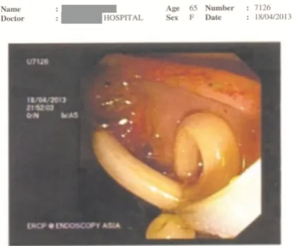
It was only during ERCP that a live roundworm (and calculi) was also seen in the CBD apart from the previous stent (Fig 2).
Fig. 2. Case 1. ERCP images showing round worm being extracted from the Common Bile Duct
The previously placed stent had to be removed piecemeal, duct clearance achieved by removal of the worm as well the calculi in the bile duct, and a fresh 10Fr stent was inserted in the CBD. This was followed by laparoscopic cholecystectomy four days later and stent removal after six weeks. Clogging of a biliary stent is a well known phenomenon, more so with the plastic stents which although affordable, clog after 3-4 months (Kenneth, 2005). The process is believed to be initiated by adherence of a biofilm of bacteria along the inner surface of the stent followed by a gel like glycocalyx formation which protects it from antibiotics, as also from the shearing effects of bile flow and the patient’s immune system. Adding to this is the mechanism believed to be mediated by the release of the enzyme beta glucoronidase from E.coli which deconjugates bilirubin glucoronide with resultant precipitation of calcium bilirubinate, which then combines with glycoprotein to form calculi (Virinder Kumar Bansal et al., 2009). An increase in the biofilm with trapping of insoluble crystals, along with refluxed duodenal contents and other cellular debris would then lead to stent occlusion. A relevant study in this context is the one by Emilio Guaglianone et al. where biliary stents in 28 patients were removed and subjected to culture, gradient gel electrophoresis and electron microscopy in an attempt to establish the role of bacterial (aerobic as well as anaerobic) and fungalcolonization of these stents with biofilm formation on its luminal aspect resulting in subsequent stent blockage. It was seen that except for one stent, all others showed a mixed microbial growth, both aerobic as well as anaerobic, including fungi, with Gram positive enterococcus fecalis being the commonest aerobic organism isolated.
It was also suggested that the production of ‘slime’ by most of the cultured enterococci played an important role in bacterial colonization which leads to an occluded stent and enterococci which carry an aggregation substance gene could be more selectively associated with colonization of biliary stents. (Emilio Guaglianone, 2010). Another similar study by Albert K Groen reported the analysis of the contents of occluded biliary stents in 21 patients. Though bacterial growth was
detected in the clogged stent it was not seen in abundance; protein, insoluble residue (plant fibres) and bile acids/lecithin constituted 25%, 20% and 15% respectively of the sludge in the blocked stent. When the protein fraction was subjected to SDS-polyacrylamide gel electrophoresis it always showed two major bands of 16 and 13k Dalton proteins, ones which bind most avidly with the stent wall. The study concluded that the initial phase of stent blockage is always protein adsorption followed by adhesion of other materials including bacteria, unconjugated bilirubin and food fibres to the stent wall (Albert K Groen et al., 1987).
While this could happen with any stent, it has been conventionally believed that effect was more marked with the latex stents as compared to those made of silicone which were less reactive. This fact has however not been conclusively proved in animal studies carried out on pigs by Koivasalo et al. and published in 1996, where biocompatibility of latex and silicone T tubes was studied after their insertion into the porcine common bile duct. In this study with 30 animals divided into 2 groups of 16 and 14, a latex or silicone T tube was inserted into the common bile duct either after a choledochotomy or a ¾ transaction of the CBD followed by suturing over the T tube respectively. Reoperation and harvesting of the ducts from the two groups after 2 and 6 weeks respectively followed by electron and light microscopy, as well as cell culture toxicity with a DNA synthesis inhibition test showed that while the latex T tubes were toxic, those made of silicone were non toxic. However final conclusions drawn at the end of the 6 week study period indicated that the tissue reactions in the bile duct wall were similar and neither material appeared to be completely harmless for the porcine CBD wall (Koivusalo et al., 1996). In fact, several randomized trials comparing Teflon stents (without side holes, the Tannenbaum stent) against standard polyethylene stents (with side holes) also have not demonstrated any improvement in the patency rates (Catalano, 2002).
Various strategies have been adopted in an attempt to prevent, or at the least retard, clogging of the stent such as use of prophylactic antibiotics, an antibiotic impregnated stent, the administration of bile altering agents such as ursodeoxycholic acid, alterations in stent design or even placement of a stent in the common bile duct with an intact sphincter of Oddi. However the only step which has seemed to have some impact
is the use of larger diameter stents (Kenneth, 2005). A “stentolith” is a term coined in this context which refers to a
large concretion of such crystals around a stent appearing like a stone,and most often blocking it, an occurrence more likely with long duration stenting or indeed a stent forgotten in the biliary tree (Gupta, 2013 and Virinder Kumar Bansal, 2009). Both, an open choledochotomy approach as well as laparoscopic removal of a stent with a ‘stenolith’ has been reported (Gupta, 2013 and Virinder Kumar Bansal, 2009). In our patientwhile the common bile duct did need to clear of calculi at the time of removal of the retained stent, a classical stentolith was not seen.
ulceration and pressure necrosis may occur following insertion of a straight stent with a long intraduodenalpart (Kenneth, 2005). Percutaneous removal of such a stent following duodenal perforation has been reported, while other similar reports by various authors relate to duodenal and small bowel perforation secondary to a migrated biliary stent (including one in a patient with an incisional hernia and even in a liver transplant patient) thereby requiring a surgical intervention (Bui, 1995; Saranga Bharathi, 2006; Storkson et al., 2000; Diller, 2003; Akimboye et al., 2006; Esterl, 1997). Incidents of other more dangerous complications due to a migrated stent have included sigmoid colon perforations reported by Anderson et al. and Eliott et al. a rare colo cutaneous fistula and even fatal necrotizing fasciitis following a migrated biliary stent reported by Marsman et al. (Anderson et al ., 2007; Elliot and Boland, 2003; Figueiras et al ., 2003 and Marsman, 1996). Needless to say, the longer a stent is left in situ, more is the chance of stent migration.
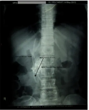
[image:4.595.349.517.116.275.2]Though the majority of biliary stenting and its complications are described for the CBD, there is a smaller subgroup of patients who undergo stenting of the pancreatic duct. Prolonged pancreatic duct stenting is also associated with complications related to recurrent cholangitis and even stent migration, though this is rare. Pancreatic duct stents can become stuck in the small bowel, especially in patients who have multiple bowel adhesions. Shapiro AM et al have reported on calcific intra pancreatic embedding of a pancreatic duct stent present in situ for a prolonged duration and warned against this as a danger of long duration endoscopic retrograde pancreatic duct stenting (Shapiro, 1999). In another of our patients a background of intermittent upper abdominal pain for 2-3 years and an acute exacerbation prompting this hospital visit with elevated amylase and lipase levels was suggestive of chronic(? relapsing) pancreatitis. Plain X ray showed shadowsof a retained bileduct stent, a possible pancreatic duct stent, as well as pancreatic head calcification (Fig. 3).
Fig.3 Case 2. Patient presented with chronic abdominal pain with past h/o endoscopy. X ray abdomen showing retained stent in
CBD and pancreatic duct and pancreatic head calcification
[image:4.595.321.547.462.613.2]A CT scan confirmed these findings including the presence of a second stent in the pancreatic duct. EUS and ERCP was performed and reported as chronic pancreatitis with evidence of a blockedCBD stent, a pancreatic duct stent and pancreatic duct calculi (Fig.4).
Fig. 4. Case 2. ERCP images showing calculus being extracted from pancreatic duct
[image:4.595.72.258.485.714.2]Cholangiogram revealed smooth narrowing of the lower CBD with upstream dilatation. No stones were seen in the CBD but multiple dense calcifications were seen in the pancreatic head region on EUS. Though the reason for the previous stenting could not be ascertained, removal of this stent, (which in all probability was non functional), was long overdue. It was indeed fortunate that the complication reported above had not occurred. Following endoscopic stent removal, MRCP confirmed chronic pancreatitis (pancreatic head calcification had already been seen on the X ray) in the form of atrophy of the proximal main pancreatic duct with a dilated mid portion of the duct (Fig. 5).
Fig. 5. Case 2. MRCP image showing dilated mid pancreatic duct
Apart from problems related to a biofilm accumulation over the stent and proliferative changes being greatly reduced, an additional benefit to the patient would be of not having to come back for a stent removal. Such a stent also offers a solution to problems of long term complications and permanency associated with the other significant advance in stent technology, i.e. the expandable metal biliary stent (Kenneth et al., 2005). Though historically biodegradable materials for surgical use have been around for several years, manufacture of stents made of these has not developed at the same pace. Hurdles which have come in the way include factors such as inadequate radial strength of the stent, an inflammatory response to the material, as well as fracture of the stent in some instances. Biodegradable coronary stents have been in vogue for some time now with the earliest reports being from Japan by Tamai et al. published as far back as in 2000 on stents made of Poly -L-Lactic Acid (PLLA) for use in coronary arteries successfully (Tamai et al., 2000). Though the retained or forgotten stent situation was never the question in coronary artery stenting, theirusefulness has been aptly described by Colombo and Karvouni as something “fulfilling the mission and stepping away” (Colombo, 2000).
Solutions for a ‘forgotten stent’ situation led to almost simultaneous research on the possible use of biodegradable stents in the urinary as well as in the biliary tract and animal studies led the way in this regard. An important study in this context is the one by Ben H Chew et al in 2013 which based its findings on an animal study involving two groups of pigs unilaterally implanted with either a biodegradable or a biostable stent in the ureter. A necropsy and histological examination 4 weeks later revealed that 90% of the biodegradable stents had totally degraded while one stent had fragmented to three pieces less than 1.5cm in size. While hematological and renal parameters were similar in the two groups, there was significantly less hydronephrosis with fewer abnormal histological findings in the group with biodegradable stents leading to the conclusion that the third generation biodegradable ureteric stent is a safe and effective alternative to the conventional polymer stent with equivalent drainage (Ben, 2013).
Studies on trying to find the ideal material for such a stent also centered on the use of Poly l-lactide-co-Glycolide (PLGA) and Poly L-Lactic Acid (PLLA), the material which had been earlier developed for biodegradable coronary stents. Several animal studies preceded attempts to use biodegradable biliary stents in human subjects Initial studies on choosing the most appropriate material for the stent included in vitro and in vivo studies using PLGA for CBD stents. In one such study PLGA, molar ratio LA/GA = 80/20, was used to prepare circular tube and dumb bell shaped stents and then tested to determine their in vitro degradation behavior in bile. Parameters used to determine suitability included morphology, weight loss, and molecular weight changes along with evaluations of the mechanical properties of the specimen. Subsequently, circular radio opaque tube-shaped stents made of PLGA were used in dogs that had undergone common bile duct exploration with primary suturing of the CBD. Results were analyzed after imaging of stents in vivo including levels of serum liver enzymes and a histological study of the CBD, which showed
that the PLGA stents exhibited the required biomedical properties.
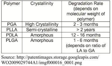
Spontaneous disappearance from CBDs was noted in 4–5 weeks time, the degradation period and function matching the requirements in repair and reconstruction of CBD, thereby reducing T-tube-related complications (Esterl et al., 1997). However comparisons between the various materials have gone in favor of PLLA as the preferred material for manufacture of biodegradable biliary stents (Fig. 6).
[image:5.595.338.530.175.288.2]Source: http://patentimages.storage.googleapis.com/ WO2009029744A1/imgf000016_0001.png
Fig. 6. Comparison between various materials used in manufacture of biodegradable biliary stents
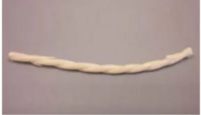
Other animal studies include those by Yigang Chen et al where techniques for safe placement of biodegradable stents in the common bile duct in rabbits had been described and Shi J et al from China who used a new paclitaxel coated PLLA biodegradable stent in mongrel dogs (Yigang Chen et al., 2013; Shi et al., 2013). Itoi et al from Japan placed a braided, self-expandable, biodegradable stent endoscopically into the pancreatic and bile duct in pigs which were then studied further after a necropsy to draw conclusions (Itoi, 2011). Moving ahead from animal studies to use in human subjects, the use of biodegradable stent materials in humans for non coronary use started with the trial by Fry and Fleisher in 1997 almost two decades ago, where a coil spring expandable stent made of a single wire of polyglycoloide was implanted in a patient with an oesophageal stricture. However the result was not good since the stent fractured proximally due to premature loss of its expansile strength and in the process led to occlusion of the oesophageal lumen requiring an endoscopic removal (Fry, 1997). Since then there has been significant advance and biodegradable stents have been used in the biliary tree in situations such as in stenting following post cholecystectomy leaks and in biliary strictures with good results. (Johanna Laukkarinen, 2007 and Mauri, 2013) A biliary stent of PLLA made of polymer strands in a tubular mesh form, with radio opacity being ensured by the incorporation of Tantalum strands in it, was initially favored for use in humans (Fig. 7).
Source:http://www.carefusion.co.uk/Images/Interventional_Specialties/IS_Dist ributors/nitinella_plus_biliary_stent_carousel_img2.jpg
Fig.7. Basic structure of the biodegradable stent
radial force exerted by these stents required balloon dilatation for expansion. Additional problems were related to bile duct lumen obstruction due to fragmentation and residue (Kenneth F Binmoeller and Biliary stenting, 2005). Modifications of these stents have been based on studies in the porcine bile duct with good results as reported in the DDW 2002, which included the use of the PLLA stent with elastometric axial runners which increases the radial force (thus avoiding a subsequent balloon expansion) as well as having significantly less complications or problems of bile duct integration (Kenneth F Binmoeller and Biliary stenting, 2005). One such device for use in humans is the “Archimedes” biodegradable stent. which was used for the first time in Asia at the UKM Medical Centre in Kuala Lumpur, Malaysia, in March 2014 on a 56 year old man suffering from acute cholangitis
(http://www.onenewspage.com/rss/latest/press+ releases.rss
03/23/14-16.13) (Fig.8).
[image:6.595.60.265.258.375.2]Source: http://stronmedical.com/images/biodegradable_product1.jpg
Fig. 8 The first biodegradable biliary stent used- “Archimedes Device”
It is reported to come with three degradation times to accommodate the various disease states that require different indwelling times for treatment; a fast absorbing (weeks), medium absorbing (months), and a long lasting one which can stay even up to six months, remaining in the biliary tree before it is slowly hydrolyzed by the body. Other modifications of such a stent are bound to come with rapid ongoing research in this area. With patent related issues and FDA and other approvals still pending, commercial use of these devices is obviously not yet widespread but this device is definitely the solution to the “forgotten stent” situation. The use of such stents is increasing and if these hurdles are overcome, this certainly is the future of biliary stenting in the days to come.
Conclusion
A “forgotten stent” is a reality in the context of biliary stenting. An entirely avoidable situation, it can be associated with severe complications often requiring another surgical or endoscopic intervention. Apart from the additional cost and hospital stay that this entails, it is also important to remember that removing such a stent may not be easy especially in a migrated stent or one with a large concretion around it. The key therefore lies in prevention of the “forgotten stent” situation. Meticulous instructions to the patient and thorough documentation regarding the presence of an indwelling stent following an endoscopic procedure can go a long way in pre empting this situation. Suggestions have been made regarding the setting up of a computerized ‘Stent Registry System’ to
track patients with a stent which would need removal and this appears to be an excellent idea (Virinder Kumar Bansal et al., 2009). As previously discussed, a solution to the problem has been suggested in the form of an electronic stent extraction reminder facility as also a computer based tracking system using a short message service based reminder to both patient and doctor (Hoscan, 2013). The use of a bar coded wrist band on the patient, details of which are scanned into a registry is also feasible (Withington et al., 2014). However, the future lies undoubtedly in the use of the biodegradable biliary stent and its wider use would obviate the need for a stent removal. At this point in time PLLA along with the modificationsdescribed appears to be the material of choice for the manufacture of the biodegradable biliary stent. Research to develop the ideal, as well as commercially viable, biodegradable stent with appropriate approval by the authorities is on, and hopefully the situation of a ‘forgotten stent’ should soon be a matter of the past.
REFERENCES
Ahmet Ali Sancaktutar, Senol Adanur, Berkan Resorlu, Abdulkadir Tepeler, Tevfiik Ziypak, Haluk Soylemez, Murat Atar, Yasar Bozkurt, Necmettin Penbegul, Adnan Tufek,SevgiYavuz. 2013. The Forgotten Ureteral Stent in Children: From Diagnosis to Treatment. The Journal of Urology, Volume 189, Issue 3, Pages 1054-1060
Akimboye, F., Lloyd, T., Hobson, S, Garcea, G. 2006. Migration of endoscopic biliary stent and small bowel perforation within an incisional hernia; Surg Laparosc Endosc Percutan Tech 2006, 16 (1): 39-40
Albert K Groen, T Out, K Huibregtse, B. Delzenne, F J Hoek, G.N.J. Tytgat. Charactrization of the content of occluded biliary endoprosthesis. Endoscopy, 04/1987;19(2):57-9 Anderson, E.M., Phillips Hughes, J., Chapman, R. 2007.
Sigmoid colonic perforation and pelvic abscess
complicating biliary stent migration. Abdom Imaging, 32(3);317-319
Aron, M., Ansari, M.S., Singh, I., Gautam, G., Kolla, S.B., Seth, A, et al . 2006. Forgotten Ureteral stents causing renal failure: multimodal endourologic treatment. J Endourol. 20:423-8
Bajbouj, M., Treiber, M., Ludwig, L., Frimberger, E., Schmid, R.M. Neu, B. 2008. Forgotten biliary endoprosthesis. “Follow up” after 10 years. Endoscopy, 40:E221
Ben, H. Chew, Ryan F. Paterson, Kenneth, W. Clinkscales, Barry, S Levine, Shalaby, W. Shalaby, Dirk 2013. Lange In vivo Evaluation of the Third Generation Biodegradable Stent: a novel approach to Avoiding the Forgotten Stent Syndrome. The Journal of Urology, February, Volume 189, Issue 2, Pages 719-725
Bui, B.T., Oliva, V.L., Ghattas, G., Daloze, P., Bourdon, F., Carignan, L. 1995. Percutaneous removal of a biliary stent after acute spontaneous duodenal perforation. Cardiovasc Intervent Radiol, 18(3):200-202
Catalano, M.F., Geenen, J.E., Lehman, G.A., et al . 2002.
“Tannenbaum” Teflon stents versus traditional
polyethylene stents for the treatment of malignant biliary stricture. GastrointestEndosc 2002.55(3):354-8
a surgical gauze from the common bile duct. Italian Journal of Gastroenertology Hepatology., 29:58-61 Colombo, A., Karvouni, E. 2000. Biodegradable stents
“fulfilling the mission and stepping away” Circulation; 102(4); 371-3
Diller, R., Senninger, N., Kautz, G., Tubergen, D. 2003. Stent
migration necessitating surgical intervention. Surg
Endosc,17(11):1803-1807
Elliot, M., Boland, S. 2003. Sigmoid colon perforation following a migrated biliary stent. ANZ. J. Surg., 2003, 73(8):669-670
Emilio Guaglianone, Rita Cardines, Claudia Vuotto, Roberta di Rosa, valentine Babini, Paola Mastrantonio 2010. Gianfranco Donelli. Microbial biofilm associated with biliary stent clogging. FEMS Immunol Med Microbiol 59, 410-420
Esterl, R.M., Jr, St. Laurent, M., Bay, M.K., Speeg, K.V., Halff, G.A. 1997. Endoscopic biliary stent migration in a liver transplant recipient. J. ClinGastroenterol,, 24 (2):106-110
Figueiras, R.G., Echart, M.O., Figueiras, A.G., Gonzalez, G.P. 2001. Colocutaneous fistula relating to the migration of a biliary stent. Eur J GastroenterolHepatol 2001.13(10): 1251-1253
Fry, S.W., Fleischer, D.E. 1997. Management of a refractory benign esophageal stricture with a new biodegradable stent. GastrointestEndosc; 45(2): 179-82.
Gupta, R., Agarwall, D.K., Choudhuri, D.D., Saraswat, V.A., Baijal, S.S. 1998. Biliary ascariasis complicating endoscopic sphincterotomy for choledocholithiasis in India, J GastroenterolHepatol, 13; 1072-3
Gupta, V., Chandra, A. Noushif, M. Singh, S.K. 2013. Giant Stentolith: Complication of a forgotten biliary stent, Endoscopy;45(S 02):E126
Hoscan, M.B., Trunckiran, A. 2013. Management of Forgotten Ureteral Stents. Nephrol-Urol Mon. 5(2): 775-6
Itoi, T.1., Kasuya, K., Abe, Y., Isayama, H. 2011. Endoscopic placement of a new short-term biodegradable pancreatic and biliary stent in an animal model: a preliminary feasibility study (with videos). J Hepatobiliary Pancreat Sci. May;18(3):463-7.
Johanna Laukkarinen, Isto Nordback, JoonasMikkonen,
PäiviKärkkäinen, Juhani Sand. 2007. A novel
biodegradable biliary stent in the endoscopic treatment of cystic-duct leakage after cholecystectomy. Gastrointestinal Endoscopy. June, Volume 65; Issue 7: Pages 1063–1068 Kaji, H., Asano, N., Tamura, h., Yuh, I. 2004. Common bile
duct stone caused by a fish bone: report of a case Surg Today., 34:268-71
Kenneth, F., Binmoeller, Biliary Stenting, 2005. Old problems and new Challenges. Gastrohep.com. 01 October 2005. Kitamura, K., Yamaguchi, T., Nakatani, H., Ichikawa, D.,
Shimotsuma, M., Yamane, T., et al . 1995. Why do cystic duct clips migrate into the common bile duct? Lancet. 346:965-6
Koivusalo, A., Makisalo, H., Talja, M., Cormio, L., Ruutu, M., Wolff, H., Hockerstedt, K. 1996. Biocompatibility of Latex and silicone T tubes in the porcine common bile duct: an experimental study. Res Exp Med (Berl) 196 (1); 53-66
Marsman, J.W., Hoedemaker, H.P. 1996. Necrotizing fasciitis: fatal complication of migrated biliary stent. Australas Radiol., 40(1): 80-83
Mauri, G.1., Michelozzi, C., Melchiorre, F., Poretti, D., Tramarin, M., Pedicini, V., Solbiati, L., Cornalba, G., Sconfienza, L.M. 2013. Biodegradable biliary stent implantation in the treatment of benign bilioplastic- refractory biliary strictures: preliminary experience. Eur Radiol. Dec; 23(12):3 3304-10.
Mehmet Odabasi, Cem Arslan, Sami Akbulut, Haci Hasan Abuoglu, Erkan Ozkan, Mehmet Kamil Yildiz, Cengiz Eris, Emre Gunay, Kemal Tekesin, and Tolga Muftuoglu. 2014. Long – term effects of forgotten biliary stents: a case series and literature review. Int. J. Clin. Exp. Med., 7(8):2045-2052
Mitchell, R., Kerr, R., Barton, J., Schmidt, A. 1991. Biliary obstruction secondary to shrapnel. American Journal of Gastroenterology; 86:1531-4
Mueller, P.R., Ferrucci, J.T. Jr, Teplick, S.K., Van Sonnenberg, E., Haskin, P.H., Butch, R.J., 1985. Papanicolaou N. Biliary stent endoprosthesis: analysis of complications in 113 patients. Radiology 1985,156 (3): 637-639
Patel, T.J., Rajput, S., Patel, K.S., Haribhakti, S.P. 2014. A forgotten biliary stent for 17 years:presented with perforated gall bladder and stentolith. J. Dig. Endosc., 5:22-3
Q3 Medical Devices Ltd: UKMMC became the first centre to deploy Archimedes Biodegradable stents in Asia. Page 300.
http://www.onenewspage.com/rss/latest/press+releases.rss 03/23/14-16.13
Saranga Bharathi, R., Rao, P., Ghosh, K. 2006. Iatrogenic duodenal perforation caused by endoscopic biliary stenting and stent migration: an update; Endoscopy, 38(12); 1271-1274
Shapiro, A.M., Scudamore, C.H., July, L.V., et al . 1999. Calcific intra-pancreatic embedding of a pancreatic stent necessitating surgical removal--a danger of chronic
endoscopic retrograde pancreatic stent placement.
Gastrointest Endosc., 50:860.
Shi, J., Lv, Y., Yu, L., Zhang, B.., Zhang, X., Fan, C., Geng, Z. 2013. Interest of a new biodegradable stent coated with paclitaxel on anastomotic wound healing after biliary reconstruction. Eur J Gastroenterol Hepatol., 25 (12) : 1415-23
Soehendra, N., Reynders- Frederix, V. 1979. Palliative biliary tract drainage. A new method for endoscopic introduction of a new drain. Dtsch Med Wochenschr,104(6)206-7 Storkson, R.H., Edwin, B., Reiertsen, O., Faerden, A.E.,
Sortland, O., Rosseland, A.R. 2000. Gut perforation caused by biliaryendoprosthesis: Endoscopy., 32 (1): 87-89 Tamai, H., Igaki, K., Kyo, E, et al. 2000. Initial and 6 month
results biodegradable and poly-L-lactic acid coronary stents in humans. Circulation:102(4);399-404
Virinder Kumar Bansal, Mahesh C Mishra, Prashant Bhowate, Subodh Kumar. 2009. Laparoscopic management of
common bile duct “Stentolith”. Tropical
Withington, J., Wong, K., Bultitude, M and O’ Brien, T. 2014. The forgotten ureteric stent: what next? BJU International, 113:850-851
Xiaoyi Xu, Tongjun Liu, Shaohui Liu, Kai Zhang, Zhen Shen, Yuxin Li, Xiabin Jing. 2009. Feasibility of biodegradable PLGA common bile duct stents: An in vitro and in vivo study. Journal of Materials Science: Materials in Medicine. May, Volume 20, Issue 5, pp 1167-1173
Yigang Chen, Jun Yan, Zhigang Wang, Song Yu, Ziming Yuan, Xiaohu Wang, Xiaonong Zhang, Qi Zheng. 2013. Technique for the safe placement of a biodegradable stent into the common bile duct of rabbits, Experimental and Therapeutic Medicine, Vol.6 ; Issue 5: 1101-1104