Original Article
Application value of diffusion weighted magnetic
resonance imaging in head and neck cancer
Si-Cong Zhang1,2*, Yang-Yang Bao1*, Shui-Hong Zhou1, De-Sheng Shang3
1Department of Otolaryngology, The First Affiliated Hospital, College of Zhejiang University, Hangzhou 310003,
Zhejiang Province, China; 2Department of Otolaryngology, People’s Hospital of Cixi City, Cixi 315300, Zhejiang
Province, China; 3Department of Radiology, The First Affiliated Hospital, College of Medicine, Zhejiang University,
Hangzhou 310003, Zhejiang Province, China. *Equal contributors.
Received January 16, 2016; Accepted July 8, 2016; Epub August 15, 2016; Published August 30, 2016
Abstract: Diffusion weighted Magnetic Resonance Imaging (DWI) is currently the only imaging technology that re-flects the diffusion motion of water molecules in living tissues. At present, DWI has been widely used in the diagno-sis and differential diagnodiagno-sis of various malignant tumors, assessment of tumor differentiation, clinical stage and therapeutic effect evaluation, with or without metastasis and recurrence, etc. This article will review the application value of DWI and the apparent diffusion coefficient (ADC) value in head and neck cancer.
Keywords: Head and neck cancer, diffusion weighted imaging, apparent diffusion coefficient
Introduction
Diffusion weighted Magnetic Resonance Im- aging (DWI) is currently the only imaging tech-nology that reflects the diffusion motion of water molecules in living tissues. At present, DWI has been widely used in the diagnosis and differential diagnosis of various malignant tumors, assessment of tumor differentiation, clinical stage and therapeutic effect evaluation, with or without metastasis and recurrence, etc. This article will review the application value of DWI and the apparent diffusion coefficient (ADC) value in head and neck cancer.
Application of DWI and ADC value in diagnosis of primary lesion in head and neck cancer
DWI is a functional imaging technique that use special sequences which highlighted the bulk phase function caused by diffusion, while in the macroscopic imaging reflects microscopic dif-fusion of water molecules in living tissues. DWI with ADC values quantitatively reflect the orga-nization diffusion properties of water mole-cules, the space distribution between the inter-nal molecular of imaging materials determines the size value of ADC. The increased tissue cell
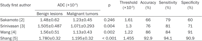
density, restriction of water molecular diffusion inside and outside of cells can lead to a drop in ADC values [1]. The malignant tumors show a high signal intensity on DWI and low ADC values due to a high malignant cell density, small inter-cellular space and low diffusion of water mole-cules. Sakamoto et al measured the ADC val-ues of 32 cases of head and neck benign lesions and 19 cases of head and neck malig-nant tumors, and found that the average ADC values of 32 cases of head and neck benign lesions and 19 cases of head and neck malig-nant tumors were 1.48±0.62×10⁻³ mm2/s and
1.23±0.45×10⁻³ mm2/s, respectively [2]. But
there was no statistical significance (P = 0.246). Srinivasan et al measured the ADC values of 17 cases of head and neck benign lesions and 16 cases of head and neck malignant tumors, and found that the average ADC values of 17 cases of head and neck benign lesions and 16 cases of head and neck malignant tumors were 1.505±0.487×10⁻³ mm2/s and 1.071±0.293×
10-3 mm2/s, respectively. There was statistical
significance (P = 0.004) [3]. They also proposed that the ADC value 1.3×10⁻³ mm2/s of 3.0 T MRI
Wang et al measured the ADC values of 22 cases of head and neck benign solid lesions and 36 cases of head and neck malignant tumors, and found that the average ADC values of 22 cases of head and neck benign solid lesions and 36 cases of head and neck malig-nant tumors were 1.56±0.51×10⁻³ mm2/s and
1.13±0.43×10⁻³ mm2/s, respectively [4]. There
was statistical significance (P = 0.002). They proposed that ADC values below 1.22×10⁻³
mm2/s forecast head and neck malignant
tumor, the accuracy, sensitivity and specificity were 86%, 84%, 91%, respectively. We have found that ADC values were lower for patients with laryngeal carcinoma (mean 1.195±0.32± 10-3 mm2/s) versus those with laryngeal
pre-cancerous lesions (mean 1.780±0.32±10-3
mm2/s; P, 0.001). The optimum threshold for
the ADC was 1.455±10-3 mm2/s, the accuracy,
sensitivity and specificity were 92.9%, 94.1%, 90.9%, respectively [5] (Table 1). The differ-ence of ADC values in different studies of head and neck malignant tumors is large, and lack of unified diagnostic criteria. The causes of this phenomenon are, first and foremost, the researchers in the selection of the research object, secondly, although in theory the ADC values are not affected by machinery and equipment and comparable between different machines, but in the actual work, hardware, scan sequence and b value selection are still influence the outcome of the ADC values. Generally, the higher the b value, the higher the lesion detection rate, but the image SNR is reduced at the same time. Blood perfusion can affect diffusion at low b value [6], while high b value inhibits the blood perfusion of richly vas-cularized tissues on the impact of diffusion [7], but b value higher than 1000 s/mm2 will lead to
poor image quality [8].
Pathological differentiation degree of tumors has certain correlation with the size of ADC val-ues, ADC values can quantify the degree of
dif-fusion of water molecules, in theory, the higher the degree of malignancy, the lower the ADC value of diseased tissue. Kato et al [9] found that the average ADC values of poorly and high-ly differentiated head and neck squamous cell carcinomas were 0.95±0.17×10⁻³ mm2/s and
1.24±0.23×10⁻³ mm2/s, respectively. There
was a significant difference (P < 0.01). Sumi et al [10] also reported that the ADC values of moderately or highly differentiated squamous carcinomas was higher than the low differenti-ated squamous carcinomas, the ADC values of poorly differentiated squamous carcinomas was close to malignant lymphomas. This is because those poorly differentiation tumors have an increased number of cells, reduced extracellular space, limited diffusion motion of water molecules, and the ADC values are rela-tively lower, while contrary to the well differenti-ated tumors.
Assessed value of DWI for the efficacy of ra
-diochemotherapy and prognosis in head and neck cancer
[image:2.612.92.522.87.175.2]How to early predict the efficacy of radiation and chemotherapy in order to individualized treatment is an increasingly concerned topic to clinical workers. DWI plays an important role in assessment and prediction of tumor curative effect, the changes of ADC values before any tumor volume reduction, ADC values measured to predict tumor early response to treatment. The measurement of ADC values can predict tumor early response to treatment. Most schol-ars [11, 12] believe that after radiotherapy: tumor cell membrane was damaged, cell apop-tosis increased, density decreased, extracellu-lar gap widened and the diffusion motion ability of water molecules enhanced, which may result in higher ADC values. It suggests that the treat-ment is effective if ADC values of tumors and lymph nodes progressively increased after the treatment; if the ADC values slightly increase or
Table 1. The differentiating diagnostic role of ADC value in the head and neck cancer in literatures
Study first author ADC (×10-3) p Threshold
(×10-3) Accuracy (%) Sensitivity (%) Specificity (%)
Benign lesions Malignant tumors
progressive decrease, showing that the tumor radiation therapy is low or no reaction; there are difference in ADC values between valid and invalid for radiotherapy of tumors, this differ-ence will become increasingly apparent as the duration of treatment [13]. There are also other studies [14] believe that in early radioactive radiation, high viscosity and abundant inflam-matory cells infiltration of necrosis tissues may led to restriction of water molecular diffusion, and ADC values will be transient decrease. Similar findings had been found in the applica-tion of DWI to evaluate the curative efficacy of neoadjuvant chemotherapy in breast cancer research [15]. Vandecaveye et al [16] found that by monitoring the ADC values of head and neck squamous cell carcinomas at 2 weeks and 4 weeks after concurrent chemoradiother-apy, the ADC values of patients with complete response compared with recurrence were sig-nificantly increased, and there was statistical significance between them. Kim et al [17] through the study of 40 patients with head and neck squamous cell carcinomas, patients with complete response and partial response before treatment, the ADC values were 1.04±0.19× 10⁻³ mm2/s and 1.35±0.30×10⁻³ mm2/s, res-
pectively. The ADC values of patients with com-plete response were significantly lower than the partial response, which was statistically signifi-cant (P < 0.05). They also found that the ADC values increased during the first week of chemoradiation, and the patients with com-plete response had a greater increase than the partial response. It indicates that the determi-nation of ADC values before treatment and the range of the ADC values change after treatment can predict the effect of head and neck malig-nant tumors to radiation and chemotherapy.
DWI and ADC value evaluate recurrence of head and neck cancer
Hypophaeyngeal cancer patients have a higher recurrence rate, how to early evaluate recur-rence after treatment is an important link of clinical research. Accurately assess the recur-rence of tumor can effectively avoid unneces-sary surgery and improve the survival rate [18]. Conventional imaging examinations for early evaluation of tumor recurrence is poorer, even if FDG PET/CT because of the high sensitivity of inflammation that results in higher false posi-tive thus affecting the correct evaluation of
early tumor recurrence [19, 20]. Abdel Razek et al [21] through the study of 30 patients with head and neck malignant tumors, the average ADC value of patients with residual or recur-rence was 1.17±0.33×10⁻³ mm2/s, significantly
lower than the change of the average ADC value 2.07±0.25×10⁻³ mm2/s after treatment, there
was statistical significance (P < 0.001). The critical point of ADC value was 1.30×10⁻³
mm2/s, as the distinction between residual or
recurrence and change after treatment, the accuracy, sensitivity, specificity, positive predic-tive value and negapredic-tive predicpredic-tive value were 87%, 84%, 90%, 94%, 76%, respectively. Tshering et al [22] through the study of 46 cases of laryngeal and hypopharyngeal cancer, the sensitivity and specificity of DWI combined conventional MRI to found cancer were 94% and 100%, respectively. The average ADC value of head and neck malignant tumor with residu-al or recurrence was 1.20±0.49×10⁻³ mm2/s,
significantly lower than the change of the aver-age ADC value 1.82±0.41×10⁻³ mm2/s after
treatment, there was statistical significance (P < 0.0002). ROC analysis to provide the best threshold value was 1.30×10⁻³ mm2/s, its
diag-nostic sensitivity, specificity and accuracy were 67%, 86% and 78% respectively. Vandecaveye et al [23] found that DWI can accurately distin-guish change after radiotherapy or chemother-apy in head and neck malignant tumor and the tumor residual or recurrence, when b value was 0, the sensitivity, specificity and accuracy were 66.2%, 60.8%, 62.4%, respectively; when b value was 1000, the sensitivity, specificity and accuracy were 71.6%, 71.3%, 71.4%, respec-tively; compared with CT, TSE-MRI and PET, DWI had a lower false positive. King et al [24] found that in the process of concurrent chemoradio-therapy, ADC values of some lesions in the treatment of early rising, late fall, or in the treat-ment of early fall, late rising, no matter what type of pathology, the decline of ADC values in the process of concurrent chemoradiotherapy strongly suggested local recurrence, decreased ADC values closely associated with local recur-rence (P = 0.00001).
Application of DWI and ADC value in evalua
-tion of cervical lymph nodes
lymph node metastasis at the visit, about 60% of the transferred sites along the internal jugu-lar vein and upper-middle cervical lymph nodes, followed by retropharyngeal lymph nodes and posterior cervical lymph nodes. Accurate ass- essment of metastatic cervical lymph nodes before the treatment helps to judge the stage of hypopharyngeal cancer, which helps to make accurate treatment plan and estimate the prog-nosis accurately. Current clinical usually use conventional CT, MRI and US to assess cervical lymph nodes, also with the help of PET/CT to evaluate cervical lymph nodes. Ashraf et al [25] used 1.0 cm in diameter as the identification standard, the negative predictive value and positive predictive value of CT scanning were 84% and 50% respectively, while 0.5 cm as the identification standard, the negative predictive value was 90%, but the positive predictive value was only 44%. Schoder et al [26] found that the sensitivity and specificity of 18F-FDG PET/CT in the early detection of regional lymph nodes were 87%-90% and 80%-93% respec-tively, better than a single conventional MRI. But because of its high price, large radiation, long time-consuming and other shortcomings that lead to certain restrictions in clinical appli-cation. DWI can be a good assessment of cervi-cal lymph nodes by noninvasive and nonradia-tive visualization and quantitanonradia-tive analysis of ADC values. Holzapfel et al [27]found the aver-age ADC values of metastatic cervical lymph nodes and benign cervical lymph nodes were 0.78±0.09×10⁻³ mm2/s and 1.24±0.16×10⁻³
mm2/s respectively, in 1.02×10⁻³ mm2/s ADC
threshold standard to identify the head and neck benign and malignant lymph nodes, the sensitivity and accuracy can reach 100.0% and 94.3% respectively. Vandecaveye et al [28] found the ADC value of the head and neck squamous carcinoma metastasis lymph nodes was 0.85±0.27×10⁻³ mm2/s, significantly lower
than the benign lymph nodes ADC value 1.19±0.22×10⁻³ mm2/s, the difference was
statistically significant (P < 0.0001), using 0.94×10⁻³ mm2/s as the best threshold, to
identify the head and neck metastatic lymph nodes and benign lymph nodes, the sensitivity, specificity and accuracy were 84%, 94% and 91% respectively. However, Sumi et al [29] found the ADC value of the head and neck metastasis lymph nodes was 1.167±0.447× 10⁻³ mm2/s, significantly higher than the benign
lymph nodes ADC value 0.652±0.101×10⁻³
mm2/s, the difference was statistically
signifi-cant (P < 0.001), this may be related to the dif-ferent selected b value and more necrosis in metastatic lymph nodes which result in higher ADC values.
Conclusion
DWI and ADC values can provide microscopic anatomical information, and has a certain application value in diagnosis of primary lesion in head and neck cancer, assessment for the efficacy of radiochemotherapy and prognosis in head and neck squamous cell carcinoma, eval-uation of recurrence in head and neck cancer and evaluation of cervical lymph nodes.
Acknowledgements
This research was supported by the National Natural Science Foundation of China (grant nos. 81372903) and Science and Technology Department of Zhejiang Province, China (No. 2016C33G2010117).
Disclosure of conflict of interest
None.
Address correspondence to: Shui-Hong Zhou, De- partment of Otolaryngology, The First Affiliated Hospital, College of Zhejiang University, Hangzhou 310003, Zhejiang Province, China. E-mail: [email protected]
References
[1] Jia H, Ma X, Zhao Y, Zhao J, Liu R, Chen Z, Chen J, Huang J, Li Y, Zhang J, Wang F. Meta-analysis of diffusion-weighted magnetic resonance im-aging in identification of colorectal cancer. Int J Clin Exp Med 2015; 8: 17333-17342.
[2] Sakamoto J, Yoshino N, Okochi K, Imaizumi A, Tetsumura A, Kurohara K, Kurabayashi T. Tissue characterization of head and neck le-sions using diffusion-weighted MR imaging with SPLICE. Eur J Radiol 2009; 69: 260-268. [3] Srinivasan A, Dvorak R, Perni K, Rohrer S,
Mukherji SK. Differentiation of benign and ma-lignant pathology in the head and neck using 3T apparent diffusion coefficient values: early experience. AJNR Am J Neuroradiol 2008; 29: 40-44.
with diffusion-weighted echo-planar MR imag-ing. Radiology 2001; 220: 621-630.
[5] Shang DS, Ruan LX, Zhou SH, Bao YY, Cheng KJ, Wang QY. Differentiating Laryngeal Car- cinomas from Precursor Lesions by Diffusion-Weighted Magnetic Resonance Imaging at 3.0 T: A Preliminary Study. PLoS One 2013; 8: e68622.
[6] Orsi G, Aradi M, Nagy SA, Perlaki G, Trauninger A, Bogner P, Janszky J, Illés Z, Dóczi T, Pfund Z, Schwarcz A. Differentiating white matter le-sions in multiple sclerosis and migraine using monoexponential and biexponential diffusion measurements. J Magn Reson Imaging 2015; 41: 676-683.
[7] Cereda CW, Christensen S, Campbell BC, Mishra NK, Mlynash M, Levi C, Straka M, Wintermark M, Bammer R, Albers GW, Parsons MW, Lansberg MG. A benchmarking tool to evaluate computer tomography perfusion in-farct core predictions against a DWI standard. J Cereb Blood Flow Metab 2015; [Epubahead of print].
[8] Colagrande S, Carbone SF, Carusi LM, Cova M, Villari N. Magnetic resonance diffusion-weight-ed imaging: extraneurological applications. Radiol Med 2006; 111: 392-419.
[9] Kato H, Kanematsu M, Tanaka O, Mizuta K, Aoki M, Shibata T, Yamashita T, Hirose Y, Hoshi H. Head and neck squamous cell carcinoma: usefulness of diffusion-weighted MR imaging in the prediction of a neoadjuvant therapeutic effect. Eur Radiol 2009; 19: 103-109. [10] Sumi M, Sakihama N, Sumi T, Morikawa M,
Uetani M, Kabasawa H, Shigeno K, Hayashi K, Takahashi H, Nakamura T. Discrimination of metastatic cervical lymph nodes with diffu-sion-weighted MR imaging in patients with head and neck cancer. AJNR Am J Neuroradiol 2003; 24: 1627-1634.
[11] Eisenhauer EA, Therasse P, Bogaerts J, Schwartz LH, Sargent D, Ford R, Dancey J, Arbuck S, Gwyther S, Mooney M, Rubinstein L, Shankar L, Dodd L, Kaplan R, Lacombe D, Verweij J. New response evaluation criteria in solid tumours: revised RECIST guideline (ver-sion 1.1). Eur J Cancer 2009; 45: 228-247. [12] Thoeny HC, Ross BD. Predicting and
monitor-ing cancer treatment response with diffusion-weighted MRI. J Magn Reson Imaging 2010; 32: 2-16.
[13] Chen J, Sheng J, Xing W, Aoun H, Chen M, Bi HL, Tian JM, Dai YM. Monitoring early response of lymph node metastases to radiotherapy in animal models: diffusion-weighted imaging vs. morphological MR imaging. Acta Radiol 2011; 52: 989-994.
[14] Hamstra DA, Lee KC, Moffat BA, Chenevert TL, Rehemtulla A, Ross BD. Diffusion magnetic
resonance imaging: an imaging treatment re-sponse biomarker to chemoradiotherapy in a mouse model of squamous cell cancer of the head and neck. Transl Oncol 2008; 1: 187-194.
[15] Bufi E, Belli P, Costantini M, Cipriani A, Di Matteo M, Bonatesta A, Franceschini G, Terribile D, Mulé A, Nardone L, Bonomo L. Role of the Apparent Diffusion Coefficient in the Prediction of Response to Neoadjuvant Chemotherapy in Patients With Locally Advanced Breast Cancer. Clin Breast Cancer 2015; 15: 370-380.
[16] Vandecaveye V, Dirix P, De Keyzer F, de Beeck KO, Vander Poorten V, Roebben I, Nuyts S, Hermans R. Predictive value of diffusion-weighted magnetic resonance imaging during chemoradiotherapy for head and neck squa-mous cell carcinoma. Eur Radiol 2010; 20: 1703-1714.
[17] Kim S, Loevner L, Quon H, Sherman E, Weinstein G, Kilger A, Poptani H. Diffusion-weighted magnetic resonance imaging for pre-dicting and detecting early response to chemo-radiation therapy of squamous cell carcinomas of the head and neck. Clin Cancer Res 2009; 15: 986-994.
[18] Putten L, Bree R, Doornaert PA, Buter J, Eerenstein SE, Rietveld DH, Kuik DJ, Leemans CR. Salvage surgery in post-chemoradiation laryngeal and hypopharyngeal carcinoma: out-come and review. Acta Otorhinolaryngol Ital 2015; 35: 162-172.
[19] Ng SH, Lin CY, Chan SC, Lin YC, Yen TC, Liao CT, Chang JT, Ko SF, Wang HM, Chang CJ, Wang JJ. Clinical utility of multimodality imaging with dy-namic contrast-enhanced MRI, diffusion-weighted MRI, and 18F-FDG PET/CT for the prediction of neck control in oropharyngeal or hypopharyngeal squamous cell carcinoma treated with chemoradiation. PLoS One 2014; 9: e115933.
[20] Oh JS, Kang BC, Roh JL, Kim JS, Cho KJ, Lee SW, Kim SB, Choi SH, Nam SY, Kim SY. Intratumor Textural Heterogeneity on Pretrea- tment (18)F-FDG PET Images Predicts Res- ponse and Survival After Chemoradiotherapy for Hypopharyngeal Cancer. Ann Surg Oncol 2015; 22: 2746-2754.
[21] Abdel Razek AA, Kandeel AY, Soliman N, Soliman N, El-shenshawy HM, Kamel Y, Nada N, Denewar A. Role of diffusion weighted echo-planar MR imaging in differentiation of residu-al or recurrent head and neck tumors and posttreatment changes. AJNR Am J Neurora- diol 2007; 28: 1146-1152.
biex-ponential fitting for the detection of recurrent or residual tumour after (chemo) radiotherapy for laryngeal and hypopharyngeal cancers. Eur Radiol 2013; 23: 562-569.
[23] Vandecaveye V, De Keyzer F, Nuyts S, Deraedt K, Dirix P, Hamaekers P, Vander Poorten V, Delaere P, Hermans R. Detection of head and neck squamous cell carcinoma with diffusion weighted MRI after (chemo) radiotherapy: cor-relation between radiologic and histopatho-logic findings. Int J Radiat Oncol Biol Phys 2007; 67: 960-971.
[24] King AD, Mo FK, Yu KH, Yeung DK, Zhou H, Bhatia KS, Tse GM, Vlantis AC, Wong JK, Ahuja AT. Squamous cell carcinoma of the head and neck: diffusion-weighted MR imaging for pre-diction and monitoring of treatment response. Eur Radiol 2010; 20: 2213-2220.
[25] Ashraf M, Biswas J, Jha J, Nayak S, Singh V, Majumdar S, Bhowmick A, Dam A. Clinical util-ity and prospective comparison of ultrasonog-raphy and computed tomogultrasonog-raphy imaging in staging of neck metastases in head and neck squamous cell cancer in an Indian setup. Int J Clin Oncol 2011; 16: 686-693.
[26] Schöder H, Yeung HW. Positron emission imag-ing of head and neck cancer, includimag-ing thyroid carcinoma. Semin Nucl Med 2004; 34: 180-197.
[27] Holzapfel K, Duetsch S, Fauser C, Eiber M, Rummeny EJ, Gaa J. Value of diffusion-weight-ed MR imaging in the differentiation between benign and malignant cervical lymph nodes. Eur J Radiol 2009; 72: 381-387.
[28] Vandecaveye V, De Keyzer F, Vander Poorten V, Dirix P, Verbeken E, Nuyts S, Hermans R. Head and neck squamous cell carcinoma: value of diffusion-weighted MR imaging for nodal stag-ing. Radiology 2009; 251: 134-46.