ORIGINAL RESEARCH
Frequency and Location of Dilated Virchow-Robin
Spaces in Elderly People: A Population-Based 3D
MR Imaging Study
Y.-C. Zhu C. Dufouil B. Mazoyer A. Soumare´ F. Ricolfi C. Tzourio H. Chabriat
BACKGROUND AND PURPOSE:dVRS have been previously associated with aging and cerebrovascular diseases. However, little is known about their prevalence and topographic distribution in the general elderly population.
MATERIALS AND METHODS: dVRS were evaluated by using high-resolution 3D MR imaging in 1826 subjects enrolled in the 3C-Dijon MR imaging study. On T1-weighted MR imaging, dVRS were detected according to 3D imaging criteria and rated by using 4-level severity scores based in the BG or in the WM. The number and anatomic location of large dVRS (ⱖ3 mm) were recorded.
RESULTS: dVRS were observed in the BG or WM in every subject. The severity of dVRS was significantly associated with higher age in both the BG and WM, whereas sex was related to the severity of dVRS only in the BG. Large dVRS were detected in 33.2% of participants. Status cribrosum was found in 1.3% of participants. dVRS were also highly prevalent within the hippocampus (44.5%) and hypothalamus (11.6%).
CONCLUSIONS:dVRS are always detected in the BG or WM in elderly people, and large dVRS are also prevalent. The topographic distribution of dVRS is not uniform within the brain and may depend on anatomic or pathologic characteristics interacting with aging and sex.
ABBREVIATIONS:BG ⫽ basal ganglia; 3C ⫽3 city; CADASIL ⫽ cerebral autosomal dominant arteriopathy with subcortical infarcts and leukoencephalopathy; CMA⫽cortical medullary arteries; dVRS⫽ dilated Virchow-Robin spaces; INSERM⫽French Institute of Health and Medical Re-search; LSA⫽lenticulostriate arteries; UMR⫽Mixed Unit of Research; WM⫽white matter
V
irchow-Robin spaces are perivascular spaces that sur-round small vessels as they extend into the brain tissue. Virchow-Robin spaces can enlarge under undetermined con-ditions and become dVRS visualized on MR imaging inhealthy subjects1or in patients with stroke.2Evidence is accu-mulating that dVRS are associated with WM lesions of vascu-lar origin,3dementia,1,4or even multiple sclerosis.5Although dVRS are widely detected in healthy individuals or in patients with stroke, their exact prevalence, severity, and topographic distribution in a large population of elderly subjects remains unknown and large epidemiologic studies of this MR imaging marker are still needed.
In previous MR imaging studies of dVRS, major technical limitations were encountered. First, most data were obtained with an MR imaging section thickness larger than 5 mm, though the in-plane resolution could reach 1⫻1 mm2in some studies.1,2 Because dVRS are usually of⬍2 mm in diameter in pathologic data and often present with a linear shape,6the limited spatial MR imaging resolution obviously hinders the interpretation of their results.2Second, almost all previous studies were based on the assessment of dVRS only in the axial planes, though 3D MR im-aging analysis is of paramount importance to disclose the typical course of dVRS along the vascular trunk and for differential di-agnosis with lacunar infarction.7
In the present study, we analyzed the prevalence, location, and severity of dVRS in a large community-based sample of elderly people. dVRS were identified and analyzed by using high-resolution 3D MR imaging and were graded by using dedicated severity scores both in the BG and within the WM.
Materials and Methods
Subjects
The 3C Study is a multicenter cohort study, conducted in 3 French cities (Bordeaux, Dijon, and Montpellier) and designed to estimate Received July 31, 2010; accepted after revision September 8.
From INSERM U708 (Y.-C.Z., C.D., A.S., C.T., H.C.), Paris, France; Universite´s Pierre et Marie Curie-Paris 6 (Y.-C.Z., C.D., A.S., C.T.), Paris, France; Department of Neurology (Y.-C.Z., C.T., H.C.), Lariboisie`re Hospital, Paris, France; INSERM UMR 740 (Y.-C.Z., H.C.), Paris, France; Department of Neurology (Y.-C.Z.), Peking Union Medical College Hospital, Beijing, China; National Center for Scientific Research-French Atomic Energy Commission-UMR 6232 (B.M.), Groupe Imagerie Neurofonctionelle, Caen, France; Universite´s Caen Basse-Nor-mandie (B.M.), Caen, France; Centre Hospitalier Universitaire de Caen (B.M.), Caen, France; Institut Universitaire de France (B.M.), Paris, France; and Department of Neuroradiology (F.R.), General Hospital, Dijon, France.
The 3C Study is conducted under a partnership agreement among INSERM, Victor Segalen–Bordeaux II University, and Sanofi-Aventis. The Fondation pour la Recherche Me´dicale funded the preparation and initiation of the study. The 3C Study is also supported by the Caisse Nationale Maladie des Travailleurs Salarie´s, Direction Ge´ne´rale de la Sante´, Multi-Generator, Institut de la Longe´vite´, Conseils Re´gionaux of Aquitaine and Bourgogne, Fondation de France, and the Ministry of Research–INSERM Programme “Cohortes et collections de donne´es biologiques.” Sponsors are involved neither in the design of the study nor in the data analyses or manuscript preparation.
Yi-Cheng Zhu is funded by the French Chinese Foundation for Science and Applications, China Scholarship Council, and Association de Recherche en Neurologie Vasculaire. Carole Dufouil has received consulting fees from Eisai pharmaceuticals. Christophe Tzourio has received investigator-initiated research funding from the French National Research Agency and has received fees from Sanofi-Synthelabo for participation in a data-safety monitoring board and from Merck-Sharp & Dohm for participation in a scientific committee.
Please address correspondence to Hugues Chabriat, MD, Assistance Publique-Hoˆpitaux de Paris, Hoˆpital Lariboisie`re, Servicede Neurologie, 2, Rue Ambroise Pare´, 75010 Paris, France; e-mail: [email protected]
Indicates open access to non-subscribers at www.ajnr.org
DOI 10.3174/ajnr.A2366
BRAIN
ORIGINAL
the risk of dementia attributable to vascular risk factors. To be en-rolled, subjects had to beⱖ65 years of age, noninstitutionalized, and registered on electoral rolls. The detailed description of the study protocol has been previously reported.8The study was approved by
the ethics committee of the University Hospital of Kremlin-Biceˆtre. Each participant signed an informed consent.
The participants from Dijon who were younger than 80 years of age and enrolled between June 1999 and September 2000 (n⫽2763) were scheduled to have a cerebral MR imaging examination. Exclu-sion criteria for the examination were the following: cardiac pace-maker; valvular prosthesis, other internal electrical/magnetic devices; history of neurosurgery/aneurysm; claustrophobia; and the presence of metal fragments (eyes, brain, and spinal cord). Although 2285 sub-jects (82.7%) agreed to participate, only 1924 subsub-jects underwent brain MR imaging because of financial limitations, among whom 1876 were available for the current analysis.9
Individuals with dementia, brain tumors, and self-reported his-tory of stroke were excluded from the current analysis (n⫽58), and the final sample size comprised 1818 subjects.
MR Imaging Data
MR imaging was performed on a 1.5T Magnetom scanner (Siemens, Erlangen, Germany). A 3D high-resolution T1-weighted brain vol-ume was acquired by using a 3D inversion-recovery fast-spoiled gra-dient-echo sequence (TR⫽97 ms; TE⫽4 ms; TI⫽600 ms; coronal acquisition). The axially reoriented 3D volume matrix size was 256⫻192⫻256 with a 1.0⫻0.98⫻0.98 mm3voxel size. There were
124 sections covering the whole brain. T2- and proton attenuation⫺ weighted brain volumes were acquired by using a 2D dual spin-echo sequence with 2 TEs (TR⫽4400 ms; TE1⫽16 ms; TE2⫽98 ms). T2 and proton-attenuation acquisitions consisted of 35 axial sections 3.5-mm thick (0.5-mm between-section spacing) having a 256⫻256 matrix size and a 0.98⫻0.98 mm2in-plane resolution.
3D MR Imaging Analysis
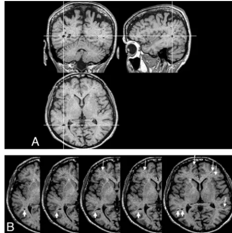
Image analysis was performed on MRIcro 1.40 (http://www.sph. sc.edu/comd/rorden/mricro.html), a free image-processing program operating on a Microsoft Windows platform. For each subject’s scan, the first step of MR imaging analysis was performed with T1-weighted images at⫻2 magnification on a 27-inch screen to visualize the char-acteristics of lesions simultaneously in axial, coronal, and sagittal planes. Thus, all suspected lesions were analyzed on the basis of their shape and location in the 3D space (Fig 1). T2- and proton attenu-ation⫺weighted images were analyzed in a second step to confirm that the signal intensity of the lesion corresponded to that of CSF.
Rating of dVRS
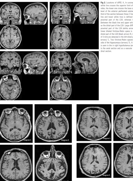
dVRS were defined as CSF-like signal-intensity (hypointense on T1 and hyperintense on T2) lesions that were round, ovoid, or linear;⬍3 mm in their maximum diameter, with smooth delineated contours; and located in areas supplied by perforating arteries (Fig 2). For le-sions fulfilling the same criteria except that their diameter wasⱖ3 mm, further effort was needed to differentiate them from infarcts by using multiplanar reformatting. Only those with a typical vascular shape and following the orientation of perforating vessels (including cystic lesions with an extension of vascular shape) were then regarded as dVRS (Fig 1).7
For each subject, all 124 axially oriented T1-weighted sections were first examined to evaluate the global burden of dVRS and to identify the section containing the largest number of dVRS in both the
BG and cerebral WM. In BG, dVRS were then rated according to a 4-level severity score in the section containing the greatest number of dVRS. The degrees of dVRS were defined as follows: degree 1, when there were⬍5 dVRS; degree 2, when there were between 5 and 10 dVRS; degree 3, when there were⬎10 dVRS but they were still count-able; and degree 4, when an innumerable number of dVRS resulted in a cribriform change in the BG (Fig 3). In cerebral WM, dVRS were scored as follows: degree 1, when there were⬍10 dVRS in the total WM; degree 2, when there were⬎10 dVRS in the total WM but⬍10 in the section containing the greatest number of dVRS; degree 3, when there were between 10 and 20 dVRS in the section containing the greatest number of dVRS; and degree 4, when there were⬎20 dVRS in the section containing the greatest number of dVRS (Fig 4). This rating scheme was adopted after testing different visual rating meth-ods, including those reported in the literature on a subset of MR imaging data from the first 150 subjects of the cohort and after per-forming step-by-step multiple rectifications. dVRS with axial diame-ters ofⱖ3 mm were defined as “large dVRS” in our study. Their presence, number, and location were systematically recorded.
All images were analyzed by the same experienced reader (Y.-C.Z.) who was blinded to all clinical data. When this reader was uncertain of the categorization of MR imaging signal-intensity abnor-malities (n⫽113), the corresponding lesions were reviewed by 2 other readers who had extensive experience in MR imaging studies (H.C. and C.T.) and a decision was made on the basis of the majority vote of the 3 readers.
Other MR Imaging Parameters
The gray matter, WM, and CSF volumes were estimated with voxel-based morphometry methods detailed elsewhere.10Total intracranial
volume was computed as the sum of the gray matter, WM, and CSF volumes and brain parenchymal fraction as the ratio of brain tissue volume to total intracranial volume.
[image:2.594.301.535.44.279.2]Statistical Analysis
Associations between qualitative variables and dVRS scores in the BG or WM were assessed by using2tests. In multivariable analyses, the
associations between qualitative variables and dVRS degree in the BG or WM were assessed by using logistic regression controlling for brain size (estimated by total intracranial volume). Associations between continuous variables and dVRS degrees in the BG or WM were
exam-ined by using analysis of covariance controlling for total intracranial volume. Potential interactions were tested by adding cross-products into the models. All analyses were performed by using SAS, Version 9.1 (SAS Institute, Cary, North Carolina).
Results
The sample comprised 1818 participants, 61.2% of whom were women. The mean age was 72.5⫾4.1 years. The intra-rater agreement for the rating of dVRS in the BG and WM was Fig 2.Locations of dVRS.A, In coronal sections, the upper white line crosses the superior limit of the caudate on both sides; the lower one crosses the base of the forebrain at the level of the anterior perforated substance. The lower one-third of the section between these 2 lines (between the black line and lower white line) is defined as belonging to the proximal part of the LSA, whereas the upper two-thirds (between the black line and upper white line) is considered as the distal part of the LSA. Large dVRS are detected in the proximal part of the LSA (white arrows) on both sides; a linear dilated Virchow-Robin space is also detected in the distal part of the LSA (black arrow).B, Large dVRS (diameter of 3 mm) are detected in the left distal part of the LSA (white arrows).C, Two Virchow-Robin spaces are seen in the left side of the hippocampus (arrows).D, A Virchow-Robin space is seen in the in right hypothalamus (arrows), as a small dot in the axial section and as a vascular shape in the longitu-dinal section.
Fig 3.Severity scores of dVRS in the BG.
[image:3.594.60.486.39.618.2] [image:3.594.303.534.368.600.2]assessed on a random sample of 100 individuals at 1-month intervals starting from the first reading. The statistics reached values of 0.77 and 0.75, respectively, indicating a good reliability.
dVRS were detected in every subject. Most participants (89%) had dVRS with scores of 1 or 2 in the BG. Degree 4 in the BG was observed in only 24 (1.3%) individuals. Within the WM, 77% of the individuals had dVRS scores of 1 or 2; only 94 subjects (5.2%) had the highest degree.
The scores of dVRS in the BG were positively associated with those obtained in the WM (2⫽100, 9df,P⬍.0001) (Table 1). However, a discrepancy ofⱖ2° between the WM and BG scores of dVRS was observed in 239 individuals (13.1%) (Table 1). Mean age increased with the severity of dVRS both in the BG (P⬍.0001, Table 2) and WM (P⫽.004, Table 2). These associations were not modified by sex (Pfor interaction,ⱕ0.51 for all tests; Table 2). The proportion of men increased with the severity of dVRS in the BG (P⫽.0002) but not in the WM (P⫽.51). There were no significant inter-actions between age and sex on the severity of dVRS (Pfor interaction,ⱕ0.40 for all tests). Additional adjustments of the models on age did not alter the results (Table 2).
dVRS were frequently detected in the hippocampus (fre-quency, 44.5%) and hypothalamus (fre(fre-quency, 11.6%). These dVRS were found to have a uniform location and shape across subjects (Fig 2). Among subjects with dVRS in the hippocam-pus, dVRS were found bilaterally in 48.8% of subjects, and among subjects with dVRS in the hypothalamus, they were found bilaterally in 45.7% of subjects.
Overall, large dVRS were detected in 603 (33.2%) subjects; 388 subjects (21.3%) had only one, 139 (7.7%) had 2, and 76
(4.2%) had ⬎2 large dVRS (there were 948 large dVRS in total). Large dVRS were observed in the following areas by decreasing frequency: proximal LSA, 19.9%; CMA, 10.2%, hippocampus, 5.8%, distal LSA, 2.8%, brain stem, 1.2%; and hypothalamus, 0.1%. Therefore, half of the large dVRS were detected in the proximal part of LSA, and one-fourth along the CMA. The prevalence of large dVRS in the proximal part of LSA, in the distal part of LSA, or along CMA significantly increased with the severity of dVRS in the BG and WM (Pⱕ .02 for all tests).
Discussion
In this sample of 1818 elderly subjects free of dementia or stroke, we found that dVRS were a common MR imaging find-ing. Using high-resolution 3D MR imaging, we found dVRS in every subject and dVRS of⬎3 mm in approximately one-third of them. The results also showed that the number of dVRS increased with age in the BG as well as in the WM and that men had more dVRS in the BG than women.
The observation of dVRS in all subjects of this cohort con-trasts with the 38% prevalence reported in MR imaging studies of 816 outpatients of wide age range and 3% of 1250 pediatric pa-tients.2,11The different spatial resolutions of imaging may be the main source of this discrepancy. Herein, we used 3D MR imaging acquisitions with a voxel size of 1.0⫻0.98⫻0.98 mm3, whereas most previous studies of dVRS including the above-mentioned were based on 2D MR imaging acquisitions and image sections of 5-mm thickness.1,2,11,12 Moreover, Groeschel et al7 found a higher detectability of dVRS on coronal T1-weighted images than on axial T2, sagittal T2, or axial fluid-attenuated inversion recov-ery images.7These data emphasized the importance of section orientation and MR imaging sequences in the assessment of dVRS. In the present study, the 3D T1 imaging analysis used for identification of dVRS may have also increased the detection of dVRS. Virchow-Robin spaces were also visible in 100% of sub-jects in another study using high-resolution 3D MR imaging in 125 healthy subjects between 1 and 30 years of age, supporting our inferences.7
[image:4.594.53.285.70.159.2]Although a diameter of 3 mm has been widely used as a cutoff value to discriminate silent brain infarcts from dilated perivascular spaces on MR imaging,13dVRS with diameters of ⬎3 mm are frequently detected.14One-third of individuals in the 3C-Dijon MR imaging sample had 1 or more dVRS larger than 3 mm. This is 3 times higher than the prevalence of lacu-nar infarctions in the same sample.9The most common loca-tion of large dVRS was found to be at the proximal part of LSA, in agreement with the most widely recognized location of di-lated perivascular spaces.6,14,15The prevalence of large dVRS was twice as high in this area as along the CMA and 10 times higher than in the distal part of LSA. Various mechanisms underlying the expansion of perivascular spaces have been previously suggested, such as the following: 1) changes in the permeability of the arterial wall,162) altered drainage and ac-cumulation of amyloid proteins along the vessels,173) spiral elongation of blood vessels,15 or 4) cerebral atrophy.18 The higher frequency of large dVRS at the proximal part of LSA may be particularly related to additional anatomic character-istics, including the angular trajectory of the vascular tree and the presence of multiple branches crossing the surface of the cerebrum in this area.
Table 1: Cross table of dVRS scores in basal ganglia and white matter
Scores of dVRS in BG
Scores of dVRS in WM, No. (%)
Total No. (%) Degree
1
Degree 2
Degree 3
Degree 4
[image:4.594.52.285.194.334.2]Degree 1 295 (16.2) 528 (29.0) 127 (7.0) 26 (1.4) 976 (53.7) Degree 2 104 (5.7) 344 (18.9) 147 (8.1) 46 (2.5) 641 (35.3) Degree 3 26 (1.4) 88 (4.8) 45 (2.5) 18 (1.0) 177 (9.7) Degree 4 4 (0.2) 10 (0.6) 6 (0.3) 4 (0.2) 24 (1.3) Total 429 (23.6) 970 (53.4) 325 (17.9) 94 (5.2) 1818 (100%)
Table 2: Distribution of scores of dVRS in the BG and WM by age and sex
Location
Scores of dVRS
No.
Mean age
(yr) (SD) PValue
% Men
(No.) PValue
BG
Degree 1 976 71.7 (3.9) ⬍.0001a/⬍.0001b 36.5 (356) .0002c/.002d Degree 2 641 73.0 (4.2) 37.8 (242)
Degree 3 177 74.2 (4.0) 50.3 (89) Degree 4 24 75.0 (3.4) 79.2 (19) WM
Degree 1 429 72.2 (4.2) .004a/.004b 33.6 (144) .51c/.53d Degree 2 970 72.4 (4.1) 39.5 (383)
Degree 3 325 72.7 (4.2) 42.2 (137) Degree 4 94 73.8 (4.1) 44.7 (42)
aComputed from analysis of covariance controlling for total intracranial volume. bComputed from analysis of covariance controlling for total intracranial volume and sex. cComputed from logistic regression controlling for total intracranial volume.
In this study, an accumulation of uncountable dVRS in the BG was observed in 1.3% of participants. A special MR imaging change of cribriform cavities in the BG was the feature of this group. An “e´tat crible´” or “status cribrosum” has been previously proved to be multiple dVRS in pathologic studies19and has been reported only among cases of vascular Parkinsonism,20 subcorti-cal ischemic vascular dementia,21,22or CADASIL23but has not been reported in the general population, to our knowledge. The present data further emphasize that the accumulation of multiple dVRS is not rare and can occur in elderly people in the absence of severe disability or dementia.
The association between age and the severity of dVRS was consistently detected in previous dVRS studies in hospitalized patients2and in patients with lacunar infarction,12as well as in specific conditions such as CADASIL.23This may suggest 1 or several potential causes of dVRS with aging.15In this study, a higher grade of dVRS in the BG was observed more frequently in men than in women. This difference was independent of the total intracranial volume. Most interesting, this sex effect was not detected for dVRS in the WM. Whether variations in ul-trastructural characteristics or permeability properties of ves-sels located within the BG are involved in the male sex associ-ation with higher severity of dVRS in the BG will require specific investigation.
In this sample, we also observed that dVRS are commonly detected in areas not usually considered for MR imaging as-sessment of dilated perivascular spaces. Although still contro-versial, the hippocampal cavities located between the cornu ammonis and dentate gyrus were found at postmortem exam-ination to be covered by a single cell layer, to contain blood vessels, and to be surrounded by intact cerebral parenchyma, which strongly suggest typical dVRS.24We found that these cavities are of uniform shape and location, with a frequency of 44.5% in the 3C-Dijon MR imaging cohort, close to the 39% reported by Sasaki et al24in 109 patients from 8 to 85 years of age. Whether their severity is related to age andapolipoprotein E4orE2alleles as suggested in small samples will need addi-tional investigations.25,26We also found that ⬎10% of pa-tients presented with dVRS in the hypothalamus, a feature never previously reported, to our knowledge.
The strengths of this study include the population-based design, the large sample size, and the use of high-resolution MR imaging and 3D image analysis. The definition of dVRS was also based on strict anatomic and imaging criteria, and the method of assessment proved to be highly reliable. The main limitations include the potential misclassification of dVRS versus small ischemic lesions despite the training of the reader in using 3D imaging analysis and the use of a semiquantitative evaluation for grading of severity.
Conclusions
The results of this study emphasize that large dVRS are highly prevalent in the healthy population and can be detected in most brain structures by using 3D high-resolution MR imag-ing. The results confirmed that the topographic distribution of dVRS is not uniform within the brain and may depend on anatomic or pathologic characteristics interacting with aging and sex. Whether the severity of dVRS at MR imaging is re-lated to vascular risk factors and/or to other imaging markers
of small vessel diseases in healthy elderly people will need ad-ditional investigation.
Acknowledgments
We thank Denis Krause, MD, from the Departments of Radi-ology and Medical Imaging, Bocage Hospital, Dijon, France, for his work in MR imaging data collection.
References
1. Maclullich AM, Wardlaw JM, Ferguson KJ, et al.Enlarged perivascular spaces are associated with cognitive function in healthy elderly men.J Neurol Neuro-surg Psychiatry2004;75:1519 –23
2. Heier LA, Bauer CJ, Schwartz L, et al.Large Virchow-Robin spaces: MR-clinical correlation.AJNR Am J Neuroradiol1989;10:929 –36
3. Udaka F, Sawada H, Kameyama M.White matter lesions and dementia: MRI-pathological correlation.Ann N Y Acad Sci2002;977:411–15
4. Patankar TF, Mitra D, Varma A, et al.Dilatation of the Virchow-Robin space is a sensitive indicator of cerebral microvascular disease: study in elderly pa-tients with dementia.AJNR Am J Neuroradiol2005;26:1512–20
5. Wuerfel J, Haertle M, Waiczies H, et al.Perivascular spaces: MRI marker of inflammatory activity in the brain?Brain2008;131:2332– 40
6. Bokura H, Kobayashi S, Yamaguchi S.Distinguishing silent lacunar infarction from enlarged Virchow-Robin spaces: a magnetic resonance imaging and pathological study.J Neurol1998;245:116 –22
7. Groeschel S, Chong WK, Surtees R, et al.Virchow-Robin spaces on magnetic resonance images: normative data, their dilatation, and a review of the liter-ature.Neuroradiology2006;48:745–54
8. Alperovitch A.Vascular factors and risk of dementia: design of the three-city study and baseline characteristics of the study population.Neuroepidemiology
2003;22:316 –25
9. Soumare A, Elbaz A, Zhu Y, et al.White matter lesions volume and motor performances in the elderly.Ann Neurol2009;65:706 –15
10. Lemaitre H, Crivello F, Grassiot B, et al.Age- and sex-related effects on the neuroanatomy of healthy elderly.Neuroimage2005;26:900 –11
11. Rollins NK, Deline C, Morriss MC.Prevalence and clinical significance of di-lated Virchow-Robin spaces in childhood.Radiology1993;189:53–57 12. Rouhl RP, van Oostenbrugge RJ, Knottnerus IL, et al.Virchow-Robin spaces
relate to cerebral small vessel disease severity.J Neurol2008;255:692–96 13. Vermeer SE, Den HT, Koudstaal PJ, et al.Incidence and risk factors of silent
brain infarcts in the population-based Rotterdam Scan Study. Stroke
2003;34:392–96
14. Takao M, Koto A, Tanahashi N, et al.Pathologic findings of silent, small hy-perintense foci in the basal ganglia and thalamus on MRI.Neurology
1999;52:666 – 68
15. Kwee RM, Kwee TC.Virchow-Robin spaces at MR imaging.Radiographics
2007;27:1071– 86
16. Pollock H, Hutchings M, Weller RO, et al.Perivascular spaces in the basal ganglia of the human brain: their relationship to lacunes.J Anat1997;191(pt 3):337– 46
17. Roher AE, Kuo YM, Esh C, et al.Cortical and leptomeningeal cerebrovascular amyloid and white matter pathology in Alzheimer’s disease.Mol Med
2003;9:112–22
18. van Swieten JC, van den Hout JH, van Ketel BA, et al.Periventricular lesions in the white matter on magnetic resonance imaging in the elderly: a morphomet-ric correlation with arteriolosclerosis and dilated perivascular spaces.Brain
1991;114(pt 2):761–74
19. Mancardi GL.Neuropathologic study of lacunae and cribriform cavities of the brain.Eur Neurol1989;29(suppl 2):16 –19
20. Fenelon G, Gray F, Wallays C, et al.Parkinsonism and dilatation of the perivas-cular spaces (e´tat crible´) of the striatum: a clinical, magnetic resonance imag-ing, and pathological study.Mov Disord1995;10:754 – 60
21. Braffman BH, Zimmerman RA, Trojanowski JQ, et al.Brain MR: pathologic correlation with gross and histopathology. 1. Lacunar infarction and Vir-chow-Robin spaces.AJR Am J Roentgenol1988;151:551–58
22. Roman GC.On the history of lacunes, e´tat crible´, and the white matter lesions of vascular dementia.Cerebrovasc Dis2002;13(suppl 2):1– 6
23. Cumurciuc R, Guichard JP, Reizine D, et al.Dilation of Virchow-Robin spaces in CADASIL.Eur J Neurol2006;13:187–90
24. Sasaki M, Sone M, Ehara S, et al.Hippocampal sulcus remnant: potential cause of change in signal intensity in the hippocampus.Radiology1993;188:743– 46 25. Barboriak DP, Doraiswamy PM, Krishnan KR, et al.Hippocampal sulcal cavi-ties on MRI: relationship to age and apolipoprotein E genotype.Neurology
2000;54:2150 –53