ORIGINAL RESEARCH
SPINE
Validation of Multisociety Combined Task Force Definitions of
Abnormal Disk Morphology
C.H. Cho, L. Hsu, M.L. Ferrone, D.A. Leonard, M.B. Harris, A.A. Zamani, and C.M. Bono
ABSTRACT
BACKGROUND AND PURPOSE: The multisociety task force descriptively defined abnormal lumbar disk morphology. We aimed to use their definitions to provide a higher level of evidence for the validation of MR imaging in the evaluation of this pathology in patients who have undergone diskectomy by retrospectively classifying their preoperative MRI.
MATERIALS AND METHODS: This retrospective, institutional review board–approved study included 54 of 86 consecutive patients (47 men; average age, 44 years) enrolled in an ongoing prospective trial of surgically treated lumbar disk herniation who had preoperative MRI and documented intraoperative classification of the abnormal disk as protrusion, extrusion, or sequestration by the treating surgeon. Preoperative MRI was classified by 2 blinded radiologists; discrepancies were resolved by a third reader. Statistical analysis of interobserver agreement and imaging compared with surgical findings was performed.
RESULTS:The readers disagreed on only 1 of the 54 cases. The third reader resolved the disagreement. Eight protrusions and 46 extrusions were found on imaging, with no sequestrations. At surgery, there were 13 protrusions and 40 extrusions, with 2 of the extrusions also containing sequestrations; the remaining case had only sequestration. There were 16 discrepancies between imaging and surgery, resulting in 70% agreement.
CONCLUSIONS: This study, which was intended to validate the multisociety combined task force definitions of abnormal disk morphology by using MR imaging with a surgical criterion standard, found 70% agreement between imaging diagnosis and surgical findings. Although reasonable, this finding highlights differences that often exist between intraoperative and preoperative imaging findings of lumbar disk herniation.
M
R imaging of the lumbar spine with defined specific MR images has gained acceptance as the standard of care for the evaluation of degenerative disk disease.1However, the interpreta-tion of these images continues to have much variability. An effort to standardize image reporting brought together multiple na-tional medical societies including the North American Spine So-ciety, American Society of Spine Radiology, and American Society of Neuroradiology. They produced and then updated a consensus document for image descriptions.2One segment of the consensus document focuses on the
mor-phology of the lumbar disk as it relates to the location of abnormal disk content with respect to the outer annulus.2MR imaging stud-ies evaluating the interobserver and intraobserver reliability of disk morphology by using the definition of the consensus docu-ment have been performed.3-5 However, these studies lacked analysis of surgical findings for correlation with a criterion stan-dard and, as such, do not provide the highest level of diagnostic evidence.
This study retrospectively classified MR imaging findings in a cohort of consecutive patients with surgically treated disk herni-ation by using the descriptive definition of abnormal lumbar disk morphology of the multisociety task force, with the aim of pro-viding a high level of evidence for validation of MR imaging by using the combined task force definitions.2
MATERIALS AND METHODS
A retrospective review was performed, with approval from the institutional review board of the study site, of the records of 86 consecutive patients (47 men, 39 women; average age, 44 years) who were enrolled between August 2009 and October 2013 in an ongoing prospective clinical trial evaluating the outcomes of sin-Received September 8, 2014; accepted after revision October 24.
From the Departments of Radiology (C.H.C., L.H., A.A.Z.) and Orthopedic Surgery (M.L.F., D.A.L., M.B.H., C.M.B.), Brigham and Women’s Hospital, Harvard Medical School, Boston, Massachusetts.
Abstract of preliminary work previously presented at: Annual Meeting of the North American Spine Society, November 12–15, 2014; San Francisco, California. Please address correspondence to Charles H. Cho, MD, MBA, Department of Radi-ology, Brigham and Women’s Hospital, Harvard Medical School, 75 Francis St, Bos-ton, MA 02115; e-mail: [email protected]
Indicates article with supplemental on-line appendix.
gle-level lumbar diskectomy. Inclusion criteria for entry into the prospective study were the presence of a symptomatic single-level lumbar disk herniation, failure of nonoperative treatment, pri-mary radicular pain, and no prior lumbar surgery. Included in prospective data collection was documentation of the intraoper-ative classification of the herniation as a protrusion, extrusion, or sequestration. For the current study, the intraoperative findings were used as the diagnostic criterion standard because the sur-geon is looking directly at the anatomy of the patient. These find-ings have been used as such in numerous other studies.6,7With continued improvement in imaging, the depiction of a patient’s anatomy is becoming clearer and more accurate and is approach-ing equivalence to lookapproach-ing at it.
Surgical cases were from the clinical practice of 3 attending orthopedic spine surgeons from a single institution. Surgeons re-corded operative findings that included classifying the disk herni-ation as a protrusion, extrusion, or sequestrherni-ation intraoperatively. In the operative field, the outer margin of the annulus was visually identified. A defect of the annulus with disk material outside the annulus was classified as “extrusion.” If there were no defect and no disk material outside the annulus, the classification of “pro-trusion” was made. “Sequestration” was determined if there was disk material separate from the annular defect with no visible attachment to the parent disk.
Of the 86 patients, 1 patient had no disk herniation noted during surgery, which was presumed to mean the disk had re-sorbed between imaging and surgery, and 3 patients had incom-plete surgical data sheets. These cases were excluded. Of the re-maining 82 cases, 54 had presurgical MR images accessible by the PACS of the study institution, so these 54 cases were included in the present study.
In preparation for the current analysis, a summary sheet (On-line Appendix 1) was created and reviewed by 3 board-certified neuroradiologists, each with a minimum of 15 years of practice experience and a combined average of⬎26 years, based on the previously described multisociety task force definitions.2Sample cases from daily practice were used in a face-to-face setting to confirm the understanding of the summary sheet definitions be-fore data collection.
The MR imaging of each subject (mean, 81⫾75 days before surgery) was reviewed by 2 board-certified radiologists who were blinded to the patient medical history and operative findings. In the evaluation, the disk level with the most substantial abnormal-ity was classified as a protrusion, extrusion, or sequestration by using both axial and sagittal images. Classifications were confined to a single disk. In cases in which⬎1 disk level had abnormalities, only the more severe level was selected. In 1 case in which there was nearly equivalent severity at 2 disk levels, the surgical level was provided to the readers to create a final, imaging-based classifica-tion that could be compared with the surgical classificaclassifica-tion. Any discrepancy between the 2 readers was resolved by a third blinded reader, and a majority consensus evaluation became the final im-aging interpretation. Of note, many of the MR images were from different, outside, referring hospitals. However, all cases included sagittal T1- and sagittal and axial T2-weighed images.
Most important, while the surgeons did have the MR imaging available at the time of surgery, they did not have the findings as
decided by the study radiologists. Additionally, the surgeons were instructed to make their classifications solely on the basis of their intraoperative observations. While having the MR imaging avail-able to surgeons may have been a potential source of bias, sur-geons always use MR imaging to plan for their surgery; thus, in practice, surgeons also have that potential source of bias. Research must be applicable to practice, and blinding surgeons to MR im-aging would not mimic practice.
All statistical analyses were performed by using STATA/SE 13.1 (StataCorp, College Station, Texas). Agreement between im-aging and surgical classifications was calculated by using the Co-hen , as was agreement between imaging assessments. The weightedwas not used because disk herniations do not always progress in a standardized manner. A standard 2⫻2 table was used to determine sensitivity, specificity, and predictive values.
RESULTS
Imaging FindingsOf the 54 cases, the 2 readers disagreed in 3. One case had a final diagnosis discrepancy between protrusion and extrusion based on all images. In the other 2 cases, there was a disagreement on disk description as protrusion or extrusion on the axial plane, but both readers described an extrusion on the sagittal images. Thus, the final diagnosis of extrusion was the same for these 2 cases. All discrepancies were resolved by the third reader. Overall, 53 of 54 cases showed agreement in the final diagnosis between readers. Interobserver agreement between the 2 readers was 98% with a of 0.93. There were 8 protrusions, 46 extrusions, and no seques-trations based on imaging findings.
Surgical Findings
Of the 54 surgical cases with MR images, there were 13 protru-sions and 40 extruprotru-sions, with 2 of the extruprotru-sions each occurring in addition to sequestrations. The 1 remaining case had only seques-tration and no extrusion.
Imaging and Surgical Agreement
Four cases with an imaging diagnosis of protrusion were found to be extrusions in surgery. Nine cases with imaging diagnoses of extrusion were found to be protrusions in surgery. Whereas im-aging data did not reveal any cases of sequestration, in 2 cases with imaging diagnoses of extrusion, surgery showed both extrusion and sequestration; these were considered discrepancies between imaging and surgical findings. In another case, imaging showed extrusion while surgery showed sequestration only. The total number of discrepancies was 16, calculated as an overall agree-ment of 70% (⫽0.19). Table 1 lists all the discrepancy cases and gives descriptions of the imaging findings.
Sensitivity and specificity of detection of a ruptured annulus were 0.90 and 0.31, respectively (Table 2). The positive predictive value was 0.80, while the negative predictive value was 0.50.
DISCUSSION
expe-rienced readers in a single academic institution; interpretation included descriptions of normal and bulge as well as protrusion and extrusion, with the most common discrepancy occurring be-tween normal and bulge.3Another study, by using readers from 1 academic center with a prestudy effort to define the review crite-ria, found near-perfect interreader agreement (summary ⫽ 0.81) for normal/bulge, protrusion, and extrusion/sequestration categories.4Other reports replicating conditions close to those of clinical practice by using radiologists from different hospitals with no prior set diagnostic criteria or training showed only moderate interobserver agreement for herniation and fair-to-moderate agreement for disk contour.5Most important, all of the above studies lacked a surgical criterion standard.
In the current study, substantial effort was made to decrease interobserver variability, because the primary goal was to evaluate the correlation with surgical findings. Before data collection, the 3 readers discussed the nomenclature by using daily clinical cases from the previous few months. There was extensive discussion about the definitions of protrusion and extrusion, especially on the sagittal images. In the end, the critical point of interpretation among the readers was the exact location of the outer annulus insertion on the vertebral body, which was crucial in
discriminat-ing protrusion from extrusion. To better understand this anat-omy, the readers reviewed studies in the literature, including pathologic and traumatic conditions, in terms of how they affect the outer annulus insertion site and how it is localized above the bony endplate on the sagittal view.8-11These studies showed that the insertion of the annulus is beyond the bone edge of the disk; this finding creates discrepancies among readers on the insertion site. With this understanding, we went back to the protrusion definition from the combined task force report2and noted that the insertion site of the annulus cannot be greater than the max-imum height of the disk space in the sagittal plane. For unifor-mity, we therefore decided to use the disk space height to define the difference between protrusion and extrusion. Thus, any disk morphology less than the maximum sagittal disk height was des-ignated as protrusion. This distinction is depicted in On-line Ap-pendix 2.
After this effort to optimize uniform understanding and agreement of the definitions among the radiologists, there was consensus in the final imaging diagnosis of 53 of 54 cases (agree-ment, 98%;⫽0.93). If the 2 other cases of discrepancy in the axial or sagittal description (which did not affect the final diagno-sis) are included, the agreement was 51 of 54, or 94%. These values are at least as good as, if not better than, those previously reported. Initially the surgical level was not provided to the readers be-cause transitional vertebral levels may have directed the attention to an incorrect surgical level by causing the readers to number the vertebrae differently. Thus, the readers were instructed to report the most severe disk level during the imaging analysis. In 1 case, L5-S1 had moderate central protrusion and L4-L5 had a small foraminal protrusion. The latter, despite appearing less severe, was in fact at the symptomatic level of radiculopathy. In this case, the correct level was indicated to the readers at the time of imaging data collection and before the analysis.
Table 1: Discrepancy cases between imaging and surgery, with a description of the imaging findings
No. Disk
Level Imaging Surgical Description
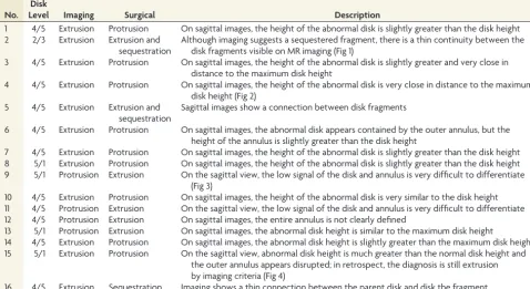
1 4/5 Extrusion Protrusion On sagittal images, the height of the abnormal disk is slightly greater than the disk height 2 2/3 Extrusion Extrusion and
sequestration
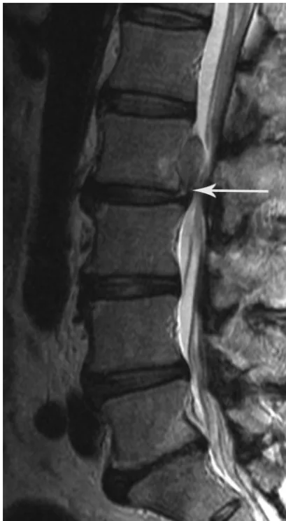
Although imaging suggests a sequestered fragment, there is a thin continuity between the disk fragments visible on MR imaging (Fig 1)
3 4/5 Extrusion Protrusion On sagittal images, the height of the abnormal disk is slightly greater and very close in distance to the maximum disk height
4 4/5 Extrusion Protrusion On sagittal images, the height of the abnormal disk is very close in distance to the maximum disk height (Fig 2)
5 4/5 Extrusion Extrusion and sequestration
Sagittal images show a connection between disk fragments
6 4/5 Extrusion Protrusion On sagittal images, the abnormal disk appears contained by the outer annulus, but the height of the annulus is slightly greater than the disk height
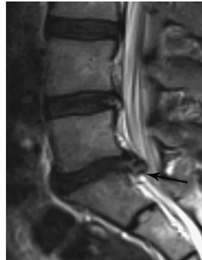
7 4/5 Extrusion Protrusion On sagittal images, the height of the abnormal disk is slightly greater than the disk height 8 5/1 Extrusion Protrusion On sagittal images, the height of the abnormal disk is slightly greater than the disk height 9 5/1 Protrusion Extrusion On the sagittal view, the low signal of the disk and annulus is very difficult to differentiate
(Fig 3)
10 4/5 Extrusion Protrusion On sagittal images, the height of the abnormal disk is very similar to the disk height 11 4/5 Protrusion Extrusion On the sagittal view, the low signal of the disk and annulus is very difficult to differentiate 12 4/5 Protrusion Extrusion On sagittal images, the entire annulus is not clearly defined
13 5/1 Protrusion Extrusion On sagittal images, the abnormal disk height is similar to the maximum disk height
14 4/5 Extrusion Protrusion On sagittal images, the abnormal disk height is slightly greater than the maximum disk height 15 5/1 Extrusion Protrusion On the sagittal view, abnormal disk height is much greater than the normal disk height and
the outer annulus appears disrupted; in retrospect, the diagnosis is still extrusion by imaging criteria (Fig 4)
[image:3.594.54.533.60.321.2]16 4/5 Extrusion Sequestration Imaging shows a thin connection between the parent disk and disk the fragment
Table 2: 2ⴛ2 contingency table for a ruptured-versus-intact annulus as determined surgically and on imaging
Imaging Assessment
Surgical Assessment
Totals (Criterion Standard)
Annulus Ruptureda
Intact Annulusb
Annulus ruptureda 37 9 41
Intact annulusb 4 4 13
Totals 46 8 54
aExtrusions and sequestrations. b
Overall, our accuracy measurements indicate that imaging is better at determining when the annulus is ruptured than it is at determining that the annulus is intact. Analysis of the discrepan-cies between imaging and surgery (Table 1) showed the following 3 observations: 1) When the height of the abnormal disk material on the sagittal plane is very close to the maximum height of the normal disk, it becomes difficult to differentiate protrusion from extrusion on imaging (Fig 2). 2) When the disk signal is low on all MR images, differentiation of an abnormal disk from the outer annulus becomes challenging and a small extruded disk cannot be differentiated from a contained (protruded) disk (Fig 3). 3) In differentiating extrusion from sequestration, imaging clearly shows a thin connecting tissue, which, by def-inition, is considered extrusion (Fig 1). The thin connecting tissue that defines a herniation as an extrusion can be clearly shown on imaging yet not observed in surgery, because the thin sliver of tissue may be very difficult to discern. All discrepancy cases are shown in On-line Appendix 3.
We believe that the effect of variation in imaging parameters on discrepant classifications was minimal in this study. Of the 16 cases with discrepancies, though the magnet strength is not noted
in all, most appear to have been performed with at least 1.5T. The TR ranged from 3000 to 5760 ms (a single case at 1000 ms); TE, from 90 to 148 ms; section thickness, between 4 and 5 mm (a single case at 3.6 mm); the FOV, from 24 to 30 cm; and the acquisition matrix, between 256 and 512 (a single case, 240⫻ 175). The overall range of imaging parameters of all these cases is within the standard for achieving high-quality images, which are also in agreement visually as determined by 2 experienced neuroradiologists. With most of the imaging findings being obvious, ultimately we neither think that these discrepancies are due to slight differences in imaging parameters nor believe that variation in imaging parameters compromises the pur-pose of this study.
Given that at surgery a defect in the posterior annulus differ-entiates extrusion from protrusion while in imaging it is the rela-tive size of the herniated disk versus the height of base of the annulus that differentiates the 2 categories, it is somewhat surpris-ing that surgery and radiology agree so often. While the approach is different on imaging versus during surgery, the goal in both cases is to determine whether the annulus is ruptured. Because this can be directly observed intraoperatively, surgical standing has been used as the criterion standard, but advancements in imaging can perhaps better define the outer annulus and
an-FIG 1. Case 2. Sagittal T2-weighted image shows a thin connection (arrow) between the level of abnormal disk and the disk fragment. With imaging, this is classified as extrusion. However, it was classified intraoperatively as extrusion and sequestration.
[image:4.594.66.271.47.419.2] [image:4.594.314.517.48.399.2]nular defects. This change would allow a truer imaging predic-tion of surgical findings. Imaging needs to be better defined so that it can more accurately and more directly determine the presence or lack of an annular defect instead of relying on morphology to provide clues as to the presence or lack of said defect. As imaging improves and this definition becomes pos-sible, it could enable better correlation of symptoms of a disk herniation to the type of herniation and prediction of surgical-versus-nonoperative outcomes for any given patient on the basis of their MR imaging.
This study has a number of limitations. The time between imaging and surgery was 81 days on the average, so progression or resorption of the disk herniation could have occurred from the time of imaging to surgery. One example of this is case 15 (Fig 4), which, on imaging, was classified as an extrusion but was intra-operatively classified as a protrusion. In addition, the number of cases of sequestration was small, thereby limiting conclusions from analysis of this category. In addition, despite the reasonable (70%) agreement between the imaging diagnosis and surgical findings, the probability of the calculated agreement occurring by chance was high owing to the distribution of classifications, which were mostly extrusions. This is reflected in the lowof 0.19. Despite this high probability, we think that the study is represen-tative of actual practice, and increasing the sample size would not have significantly altered the pathology distribution. Addition-ally, agreement on a surgical classification is generally moderate at best.12 For disk classification, the agreement on the difference between protrusion and extrusion/sequestration would likely be good, but the agreement on the difference between extrusion and sequestration would likely be moderate at best. However, due to the pathology distribution and our resulting focus on the distinc-tion between protrusion and sequestradistinc-tion/extrusion rather than
the distinction between sequestration and extrusion, the effect of this limitation is unlikely to be substantial.
CONCLUSIONS
In this study, which was intended to validate the multisociety combined task force definitions of abnormal disk morphology by using MR imaging with a surgical criterion standard, there was 70% agreement between the imaging diagnosis and surgi-cal findings. Common trends for the discrepancy are de-scribed. Future effort may yield better agreement between sur-geons and radiologists as to how they describe disk herniation and abnormalities.
Disclosures: Charles H. Cho—UNRELATED: Board Membership: North American Spine Society (no money). Mitchel B. Harris—UNRELATED:Board Membership: North American Spine Society (no money). Christopher M. Bono—UNRELATED:
Board Membership:North American Spine Society (nonmonetary, volunteer); Con-sultancy: United Healthcare;Expert Testimony: CRICO;Royalties: Wolters Kluwer, for edited books;Other:Journal of the American Academy of Orthopaedic Sur-geons,Comments: stipend for being Deputy Editor.
REFERENCES
1. Modic MT, Ross JS.Lumbar degenerative disk disease.Radiology
2007;245:43– 61
2. Fardon DF, Williams AL, Dohring EJ, et al.Lumbar disc nomen-clature: version 2.0, recommendations of the combined task forces of the North American Spine Society, the American Society of Spine Radiology, and the American Society of Neuroradiology.Spine (Phila Pa 1976)2014;39:E1448 – 65
3. Brant-Zawadzki MN, Jensen MC, Obuchowski N, et al. Interob-server and intraobInterob-server variability in interpretation of lumbar disc abnormalities: a comparison of two nomenclatures.Spine (Phila Pa 1976)1995;20:1257– 63; discussion 1264
4. Lurie JD, Tosteson AN, Tosteson TD, et al.Reliability of magnetic
FIG 3. Case 9. Sagittal T2-weighted image shows decreased signal of the disk material, which makes differentiation of the disk from the annular margin difficult.
[image:5.594.66.271.45.289.2] [image:5.594.313.519.47.312.2]resonance imaging readings for lumbar disc herniation in the Spine Patient Outcomes Research Trial.Spine (Phila Pa 1976)
2008;33:991–98
5. Arana E, Kovacs FM, Royuela A, et al.Influence of nomenclature in the interpretation of lumbar disk countour on MR imaging: a comparison of the agreement using the combined task force and the Nordic nomenclatures. AJNR Am J Neuroradiol 2011;32: 1143– 48
6. Zhuge W, Ben-Galim P, Hipp JA.Efficacy of MRI for assessment of spinal trauma: correlation with intra-operative findings.J Spinal Disord Tech2013 Nov 8. [Epub ahead of print]
7. Hedberg MC, Drayer BP, Flom RA, et al.Gradient echo (GRASS) MR imaging in cervical radiculopathy. AJR Am J Roentgenol 1988; 150:683– 89
8. Humzah MD, Soames RW.Human intervertebral disc: structure and function.Anat Rec1988;220:337–56
9. Rauschning W. Computed tomography and cryomicrotomy of lumbar spine specimens: a new technique for multiplanar ana-tomic correlation.Spine (Phila Pa 1976)1983;8:170 – 80
10. Tajima N, Kawano K.Cryomicrotomy of the lumbar spine.Spine (Phila Pa 1976)1986;11:376 –79
11. Fredrickson BE, Edwards WT, Rauschning W, et al.Vertebral burst fractures: an experimental, morphologic, and radiographic study.
Spine (Phila Pa 1976)1992;17:1012–21
12. Brumback RJ, Jones AL.Interobserver agreement in the classifica-tion of open fractures of the tibia: the results of a survey of two hundred and forty-five orthopaedic surgeons.J Bone Joint Surg Am