warwick.ac.uk/lib-publications
A Thesis Submitted for the Degree of PhD at the University of Warwick
Permanent WRAP URL:
http://wrap.warwick.ac.uk/80032
Copyright and reuse:
This thesis is made available online and is protected by original copyright.
Please scroll down to view the document itself.
Please refer to the repository record for this item for information to help you to cite it.
Our policy information is available from the repository home page.
Doctoral Thesis
Temporal and spatial factors affecting
synaptic transmission in cortex
Author:
Alex D. Bird
Supervisors:
Magnus J. E. Richardson Mark J. Wall
A thesis submitted in fulfilment of the requirements
for the degree of Doctor of Philosophy
Theoretical Neuroscience Group
Warwick Systems Biology Doctoral Training Centre
Warwick Systems Biology Centre
School of Life Sciences
I, Alex Bird, declare that this thesis titled, ‘Temporal and spatial factors affecting
synap-tic transmission in cortex’ and the work presented in it are my own. I confirm that:
This work was done wholly or mainly while in candidature for a research degree
at this University.
Where any part of this thesis has previously been submitted for a degree or any
other qualification at this University or any other institution, this has been clearly
stated.
Where I have consulted the published work of others, this is always clearly
at-tributed.
Where I have quoted from the work of others, the source is always given. With
the exception of such quotations, this thesis is entirely my own work.
I have acknowledged all main sources of help.
Where the thesis is based on work done by myself jointly with others, I have made
clear exactly what was done by others and what I have contributed myself.
Signed:
Date:
Abstract
Synaptic transmission in cortex depends on both the history of synaptic activity and the location of indi-vidual anatomical contacts within the dendritic tree. This thesis analyses key aspects of the roles of both these factors and, in particular, extends many of the results for deterministic synaptic transmission to a more naturalistic stochastic framework.
Firstly, I consider how correlations in neurotransmitter vesicle occupancy arising from synchronous activity in a presynaptic population interact with the number of independent release sites, a parameter recently shown to be modified during long-term plasticity. I study a model of multiple-release-site short-term plas-ticity and derive exact results for the postsynaptic voltage variance. Using approximate results for the postsynaptic firing rate in the limits of low and high correlations, I demonstrate that short-term depression leads to a maximum response for an intermediate number of presynaptic release sites, and that this in turn leads to a tuning-curve response peaked at an optimal presynaptic synchrony set by the number of neu-rotransmitter release sites per presynaptic neuron. As the nervous system operates under constraints of efficient metabolism it is likely that this phenomenon provides an activity-dependent constraint on network architecture.
Secondly, I consider how synapses exhibiting short-term plasticity transmit spike trains when spike times are autocorrelated. I derive exact results for vesicle occupancy and postsynaptic voltage variance in the case that spiking is a renewal process, with uncorrelated interspike intervals (ISIs). The vesicle occupancy predictions are tested experimentally and shown to be in good agreement with the theory. I demonstrate that neurotransmitter is released at a higher rate when the presynaptic spike train is more regular, but that positively autocorrelated spike trains are better drivers of the postsynaptic voltage when the vesicle release probability is low. I provide accurate approximations to the postsynaptic firing rate, allowing future studies of neuronal circuits and networks with dynamic synapses to incorporate physiologically relevant spiking statistics.
Thirdly, I develop a Bayesian inference method for synaptic parameters. This expands on recent Bayesian approaches in that the likelihood function is exact for both the quantal and dynamic synaptic parameters. This means that it can be used to directly estimate parameters for common synaptic models with few release sites. I apply the method to simulated and real data; demonstrating a substantial improvement over analysis techniques that are based around the mean and variance.
T
hanks to Magnus Richardson and Mark Wall for continued guidance and support throughout the course of my PhD, they have made the time I havespent on this project both productive and enjoyable. They have been models
of professionalism and shown how well experimental and theoretical
collab-oration in neuroscience can work. I would also like to thank Hermann Cuntz for the
help with and time spent on our external collaboration.
I would like to thank the rest of the Theoretical Neuroscience Group for their help and
companionship; in particular Ruth Harbord, Tim Kaufmann, and Michael Fisher for
their expertise on the experimental side of the project. Thanks to my PhD committee
members Yulia Timofeeva, Marco van den Top, and Colm Connaughton for their helpful
advice over the years.
I would also like to thank the Warwick Systems Biology DTC for giving me the
oppor-tunity to undertake this project, and the studentship made available by the BBSRC.
Travel grants from ACCN, OCNS, Guarantors of Brain, FIAS, and ISN* have helped
me attend workshops and conferences and enriched my studies.
It has been a fantastic experience to spend time working at the Systems Biology Centre
and I would like to thank the staff and students whom I have had the privilege of getting
to know during my time at Warwick.
Finally thank you to my family for supporting me throughout my many, many years of
study, and to Liz, for putting up with me over the last couple of months and bringing
me cups of tea.
space
space
space
space
space
*Biotechnology and Biological Sciences Research Council, Advanced Course in
Compu-tational Neuroscience, Organisation of CompuCompu-tational Neuroscience Societies, Frankfurt
synaptic transmission in cortex
Theoretical Neuroscience Group
Systems Biology Doctoral Training Centre
Systems Biology Centre
School of Life Sciences
Doctor of Philosophy
Alex D.Bird
Published Material
The following papers were published in the course of this study:
Bird A. D. and M. J. E. Richardson. Long-term plasticity determines the
postsynap-tic response to correlated afferents with multivesicular short-term synappostsynap-tic depression.
Declaration of Authorship i
Abstract ii
Acknowledgements iii
Published material iv
Contents v
List of Figures viii
List of Tables x
Abbreviations xi
Symbols xii
1 Introduction 1
1.1 Introduction . . . 1
1.1.1 Basic neuronal physiology . . . 1
1.1.2 Brain architecture and cortical structure . . . 4
1.2 Synaptic transmission . . . 7
1.2.1 Structure of a chemical synapse . . . 7
1.2.2 Function of a chemical synapse . . . 10
1.3 Synaptic plasticity . . . 11
1.3.1 Short-term synaptic plasticity . . . 11
1.3.2 Long-term synaptic plasticity . . . 14
1.3.3 Spike-train statistics . . . 14
1.3.4 Neuromodulators and retrograde transmission . . . 16
1.3.5 Paired whole-cell patch-clamp recording . . . 16
1.4 Neurons . . . 17
1.4.1 Thick-tufted Layer V pyramidal cell . . . 17
1.4.2 Other neurons . . . 19
1.5 Optimality in dendritic structure . . . 21
1.5.1 Spatially extended neuron models . . . 22
1.5.2 Synaptic equalisation and ‘dendritic democracy’ . . . 23
1.6 Thesis Structure . . . 23
2 Long-term plasticity determines the postsynaptic response to corre-lated afferents with multivesicular short-term synaptic depression 25 2.1 Overview . . . 25
2.1.1 Author contributions . . . 26
2.2 Intoduction . . . 27
2.3 Methods . . . 28
2.4 Results . . . 29
2.4.1 Vesicle occupancy crosscorrelations . . . 30
2.4.2 Neurotransmitter release crosscorrelations . . . 30
2.4.3 Membrane voltage mean and variance . . . 31
2.4.4 Release site number and postsynaptic rate . . . 32
2.4.5 Long-term plasticity and response to synchrony . . . 33
2.4.6 Optimal response and synchrony jitter . . . 33
2.4.7 Optimal-response curves are a robust feature of synaptic homeostasis 34 2.5 Discussion . . . 34
2.6 References . . . 35
2.7 Appendix . . . 37
2.7.1 Derivation of the voltage variance . . . 37
2.8 Additional Discussion . . . 38
2.8.1 Synaptic kernels . . . 38
2.8.2 Location of the maximum in the postsynaptic voltage variance . . 38
2.8.3 Validity of the lownapproximation . . . 40
2.8.4 The limit Rr→ ∞ . . . 40
2.8.5 Setting tuning curves with release probabilityp . . . 40
3 Synaptic transmission of spike trains with arbitrary interspike interval statistics 42 3.1 Overview . . . 42
3.1.1 Author contributions . . . 43
3.2 Introduction . . . 44
3.3 Methods and Models . . . 45
3.4 Results . . . 48
3.4.1 Prespike and overall occupancy means for a renewal process . . . . 49
3.4.2 Gamma ISIs . . . 50
3.4.3 Experimental validation of predictedhxi∞ values . . . 51
3.4.4 Fluctuation-driven ISIs . . . 51
3.4.5 Autocorrelations in vesicle release . . . 53
3.4.6 The postsynaptic response . . . 56
3.4.7 Effect of multiple release sites . . . 57
3.4.8 Postsynaptic firing rates estimated by the matched-variance method 59 3.5 Discussion . . . 60
3.5.1 Extensions . . . 62
4.1.1 Author contributions . . . 64
4.2 Introduction . . . 66
4.3 Methods . . . 68
4.4 Results . . . 74
4.4.1 Computing exact likelihoods . . . 74
4.4.2 Example likelihood calculation . . . 78
4.4.3 Identifiability of number of release site numbernand release prob-abilityp0 . . . 80
4.4.4 Depression at juvenile synapses for noisy connections . . . 80
4.4.5 Adenosine at juvenile synapses . . . 82
4.5 Discussion . . . 82
4.5.1 Extensions . . . 84
5 Optimal current transfer in dendrites 85 5.1 Overview . . . 85
5.1.1 Author contributions . . . 85
5.2 Optimal current transfer in dendrites . . . 86
5.3 Supplementary material: Overview . . . 93
5.4 Derivation of the cable equation for an arbitrary dendritic radius profile . 93 5.5 First-order multiple scales approximation . . . 95
5.5.1 Homogenous solution . . . 95
5.5.2 Current injection . . . 96
5.5.3 Proximal voltage propagation . . . 97
5.6 Validity of approximation in real dendrites . . . 98
5.7 Extending the approximation . . . 98
5.7.1 Higher-order terms . . . 98
5.7.2 Transient solution . . . 99
5.7.3 Complex dendritic structures . . . 100
5.8 Optimality of current transfer . . . 101
5.8.1 Algorithm for optimising an arbitrary dendritic tree . . . 103
5.9 Morphological data and numerical methods . . . 103
5.9.1 Dendritic morphologies and passive parameters . . . 103
5.9.2 Numerical methods . . . 104
6 Discussion 105 6.1 Introduction . . . 105
6.2 Extensions . . . 107
6.3 Concluding Remarks . . . 108
1.1 Cortical neurons, chemical synapses, and distributed inputs . . . 2
1.2 Basic neuronal physiology . . . 5
1.3 Brain architecture and cortical structure . . . 7
1.4 Structure of a chemical synapse . . . 10
1.5 Short-term synaptic plasticity . . . 11
1.6 Correlations between neurons and within spike trains . . . 15
1.7 Thick-tufted Layer V pyramidal cell . . . 18
1.8 Other neuronal morphologies . . . 21
2.1 Stochastic quantal model . . . 28
2.2 Release-site occupancy is correlated, neurotransmitter release events are anticorrelated . . . 31
2.3 Exact voltage variance for a postsynaptic neuron receiving multiple de-pressing synaptic contacts from a presynaptic population . . . 32
2.4 Membrane voltage distributions become markedly non-Gaussian as cor-relations increase . . . 32
2.5 The postsynaptic firing rate exhibits a maximum as a function of the number of pre-to-post release sites n . . . 33
2.6 Long-term plasticity that alters release-site numbernsets the sensitivity to presynaptic sunchrony . . . 34
2.7 Impact of synchrony jitter on the optimal response curves . . . 34
2.8 Curves with a maximal postsynaptic rate at intermediate nare a robust feature . . . 35
3.1 Gamma interspike intervals demonstrate the effects of regularity on the-oretical vesicle occupancy . . . 50
3.2 Changes in mean EPSP amplitude for experimental paired-cell recordings 52 3.3 Theoretical vesicle occupancy for noise-driven LIF neurons . . . 54
3.4 Theoretical release autocorrelations and voltage variances . . . 55
3.5 The theoretical effect of multiple release sites per presynaptic neuron . . . 60
3.6 Simulated postsynaptic firing rates are well-approximated by the matched-variance method . . . 61
4.1 An illustration of the procedure for calculating exact likelihoods . . . 76
4.2 Identifiability of parameters in a quantal model with no uncertainty in ‘mini’ quantal size (simulated data) . . . 77
4.3 Joint distribution of release-site numbernand release probabilityp0 (Sim-ulated data) . . . 78
4.4 DEP model applied to a noisy juvenile synapse (experimental data) . . . 81
4.5 Change in synaptic dynamics after application of adenosine (experimental data) . . . 83
5.1 Cable theory with arbitrary diameters: Accuracy of the asymptotic ap-proximation . . . 87 5.2 Diameter profiles to optimise current transfer . . . 88 5.3 Real dendrites are constrained by current transfer optimality . . . 91 5.4 Regions of reconstructed dendrites where the asymptotic approximation
2.1 Typical parameters used for figures . . . 30
3.1 Typical parameter values used for figures . . . 48
4.1 Table of synaptic models used and their inferred parameters . . . 70 4.2 Table of inferred, fixed, and dynamic, synaptic parameters used in the
model and their ranges . . . 73
AHP Afterhyperpolarisation
AMPA α-amino-3-hydroxy-5-methyl-4-isoxazoleproponic acid
AMPAR α-amino-3-hydroxy-5-methyl-4-isoxazoleproponic acid receptor
Ca2+ Calcium anion
Cl− Chloride cation
EPSP Excitatory postsynaptic potential
GABA γ-aminobutyric acid
HS Horizontal system (of the blowfly lobula plate)
K+ Potassium anion
ISI Interspike interval
LIF Leaky integrate-and-fire (neuron model)
mGluR Metabotropic glutamate receptor
Na+ Sodium anion
NMDA N-methyl-D-aspartate
NMDAR N-methyl-D-aspartate receptor
VS Vertical system (of the blowfly lobula plate)
a amplitude of voltage increase induced by a single neurotransmitter vesicle mV
ae mean amplitude of shot noise mV
E0 resting membrane voltage mV
gl membrane conductance per unit area Ω−1m−2
Ih hyperpolarisation-activated depolarising current A
M total number of presynaptic vesicle release sites (nN)
n number of vesicle release sites at a single synapse
N number of neurons that synapse onto a single postsynaptic cell
p neurotransmitter release probability
ra axial resistance of a neurite Ωm
Ra rate of presynaptic spiking Hz
Re rate of excitatory shot noise drive Hz
Rr rate of vesicle replacement (τ1
D) Hz
v membrane voltage above rest (V −E0) mV
V membrane voltage mV
Vre membrane reset voltage after a spike mV
Vth membrane threshold voltage for spike initiation mV
x occupancy of a vesicle release site
y total occupancy of all release sites at a synapse
α shape parameter of a gamma distribution
ζ neurotransmitter release train from presynaptic population Hz
µf mean interspike interval s
σv strength of gaussian noise mV
τ membrane time constant (see τL) s
τD depression time constant (R1r) s
τl membrane time constant (see τ) s
χ neurotransmitter release train from single site Hz
Note: Additional symbols and units are used in the discussion of various forms of
Introduction
1.1
Introduction
T
heneocortex is the largest part of the human brain and is involved in higherfunctions such as sensory perception, generation of motor commands, spatial
reasoning, conscious thought, and language. The complexity that enables
it to perform these roles arises largely from the diversity of the hundreds of
trillions of connections between a hundred billion excitatory neurons (Fig. 1.1). These
connections, or synapses, adapt to activity on timescales ranging from milliseconds to
days. In addition, they are located on a spatially extended dendritic tree and are often
hundreds of microns from the postsynaptic cell body, introducing potential for synaptic
signals to decay before they reach the body of the cell.
This thesis examines how the properties of chemical synapses govern transmission of
signals across the cortex. The study follows two complementary routes, analysing both
the temporal dynamics and spatial distribution of synaptic connections. The key factors
considered are short-term synaptic plasticity, activity-dependent changes in synaptic
efficacy, and current transfer, the location-dependent flow of synaptic currents to the
body of the cell.
1.1.1 Basic neuronal physiology
Neurons store and process information by holding a voltage across their cell membranes.
This is achieved through maintaining a high concentration of Na+ sodium and Ca2+
calcium ions outside the cell, balanced to some extent by a higher concentration of K+
potassium inside (Fig. 1.2a). Cl−chloride ions are also important and are more
concen-trated outside the cell. Structurally, the membrane consists of a lipid bilayer, which is
A B
C
[image:18.596.129.509.82.324.2]D
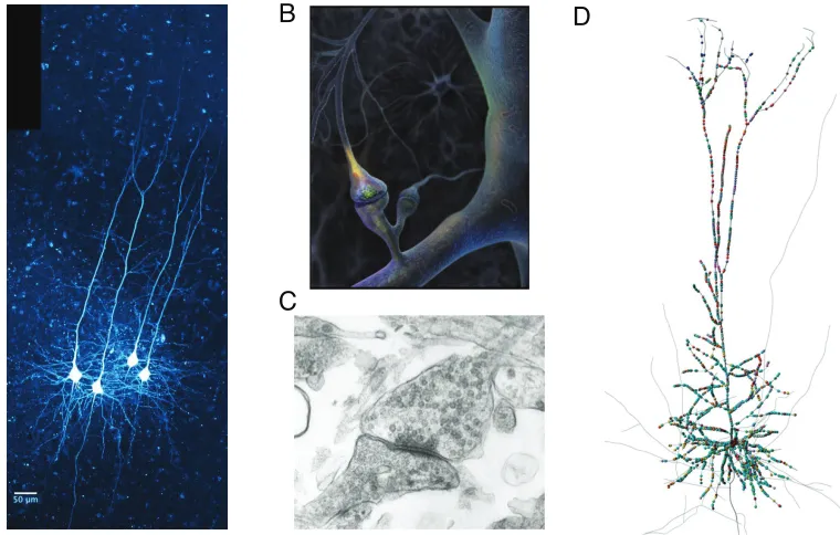
Figure 1.1: Cortical neurons, chemical synapses, and distributed inputs. (A)Four layer-V pyramidal neurons filled with fluorescent dye. This class of neuron is a major subject of this thesis. (B)A schematic of a chemical synapse. Presynaptic action potentials arrive in the bouton (top), released neurotransmitter diffuses across the synaptic cleft and binds to receptors on the postsynaptic density of a dendritic spine (bottom), causing a local change in conductance which allows a synaptic current to flow. (C)An electron microscope image of a synapse. Neurotransmitter vesicles are visible in the presynaptic terminal (right). (D) Predicted synaptic inputs onto a reconstruction of a layer-V pyramidal neuron. Panels from: AR Harbord, University of Warwick,BG Johnson, Science,C NeuralNoises, andD(Hill et al. 2012).
relatively impermeable to charged particles, containing embedded proteins which allow
ions to pass through either passively under a voltage, actively against a voltage or
pas-sively only once a certain voltage has been exceeded. The lipid bilayer of the membrane
therefore stores charge, acting as a capacitor, with ion channels acting as resistors in
parallel for different ionic species (Fig. 1.2a).
The Na+−K+ pump actively transports Na+ out of the cell, against the electrochem-ical gradient, and K+ into the cell; constructing the ionic imbalance that causes the
transmembrane voltage. A higher concentration of K+ channels means that the
effec-tive permeability of the membrane is higher for K+ ions than for Na+, allowing them
to diffuse across under the electrochemical gradient. Perturbations to the resting
po-tential across a membrane decay with a timescale τl. A typical resting value for the
membrane potential of a cortical Layer V cell is −70mV and τl is around 10ms. If the
voltage is above rest the cell is said to be depolarised and if the voltage is below it is
hyperpolarised.
Layer V pyramidal cells) causes voltage-gated Na+channels to open and a fast influx of
positive ions increases the membrane voltage by around 100mV, before slower-opening
voltage-gated K+ channels return the cell to rest (Fig. 1.2b). This is referred to as an
action potential, or spike, and the cell is said to have spiked, or fired (Hodgkin et al.
1952). The whole process takes less than a few milliseconds. The action potential is the
most important feature of a neuron and allows individual cells to integrate inputs before
giving rise to an ‘all-or-nothing’ event which is transmitted to other cells. The decay
of voltage fluctuations from the resting potential mean that inputs must be temporally
close to be successfully combined into a deviation sufficient to cause a spike. Neurons are
assumed to transmit signals through either the overall rate at which they spike, or with
the precise timing of their action potentials (de Charms and Merzenich 1996; Averbeck
et al. 2006); a succession of action potentials is referred to as a spike train. The times
between spikes in a train are called interspike intervals (ISIs). The importance of spiking
sometimes allows neurons to be abstracted as a decaying membrane voltage equipped
with a mechanism to record a spike and reset the voltage when a threshold is reached.
This is the influential leaky integrate-and-fire (LIF) model (Lapicque 1907; Brunel and
van Rossum 2007) that is used throughout the first two chapters of the thesis.
Neurons are typically spatially extended, making connections far from the cell body, or
soma. The processes branching from the cell body are collectively referred to as neurites
and are grouped into two broad classes: dendrites and axons. Dendrites typically receive
connections from other cells and axons typically make them (Fig. 1.2c). These are broad
categories and the existence has been noted, alongside the ‘classical’ axo-dendritic and
axo-somatic contacts, of dendro-dendritic (Rall et al. 1966), axo-axonal (Atwood and
Morin 1970) and dendro-axonal (Gobell 1976) connections. Connections are referred to
as synapses (Sherrington 1906).
Action potentials are initiated in the soma or segments of axon immediately adjacent to
it and propagate largely by active means. Axons in the peripheral nervous system can
extend up to 1m (from the base of the spinal column to the foot), but in the brain are
mostly a few hundred microns in length. Longer sections are often myelinated: wrapped
in an additional lipid sheath to insulate the neurite, reducing energy loss and delay
when transmitting spikes. Dendrites are also often capable of initiating active processes
when the local voltage reaches a certain threshold (Llin´as 1988). These features help
to propagate distant synaptic signals to the soma and allow regions of the dendrite
to separately process synaptic inputs, making a single neuron capable of performing
substantial computations (London and H¨ausser 2005). Regions of neurite far from the
Synapses are typically grouped into excitatory, which increase the likelihood of the
post-synaptic cell generating an action potential, and inhibitory which reduce this likelihood.
Excitatory synapses achieve this through opening ion channels that allow depolarising
current to flow into the cell, whereas inhibitory synapses either hyperpolarise the cell or
increase the permeability of the membrane, allowing excitatory currents to decay faster
and reducing the ‘window’ within which excitatory events can be integrated. The
sec-ond type of inhibition is referred to as ‘shunting’. This description is again simplified
slightly as an effect known as post-inhibitory rebound (Sherrington 1913; Granit 1956)
can cause cells to fire after receiving a barrage of inhibition due to delayed compensation
by depolarising currents.
1.1.2 Brain architecture and cortical structure
Different regions of the brain have different roles and so contain very different classes of
neurons. This thesis is mostly concerned with the neocortex, but many of the results
also apply to other brain regions. An overview of brain architecture helps to relate the
local functionality discussed here with the broader computational role of the brain.
The central nervous system generally receives sensory signals as inputs and generates
motor signals as outputs. The basic functionality necessary to maintain life is found in
the brain stem, which regulates cardiac rhythms and breathing. The brain stem forms
part of the hindbrain alongside the cerebellum, or ‘little brain’, which is associated with
balance and motor coordination (Fig. 1.3a). A unique feature of the cerebellum is
that the output to other brain regions is made up entirely of inhibitory synapses. The
midbrain acts as a relay for sensory information arriving from the peripheral nervous
system via the pons in the brainstem and is also associated with emotion and control of
hormones. Two midbrain regions of note here are the hippocampus, which is associated
with navigation and memory formation, and the thalamus, which synapses onto the
cerebral cortex.
The outer layer of the cerebral cortex is the evolutionarily youngest part of the cortex,
and is referred to as the neocortex. It is this region that is associated with higher
cog-nitive functions such as processing sensory information, generating motor commands,
storing of memories, and reasoning. Different spatial regions have different functions,
for example processing visual input or generating motor output, and slightly different
structures, but the neocortex has remarkably well-conserved features across its
consider-able area (Silberberg et al. 2002). The mammalian neocortex, henceforth shortened to
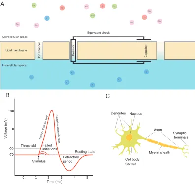
Na+ Na+ Na+ Na+ Na+ Na+ Ca2+ Ca2+ Ca2+ Cl
-K+ Cl
-Cl -K+ K+ K+ K+ K+ K+ C a p a ci to r R e si st o r Extracellular space Intracellular space Lipid membrane Io n ch a n n e l Equivalent circuit
A
Time (ms) Stimulus Failed initiations Threshold V o lt a g e ( m V ) Refractory period Resting state So diu m ch an ne ls o [image:21.596.117.513.80.453.2]pe n Po ta ssi um ch a nn e ls op e n Axon Myelin sheath Cell body (soma) Dendrites Nucleus Synaptic terminals
B
C
Figure 1.2: Basic neuronal physiology. (A)Schematic of neuronal membrane and equiva-lent circuit. K+is more concentrated within the cell and Na+
is more concentrated outside, creating a potential difference across the membrane. The lipid bilayer acts as a capacitor, storing charge, and the ion-specific transmembrane channels act as resistors. (B)Schematic of an action potential. Stimulus below the threshold fails to initiate an ‘all-or-nothing’ ac-tion potential. Once the threshold is crossed, Na+ channels open, allowing a fast influx of current. This is counteracted by the slower opening of K+ channels that allow current to leave the cell. After an action potential, the neuron is temporarily unable to initiate another due to the effect of the K+and undergoes a refractory afterhyperpolarisation (AHP). Panel adapted fromwww.innovateus.net. (C)Spatially extended neuron, showing axon, dendrite, and soma. Panel adapted form C Boeree, Shippensburg University
found there (Fig. 1.3c). The basic structure and function of these layers are discussed
below:
Layer I. The uppermost layer, Layer I, contains very few cell bodies. Dendrites from
neurons with soma in deeper cortical regions branch extensively in this region, where
they receive excitatory and inhibitory synapses. Often the excitatory synapses are from
cells with soma in a different layer to that of the postsynaptic cell, allowing interlaminar
communication, or are from different cortical regions or the thalamus (Rubio-Garrido et
Layer II/III.Layers II and III are often considered together as they differ strongly only
in the cortical areas processing visual input. Excitatory neurons in this layer receive
excitatory synapses from Layer IV and synapse both intralaminarly and onto cells with
bodies in Layer V (Feldmeyer et al. 2002).
Layer IV. Layer IV is the major target of long-range connections from both different
cortical areas and the thalamus. It is notably absent in the region of cortex
associ-ated with generating motor commands. The neurons here make excitatory interlaminar
connections to Layer II/III.
Layer V. Layer V is the cortical layer most considered in this thesis. Layer V provides
the major outputs to other areas of cortex and brain. The cell class most discussed
here, the thick-tufted Layer-V neuron, is found mostly in the lower portion of this layer,
where they receive long-range excitatory input from the thalamus, via Layers IV and
II/III, and inhibitory input from interneurons with soma in Layer V. They also form
well-structured local networks with other Layer V pyramidal cells (Song et al. 2005;
Perin et al. 2011), with synapses located mostly within Layer V (Markram et al. 1997).
Layer VI. The deepest cortical layer is commonly referred to as the polymorphic layer
as the cells found here are more heterogeneous than in other layers (Fig. 1.3c). The
functions of these cells are less easily categorised than those in other layers, but an
important output is to the thalamus, providing cortico-thalamic feedback to the strong
thalamo-cotical inputs (Crandall et al. 2015).
Alongside this vertical layered structure, the cortex is organised horizontally around
columns that span all six layers (Mountcastle et al. 1957). Connections are more
probable between neurons sharing a column and inputs to and outputs from a column
tend to be shared. The region of cortex that processes sensory inputs that are not visual
or auditory is called the somatosensory cortex. In a part of this region in the rodent
brain, cortical columns correspond precisely with individual whiskers: a single whisker
induces activity in a single column (Woosley and van der Loos 1970).
The final subdivision of the cortex is the microcircuit. Neocortical microcircuits are
defined by stereotyped intracolumnar, interlaminar connectivity (Douglas et al. 1989).
They are conserved across brain regions and species and are well-studied as a potential
A B
[image:23.596.120.506.80.343.2]C
Figure 1.3: Brain architecture and cortical structure. (A)Schematic of the human brain, with areas discussed in the text labelled. Panel from D McDaniel, Arizona State University. (B)Schematic of the neocortex, showing key excitatory and inhibitory connections between and within cortical layers. Panel from M Richardson, University of Warwick. (C)Diversity of excitatory cell classes by layer. Thick-tufted Layer V pyramidal cells are third from the right. Panel from (Oberlaender et al. 2012).
1.2
Synaptic transmission
Neurons are generally insulated from one another (Waldeyer 1891; Cajal 1899); changes
in voltage cannot propagate between cells without synaptic intermediaries. The more
evolutionarily and structurally primitive method of sharing voltage between cells is an
electrical synapse or gap junction: a partially uninsulated section of membrane between
two neurons. This allows a flow of current between the two cells and provides a fast
and simple method of transmitting information. In the mammalian cortex, however, the
vast majority of synapses are chemical and rely on an intermediary neurotransmitter to
communicate. The structure and functional roles of this form of synaptic transmission
are discussed below, followed by an introduction to rate- and timing-based neuronal
codes.
1.2.1 Structure of a chemical synapse
A synapse is defined as the connection between a presynaptic neuron and a postsynaptic
neuron (Sherrington 1906). This connection generally consists of a number of anatomical
that function via neurotransmitter and others via direct electrical coupling
(Hamzei-Sichani et al. 2012), but most consist entirely of neurotransmitter-mediated contacts.
A chemical synaptic contact is made up of a presynaptic terminal, a synaptic cleft, and
a postsynaptic density (Fig. 1.4).
Within the presynaptic terminal, the neurotransmitter is contained within a lipid bilayer
membrane, forming a structure known as a vesicle around 40nm in diameter (S¨udhof
2004). The presynaptic terminal has a number of active, or release, sites on the
mem-brane where neurotransmitter vesicles can dock (Schikorski and Stevens 2001). When
the presynaptic cell spikes, voltage-gated calcium channels in the presynaptic terminal
open and allow an influx of Ca2+. The Ca2+ has the potential to trigger a release of
neurotransmitter from vesicles docked at release sites (Fatt and Katz 1954; Katona and
Freund 1969; Neher and Sakaba 2008; Nakamura et al. 2015). The probability that a
single docked vesicle will release neurotransmitter on arrival of an action potential is
typically quite low, often less than half. Synapses are thus ‘reliably unreliable’ (Goda
and S¨udhof 1997; Ribrault et al. 2011).
The synaptic cleft is on the order of 20nm wide and neurotransmitter molecules pass
across it by diffusion. On reaching the postsynaptic density, the neurotransmitter
molecules bind to transmitter-gated ion channels, causing them to open and thus
al-ter the conductance of the postsynaptic cell. Transmital-ter molecules do not remain in
the cleft for long, they are either broken down by enzymes, taken up by non-neuronal
glial cells, or recycled into vesicles in the presynaptic cell for re-use. Vesicles filled with
recycled or newly synthesised neurotransmitter are trafficked to empty release sites.
Re-cent experimental techniques allowing manipulation of individual vesicles have greatly
enhanced understanding of the details of this process (Trigo et al. 2012; Park et al.
2012).
At the postsynaptic cell, open transmitter-gated ion channels can have a variety of
ef-fects. Excitatory neurotransmitters typically open sodium and potassium channels
caus-ing an influx of Na+ anions that depolarise the postsynaptic cell causing an excitatory
postsynaptic potential (EPSP). The fast opening and slow closing of transmitter-gated
ion channels means that the EPSP takes the form of a difference of exponentials (Eccles
et al. 1941; Richardson and Silberberg 2008). Inhibitory neurotransmitters typically
target K+ or Cl− channels, allowing K+ to diffuse down its concentration gradient out
of the cell or Cl− to flow down its concentration gradient into the cell; both have a
hy-perpolarising effect. As neurotransmitters increase the conductance of the membrane,
they allow excitatory currents to decay more quickly by reducing the effective membrane
time constant τl. This reduces the time window over which temporally separated
‘shunting’, inhibition (Eccles 1964). An important feature of excitatory synapses is that
due to the equilibrium potential of Na+ being much higher than the resting potential of
the cell, the voltages induced by excitatory neurotransmitters from different vesicles can
be well-approximated by a linear sum (Burke 1967; Bekkers 2003). This approximation
is used throughout the thesis.
The fact that neurotransmitter is released from vesicles means that EPSPs are ‘built up
statistically of small all-or-none units which are identical in size with the spontaneous
‘miniature” postsynaptic potentials (del Castillo and Katz 1954). Synaptic transmission
is therefore quantal (Boyd and Martin 1956; Liley 1956), and the EPSP induced by a
single quanta of neurotransmitter is referred to as a ‘mini’ EPSP, following del Castillo
and Katz.
Neurotransmitters were first identified by Otto Loewi in 1921. In cortex, the most
prevalent excitatory and inhibitory neurotransmitters are glutamate andγ-aminobutyric
acid (GABA) respectively. Synapses that use glutamate are referred to as
glutamater-gic and those that use GABA are referred to as GABAerglutamater-gic. Glutamate receptors
for transmitter-gated ion channels in the postsynaptic density include the α
-amino-3-hydroxy-5-methyl-4-isoxazoleproponic acid receptor (AMPAR) and N-methyl-D-aspartate
receptor (NMDAR). Both are implicated in long term changes to the strength of
synapses, discussed below. In addition to the fast action of ion-channel-linked (ionotropic)
receptors, other, metabotropic, neurotransmitter receptors indirectly influence neuronal
function through second-messenger signalling: triggering the release of ‘second’
messen-ger molecules from intracellular stores. An important class is the metabotropic
glu-tamate receptors (mGluRs), which have slower and longer-lasting actions than those
triggered by AMPARs and NMDARs (Nakanishi 1994). mGluRs have a variety of
ef-fects, causing excitation or inhibition or modulating the efficacy of synapses onwards
from the postsynaptic cell (Pin and Duvoisin 1995). Neurotransmitters are generally
abundant and easy to synthesise: glutamate, for example, is an amino acid and GABA
is directly synthesised from it. This helps to mitigate the high metabolic costs of synaptic
transmission (Harris et al. 2012).
It is important to note that the number of anatomical contacts forming a synapse is
in general not equal to the number of active sites from which neurotransmitter can be
released. This ‘single-vesicle’ hypothesis was widely held (Kuno 1971; Korn et al. 1981;
Redman 1990), particularly in cortex (Silver 2003), but more recent work has found that
the distribution of cortical postsynaptic responses observed (Song et al. 2005; Lefort et
al. 2009) is best explained by a number of release sites that can be substantially higher
Figure 1.4: Structure of a chemical synapse. The presynaptic terminal (top) contains a number of active sites to which vesicles containing neurotransmitter can dock. On arrival of an action potential, neurotransmitter is probabilistically released and diffuses to receptors in the postsynaptic density (bottom). The site from which neurotransmitter has been released is inactive until a full vesicle is trafficked to the site. Panel from Wikipedia.
1.2.2 Function of a chemical synapse
Chemical synapses have a number of functional advantages over electrical synapses,
de-spite growing evidence of interactions between the two modes of synaptic transmission
(Pereda 2014) and the fact that electrical synapses have their own complexity
(Landis-man and Connors 2005). Chemical synapses transmit neuronal codes based either on
a firing-rate or precise spike-timing (de Charms and Merzenich 1996) and are able to
reverse the sign of transmission through use of inhibitory neurotransmitters; they also
have an important gating role in only feeding a signal forward after the presynaptic cell
has integrated its own synaptic inputs.
Chemical synapses are plastic: their strength alters through time. These changes occur
on both short and long timescales and are a major subject of Chapters 2, 3, and 4.
Synaptic plasticity is discussed in depth below.
Finally, chemical synapses are stochastic: an action potential is unlikely to induce release
of neurotransmitter from all docked vesicles and will sometimes induce release from
none (Kuno and Weakly 1972; Goda and S¨udhof 1997; White et al. 2000; Faisal et
al. 2008; Ribrault et al. 2011). This feature of synaptic function is surprising and has
been well studied. From an information-theoretic point of view, synaptic unreliability
enhances the information content of some codes (Zador 1998) and reduces the metabolic
requirements of many others (Levy and Baxter 2002; Goldman 2004). The substantial
‘noise’ introduced by variability in synaptic transmission will also have a potentially
critical effect on the firing-rate dynamics of a large-scale neuronal network (Salinas and
Sejnowski 2000; Kuhn 2004; de la Rocha and Parga 2005). At an individual synapse,
transmission will show a great deal of trial-to-trial variability and the models in Chapters
2, 3, and 4 specifically account for this.
For the rest of this thesis, the term ‘synapse’ without qualification will refer to a chemical
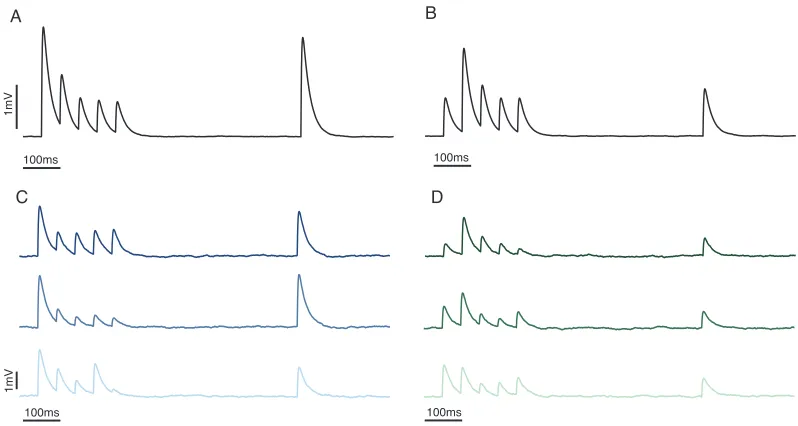
100ms 100ms
100ms 100ms
1
mV
1
mV
A
C
B
[image:27.596.117.516.80.293.2]D
Figure 1.5: Short-term synaptic plasticity. Simulated EPSPs showing effects of vesicle depletion depression (left) and facilitation (right). (A), (B) The average responses over 50 trials, showing the typical effects of depression and facilitation. (C), (D) Individual postsynaptic responses, displaying considerable trial-to-trial variability.
1.3
Synaptic plasticity
Chapters 2, 3, and 4 of this thesis study the effects of short-term synaptic plasticity.
The current section first introduces the basic ideas behind this phenomenon, before
examining longer-term changes in synaptic strength which are an important part of
the work introduced in Chapter 2. The next subsection discusses patterns of spiking
activity seenin vivo, which are considered alongside short-term plasticity in Chapters 2
and 3. The penultimate subsection introduces the idea of neuromodulators, which alter
neuronal and synaptic function over wider areas, one of which is studied in Chapter
4. The final subsection here briefly describes paired whole-cell patch-clamping, the
experimental technique used to gather data about synaptic function.
1.3.1 Short-term synaptic plasticity
Changes in synaptic efficacy due to the immediate history of a synapse are referred to as
short-term synaptic plasticity (Zucker and Regehr 2002). The most commonly seen and
studied forms are vesicle depletion depression and facilitation (Fig. 1.5). Vesicle
deple-tion depression occurs after an acdeple-tion potential has induced release from docked vesicles
at presynaptic active zones, leaving those zones without the ability to release
neurotrans-mitter until a new vesicle has been trafficked to the site, a process that typically takes a
few hundred milliseconds (Eccles et al. 1941; Liley 1956; Vere-Jones 1966; S¨udhof 2004;
terminal during persistent activity (Dudel and Kuffler 1961; Katona and Freund 1969).
This residual calcium increases the probability that neurotransmitter will be released by
subsequent action potentials and can increase the size of the second EPSP in a
closely-spaced pair to five times the size of the first (Zucker and Regehr 2002). Together the two
processes gives the synapse a ‘memory’ that lasts around ten to a hundred times longer
than voltage fluctuations in the postsynaptic membrane (Mongillo et al. 2008), and have
a variety of postulated computational roles (Abbott and Regehr 2004), including: gain
control (Abbott 1997; Rothman et al. 2009), increasing information transmission (Zador
1998; Kilpatrick 2013; Scott et al. 2012), sensory adaptation (Furukawa et al. 1982;
Hallermann and Silver 2013), and filtering inputs (Fortune and Rose 2001; Lindner et
al. 2009; Rosenbaum et al. 2012; Nagel et al. 2015). The results presented in Chapters
2, 3, and 4 do not rely on a specific interpretation of the function of vesicle depletion
depression, focussing mainly on the implications for firing rates in the postsynaptic cell
and recovering information about synaptic parameters from experiments.
Different regions of cortex and different developmental states imply different ratios of
depression and facilitation. Depression is more prevalent in cortical areas associated
with sensory input (Thomson 1997) and in thalamocortical synapses (Boudreau and
Ferster 2005), whereas facilitation becomes more prevalent in non-sensory cortical areas
(Mongillo et al. 2008). Age is also an important factor, with a shift from predominately
depressing to facilitating synapses throughout development in rodent models (Reyes
and Sakmann 1999), although some regions of mature human cortex have been observed
to exclusively depress (Testa-Silva et al. 2014). The parameter that most determines
the ratio between depression and facilitation is the probability that an individual docked
vesicle will release neurotransmitter on arrival of an isolated spike,p. A lowpis likely to
lead to facilitation dominating as a relatively low proportion of vesicles are released after
the first spike and increases in release probability will have more of an impact. There
is also a dependence on the spike-train that is being transmitted and some neurons
are believed to route spike-trains with different statistics to different targets through
different synaptic responses, with very high frequency spike trains causing depression
and lower frequency trains inducing facilitation (Middleton et al. 2011; Nagel et al.
2015; Crandall et al. 2015).
The most influential models of short-term depression and facilitation are those of Abbott
(1997) and Tsodyks and Markram (1997). These phenomenological models have enabled
understanding of the dynamics of large networks of cells, revealing emergent behaviours
that do not arise in models of static synapses (Tsodyks et al. 1998; Cortes et al. 2013).
These models consider synaptic ‘resources’, which are deterministically depleted by a
presynaptic action potential and recover at a fixed rate in the absence of stimulus. They
stochasticity in both probabilistic vesicle release and variable vesicle recovery times,
as well as the quantal nature of transmission (Fig. 1.5c and d). Models taking full
account of this stochasticity have been applied where effects of trial-to-trial variability are
particularly important (Fuhrmann et al. 2002; de la Rocha and Parga 2005; Rosenbaum
et al. 2012); Chapters 2, 3, and 4 of this thesis apply a stochastic quantal model.
Whilst vesicle depletion depression and facilitation are the most commonly observed
forms of short-term plasticity, the diversity and complexity of synapses (O’Rourke et al.
2012) mean that many other forms have also been described. Facilitation due to residual
calcium decays on timescales with orders of hundreds of milliseconds, but less substantial
increases in postsynaptic responses have been observed with timescales that are much
longer. Augmentation has timescales of seconds to tens of seconds and post-tetanic
potentiation has timescales of minutes; both can be explained by long-lived changes to
the efficacy of the ion pumps at presynaptic terminals during stimulus, which alter the
amount of Ca2+ that can enter during a subsequent spike (Regehr 1997; Zhong et al.
2001), and by effects on enzyme concentration (Fioravante and Regehr 2011). These
processes occur on slightly longer timescales than those modelled in this thesis.
A more relevant alternative form of plasticity is release-independent depression (RID)
(Dobrunz et al. 1997; Thomson and Bannister 1999), where the reduction in size of
a second EPSP is uncorrelated with the size of the first. The mechanism for this is
not entirely clear, but is believed to be a stimulus-dependent reduction in the rate at
which Ca2+ can enter the terminal (Catterall and Few 2008; Regehr 2012) and is best
modelled by a decrease in the release probability for successive spikes (Fuhrmann 2004).
RID initially decays on timescales around 500ms, but is highly-correlated with
frequency-dependent recovery (FDR), a reduction in the decay timescale during sustained activity.
Both mechanisms are incorporated in the models fitted to synaptic datasets in Chapter
4.
Another feature that can reduce the impact of depression is a stimulation-dependent
increase in the rate at which fresh vesicles are trafficked to empty release sites. The
mechanism behind this is unclear but is believed to relate to the build-up of calcium in
the presynaptic terminal (Wang and Kaczmarek 1998). The recovery of vesicles under
intense stimulus follows a double exponential, suggesting two distinct processes, with
a slow timescale that is invariant to stimulus and a fast timescale that is
stimulus-dependent (Hosoi et al. 2007). This is believed to be an important factor in maintaining
synaptic transmission at high-frequencies (Fioravante and Regehr 2011; Hallermann and
Silver 2013).
Synaptic depression is sometimes also attributed to saturation and desensitisation of the
vesicle depletion. The two effects are indistinguishable under the phenomenological
models of Abbott (1997) and Tsodyks and Markram (1997), but would have an impact
on the interpretation of quantal models of release. Experiments to distinguish between
these pre- and postsynaptic modes of depression have shown that neuronal mechanisms
can specifically counteract receptor desensitisation (DiGregorio et al. 2007; Heine et al.
2008), that desensitisation is relatively unimportant for presynaptic firing rates on the
order of 10Hz, and that vesicle depletion accounts for the majority of observed depression
(Wong et al. 2003; Foster and Regehr 2004).
1.3.2 Long-term synaptic plasticity
Long-term changes in synaptic strength are the physiological correlate of memory
for-mation (Hebb 1949). The factors that induce this are varied and beyond the scope of
this thesis (Abbott and Nelson 2000; Markram et al. 2011), but the mechanism behind
changing weights at excitatory glutamatergic synapses is important for Chapter 2. The
glutamate receptors AMPAR and NMDAR can, under certain conditions, activate
ini-tially blocked channels in the postsynaptic density, increasing synaptic strength quickly,
and induce changes in genetic transcription that strengthen the synapse over longer
timescales (Kandel 2001; Bayazitov et al. 2007). These post-synaptic changes can be
matched presynaptically by an increase in the number of vesicle release sites so that the
average ‘mini’ EPSP amplitude is unchanged, but the total synaptic strength increases
over a timescale of hours (Loebel et al. 2013).
Long-term depression has a similar mechanism, where the ‘mini’ amplitude is unchanged
but the total strength decreases due to matched reductions in presynaptic release sites
and postsynaptic receptors. As the long-term plasticity that gives rise to memories can
provide positive-feedback, where synapses between co-active neurons strengthen
repeat-edly (Hebb 1949), a form of homeostatic plasticity is observed to balance ‘runaway’
connection strengths by weakening others (Turrigiano and Nelson 2004). This
homeo-static plasticity is another consideration in Chapter 2.
1.3.3 Spike-train statistics
When dealing with stochastic quantal models of synaptic function it is often
mathemat-ically convenient to make the assumption that presynaptic spike times are uncorrelated
(Fig. 1.6a) (Fuhrmann et al. 2002; Rosenbaum et al. 2012). This is not necessarily the
case and analysis of in vivo spiking reveals correlations both between neurons (Aertsen
et al. 1989; Schneidman et al. 2006) and autocorrelations within a single spike train
N
e
u
ro
n
N
e
u
ro
n
N
e
u
ro
n
N
e
u
ro
n
200ms
200ms 200ms
200ms
A B
C D
Uncorrelated Crosscorrelated (synchronous)
[image:31.596.113.516.79.420.2]Positively autocorrelated (bursty) Negatively autocorrelated (periodic)
Figure 1.6: Correlations between neurons and within spike trains. Raster plots displaying the spike times of one hundred neurons under different types of correlation. (A) Indepen-dent neurons with Poisson spike trains. (B) Crosscorrelated (synchronous) neurons with individually Poisson spike trains. (C) Independent neurons, each firing as a positively autocorrelated spike (bursty) train. (D)Independent neurons, each with a negatively auto-correlated (periodic) spike train.
The results in Chapter 2 involve correlated spiking between presynaptic neurons, and
those in Chapter 3 consider spike trains that are autocorrelated.
Correlations between spike trains (Fig. 1.6b) have relevance for encoding sensory
infor-mation (von der Malsburg 1981; de Charms and Merzenich 1996; Averbeck et al. 2006),
motor control (Baker et al. 2001; Capaday et al. 2013) and decision making (Cohen
and Newsome 2008; Cain and Shea-Brown 2013). Recent work suggests that
modula-tion of correlamodula-tions can be more significant for neuronal coding than alteramodula-tions in the
presynaptic firing rate (Seri`es et al. 2004; Mitchell et al. 2009; Cohen and Kohn 2011).
Correlations within spike trains (Fig. 1.6 c and d) are an important part of neuronal
codes (Ramcharan et al. 2000; Sherman 2001) and have substantial effects on how well
spike trains propagate (Cateau and Reyes 2006; Lindner 2006; Reich and Rosenbaum
2013; Pipa et al. 2013; Dummer et al. 2014). Further effects are discussed in detail in
1.3.4 Neuromodulators and retrograde transmission
In addition to the very local effects of neurotransmitters, neuronal and synaptic
prop-erties are also affected on a larger scale by neuromodulators (see Marder 2012, for a
review). These are typically released during activity and can have profound effects on
the effective connectivity and function of neuronal networks. Recent work by Kerr et
al. (2013) has implicated the neuromodulator adenosine (Boison 2006) in the
develop-mental shift in glutamatergic synaptic properties from depressing to facilitating (Reyes
and Sakmann 1999; Chen and Buonomano 2012); the analysis introduced in Chapter 4
is applied to the effects of adenosine on synaptic function.
Synapses are also modulated locally by retrograde signalling; where the postsynaptic
cell releases a transmitter that alters synaptic efficacy. This allows the postsynaptic
cell to control afferent signals from selected presynaptic neurons. Endocannabinoids
are retrograde transmitters that temporally reduce the amount of conventional
neuro-transmitter released by the presynaptic cell (Katz 2012) and so can alter the dominant
short-term plasticity displayed presynaptically from depression to facilitation (Brenowitz
and Regehr 2005).
1.3.5 Paired whole-cell patch-clamp recording
Data on synaptic properties is gathered through paired-cell patch clamping. This
pro-cedure is described exactly in the Methods sections of Chapters 3 and 4, but a brief
overview here may clarify some points about synaptic datasets. Glass electrodes are
placed against the membrane of two cells and suction is applied until the membranes
break and the electrodes can record from the interior of the cells; the process is known
as whole-cell patch clamping (Neher 1991; Sakmann 1991). Current is injected into one
cell to induce it to fire and the voltage of the other cell is monitored to detect an EPSP.
In particular, this method does not distinguish between synaptic currents induced at
individual anatomical contacts.
Paired whole-cell recording has advantages over experimental protocols using
extracel-lular stimulating electrodes and intracelextracel-lular recording electrodes as it is possible to
precisely stimulate a single presynaptic cell whilst recording its voltage. Precise
stim-ulation means that EPSPs are certain to come from a single synapse and presynaptic
voltage recording allows failed action potential initiation to be distinguished from failed
neurotransmitter release. Experiments are done in vitro on slices of tissue typically
Tissue slices are ‘quiet’, they have very little background spiking activity compared to
a living brain. This must be considered when extrapolating from in vitro slice data to
in vivo living systems; in particular it means that stimulated activity in slice causes
additional initial depression and facilitation compared to the same activityin vivo. In
practice theoretical studies account for this easily by considering steady-state behaviour
or explicit transitions in the activity levels (Tsodyks and Markram 1997). Inference
of dynamic synaptic properties is also more straightforward in a quiet slice due to the
larger initial changes in EPSP amplitude on stimulation onset (Eccles et al. 1941;
Bekkers 1994).
1.4
Neurons
So far this chapter has discussed the broad structure and basic behaviour of neurons
without much emphasis on the specifics of different cell classes. An understanding of
the functionality discussed in this thesis does require consideration of the properties
of the different cell classes discussed. The broadest categorisation of cells comes from
the effect of the synapses they form: excitatory neurons form excitatory synapses and
inhibitory neurons form inhibitory synapses. Beyond this, the brain region in which the
cells are found and their location within this region, alongside morphological, genetic,
and electrophysiological properties are used to define neuron classes (Wang et al. 2001;
Oberlaender et al. 2012).
The major class of neuron discussed in this paper, and that for which experimental
evidence is gathered in Chapters 3 and 4, is the thick-tufted pyramidal cell from Layer
V of the cerebral cortex. The first subsection below describes the properties of this
cell class in some detail and the second outlines unique properties of other cell classes
discussed in Chapter 5.
1.4.1 Thick-tufted Layer V pyramidal cell
Layer V pyramidal cells are a major output cell of the cortex and are heavily implicated
in complex computation (Markram et al. 1997; London and H¨ausser 2005; Spruston
2008; Ramaswamy and Markram 2015). These cells have soma lying in Layer V of the
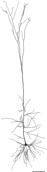
cerebral cortex, typically in the lower half, often referred to as Layer Vb (Fig. 1.7). They
have a large dendrite that heads vertically up through the cortical layers to Layer I, where
it branches extensively. This main dendrite is referred to as the apical dendrite, and the
branches are referred to as tuft dendrites. The apical and tuft dendrites are over 1000µm
Figure 1.7: Thick-tufted Layer V pyramidal cell. Scale bar 100µm. Morphology taken from (Hay et al. 2011), with all dendritic radii increased by 0.5µmfor ease of visualisation.
plane of Layer V and are shorter, typically spanning around 300µm. The soma is around
25µm in diameter (all measurements from Markram et al., 1997). A single axon leaves
the soma and branches extensively, occasionally reaching up to higher layers within a
cortical column, targeting basal dendrites of other pyramidal cells, and projecting down
out of the cortex as a major output pathway. The characteristic pyramidal shape is
found throughout the nervous systems of mammals, birds, fish, and reptiles suggesting
a highly conserved computational role (Spruston 2008).
Layer V pyramidal cells receive synapses across their dendritic trees, with fairly
stereo-typed presynaptic locations. Excitatory synapses on the tuft dendrites tend to be
long-range, from different cortical areas or the thalamus (Rubio-Garrido et al. 2009; Meyer et
al. 2010). The basal dendrites are targets of short-range glutamergic synaptic
connec-tions from other Layer V cells within a cortical column and can be considered short-range
(Markram et al. 1997; Hay et al. 2011). There is evidence that anatomical contacts
on basal dendrites further from the postsynaptic soma are stronger to equalise their
5000 synapses onto its basal dendrites (O’Kusky and Colonnier 1982; Spruston 2008).
Pyramidal cells also receive a complex pattern of inhibition from a variety of inhibitory
cells within the cortical microcircuit that are not discussed in detail here, but have rich
and varied effects on the excitability of the pyramidal cell (London and H¨ausser 2005;
Silberberg and Markram 2007; Fino and Yuste 2011; Hill et al. 2012; Gidon and Segev
2012).
Layer V pyramidal cells have active dendrites. The most striking aspect of this is a
concentration of voltage-gated calcium channels near the top of the apical dendrites,
which instigate calcium spikes that propagate to the soma when the voltage in the
calcium channel region exceeds a threshold (Williams and Stuart 2002; London and
H¨ausser 2005; Hay et al. 2011). This helps transmit long-range signals to the soma. A
hyperpolarisation-activated depolarising current denotedIh is also present, with the Ih
cation channels decreasing in density towards the soma. The relatively slow timescale
of these channels means that EPSPs recorded at the soma are succeeded by a
‘sag-rebound’ in voltage (Berger et al. 2001; Silberberg and Markram 2007). A third active
process is the NMDA spike, which has only been observed in the thinner basal and
tuft dendrites and requires direct glutamatergic synaptic input (Schiller et al. 2000;
Antic et al. 2010). Action potentials initiated in the soma can backpropagate into the
dendrites (Stuart and Sakmann 1994), a function that appears to play a crucial role in
long-term synaptic plasticity (Spruston 2008). Together with ion channels in the soma,
these active processes play a part in spike-frequency adaptation (Fuhrmann et al. 2002).
Adaptation is a modulation of a cell’s response to persistent stimulation (Wang 1998;
Benda et al 2005; Peron and Gabbiani 2009). A key feature of adaptation currents
is in the creation of correlations between interspike intervals (Schwalger et al. 2010;
Schwalger and Lindner 2013), generating non-renewal spike trains which are discussed
in Chapter 3.
1.4.2 Other neurons
Whilst the mathematical results in Chapters 2, 3, and 4 are general and can be applied
to any synaptic contacts, both the experimental verification and main interpretations
are centred around Layer V pyramidal cells. In Chapter 5, a morphological constraint is
derived and experimental validation requires an analysis of the geometries of a number
of other cell classes. Their structure and proposed functions are outlined below.
Purkinje cell. Purkinje cells (1.8a) are the major output neuron of the cerebellum. Like
the cortex the cerebellum has a layered structure, but with an even greater degree of
branched dendrites receive excitatory synaptic inputs from two main sources. Climbing
fibres from the medulla in the brainstem make strong excitatory synapses on proximal
regions of dendrite. Each Purkinje cell receives climbing fibre input from a single
presy-naptic cell, and the activation of this synapse induces a complex spike, where a large
initial action potential is succeeded by a burst of action potentials with lower
ampli-tudes. Purkinje cells can also fire simple spikes, which are individual action potentials.
Parallel fibres from cerebellar granule cells make a very large number, in the region of
200,000, of weak glutamatergic synapses to the distal regions of the dendrite, and can
induce simple spikes. The dendrites of Purkinje cells allow for many active processes
(Roth and H¨ausser 2001).
The site of action potential initiation is relatively far down the axon compared to the
ma-jority of neurons (Stuart and H¨ausser 1994). The output of Purkinje cells is inhibitory,
and axon collaterals branch off from the main tract to specifically target proximal
re-gions of neighbouring Purkinje cells with GABAergic synapses. Purkinje cells have a
notably different genetic profile to many other neurons (Kirsch et al. 2012).
Dentate gyrus granule cell. The dentate gyrus lies within the hippocampus. Granule
cells (1.8b) have small bodies (∼ 10µm in diameter) and their conical arrangement of dendrites receives input from Layer II of the cortical region associated with memory
formation and navigation (Schmidt-Hieber et al. 2007). Their activity is relatively
sparse and strongly associated with spatial exploration. They are one of the few neuronal
classes known to undergo adult neurogenesis (Cameron and Mckay 2001).
Blowfly lobula plate HS and VS neurons. The structure of invertebrate brains is very
different to that of mammals, but the constraints in Chapter 5 are also shown to apply to
neurons from the fly central nervous system. The lobula plate of the blowfly calliphora
vicinalies within the region of the brain that receives visual inputs and consists of around
a dozen giant neurons; three of which, the HS cells (1.8c), process horizontal motion, and
nine to eleven of which, the VS cells (1.8d), process vertical motion (Hausen et al. 1980;
Scott et al. 2002). These cells are large and have a membrane conductance a factor of ten
higher than many mammalian neurons. The layout of these cells is highly-stereotyped
and the dendritic morphology is not experience-dependent, suggesting that the cells
are genetically relatively hard-coded (Karmeier et al. 2001). The lobula plate has an
important computational role, receiving inputs from a large number of presynaptic cells
that retain a direct spatial mapping from the retina and producing the outputs necessary
A
B
[image:37.596.142.443.78.458.2]C
D
Figure 1.8: Other neuronal morphologies. (A) Purkinje cell (Roth and H¨ausser 2001). (B) Dentate gyrus granule cell (Schmidt-Hieber et al. 2007). (C), (D) Blowfly lobula plate HS (C) and VS (D) neurons (Cuntz et al. 2010). All scale bars 100µmand all radii increased by 0.5µmfor ease of visualisation.
1.5
Optimality in dendritic structure
The results presented in Chapter 5 involve studying a spatially extended neuron model,
in contrast to the point neurons in Chapters 2, 3, and 4 that allow analytical results even
when the input is stochastic and temporally complex (Brunel and Hakim 1999; Gerstner
2000; Fourcaud-Trocm´e et al 2003). Spatially extended models attempt to account for
the full morphological properties of real cells and are an important consideration in
understanding synaptic function. The results in Chapter 5 consider dendritic optimality:
how well dendrites are adapted for the task of propagating synaptic currents passively to
the soma. To put this in context, I will introduce the ideas behind a spatially extended
an idea known as ‘dendritic democracy’ (Jaffe and Carnevale 1999; Magee and Cook
2000; H¨ausser 2001).
1.5.1 Spatially extended neuron models
The results of Wilfrid Rall in describing the properties of voltage in dendritic trees
(Rall 1959; Rall 1969; Rall and Rinzel 1973) were critical in illustrating the function
of extended dendrites. Previous work had neglected the importance of dendritic trees,
overestimating the attenuation of voltage with distance (Eccles 1957). Rall was able to
demonstrate that physiological values of dendritic radius and passive electrical
proper-ties meant that dendritic trees would have a substantial effect on somatic voltage (Rall
1959); thereby introducing neuronal cable theory, the study of voltage and current in
leaky neurites. The governing cable equation is derived in the supplementary material
to Chapter 5 for a cable with arbitrary radius profile; the classical form is for constant
radius. The lengthscale of a cable depends on its radius and passive properties and is
referred to as electrotonic length. A further innovation of Rall was to allow a
branch-ing dendritic tree to be modelled as a sbranch-ingle cylinder under certain conditions. These
conditions are rarely met in real neurons (Whitehead and Rosenberg 1993) (but see
Desmond and Levy, 1984), but the ‘equivalent cable’ allowed an intuitive mathematical
understanding of complex dendritic structures (Segev et al. 1995).
Further analytical approaches to the problem of resolving voltages in branched dendrites
applied various transforms to allow efficient computation (Butz and Cowan 1974; Koch
and Poggio 1985; Abbott et al. 1991; Zador et al. 1995), but arguably lacked the
intuitive value of an equivalent cable interpretation (Whitehead and Rosenberg 1993).
The equivalent cable idea was therefore extended to relax many of the original constraints
(Schierwagen 1989; Poznanski 1991; Jaffe and Carnevale 1999; Lindsay et al. 2003),
and solutions were found for many types of non-cylindrical or passively non-uniform
dendritic cable (Goldstein and Rall 1974; Holmes and Rall 1992; London et al. 1999).
These solutions did, however, lack complete generality, leaving room for the asymptotic
solution to arbitrary taper introduced in Chapter 5.
The active processes in many dendrites, combined with the branching complexity, has
led to widespread adoption of numerical methods for resolving voltages in realistic
mor-phologies (Hines and Carnevale 1997), but analytical studies of passive properties do still
have the potential to generate important new insights into dendritic function (Timofeeva