Original Article
Expression of MMP-9/TIMP-2 in nasal
polyps and its functional implications
Xuechang Li1,2, Yanli Tao2, Xuezhong Li1
1Department of Otolaryngolgogy, Qilu Hospital of Shandong University, Jinan 250012, China; 2Department of Otorhinolaryngology, Weifang People’s Hospital, Weifang 261041, Shandong, China
Received May 19, 2015; Accepted June 28, 2015; Epub November 1, 2015; Published November 15, 2015
Abstract: Nasal polyps (NP) involve tissue repair and structural remodel, both of which require the extracellular matrix. Matrix metalloproteinase (MMP) and tissue inhibitor of metalloproteinase (TIMP) are known regulators for
tissue reconstruction. This study therefore aimed to analyze the expressional profile of MMP-9 and TIMP-2 in NP
patients, with further investigation of their roles in pathogenesis. A total of 60 NP tissue samples (including 15 type I, 21 type II and 24 type III) were collected from surgeries in our hospital, in addition to 6 normal ethmoid sinus
mucosa samples. The mRNA and protein expression levels of MMP-9/TIMP-2 were quantified by real-time PCR and
Western blotting, respectively. Serum levels were also checked by enzyme-linked immunosorbent assay (ELISA).
Both mRNA and protein levels of MMP-9 in NP tissues or serum were significantly elevated compared to those
in control ones (P<0.05) while the TIMP-2 expression was suppressed (P<0.05). In patients with more advanced stage, MMP-9 expression was further elevated, with lowered TIMP-2 levels (P<0.05 in both cases). Pathogenesis and progression of NP is closely related with elevated MMP-9 and suppressed TIMP-2 expression, suggesting the role of those factors as indexes for evaluating NP stage. Our results also provide evidences for further studies of pathogenesis and drug targets of NP.
Keywords: Nasal polyps, chronic sinusitis, MMP-9, TIMP-2
Introduction
As a common chronic disease, nasal polyps (NP) are manifested as persistent course and frequent recurrence, thereby severely affecting the life quality of patients [1]. The pathogenesis mechanism has not been fully illustrated so far, but most scholars agree that a cascade involv-ing various factors, genes and signals are involved. In brief, pathogens including fungi, virus and bacteria stimulate the nasal mucous
inflammation, which may increase the vascular
permeability, leading to tissue edema and deg-radation. Such damages of epithelial tissues can cause the reformation of epithelial layers involving the de novo formation of glands and
vessels, finally leading to NP [2-4]. Therefore
pathogenesis of NP is dependent on structural remodeling [5], which involves the participation of extracellular matrix (ECM) including collagen
and fibrin and inflammatory factors. The crucial
role of ECM in tissue remodeling of both normal tissues and pathological polyps has been reported [6, 7].
Matrix metalloproteinase (MMP) is a family of calcium-dependent endogenous proteinase and can degrade protein components of ECM and basal membrane, thereby playing a key function in the homeostasis of ECM under nor-mal physiological conditions [8, 9]. MMP can be
specifically inhibited by tissue inhibitor of metal -loproteinase (TIMP) [10]. Thus both MMP and TIMP maintain the integrity of ECM and cellular basal membrane via the dynamic balance of those two factors. The pathological tissue remodeling may occur as a result of imbalance between MMP and TIMP, both of which break the integrity of ECM and basal membrane, thereby impeding the normal tissue repair in various diseases including auto-immune
dys-function, tumor metastasis and inflammation
[11, 12]. However, no study has been performed regarding the expression of MMP-9 and TIMP-2 in NP and their implications. This study there-fore aimed to investigate the role of those two factors in disease pathogenesis via the
Methods and materials
Research subjects and sampling
A total of 60 NP patients (29 males, 31 females; aging between 26~60 years old, aver-age=38.2±22.7 years old) admitted in our hos-pital between Jan 2013 and Dec 2014 were recruited in this study. Clinical grading was per-formed based on nasal endoscopy and nasal sinus CT scan results, along with patient’s med-ical history [13]. There were 15 cases of type I, 21 type II and 24 type III cases. Tissue samples
were collected from inflammatory mucous of
type I lesion and polyps of type II/III lesion. Meantime, 6 normal sinus mucosa tissue sam-ples were collected from nasal septal recon-struction surgery as control group. Samples were collected, divided into small pieces (1 mm3) and stored in liquid nitrogen. All patients had not received any glucocorticoid medication within 4 weeks before the surgery. General information including sex and age had no
sig-nificant difference across all groups (P>0.05). This study has been pre-approved by the ethi-cal committee of our hospital and written con-sents have been obtained from all partici- pants.
Western blotting for MMP-9 and TIMP-2
pro-tein quantification
Tissues were lysed on ice for 30 min using lysis buffer (Beyotime Biotech, China). Cells were further ruptured in ultrasound (5 sec, 4 times) and were centrifuged at 10 000 g for 15 min. Supernatants were transferred to new tubes
and quantified. 10% SDS-PAGE was used to
separate proteins, which were transferred onto PVDF membrane (Pall Life, US) by semi-dry
Germany), TIMP-2 (1:1000, DPC Biermann,
Germany) or β-actin (1:1000, Santa Cruz, US)
overnight. After washing with PBST, mouse anti-rabbit IgG conjugated with horseradish peroxidase (1:2000, Cell signaling, US) was added for 30-min incubation. Chemical chro-mogenic substrates (Beyotime Biotech, China) were used to develop the membrane for 1 min, followed by X-ray visualization. The images were captured by Quantity One software (BioRad, US). Relative optical density (OD) values were
measured by Image J software. β-actin was
used as an internal reference. All experiments were repeated for four times (N=4) for statisti-cal analysis.
Real-time PCR for mRNA quantification
NP tissues were intensively grinded and were
eluted repeatedly using 150 μl buffer until
obtained a clear suspension. Tissue elute was further centrifuged at 10,000 g for 2 min and added with Trizol (Invitrogen, US) to extract total RNA according to the manual instruction. cDNA
was synthesized using specific primers (Table 1). Target gene was quantified using real-time
PCR in buffers (Invitrogen, US) using the follow-ing condition: 52°C for 1 min; 90°C denature for 30 sec, 58°C annealing for 50 sec, plus 72°C elongation for 35 sec. The cycle repeated for 35 times. Fluorescent quantitative PCR reactor collected the data. Standard curves were plotted using internal reference gene GAPDH. Relative mRNA levels were determined using 2-ΔCt method.
Enzyme-linked immunosorbent assay (ELISA)
Peripheral blood samples were collected from elbow veins before the surgery. After centrifu-gation at 3,000 g for 15 min, serum was trans-ferred to new tubes and stored at -80°C. ELISA was used to quantify serum MMP-9 and TIMP-2
levels using test kits (RD, US). In brief, 50 μl
serial diluted standard samples were tested to plot the standard curve. Samples were then added into 96-well plate. After gentle washing,
[image:2.629.99.298.181.237.2]50 μl enzyme-linked reagents were added into Table 1. Real-time PCR primer sequences
Gene Forward primer (5’-3’) Reverse primer (5’-3’) GADPH AGTGC CAGCC TCGTC TCATAG CGTTG AACTT GCCGT GGGTA G MMP-9 GATCT ACGCA GCGAA GAACT T CTCTG GGAC ATCTC CGTCA TIMP-2 CTACG GAAGA TCTCA ATAGC G GGGAC TCTCA ATCCT CGTC
method. Non-specific binding sites were blocked by 5% defat -ted milk powders for 2 hour at room temperature. The mem-brane was then incubated with rabbit primary antibody against MMP-9 (1:500, DPC Biermann,
Table 2. General information of NP patients
each well, followed by incubation at 37°C for 30 min. After washing, chromogenic substrate A and B were added into each well. The reaction was stopped after 10-min incubation by stop-ping buffer. OD values at 450 nm were mea-sured in each well. Concentration of samples was deduced based on the standard curve.
Statistical analysis
All collected data were processed using SPSS 13.0 software package. Numeration data were tested by chi-square method while measure-ment data were expressed as mean ± standard deviation (SD). Analysis of variance (ANOVA) was employed to compare data across multiple
groups. A statistical significance was defined
when P<0.05.
Results
General information of NP patients
Similar onset ages existed across different types of NP patients. Overall speaking, the average age is 38.2±22.7 years old, with simi-lar incidence between males and females (Table 2). All those parameters showed no
sig-nificant difference (P<0.05).
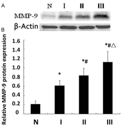
Expression of MMP-9 proteins
Western blotting showed lower MMP-9 proteins in normal mucous tissues. In NP samples, there
were significantly more MMP-9 proteins
(P<0.05). Further comparisons across different sub-types of NP showed that type III NP tissues
had significantly more MMP-9 compared to
those in type II, which are higher than type I (Figure 1, P<0.05 in both cases).
Expressional profiles of TIMP-2 proteins in NP
A similar study was also performed on TIMP-2 protein levels. Results (Figure 2) showed an opposite pattern of this protein when compared to MMP-9: the highest level of TIMP-2 occurred in normal tissues, and was gradually decreased in type I, II and III NP samples. Statistical
analy-sis showed that type I NP tissues had signifi -cantly lowered TIMP-2 proteins compared to control ones (P<0.05). Type II/III NP had signifi -cantly suppressed TIMP-2 when compared to type I patients (P<0.05). No significant differ
-ence, however, was identified between type II
[image:3.629.99.295.77.282.2]and III tissues (P>0.05). Such results of TIMP-2 along with MMP-9 proteins as abovementioned illustrated the occurrence of potentiated Figure 1. Expression of MMP-9 in NP tissues. A.
Showed representative bands of Western blotting; B. Showed quantified expression levels of
MMP-9 proteins (using actin as the normalized level). N, control group; I, II and III represented sub-types of NP. *, P<0.05 compared to control ones; #, P<0.05
[image:3.629.333.527.77.288.2]compared to type I; Δ, P<0.05 compared to type II.
Figure 2. Expression of TIMP-2 in NP tissues. A. Showed representative bands of Western blotting; B. Showed quantified expression levels of
MMP-9 and suppressed TIMP-2 expression dur-ing the occurrence of NP.
mRNA levels of MMP-9 and TIMP-2 in different subtypes of NP
We further extracted mRNA from NP tissue
samples and quantified their expressions of
MMP-9 and TIMP-2 by semi-quantitative real-time PCR. Results (Figure 3) showed consistent patterns with those in Western blotting: MMP-9 mRNA levels were gradually potentiated in type I, II and III NP patients, when compared to nor-mal ones (P<0.05 in all paired comparisons).
TIMP-2 mRNA level, however, was significantly
decreased in NP tissues (P<0.05).
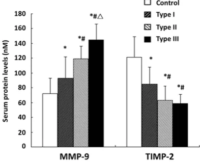
Serum MMP-9 and TIMP-2 levels in different subtypes of NP patients
We further used ELISA to quantify the serum protein levels of MMP-9 and TIMP-2 in different NP patients. As shown in Figure 4, MMP-9
lev-els in NP patients were significantly elevated
compared to control ones (P<0.05), with signifi -cant difference across different subtypes.
Meantime, TIMP-2 levels were significantly sup -pressed in all NP patient’s serum (P<0.05). All those patterns were consistent with those in Western blotting and real-time PCR, thereby suggesting that the dynamic balance between MMP-9 and TIMP-2 was impaired in NP, sug-gesting the involvement of tissue repair and reconstruction. Our results also point the potency of those two indexes as reference for clinical diagnosis.
Discussion
NP is manifested with nasal congestion, head-ache and dysosmia. It can aggravate the respi-ratory infection symptoms, compromising patient’s life quality or even inducing intracra-nial complications [13]. Classical views believe that nasitis, nasosinusitis and NP are three independent diseases. Recent pathological studies, however, suggested the correlation among those diseases as they share the
com-mon disposing factor of inflammation.
Moreover, there is a gradual progression from nasitis to nasosinusitis and NP. In brief, NP orig-inated from nasitis, which may develop edema of mucous, vessel proliferation, increased per-meability and formation of glands, contributing
to the inflammatory lesion, i.e. nasal polyps [14,
15]. However, no clear explanation of NP’s pathogenesis has been reported so far. Most scholars believe that NP is caused by a cas-cade reaction involving multiple factors includ-ing bacteria, virus, allergen and air pollution, all of which activate epithelial cells and stimulate
the release of inflammatory cytokines, thereby facilitating infiltration in inflammatory cells [16].
[image:4.629.99.296.79.223.2]Recent studies showed that the tissue remodel and degradation of basal membrane is one key step in the formation of NP [6, 7]. MMP-9, as one important member of MMP family for the degradation of ECM, exists in various body tis-sues. Under normal physiological conditions, it can regulate cell-to-cell adhesion via modulat-ing ECM breakdown and tissue remodel by its Figure 3. mRNA levels of MMP-9 and TIMP-2 in NP
tissues. *, P<0.05 compared to control ones; #,
P<0.05 compared to type I. Δ, P<0.05 compared to type II.
Figure 4. Serum levels of MMP-9 and TIMP-2 in NP tissues. *, P<0.05 compared to control ones; #,
[image:4.629.98.295.302.461.2]actions on substrate type IV collagen [17, 18]. MMP-9 can modulate tissue reconstruction, embryonic development and injury repair by its regulation on extracellular components, with the help of zinc ions [19]. The over-activation of MMP-9, however, may cause the intensive breakdown of ECM, thereby facilitating tumor
infiltration/metastasis, and auto-immune dis -ease including osteoarthritis and rheumatoid
arthritis. TIMP-2, an endogenous specific MMP
inhibitor, can prevent MMP-9 from over-activa-tion; thereby maintain homeostasis of ECM [20].
In this study, we found less MMP-9 but more TIMP-2 proteins in normal tissues. In NP
sam-ples, MMP-9 protein level was significantly ele -vated while TIMP-2 expression was suppressed. Further analysis revealed that with more advanced NP type, MMP-9 expression (both mRNA and protein) was gradually increased while TIMP-2 was decreased. All those results suggest that the facilitation of MMP-9 and inhi-bition of TIMP-2 can potentiate the disease course. This can be explained as abnormally higher MMP-9 and lower inhibitor TIMP-2 can elevate degradation of ECM, breaking the basal membrane, thereby loosening the connective tissue of nasal lamina propria. All these events can facilitate the formation of edema, break down vascular basal membrane and increase the vessel permeability, thus further aggravat-ing the edema of nasal mucosa and formation of polyps [21, 22]. Further assays in serum also
identified similar patterns of those two pro
-teins, confirming the imbalance of MMP-9 and
TIMP-2 in NP patients and suggesting the potency of those factors as reference indexes for disease evaluation.
In summary, the occurrence and progression of NP are closely related with elevated MMP-9 and suppressed TIMP-2 proteins. Those two factors may work to evaluate the clinical sub-type/stage of NP. Our results also provide evi-dences for further studies regarding NP patho-genesis and development of drug targets.
Disclosure of conflict of interest
None.
Address correspondence to: Dr. Xuezhong Li, Department of Otolaryngolgogy, Qilu Hospital of Shandong University, 107 West Wenhua Road, Jinan 250012, China. Tel: (86)-531-88565657; E-mail: [email protected]
References
[1] Rosenfeld RM, Piccirillo JF, Chandrasekhar SS, Brook I, Ashok Kumar K, Kramper M, Orlandi RR, Palmer JN, Patel ZM, Peters A, Walsh SA, Corrigan MD. Clinical practice guideline (up-date): adult sinusitis. Otolaryngol Head Neck Surg 2015; 152 Suppl 2: S1-s39.
[2] Poposki JA, Peterson S, Welch K, Schleimer RP, Hulse KE, Peters AT, Norton J, Suh LA, Carter R, Harris KE, Grammer LC, Tan BK, Chandra RK, Conley DB, Kern RC, Kato A. Elevated presence of myeloid dendritic cells in nasal polyps of pa-tients with chronic rhinosinusitis. Clin Exp Allergy 2015; 45: 384-93.
[3] Bai J, Miao B, Wu X, Luo X, Ma R, Zhang J, Li L, Shi J, Li H. Enhanced expression of SAM-pointed domain-containing Ets-like factor in chronic rhinosinusitis with nasal polyps. Laryngoscope 2015; 125: E97-103.
[4] Cho JS, Han IH, Lee HR, Lee HM. Prostaglandin E2 Induces IL-6 and IL-8 Production by the EP Receptors/Akt/NF-kappaB Pathways in Nasal Polyp-Derived Fibroblasts. Allergy Asthma Immunol Res 2014; 6: 449-57.
[5] Lee KI, Kim DW, Kim EH, Kim JH, Samivel R, Kwon JE, Ahn JC, Chung YJ, Mo JH. Cigarette
smoke promotes eosinophilic inflammation,
airway remodeling, and nasal polyps in a mu-rine polyp model. Am J Rhinol Allergy 2014; 28: 208-14.
[6] Pezato R, Voegels RL, Pinto Bezerra TF, Perez-Novo C, Stamm AC, Gregorio LC. Mechanical disfunction in the mucosal oedema formation of patients with nasal polyps. Rhinology 2014; 52: 162-6.
[7] Niarakis A, Giannopoulou E, Ravazoula P, Panagiotopoulos E, Zarkadis IK, Aletras AJ. Detection of a latent soluble form of mem-brane type 1 matrix metalloprotease bound with tissue inhibitor of matrix
metalloprotein-ases-2 in periprosthetic tissues and fluids from
loose arthroplasty endoprostheses. FASEB J 2013; 280: 6541-55.
[8] Zhang L, Wu Z, Qin H, Chen W, Zhang G. Changes of Transforming Growth Factor-beta1 and Extracellular Matrix in the Wound Healing Process of Rats Infected With Pseudomonas aeruginosa. Wounds 2014; 26: 293-300. [9] Lee GR, Jang SH, Kim CJ, Kim AR, Yoon DJ,
Park NH, Han IS. Capsaicin suppresses the mi-gration of cholangiocarcinoma cells by down-regulating matrix metalloproteinase-9 expres-sion via the AMPK-NF-kappaB signaling path-way. Clin Exp Metastasis 2014; 31: 897-907. [10] Mavrogonatou E, Angelopoulou MT, Kletsas D.
the TNFalpha-mediated up-regulation of MMP-3. J Orthop Res 2014; 32: 1701-7.
[11] Hwang KE, Shon YJ, Cha BK, Park MJ, Chu MS, Kim YJ, Jeong ET, Kim HR. Tissue inhibitor of metalloproteinase-1 is responsible for residual pleural thickening in pleural tuberculosis. Tohoku J Exp Med 2015; 235: 327-33.
[12] Zhang C, Chen L, Gu Y. Polymorphisms of MMP-1 and MMP-3 and susceptibility to rheu-matoid arthritis: A meta-analysis. Z Rheumatol 2015; 74: 258-262.
[13] Cui XY, Miao JL, Lu HQ, Qi QH, Chen XI, Xu J, Lin ZP, Chen ZB, Yin M, Cheng L. Serum levels of
specific IgE to enterotoxins in patients with
chronic rhinosinusitis. Exp Ther Med 2015; 9: 1523-1527.
[14] Sommer JU, Schultz JD, Grossbaier J, Stern-Straeter J, Hörmann K, Sauter A. In vitro
ef-fects of doxycycline on inflammatory cytokines
and gelatinases in chronic rhinosinusitis. In Vivo 2012; 26: 369-74.
[15] Ren XF, Zhao H, Gong XC, Wang LN, Ma JF. RLN2 regulates in vitro invasion and viability of osteosarcoma MG-63 cells via S100A4/MMP-9 signal. Eur Rev Med Pharmacol Sci 2015; 19: 1030-6.
[16] Uluyol S, Arslan IB, Demir A, Mercan GC, Dogan O, Çukurova I. The role of the uncinate process in sinusitis aetiology: isolated agenesis versus maxillary sinus hypoplasia. J Laryngol Otol 2015; 129: 458-61.
[17] Hatten KM, Palmer JN, Lee RJ, Adappa ND, Kennedy DW, Cohen NA. Corticosteroid Use Does Not Alter Nasal Mucus Glucose in Chronic Rhinosinusitis. Otolaryngol Head Neck Surg 2015; 152: 1140-4.
[18] Wang LF, Chien CY, Chiang FY, Chai CY, Tai CF. Expression of matrix metalloproteinase-2 and matrix metalloproteinase-9 in recurrent chron-ic rhinosinusitis with nasal polyposis. Kaohsiung J Med Sci 2013; 29: 26-31.
[19] Malinsky RR, Valera FC, Cavallari FE, Küpper DS, Milaneze C, Silva JS, Tamashiro E, Anselmo-Lima WT. Matrix metalloproteinases and their impact on sinusal extension in chron-ic rhinosinusitis with nasal polyps. Eur Arch Otorhinolaryngol 2013; 270: 1345-8.
[20] Desmeules P, Trudel D, Turcotte S, Sirois J, Plante M, Grégoire J, Renaud MC, Orain M,
Têtu B, Bairati I. Prognostic significance of
TIMP-2, MMP-2, and MMP-9 on high-grade se-rous ovarian carcinoma using digital image analysis. Hum Pathol 2015; 46: 739-45. [21] Katainen E, Kostamo K, Virkkula P, Sorsa T,
Tervahartiala T, Haapaniemi A, Toskala E. Local and systemic proteolytic responses in chronic rhinosinusitis with nasal polyposis and asthma. Int Forum Allergy Rhinol 2015; 5: 294-302.
[22] Cho JS, Kang JH, Um JY, Han IH, Park IH, Lee
HM. Lipopolysaccharide induces pro-inflam -matory cytokines and MMP production via