SHORT COMMUNICATION
Visual acuity and signal color pattern in an
Anolis
lizard
Leo J. Fleishman§, Anna I. Yeo* and Carley W. Perez‡
ABSTRACT
Anolislizards communicate with colorful dewlaps that often include detailed patterns. We measured the visual acuity ofAnolis sagrei.
Lizards viewed a checkerboard pattern of red and yellow–green squares that were too small to resolve, and thus appeared uniform in color. We quickly replaced the center portion of the display with a pattern of larger squares. If the new pattern could be resolved, the lizards perceived a change in color and reflexively shifted their gaze toward the target. The acuity threshold was 1.21 cycles deg−1. We also calculated acuity based on published anatomical data for
Anolis carolinensis. It was similar to that ofA. sagreifor the visual periphery. Foveal acuity was 10 times greater. We approximated the effects of viewing conditions on the visibility of fine details of a conspecific’s dewlap. For peripheral vision, no detailed patterns were visible at≥0.5 m. For foveal vision, color-pattern details were visible at 1.0 m.
KEY WORDS: Dewlap, Vision, Communication, Color
INTRODUCTION
Many animals rely on complex color patterns for communication. The visibility of fine-scale patterns depends on the receiver’s visual resolution. Male anoline lizards signal to conspecifics by expanding a dewlap that, in many species, exhibits detailed color patterns. The effects of viewing distance and angle on the visibility of these patterns is not known. There is tremendous interest in the appearance of dewlap colors to conspecifics because anoles are an important model system for studies of the evolution of animal signal diversity and its role in speciation (Losos, 2009).
Anolis vision has been studied extensively (Fleishman, 1992;
Fleishman et al., 1993, 2016a,b; Leal and Fleishman, 2002, 2004) but little is known about lizard visual acuity (but see New and Bull, 2011). Although Anolis species vary widely in social behavior, habitat preference and ecology, their visual systems are very similar (Fite and Lister, 1981; Fleishman et al., 1995, 1997; Persons et al., 1999; Loew et al., 2002). Thus, detailed knowledge of visual performance from one species can be usefully applied to the genus as a whole.
We carried out behavioral tests of visual acuity forAnolis sagrei
(Duméril and Bibron 1837) and used published anatomical data to calculate visual acuity forAnolis carolinensisVoigt 1832 (Makaretz and Levine, 1980). We then assessed the impact of viewing conditions on the appearance of color-pattern details of a conspecific dewlap.
MATERIALS AND METHODS
All applicable international and institutional guidelines for the care and use of animals were followed. Procedures involving live animals were in accordance with the ethical standards of Union College and were approved in advance by the IACUC committee.
We used adult male A. sagrei, wild-collected in Florida by a commercial supplier (Snakes at Sunset, Miami, FL, USA). For our first experiment, 10 individuals were obtained on 7 September 2013 and maintained in our laboratory in a constant temperature and humidity room (28°C, 50% relative humidity) for 3 weeks prior to and 7 weeks during the experiment. For our second experiment, 10 individuals were obtained on 28 December 2013, and were maintained for 2.5 weeks prior to and 5 weeks during the experiment. During the second experiment, two individuals died and the data from these individuals was discarded.
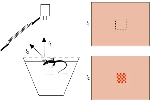
The experimental set-up is illustrated in Fig. 1. A trial was initiated only when the test animal was voluntarily perched lengthwise on a wooden dowel at the front of the cage. Prior to each trial, a cart holding the stimulus apparatus was positioned in front of the lizard’s home cage, based on the location of the lizard’s outward-facing eye, and the experimenter left the room. We waited until the lizard’s monocular gaze was directed straight outward towards a remotely monitored video camera. We then waited 2 min before initiating a trial. If the lizard was not in position after 10 min, no trial was attempted and we moved on to a new individual, retesting this individual later in the same session.
The stimulus (Fig. 1) consisted of a 14×11 cm (w×h) cardboard
‘background’ with a 2×2 cm square opening in the center. The center was positioned 21.0 cm from the lizard’s eye at an angle of 30 deg relative to the direction of gaze straight out from the perch. The background pattern consisted of tiny yellow–green and red squares that appeared to be a uniform orange color because the squares could not be individually resolved. Immediately behind the background card, we positioned a‘stimulus’card. The lower half of each stimulus card was identical to the background. The upper half contained one of five different checkerboard patterns of the same colors as the background, with squares of different sizes. In the control, the squares were the same size as the background. The other four stimuli were made up of checkerboard patterns of increasing size. In each experimental trial, the stimulus card was shifted downward, in 20 ms, to a new position that placed the upper checkerboard pattern behind the square opening. The card movement was faster than the lizard’s temporal resolution and thus did not create a visual motion stimulus. If the lizard visual system could resolve the new, larger spatial pattern, the visual stimulus would appear to change in appearance and color. If not, there would be no change in appearance when the card was moved. We assessed detection of the change in pattern by observing whether the lizard shifted its gaze toward the stimulus within 3 s. This reflexive gaze shift, known as the ‘visual-grasp’ reflex (Fleishman, 1986), is unambiguous and easy to observe because the eyelid moves with the eye. It has been frequently used as an assay of stimulus visibility (e.g. Fleishman, 1986, 1992; Fleishman Received 28 September 2016; Accepted 31 March 2017
Department of Biology, Union College, Schenectady, NY 12309, USA. *Present address: 20 Harcourt Road, Plainview, NY 11803.‡Present address: 32 Indian Ledge Road, Voorheesville, NY 12186, USA.
§
Author for correspondence ([email protected])
L.J.F., 0000-0002-2309-7496
Journal
of
Experimental
and Persons, 2001; Fleishman et al., 2016b; Nava et al., 2009; Steinberg and Leal, 2016).
We defined our stimuli based on the width of one complete cycle of the two differently colored squares. For the background and the smallest stimulus pattern, one cycle equaled 0.10 cm, or 0.27 deg at the front of the lizard’s eye. For the four other test stimuli, one cycle equaled 0.20 cm (0.55 deg), 0.36 cm (0.98 deg), 0.70 cm (1.91 deg) and 1.40 cm (3.81 deg).
Two experiments were carried out: high light (irradiance striking the stimulus surface=30.7 µmol m−2 s−1) and low light (0.41 µmol m−2s−1) (see Fig. S1). For the high light experiment, the front of each cage and the stimulus surface were illuminated with a single 50 W Solux broad-spectrum tungsten lamp with a glass diffuser, positioned above the front edge of each cage and directed downward. For the low light trials, the cage-front lights were turned off (during testing sessions only) and three uniformly spaced 50 W Solux lamps with diffusers aimed at the white ceiling of the lizard room were turned on, so that they diffusely illuminated the entire test room. The high light intensity is typical of a partially shaded habitat under full sun, within the range of light typical in the
A. sagreihabitat. The low light intensity is typical of a fully shaded
forest: considerably darker than the typical A. sagrei habitat (Jenssen and Swenson, 1974; Fleishman et al., 1997). In both cases, the illumination was highly diffuse and produced no glare on the stimulus surface.
All lizards were tested during each 1–2 h experimental session sometime between 09:00 h and 16:00 h. Normally, every lizard was tested twice in one day, with at least 5 min between each test. Lizards were never tested 2 days in a row. Before a trial, a lizard was given at least 15 min to acclimate to the experimental lighting conditions.
Each lizard viewed five different stimulus patterns in a random order that differed for each lizard. For each lizard, no stimulus was repeated until all five had been presented. The set of five stimuli was repeated five times for each individual.
We determined spectral radiance of the yellow–green and red stimulus squares by illuminating them under the experimental light conditions and measuring with a 4 deg acceptance angle radiance probe (collimating lens) input to an Ocean Optics Jaz fiber-optic
spectroradiometer, calibrated for radiance measurement with a LI-COR LI-1800-02 optical radiation calibrator. We multiplied these values by the well-established spectral sensitivity function of
A. sagrei(see Fleishman et al., 2016b, for details). The ratio of red:
yellow–green luminance was 0.7 (±1%), providing both luminance and chromatic contrast between the colored squares.
Statistical analysis
The two experiments occurred at different times with different lizards and were analyzed separately.‘Lizard identity’was analyzed as a random effect.‘Stimulus pattern’and‘days in captivity’were analyzed as fixed effects. The proportion of positive responses by each individual to each of the five stimuli was the response variable. We used a logistic model (logit transformation), following procedures outlined in Warton and Hui (2011). We used the GLIMMIX procedure in SAS statistical software. We used a generalized linear mixed model assuming a binomial distribution with logit link. Least squares means and standard errors, used for descriptive statistics of the results, were estimated using an inverse link transformation. We tested for a significant increase in response probability for each stimulus pattern relative to the control pattern using the Dunnett–Hsu correction for multiple comparisons.
We defined the visual acuity threshold as the stimulus visual angle (for one cycle) at which the proportion of positive responses was equal to half of the maximum positive response rate, determined by linear interpolation between the two closest response values.
Visual acuity estimate forA. carolinensis
Makaretz and Levine (1980) measured the cone and ganglion cell density in the retina ofA. carolinensis. The ratio of the two cell classes was close to 1.0 throughout the retina, making it possible to estimate grating acuity from cone densities (Land and Nilsson, 2012), which averaged 290,000 mm−2 in the central fovea and 3200 mm−2in the middle of the peripheral retina. We assumed an eye size of 4 mm (based on their fig. 1, Makaretz and Levine, 1980) and assumed that the posterior nodal distance (PND) of the eye is 2/ 3 of this length. One cycle of spatial stimulus must cover two receptive fields in order to be reliably resolved (Nyquist sampling theorem: Land and Nilsson, 2012). If cone density=D, the grating acuity for a repetitive pattern in units of cycles deg−1is given by (Pettigrew et al., 1988):
Acuity¼2pPND
360
p
D
2 : ð1Þ
Estimating dewlap appearance
The variations in light falling within one retinal receptive field are averaged by the visual system in a Gaussian fashion (Cronin et al., 2014). The limiting effects of resolution limits can therefore be modeled by blurring an image with a two-dimensional Gaussian filter. We applied a Gaussian blur filter (Adobe Photoshop CS6), with a diameter determined by the visual acuity limit, to a high-resolution photograph of a displaying maleA. sagreiin order to estimate the detail visible for a viewer at different distances.
RESULTS AND DISCUSSION
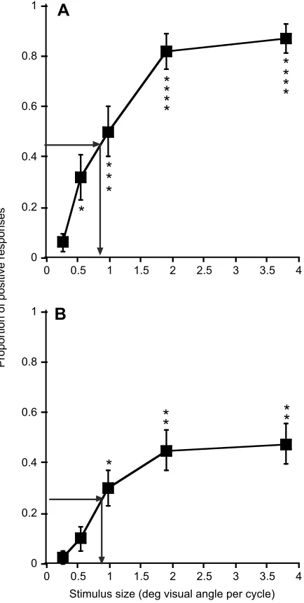
The mean proportion of positive responses versus stimulus pattern size is summarized in Fig. 2. For high light conditions (Fig. 2A), there were significant effects of lizard identity (P<0.0002,χ2test,
d.f.=1) and stimulus pattern (P<0.0001,F=14.49, d.f.=4,36). There
t2
t1
t1
[image:2.612.50.300.55.220.2]t2
Fig. 1. Schematic drawing of the experimental set-up and the lizard’s home cage.A trial started when the lizard’s body was oriented parallel to the cage front, with its monocular gaze directed straight out toward the camera (t1). The position of the new pattern on the stimulus card was rapidly brought into view (t2), and the experimenter observed whether there was a shift of gaze of the outward-facing eye towards the novel stimulus within 3 s. The distance from the lizard’s eye to the stimulus card was 21.0 cm.
Journal
of
Experimental
were no significant effects (P>0.05) of days in captivity (F=0.07, d.f.=1231) or the interaction days in captivity×stimulus pattern (F=2.35, d.f.=4231). For low light conditions (Fig. 2B), there were significant effects of stimulus pattern (P<0.02,F=4.6, d.f.=3,21) and days in captivity (P<0.001, F=18.28, d.f.=1152) and no significant effects (P>0.05) of lizard identity (χ2test, d.f.=1) or the
interaction days in captivity×stimulus pattern (F=0.77, d.f.=3152). We discuss the reduction in response with time for low light in the Appendix. The lizards were less responsive in low than in high light, which could be due either to light level differences or to differences in response rates for the two cohorts.
Our stimulus patterns had both chromatic and luminance contrast components. Acuity is generally higher for luminance contrast, and this probably determined the threshold.
Threshold-estimated minimum resolvable visual angle for the high light experiment was 0.82 deg (grating acuity=1.22 cycles deg−1) and for low light it was slightly lower at 0.85 deg (grating acuity=1.18 cycles deg−1). The difference was smaller than we expected, as spatial summation is often an important mechanism for maintenance of sensitivity in low light (Olsson et al., 2017). Anoles possess small eyes and are highly dependent on high acuity vision. For the two light levels tested, at least in the visual periphery, they do not appear to rely heavily on spatial summation, perhaps because resolution in the periphery is already quite reduced (relative to the fovea) and further increases in receptive field size would sacrifice too much resolution (see below). Fleishman et al. (1995) demonstrated that anoles rely on temporal summation at low light levels, which is an alternative that does not require loss of spatial acuity.
For theA. carolinensiscentral fovea, we estimated a minimum resolvable angle of 0.08 deg (grating acuity=12.5 cycles deg−1) and 0.8 deg for the periphery (grating acuity=1.25 cycles deg−1), which is close to our estimated value forA. sagrei (1.22 cycles deg−1). Other literature estimates for reptilian visual acuity include 6.8 cycles deg−1for the slow-moving skinkTiliqua rugosa(based on retinal anatomy; New and Bull, 2011), 6.1 cycles deg−1for the turtle Pseudemys scripta elegans (based on evoked potentials; Northmore and Granda, 1991) and 4.9 cycles deg−1for the water snakeNerodia sipedon(evoked potentials; Baker et al., 2007). The central foveal estimate forA. carolinensisis twice as high as the peak value for these other reptiles. Although anoles are small, and have small eyes, they eat small insect prey and rely more extensively on high-acuity vision than the other species that have been tested.
The visual motion detection threshold has been measured in five
Anolisspecies (including A. sagrei) (Fleishman, 1986; Steinberg
[image:3.612.68.285.188.619.2]and Leal, 2016), and found to be approximately 0.25 deg. This is three times as fine as our estimated stationary acuity threshold. Minimum detection angle for visual motion is usually smaller than for stationary acuity. For example, the minimum motion distance detectable by humans is half the size of the stationary threshold (Davson, 1977). The anoline visual system is highly specialized for motion perception (Fleishman, 1992), which may explain why this difference is even greater inAnolis.
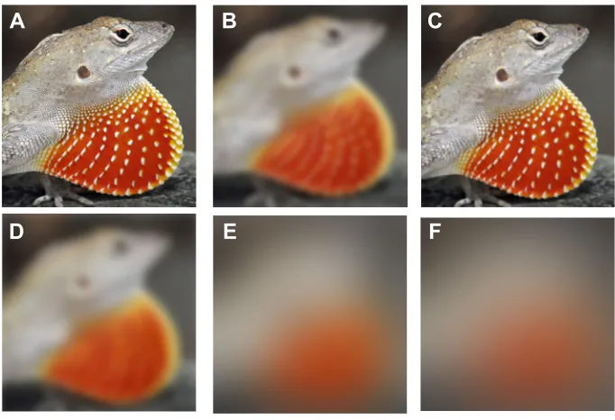
Fig. 3 shows the spatial detail visible to a conspecificA. sagrei
viewing a dewlap from different distances. For foveal vision, the fine dewlap patterns are visible from 1 m. For peripheral vision, the dot pattern disappears at 10 cm, and at 0.5 m the yellow rim of the dewlap is no longer visible. At 1 m, only a blur of orange color is visible.Anolis sagrei males typically have territory sizes that are 3.0 m2in area or greater (Schoener and Schoener, 1982), and their
displays are frequently directed at rivals and potential mates well outside the territory, more than a meter away.
These results suggest that distinctly different aspects of the dewlap color signal come into play in different behavioral contexts. Male anoles frequently give spontaneous dewlap displays directed toward inattentive conspecifics several meters away, to attract females or to deter potential rivals from approaching the territory (Fleishman, 1992). These observers will see a spatial average of the various color markings of the dewlap, and will not perceive detailed patterns such as the fine dots or the thin outer ring of theA. sagreidewlap. Only when viewed from close range and/or with foveal vision–during agonistic interactions or courtship, for example–will these more complex color patterns have the potential to be important signal features.
0 0.2 0.4 0.6 0.8
1
B
*
*
*
*
*
0 0.2 0.4 0.6 0.8 1
0 0.5 1 1.5 2 2.5 3 3.5 4
Proportion of positive responses
0 0.5 1 1.5 2 2.5 3 3.5 4
Stimulus size (deg visual angle per cycle)
A
*
*
*
*
*
*
*
*
*
*
*
*
Fig. 2. The results of visual-grasp experiments under different light intensities.(A) High light intensity. (B) Low light intensity. The mean (±s.e.m.) proportion of positive responses out of five trials is indicated. For high light intensity,n=10; for low light intensity,n=8 (sample sizes constrained by the number of available cages). Asterisks indicate significant differences (see Materials and methods for details) for each trial versus the control (smallest stimulus visual angle): *P<0.05, **P<0.01, ***P<0.005, ****P<0.0001. Arrows show the detection threshold, which is estimated as the visual angle that elicits a response rate that is half the maximum observed response rate. These graphs are based on results averaged over the duration of the experiment. The effects of‘days in captivity’(for the low light condition) are considered in detail
in the Appendix.
Journal
of
Experimental
APPENDIX
Effects of time in captivity
For the low light intensity experiment there was a significant reduction in response across all stimuli with days in captivity. The effect of time for the low light experiment is summarized in Fig. S2. As the figure illustrates, inclusion of the effects of reduced response due to time in captivity had a small impact on our estimate for detection threshold. The value presented in the Results and discussion is based on average responses across time.
We consider here two general hypotheses for the loss of responsiveness with time. There may have been (1) some change through time in the response properties of the visual system, or (2) a general loss of responsiveness to novel stimuli with time in captivity unrelated to peripheral visual perception. One potential mechanism that could conceivably result in loss of responsiveness to our stimuli is a change in the perceived brightness contrast of the two color patterns that made up our stimuli. This could, in principle, arise from changes in the spectral response characteristics of the long-wavelength-sensitive (LWS) double cones that are responsible for perception of luminance in lizards (Fleishman et al. 2016a). The likeliest cause of such a change would be an alteration in the transmission properties of the oil droplets and/or the dispersed pigment that filter input to these cones. It has been shown for various species of birds that changes in diet or light environment can induce changes in oil droplet density, which can alter the spectral transmission properties (e.g. Nott et al. 2009), and thereby alter the spectral response of the LWS double cones.
We do not believe, however, that this caused changes in response in this study. First of all, diet or light-induced changes in lizard oil droplet properties have not been reported in the literature forAnolis.
Recognizing that this lack of evidence might be due to a lack of studies, we consulted Dr Ellis Loew of Cornell University, USA, who has made measured lizard oil droplets and cone outer segments using microspectrophotometry. He reported to us that he has made extensive measurements of oil droplets of A. sagrei, and these measurements included animals newly captured from the wild and animals that have been in captivity for several months. He has not observed any differences in the oil droplet spectra resulting from time in captivity (E. R. Loew, personal communication).
Our second line of evidence that changes in oil droplet spectral properties did not influence our results is based on modeling of photoreceptor spectral absorption properties. In order to estimate the luminance contrast of our stimuli, we modeled spectral absorption of
theA. sagreiLWS photoreceptor using Lamb’s Pigment Template
(Lamb, 1995; see also Fleishman et al., 2016a). We modeled the effects of oil droplet transmission on the cone absorption spectrum following Hart and Vorobyev (2005) with a value for their parameter
b=0.05. To represent normal, baseline, spectral sensitivity, we used published values for A. sagrei for maximum photoreceptor absorption (=567 nm) and oil droplet λ0 (=483 nm), based on
Loew et al. (2002). We also created model absorption spectra withλ0
shifted to shorter wavelengths by 20 nm (λ0=463 nm) andλ0shifted
to longer wavelengths by 20 nm (λ0=503 nm). We multiplied the
radiance of our two spectral colors by the three normalized absorptions based on the three different oil droplet spectra. We then estimated luminance contrast for our two stimulus colors for each case. The ratio of the brighter (yellow−green) to the darker (red) color for the three different oil droplet values was: 0.697, 0.699 and 0.702. The reason why these oil droplet shifts had so little impact on luminance contrast of these colors is that the cut-off wavelengths of
F
A
B
C
[image:4.612.135.476.56.289.2]D
E
Fig. 3. An illustration of detail visible to a conspecific viewer of anAnolis sagreidewlap viewed from different distances with foveal and peripheral vision.For each viewing condition, the width of the threshold viewing angle was calculated relative to a known measurement on the lizard’s body ( jawline to top of eye). The image was then subjected to a Gaussian blur function in Adobe Photoshop (for a detailed description of this function, see Leong et al., 2003), with a blur diameter equal to the minimum threshold angle determined from the high light intensity behavior experiment. Foveal acuity was assumed to be 10 times greater. These pictures are not meant to accurately reflect the appearance of the overall scene because (1) the size of the image was kept constant, whereas in nature the image would get smaller with greater distance, and (2) the edge-enhancement effects of center-surround receptive fields, which would make the edges of large objects such as the animal’s body clearer, are not accounted for. These pictures are designed to accurately illustrate the extent to which fine-pattern details, such as the dots on the dewlap, are visible. Viewing conditions shown are: (A) foveal vision at 0.1 m; (B) foveal vision at 1.0 m; (C) peripheral vision at 0.1 m; (D) peripheral vision at 0.2 m; (E) peripheral vision at 0.5 m; (F) peripheral vision at 1.0 m. Photos by Gary Nafis, CaliforniaHerps. com, used with permission.
Journal
of
Experimental
the oil droplets are positioned near the short-wavelength edge of the L cone absorption spectra inA. sagrei, and the stimulus we used employed colors with most of their energy in the long-wavelength portion of the spectral absorption curve. Thus, shifts in oil droplet sensitivities are unlikely to account for changes in response.
More generally, we doubt that visual system changes accounted for the reduced response. The lizards were exposed to broad-spectrum light between trials and were fed vitamin-supplemented food. Thus, the conditions that have been shown to alter visual system performance (dietary restriction and limited light environment) in other species were not present.
We believe that the reduction in response to the low light stimuli arose from a general loss of wariness and attentiveness, likely due to either to a slow loss of fear of novel conditions in their captive surroundings or more specifically to a slow habituation to the stimuli used in the experiments. The fact that the response was reduced over time for the low light but not the high light condition is difficult to explain. We suspect, however, that the high light stimuli represented a stronger visual stimulus than the low light stimuli, and the loss of attentiveness through time was much greater for the weaker stimulus.
Acknowledgements
We thank Erin Leone and Roger Hoerl for assistance with statistical analysis and two anonymous reviewers for suggestions on the manuscript.
Competing interests
The authors declare no competing or financial interests.
Author contributions
Conceptualization: L.J.F.; Methodology: L.J.F.; Validation: L.J.F.; Formal analysis: L.J.F.; Investigation: A.I.Y., C.W.P.; Resources: L.J.F.; Writing - original draft: L.J.F., A.I.Y., C.W.P.; Writing - review & editing: L.J.F., A.I.Y., C.W.P.; Supervision: L.J.F.; Funding acquisition: L.J.F.
Funding
This research was funded by a National Science Foundation grant (IOS-1051796) to L.J.F.
Supplementary information
Supplementary information available online at
http://jeb.biologists.org/lookup/doi/10.1242/jeb.150458.supplemental
References
Baker, R. A., Gawne,T. J., Loop, M. S. and Pullman,S.(2007). Visual acuity of the midland banded water snake estimated from evoked telencephalic potentials. J. Comp. Physiol. A193, 865-870.
Cronin, T. W., Johnsen, S., Marshall, J. and Warrant, E. J.(2014).Visual Ecology. Princeton, USA: Princeton University Press.
Davson, H. (1977).The Physiology of the Eye 3rd Edition. New York, USA: Academic Press.
Fite, K. V. and Lister, B. C.(1981). Bifoveal vision inAnolislizards.Brain Behav. Evol. 19, 144-154.
Fleishman, L. J. (1986). Motion detection in the presence and absence of background motion in anAnolislizard.J. Comp. Physiol. A159, 711-720. Fleishman, L. J.(1992). The influence of the sensory system and the environment
on motion patterns in the visual displays of anoline lizards and other vertebrates. Am. Nat.139, S36-S61.
Fleishman, L. J. and Persons, M. (2001). The influence of stimulus and background colour on signal visibility in the lizardAnolis cristatellus.J. Exp. Biol.204, 1559-1575.
Fleishman, L. J., Loew, E. R. and Leal, M.(1993). Ultraviolet vision in lizards. Nature365, 397.
Fleishman, L. J., Marshall, C. J. and Hertz, P.(1995). Comparative study of temporal response properties of the visual system of three species of anoline lizards.Copeia1995, 422-431.
Fleishman, L. J., Bowman, M., Saunders, D., Miller, W. E., Rury, M. J. and Loew, E. R.(1997). The visual ecology of Puerto Rican anoline lizards: habitat light and spectal sensitivity.J. Comp. Physiol. A181, 446-460.
Fleishman, L. J., Ogas, B., Steinberg, D. and Leal, M.(2016a). Why doAnolis dewlaps glow? An analysis of a translucent visual signal.Funct. Ecol.30, 345-355. Fleishman, L. J., Perez, C. W., Yeo, A. I., Cummings, K. J., Dick, S. and Almonte, E.(2016b). Perceptual distance between colored stimuli in the lizardAnolis sagrei: comparing visual system models to empirical results.Behav. Ecol. Sociobiol.70, 541-555.
Hart, N. S. and Vorobyev, M.(2005). Modeling oil droplet absorption spectra and spectral sensitivities of bird cone photoreceptors.J. Comp. Physiol. A,191, 381-392.
Jenssen, T. A. and Swenson, B.(1974). An ecological correlate of critical flicker-fusion frequencies for someAnolislizards.Vis. Res.14, 965-970.
Knott, B., Berg, M.L., Morgan E.R., Buchanan K.L., Bowmaker J.K. and Bennett, A.T.D. (2010). Avian retinal oil droplets: dietary manipulation of colour vision?Proc. R. Soc. B.277, 953-962.
Lamb, T. D.(1995). Photoreceptor spectral sensitivities: common shape in the long wavelength region.Vis. Res.35, 3083-3091.
Land, M. F. and Nilsson, D.-E.(2012).Animal Eyes, 2nd Edn. Oxford, UK: Oxford University Press.
Leal, M. and Fleishman, L. J.(2002). Evidence for habitat partitioning based on adaptation to environmental light in a pair of sympatric lizard species. Proc. R. Soc. Lond. B Biol. Sci.269, 351-359.
Leal, M. and Fleishman, L. J.(2004). Differences in visual signal design and detectability between allopatric populations ofAnolislizards.Am. Nat.163, 26-39. Leong, F. J. W.-M., Brady, M. and McGee, J. O’D.(2003). Correction of uneven illumination (vignetting) in digital microscopy images.J. Clin. Pathol.56, 619-621. Loew, E. R., Fleishman, L. J., Foster, R. G. and Provencio, I.(2002). Visual pigments and oil droplet in diurnal lizards: a comparative study of Caribbean anoles.J. Exp. Biol.205, 927-938.
Losos, J. B. (2009).Lizards in an Evolutionary Tree: Ecology and Adaptive Radiation of Anoles. Berkeley, USA: University of California Press.
Makaretz, M. and Levine, R. L.(1980). A light microscopic study of the bifoveate retina in the lizardAnolis carolinensis: general observations and convergence ratios.Vis. Res.20, 679-686.
Nava, S. S., Conway, M. and Martins, E. P. (2009). Sexual-specific visual performance: female lizards outperform males in motion detection.Biol. Lett.5, 732-734.
New, S. T. D. and Bull, M.(2011). Retinal ganglion cell topography and visual acuity of the sleepy lizard (Tiliqua rugosa).J. Comp. Physiol. A197, 703-709. Northmore, D. P. M. and Granda, A. M.(1991). Refractive state, contrast sensitivity,
and resolution in the freshwater turtle,Pseudemys scripta elegans, determined by tectal visual-evoked potentials.Vis. Neurosci.7, 619-625.
Olsson, P., Wilby, D. and Kelber, A.(2017). Spatial summation improves bird color vision in low light.Vis. Res.130, 1-8.
Persons, M. H., Fleishman, L. J., Frye, M. A. and Stimphil, M. E.(1999). Sensory response patterns and the evolution of visual signal design in anoline lizards. J. Comp. Physiol.184, 585-607.
Pettigrew, J. D., Dreher, B., Hopkins, C. S., McCall, M. J. and Brown, M.(1988). Peak density and distribution of ganglion cells in the retinae of microchiropteran bats: implications for visual acuity.Brain Behav. and Evol.32, 38-56.
Schoener, T. W. and Schoener, A.(1982). Intraspecific variation in home-range size in someAnolislizards.Ecology63, 809-823.
Steinberg, D. S. and Leal, M. (2016). Visual motion detection and habitat preference inAnolislizards.J. Comp. Physiol. A.202, 783-790
Warton, D. I. and Hui, F. K. C.(2011). The arcsine is asinine: the analysis of proportions in ecology.Ecology92, 3-10.