Original Article
Grb2-associated binder 2 silencing impairs growth
and migration of H1975 cells via modulation
of PI3K-Akt signaling
Wen Jie Wang1, Kun Mou1, Xi Feng Wu2, Jin Zhong Zhang1, Gang Ren1, Jiu De Qi1, Yi-Fu Xu3, Xin Yao3 1Department of Oncology, People’s Hospital of Laiwu City, No. 1 Xuehu Street, Changshao Road, Laiwu,
Shandong, China; 2Department of Hematology, People’s Hospital of Laiwu City, No. 1 Xuehu Street, Changshao
Road, Laiwu, Shandong, China; 3Department of Pharmacy, First Affiliated Hospital of Soochow University, Suzhou,
Jiangsu Province, China
Received July 19, 2015; Accepted August 25, 2015; Epub September 1, 2015; Published September 15, 2015
Abstract: Non-small cell lung cancer (NSCLC) is a leading cause of cancer-related death and often has a poor prog-nosis. Investigation of NSCLC cancer cell migration, invasion and development of strategies to block this process is essential to improve the disease prognosis. In this study, we tested our hypothesis that Grb2-associated binder 2 (Gab2) regulate NSCLC cancer cell H1975 malignant biological behaviors, and silencing Gab2 reduced H1975 cel-lular colony forming ability, migration and invasion. Moreover, silenced cells present defects in phosphatidylinositol 3-kinase (PI3K)-serine/threonine kinase (Akt) signaling, and reduced expression/activity of matrix metallopepti-dase (MMP)-2/9. Furthermore, in Gab2 siRNA-transfected cells, we detected a decrease in signal transducer and activator of transcription 3 (STAT3) phosphorylation and nuclear translocation. In vivo, Gab2 siRNA cells inoculated subcutaneously in nude mice demonstrated decreased tumor growth and PI3K-Akt signaling inhibition. These re-sults indicate that Gab2 is a key factor in H1975 tumor migration, invasion, suggesting that Gab2 can be a novel therapeutic target in NSCLC.
Keywords: Gab2, invasion, STAT3, non-small cell lung cancer
Introduction
Lung cancer is the leading cause of cancer death worldwide and more than 80% of lung cancers are non-small cell lung carcinoma (NSCLC) [1]. This cancer has a very poor prog-nosis, principally because it is most commonly diagnosed during its later stages, and it is often characterized by aggressive local invasion, early metastasis and poor response to chemo-therapy. In the last few decades, our under-standing of the molecular biology of NSCLC metastasis has increased tremendously. Future, continue to identify the key molecules that control tumor deteriorate and develop-ment of novel treatdevelop-ments than can block or inhibit invasion and/or metastasis is important for improving the prognosis of NSCLC [2]. The Grb2-associated binder (Gab) proteins including mammalian Gab1, Gab2 and Gab3, comprise a family of scaffolding or docking
metastasis in a variety of cancers [6]. In con-trast, many of the functions of Gab2 in NSCLC progression, invasion, and metastasis remain unclear.
In the present study, to investigate how Gab2 regulates NSCLC cancer migration and inva-sion, we examined effects of transient Gab2 silencing by RNA interference on NSCLC carci-noma cell line H1975. We found that silencing Gab2 decreased the H1975 cells migration and invasion abilities, and reduced the phosphory-lated PI3K (p-PI3K), phosphoryphosphory-lated Akt (p-Akt) and STAT3 transcriptional activation. Fur- thermore, silencing of Gab2 decreased MMP-2/9 levels and activity in H1975. These effects are consequence of a down-regulation of PI3K-Akt signaling pathway, and eventually lead to an impairment of in vivo tumorigenesis.
Materials and methods
Cell culture and transfections
The human NSCLC lines (H1650, H1975, A549, HCC827, H1299) and SV40-immortalized non-tumorigenic human bronchial epithelial cells BEAS-2B used as control were obtained from the Chinese Academy of Sciences Cell Bank of Type Culture Collection (CBTCCCAS, Shanghai, China). H1975 cells were transfected with Gab2 siRNA or control siRNA (Santa Cruz Biote- chnology), respectively, using the Lipofectamine RNAi MAX reagent (Invitrogen), following the manufacturer’s instructions. After 48 h of transfection, the culture medium was replaced by RPMI-1640 Medium and was incubated for tests [7].
Cell proliferation assay
The cell viability was determined by CellTiter 96® Aqueous One Solution cell proliferation assay (Promega). H1975 cells (2.5 × 10³ cells/ well) transfected with either control- or Gab2-siRNA and were seeded in 96-well cell culture plates and treated with indicated agents. After incubation for indicated times, 20 μL of One Solution reagent were added to each well and incubation was continued for additional 4 h. The absorbance was measured at 490 nm using Synergy™ HT Multi-Mode Microplate Reader (Bio-Tek). The effect of siRNA Gab2 on cell viability was assessed as the percent of cell viability compared with untreated control
cells, which were arbitrarily assigned 100% viability [8]. Experiment was performed in triplicate.
Lactate dehydrogenase (LDH) toxicity assay
The LDH released into cell cultures is an index of cytotoxicity and evaluation of the permeabil-ity of cell membrane. H1975 cells were trans-fected with Gab2 siRNA or control siRNA, respectively, and were seeded in 96-well plate at a density of 3 × 10³ cells per well. Other two groups of H1975 cells were incubated with vehicle (0.1% DMSO, as untreated cells ) or 1% Triton X-100 (as positive group) for 24 h, and, all group cell supernatants were collected and analyzed for LDH activity using LDH cyto-toxici-ty assay kit from Keygen biotech [9].
Anchorage-independent cell growth in soft agar
After transfection, H1975 cells (5 × 10⁴) sus-pended in 2 ml medium, 0.3% agar, and 10% FBS were plated over the bottom agar pre-solidified with 3 ml medium containing 0.6% agar and 10% FBS. Colony formation was then carried out by photographing through the micro-scope after 14 days [10]. Five random fields under microscope were taken and colony for-mation unities were counted.
Wound healing assay
We examined the migration of control- and Gab2-siRNA-transfected H1975 using a wound-healing assay. Briefly, the transfected cells were each grown on 3.5-cm plates with their respective culture media. After the growing cell layers had reached confluence, we inflicted a uniform wound (i.e., in a straight line) in each plate using a pipette tip, and washed the wounded layers with PBS to remove all cell debris. Then, we evaluated the closure at 48 h using bright-field microscopy (Nikon) [11]. All experiments were performed in triplicate.
Migration assay
can-cer cells (5 × 10⁴) in serum-free medium was placed in each upper chamber. After incubation for 24 h, the cells were fixed with 70% ethanol and stained with crystal violet [12]. Cell count-ing was then carried out by photographcount-ing the membrane through the microscope. Five ran-dom fields under microscope were taken.
Invasion assay
H1975 invasion assays were performed with Matrigel-coated chambers from a BioCoat Matrigel Invasion Chamber Kit (BD Biosciences) according to the manufacturer’s instructions. In brief, 1 × 10⁵ control- and Gab2-siRNA-transfected H1975 with 500 µl in serum-free medium were added into the upper chamber and complete medium was added into the lower chamber. After incubation for 24 h, the non-invasive cells in the upper surface of the membrane were removed by “scrubbing” with cotton tipped swab and the cells invasion to the lower surface of the membrane were fixed and stained with DiO (3,3’-dioctadecyloxacarbocya-nine perchlorate) for 20 min. Cell counting was then carried out by photographing the mem-brane through the microscope [12] and five ran-dom fields were taken.
Western blot analysis
All cells were harvested after indicated control- and Gab2-siRNA treatments. Whole-cell lysates were prepared with RIPA buffer containing pro-tease and phosphatase inhibitors. Nuclear and cytoplasmic cell extracts were prepared using the NE-PER Nuclear and Cytoplasmic Extraction kit (Thermo). Equal amounts of cell lysates (25 μg) were loaded on 10% SDS-PAGE and trans-ferred onto PVDF membranes. After mem-branes were blocked, they were incubated with monoclonal antibody against Gab2 (1:1000, Santa Cruz), MMP-2 and MMP-9 (1:1000, CST), PI3k p85 and phosphor-PI3K p85 Tyr458 (1:10000, CST), STAT3 and phosphor-STAT3 Tyr705 (1:1000, CST), Akt and phosphor-Akt Thr308 (1:1000, CST), GPADH (1:5000, Bioworld Technology) and Lamin A/C (1:5000, Epitomics) followed by incubation with horse-radish peroxidase-conjugated IgGs (1:10000, Bioworld Biotechnology). Target proteins were detected by the ECL system (Millipore) and visu-alized with the ChemiDoc XRS system (Bio-Rad) [12].
Matrix metalloprotein (MMP) activity assay
The activity of MMP-9 and MMP-2 were deter-mined by QuickZyme MMPs activity assay (QucikZyme BioSciences) according to the manufacturer’s instructions [7]. Briefly, after transfection for 48 h, H1975 were washed with fresh medium and replaced with serum-free medium. After additional 24 h, the medium was collected and centrifuged at 10000 g for 10 min. Respective supernatant was added to the 96-well strip coated with MMP-9 antibody or MMP-2 antibody and incubated at 4°C over-night. After washing with wash buffer for 3 times, 50 µl assay buffer was added into the well, followed by adding 50 µl detection reagent. After incubation at 37°C for 1 h, OD405 was measured with Microplate Reader (Bio-Tek).
Real-time PCR
Total RNA was isolated using TRIzol according to the manufacturer’s instructions (Invitrogen, USA) and the concentration of total RNA was detected by spectrophotometry at OD260. Reverse transcription was carried out using superscript III reverse transcriptase (Invitrogen) as described in the manufacturer’s manual [13]. The real-time PCR was performed on ABI Prism 7500 Sequence detection system (Applied Biosystems) with the KAPA SYBR® qPCR Kit (KAPA Biosystems) according to the manufacturer’s instructions. The conditions for real-time PCR amplification were as follows: 2 min at 50°C, 1 min at 95°C, 40 cycles at 95°C for 3 s and 60°C for 1 min, followed by melting curve analysis at the end of each run from 60°C to 95°C. The primers used were as follows: Gab2 (Forward: 5’-ACAGTACCTACGACCTCCCC- 3’, Reverse: 5’-CTGGGCGTCTTGAAGGTGTA-3’), β-actin (Forward: 5’-GCT CTT TTC CAG CCT TCCTT-3’, Reverse: 5’-TGA TCC ACA TCT GCT GGAAG-3’). The target mRNA level of control cells normalized to the level of β-actin mRNA, was defined as 1. Results were obtained from three independent experiments.
Immunocytochemistry
against phosphor-STAT3 Tyr705 (1:500) over-night at 4°C. After being washed with PBS, cells were incubated with FITC-conjugated goat anti-rabbit secondary antibody (1:60, Boster Biotechnology). Nuclei were stained with 4’, 6-diamino-2-phenylindole (DAPI; Invitrogen) [14]. Images were taken and analyzed using the ZEN 2011 imaging software on a Zeiss invert microscope (CarlZeiss).
Subcutaneous xenograft models
After transfected with either control- or Gab2-siRNA, H1975 cells (3 × 10⁶) were subcutane-ously implanted into female, BALB/c nude mice to build non-small cell lung cancer xenograft.
[image:4.612.90.518.67.413.2]compliance with the Guidelines for the Institute for Experimental Animals, Center of Hunan University Of Chinese Medicine.
Statistical analysis
The data were presented as mean ± SD. Differences in the results of two groups were evaluated using either two-tailed Student’s t test or one-way ANOVA followed by post-hoc
Dunnett’s test. The differences with P < 0.05 were considered statistically significant.
Results
Gab2 silencing reduced clone formation of non-small cell lung cancer cell
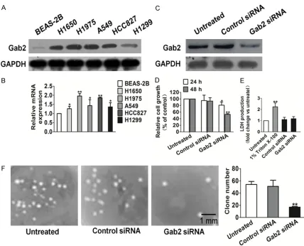
[image:5.612.86.521.67.485.2]five non-small cell lung cancer cell lines (H1650, H1975, A549, HCC827, H1299) and human bronchial normal epithelial cells BEAS-2B. Gab2 mRNA and protein expression status were up-regulated in all NSCLC cell lines, espe-cially in H1975 cells compared with that in BEAS-2B (Figure 1A and 1B). To determine the temporal pattern of Gab2 gene silencing, we transfected H1975 with either Gab2- or control-siRNA for comparison. As shown in Figure 1C, Gab2 silencing achieved significantly knock-down at 48 h after transfection in H1975 cells. Then, we observed cell proliferation of Gab2 siRNA, control-siRNA transfected H1975 and untreated cells for 24 h and 48 h, respectively, and detected significantly growth inhibition in Gab2-siRNA-transfected cells (Figure 1D). To validate whether Gab2 siRNA would result in toxicity effects on H1975, LDH cytotoxicity assay was carried out. As shown in Figure 1E,
0.1% Triton X-100 significantly increased LDH release and Gab2 siRNA transfection resulted in little toxic effects on H1975 cells when com-pared to untreated control cells. In anchorage-independent cell growth assay, only untreated and control-siRNA transfected H1975 cells could form more colonies in soft agar, an in vitro hallmark of cell transformation. Strikingly, however, the ability of H1975 to form colonies was completely abrogated by Gab2 silencing (Figure 1F).
Gab2 silencing reduced migration and inva-sion of H1975
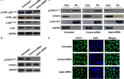
[image:6.612.89.521.77.353.2]Cellular invasion, a characteristic of metastatic tumors, involves cell attachment to extracellu-lar matrix (ECM), ECM degradation and cell migration. To determine if loss function of Gab2 affects cell migration, wound-healing assay Figure 3. Effects of Gab2 silencing on the PI3K/Akt pathway. A. Expression of PI3K and Akt in Gab2-silenced cells. After transfection of cells with either control- or Gab2-siRNA for 48 h (or cells were left untreated), the total cells were isolated and PI3K/Akt expression levels were detected by western blotting. GAPDH was used as a loading control. B. H1975 cells transfected using the same conditions as described above and total protein was extracted, the total and phosphorylated (p) STAT3 were detected. C. After Gab2 silencing, cytoplasmic (Cyto.) and nuclear (Nu.) extracts were prepared and then subjected to western blot for measuring protein level of phosphor-STAT3Tyr705 and
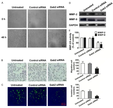
was performed. Compared to untreated cells, disruption of Gab2 significantly reduced cell wound healing (Figure 2A). Then we performed Transwell assay to confirm the inhibitory effect of Gab2 silencing on cell migration. In contrast with untreated H1975, Gab2 silencing decreased the migration of H1975 (Figure 2B). Furthermore, we determined that Gab2 silenc-ing affected cell invasion similarly to migration of the NSCLC cancer line. Consistently, there were less Gab2-silenced H1975 that invasive across the Matrigel-coated chamber compared to the control cells (Figure 2C).
Taking into account that MMPs such as MMP-2 and MMP-9 can be involved in the development
[image:7.612.89.523.69.407.2]of several human malignancies, as degradation of collagen IV in basement membrane and extracellular matrix facilitates tumor progres-sion, including invaprogres-sion, metastasis, and angio-genesis, we analyzed their levels and activity. A significant drop in MMP-2 and MMP-9 expres-sion levels were observed in Gab2 silenced cells (Figure 2D). Quantification of MMP-2 and MMP-9 activities using a fluorogenic assay showed a significantly decrease in extracellular MMP-2 and MMP-9 activity in H1975 Gab2 siRNA cells compared to control-siRNA or untreated cells (Figure 2E). In summary, disrup-tion of Gab2 led to reduced cell migradisrup-tion and significantly impaired the invasion of H1975 cells.
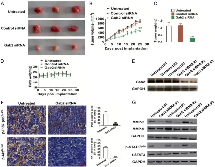
Figure 4. In vivo tumorigenicity of Gab2-siRNA H1975 and untreated H1975 cells. (A) Representative pictures of tumor masses from mice were taken after 25 days of implanted with H1975 cells or with siRNA-Gab2 H1975 cells. Scale bars: 1 cm (B). Gab2 silencing resulted in significantly H1975 tumor growth inhibition versus mice injected untreated H1975 cells. (C) After surgical excision, significant differences were also evident in tumor weight. (D) There was no significant difference in body weight in the three groups. (E) Total tumor tissue lysates were isolated and Gab2 expression levels were detected by western blotting. #, representatives mice number. (F) Tumor tissues were prepared for immunohistochemistry detection with antibodies against p-PI3K p85Tyr458 and p-AKTThr308, and the
p-PI3K p85Tyr458 and p-AKTThr308 positive cells were statistical analyzed. Scale bars: 50 µm (G) Shows western blot of
Gab2 silencing induced degradation of STAT3 and suppressed phosphorylation of both PI3K and Akt
It has been reported that Gab2 plays a role in breast, colon, and gastric cancer cells by inter-acting with PI3K/Akt. Accordingly, we investi-gated the signaling pathway that was mediated by Gab2 and that was associated with cell motility. Specifically, we first confirmed whether Gab2 modulates PI3K/Akt phosphorylation in H1975 cells by western blotting. We found that the expression levels of both proteins remained unchanged after silencing Gab2, whereas the phosphorylated forms of PI3K and Akt were strongly reduced by this transfection (Figure 3A).
To further investigate the signaling pathway that mediated STAT3 expression, we assessed the expression of total and phosphorylated STAT3 in cells transfected with either control-or Gab2-siRNA by western blotting. We found that the STAT3 total expression level remained unchanged after Gab2 silencing, whereas STAT3 phosphorylation was inhibited by this transfection (Figure 3B).
Additionally, the activity of STAT3 was also regu-lated by subcellular localization; we therefore attempted to explore the effect of Gab2-siRNA in different cell fractions. The experiment showed that the nuclear STAT3 phosphoryla-tion in H1975 cells was evidently abrogated by Gab2-siRNA (Figure 3C). We also examined the subcellular distribution of STAT3 in both con-trol- and Gab2-siRNA-transfected H1975 cells by immunofluorescence staining and confocal microscopy. As illustrated in Figure 3D, treat-ment with Gab2-siRNA hardly decreased broad nuclear translocation of STAT3, while untreated and control-siRNA transfection rendered STAT3 stability and general nuclear distribution.
Gab2 siRNA impaired the growth of H1975 in vivo
To further investigate the role of Gab2 in tumor-al growth in vivo, Gab2-siRNA, control-siRNA, and un-transfected H1975 cells were injected subcutaneously into the backs of female nude mice, respectively (6 mice in each group), and tumor growth was monitored for 25 days (Figure 4A). According to our in vitro findings, the mean tumoral volume (Figure 4B) and
weight (Figure 4C) of the Gab2-siRNA group was significantly smaller compared to the con-trol-siRNA and untreated. Furthermore, there was no significant difference in weight between control group and Gab2-siRNA treated groups (Figure 4D). Gab2 expression was checked on tumor tissue after H175 injection into nude mice to confirm that Gab2 remained silenced (Figure 4E). Immunohistochemistry staining (IHC) of p-PI3K p85Tyr458 and p-AktThr308 with tumoral tissues in vivo clearly showed decreased immunostaining for PI3K and Akt activity in the Gab2-siRNA derived tumors com-pared with control-siRNA transfected H1975 injected mice (Figure 4F). In addition, Gab2-siRNA also resulted in down-regulation of PI3K-Akt downstream molecules including MMP-2 and MMP-9 (Figure 4G). At the same time, we observed a significant decrease in the phos-phorylation of STAT3 in the mice injected Gab2-siRNA H1975 (Figure 4G), which was consistent with the results of H1975 cells in vitro.
Discussion
associ-ated with H1975 cancer cell motility. In this study we examined whether Gab2 silencing affected MMP-2/9 expression in H1975. We found that Gab2-siRNA-transfected cells from H1975 had drastically reduced MMP-2/9 levels and activity compared with corresponding con-trol cells.
Gab2 undergoes tyrosine phosphorylation, cre-ating a number of docking sites to mediate interactions with the p85 subunit of PI3K [5]. The interaction of Gab2 with p85 subunit of PI3K is crucial in mediating the PI3K-Akt signal-ing. In this study, using western blotting experi-ment, we determined that the levels of p-Akt and p-PI3K decreased in siRNA-Gab2 H1975. It has been demonstrated that phosphorylated Akt activates its transcription factor STAT3, thereby activates specific target genes, includ-ing those that encode c-Myc, cyclin D1, and MMP-2/9 [20]. STAT3, which acts as transcrip-tion activator, is reported plays a role in cell, proliferation, invasion, angiogenesis, and sur-vival. Therefore, we performed immunofluores-cence and western blot from control- and Gab2-siRNA-transfected H1975 cells, and found that STAT3 phosphorylation was significantly decreased in Gab2-silenced cells.
More convincingly, Gab2 silencing markedly impaired the growth of H1975 cells in vivo. Histological studies of the tumor sections revealed that Gab2 silencing also significantly reduced PI3K/Akt activity. Meanwhile, western blot results show that Gab2 silencing could obviously attenuate expressions of MMP-2/9 expression in tumor tissue. All these data pre-sented that Gab2 is involved in the growth of NSCLC cancer cell H1975. Due to the impor-tant roles of Gab2 in the growth and migration of NSCLC H1975 cells, it may serve as an attractive target for molecular targeting cancer therapy.
Disclosure of conflict of interest None.
Address correspondence to: Dr. Jiu De Qi, Depart- ment of Oncology, People’s Hospital of Laiwu City, No. 1 Xuehu Street, Changshao Road, Laiwu, Shandong, China. E-mail: [email protected]
References
[1] Shtivelman E, Hensing T, Simon GR. Molecular pathways and therapeutic targets in lung can-cer. Oncotarget 2014; 5: 1392-1433.
[2] Forde PM, Ettinger DS. Targeted therapy for non-small-cell lung cancer: past, present and future. Expert Rev Anticancer Ther 2013; 13: 745-758.
[3] Shi L, Sun X, Zhang J, Zhao C, Li H, Liu Z, Fang C, Wang X, Zhao C, Zhang X, Zhou F, Lu S, Luo R, Zhang B. Gab2 expression in glioma and its implications for tumor invasion. Acta Oncol 2013; 52: 1739-1750.
[4] Fleuren ED, O’Toole S, Millar EK. Overexpression of the oncogenic signal transducer Gab2 oc-curs early in breast cancer development. Int J Cancer 2010; 127: 1486-1492.
[5] Wang Y, Sheng Q, Spillman MA. Gab2 regu-lates the migratory behaviors and E-cadherin expression via activation of the PI3K pathway in ovarian cancer cells. Oncogene 2012; 31: 2512-2520.
[6] Ortiz-Padilla C, Gallego-Ortega D, Browne BC. Functional characterization of cancer-associat-ed Gab1 mutations. Oncogene 2013; 32: 2696-2702.
[7] Santiago-Gómeza A, Barrasaa JI, Olmoa N, Lecona E, Burghardt H, Palacín M, Lizarbe MA, Turnay J. 4F2hc-silencing impairs tumorigenic-ity of HeLa cells via modulation of galectin-3 and β-catenin signaling, and MMP-2 expres-sion. Biochim Biophys Acta 2013; 1833: 2045-2056.
[8] Kobayashi T, Shimura T, Yajima T. Transient gene silencing of galectin-3 suppresses pan-creatic cancer cell migration and invasion through degradation of β-catenin. Int J Cancer 2011; 129: 2775-2786.
[9] Youm I, Yang XY, Murowchick JB, Youan BB. Encapsulation of docetaxel in oily core polyes-ter nanocapsules intended for breast cancer therapy. Nanoscale Res Lett 2011; 6: 630. [10] Kim A, Im M, Yim NH, Ma JY. Reduction of
met-astatic and angiogenic potency of malignant cancer by Eupatorium fortunei via suppression of MMP-9 activity and VEGF production. Sci Rep 2014; 4: 6994.
[11] Zhu K, Pan Q, Jia LQ, Dai Z, Ke AW, Zeng HY, Tang ZY, Fan J, Zhou J. MiR-302c inhibits tumor growth of hepatocellular carcinoma by sup-pressing the endothelial-mesenchymal transi-tion of endothelial cells. Sci Rep 2014; 4: 5524.
[12] Dong Y, Zhang T, Li J, Deng H, Song Y, Zhai D, Peng Y, Lu X, Liu M, Zhao Y, Yi Z. Oridonin Inhibits Tumor Growth and Metastasis through Anti-Angiogenesis by Blocking the Notch Signaling. PLoS One 2014; 9: e113830. [13] Zhang D, Ni Z, Xu X, Xiao J. MiR-32 Functions
as a Tumor Suppressor and Directly Targets EZH2 in Human Oral Squamous Cell Carcinoma. Med Sci Monit 2014; 20: 2527-2535.
Oral Squamous Cell Carcinoma Cells to Cisplatin by Targeting STAT3/miR-21 axis. Sci Rep 2014; 4: 7461.
[15] Aftab BT, Dobromilskaya I, Liu JO, Rudin CM. Itraconazole inhibits angiogenesis and tumor growth in non-small cell lung cancer. Cancer Res 2011; 71: 6764-6772.
[16] Wöhrle FU, Halbach S, Aumann K, Schwemmers S, Braun S, Auberger P, Schramek D, Penninger JM, Laßmann S, Werner M, Waller CF, Pahl HL, Zeiser R, Daly RJ, Brummer T. Gab2 signaling in chronic myeloid leukemia cells confers resis-tance to multiple Bcr-Abl inhibitors. Leukemia 2013; 27: 118-129.
[17] Hoeben A, Martin D, Clement PM, Cools J, Gutkind JS. Role of GRB2-associated binder 1 in epidermal growth factor receptor-induced signaling in head and neck squamous cell car-cinoma. Int J Cancer 2013; 132: 1042-1050. [18] Nyga R, Pecquet C, Harir N, Gu H,
Dhennin-Duthille I, Régnier A, Gouilleux-Gruart V, Lassoued K, Gouilleux F. Activated STAT5 pro-teins induce activation of the PI3-kinase/Akt and Ras/MAPK pathways via the Gab2 scaf-folding adapter. Biochem J 2005; 390: 359-366.
[19] Samanta AK, Lin H. Bcr-Abl regulates c-Myc ex-pression in chronic myelogenous leukemia through a Bcr-Abl/Jak2 network of proteins in-volving Gab2, PI-3 kinase, NF-κB and GSK3β. Cancer Res 2006; 66: 968
[20] Matsumura T, Sugimachi K, Takahashi Y, Uchi R, Sawada G, Ueda M, Hirata H, Sakimura S, Ueo H, Takano Y, Kurashige J, Shinden Y, Eguchi H, Sudo T, Yamamoto H, Doki Y, Mori M, Mimori K. Clinical Significance of GAB2, a Scaffolding/Docking Protein Acting Down- stream of EGFR in Human Colorectal Cancer. Ann Surg Oncol 2014; 21 Suppl 4: S743-9. [21] Yang L, Han S, Sun Y. An IL6-STAT3 loop